© 1986 by The University of Chicago. All rights reserved. 0022-1899/86/5302-0020$01.00
Association of Symptomatic· Human Infection with
Toxoplasma gondii
with
Imbalance of Monocytes alldAntigen-Specific T Cell Subsets
Ivo Sklenar, Thomas C. Jones, Sefik Alkan,and Peter Erb
From the Institute for Microbiology and Hygiene, University of Basel; the Pharmaceuticals Division, Ciba-Geigy, Basel, Switzerland; and the Division of International Medicine, Cornell Medical College, New York, New York
During. recent symptomatic toxoplasmosis, alterations in quantity and function of mononuclear cellsin peripheral blood were observed. Flow cytofluorometric analysis and differential leukocyte counts revealed increased absolute numbers of T8 + cells, Leu 7 + (natural killer/killer) cells, and monocytes. T4+ cells and HLA-DR+ cells were not sig-nificantly changed. T4/T8 cell ratios were reversed in symptomatic toxoplasmosis (0.7 ± 0.3) and normal in chronic infection (1.7 ± 0.5). Toxoplasma antigen induced higher numbers of T8-+- aridTQl + cells in four T cell lines from two individuals with sympto-matic infection than in five T cell lines from three individuals with asymptosympto-matic infec-tion. Eight cloned T cell lines producedyinterferon in an antigen-specific fashion and in higher amounts when they originated from an asymptomatic subject than from a symp-tomatic subject. These results indicate that marked alterations in properties of im-munoregulatory cells are characteristic of recent symptomatic toxoplasmosis. The tran-sient immune dysfunction may be a major part of the observed disease and/or a feature of successful parasitism.
Infection with the coccidian protozoanToxoplasma gondiicauses no illness in most individuals. In about one of five infected individuals, various degrees of fatigue, malaise, fever, and lymph node enlargement are recorded [1]. These subjects recover without re-sidual signs of disease; this distinguishes them from those with documented immune deficiency, in whom toxoplasmosis is often severe and even fatal [2].It
has also been noted that evidence of cellular immune response to toxoplasma antigens is delayed in ap-pearance in some patients, compared with the ki-netics of response to other antigens [1, 3, 4]. In an effort to understand better immune responses dur-ing acute symptomatic infection, we examined total T cell subsets and toxoplasma antigen-induced T cell subsets during toxoplasmosis.
Received for publication 22 May 1985 and in revised form 12 August 1985.
Informed consent was obtained from all human subjects studied.
This work was supported by grant 3.016.-0.84 from the Swiss National Science Foundation.
We thank MartinWesp (Ciba-Geigy, Basel, Switzerland) for help with the Cytofluorograph analyses and Danielle Rieder for technical assistance.
Please address requests for reprints to Dr. Ivo Sklenar, Insti-tute for Microbiology and Hygiene, University of Basel, Peter-splatz 10, CH-4003 Basel, Switzerland.
315
Subjects and Methods
Subjects. A total of 22 individuals was studied. T cell surface markers of peripheral blood lympho-cytes (PBLs) were studied in 19 individuals with use of monoclonal antibodies and a Cytofluorographs' (Ortho Instruments, West Wood, Mass). According to the history of infection byT.gondii,three groups of subjects were defined. Six Iloninfected individu-als (mean ± SD age, 29 ± 11 years) were compared with five chronically infected individuals (mean ±
SD age, 33 ± 7 years) and eight patients with recent symptomatic toxoplasmosis (mean ± SD age, 26 ±
6 years).
The noninfected individuals were negative for IgG antibody to Toxoplasmaby the immunofluorescence test at a serum dilution of 1:5. The chronically in-fected individuals were positive for IgG (mean titer, 1:300),w~renegativefor IgM by conventional in-direct immunofluorescence test, and had had no c1in-icallymanifest toxoplasmosis in the past. All the sub-jectswith recent symptomatic toxoplasmosis had IYl!1pll.Cldenopathy of more than one week in dura-tiBn,andseveilof the eight also had elevated tem-Q~gt!}!I£~{~7=-~8
CJ.
and malaise. In each of these iIlgiYH;ll!~J~,l1igllor rising titers of IgG antibodiest01bxoplasma
and a titer of IgM antibodies of~1:50 weredemonstrated by the immunofluorescence test.316
These subjects reported fluctuations in lymph node size for up to six months after infection.
Toxoplasma antigen-specific T cell lines were generated from five subjects. The two designated by A had a history of recent symptomatic toxoplasmo-sis. Subject Al was a 22-year-old man with lymph-adenopathy of two weeks in duration, elevated tem-perature, and malaise who was positive for IgM antibody toToxoplasma and had a titer of IgO anti-body of 1:3,200 by the indirect immunofluorescence test. Subject A2 was a 36-year-old woman with a his-tory of a short period of fever, followed by malaise and fatigue, associated with submandibular adenopathy that progressed to generalized lymph-adenopathy in the cervical, axillary, and inguinal regions. The enlarged lymph nodes persisted for four months. The titer of IgM antibody to Toxoplasma was 1:64 and that of IgO antibody was 1:4,000. This patient was chosen for study even 21 months after infection because she was one of those rare individ-uals without known underlying immunodeficiency who develop severe toxoplasmosis. Two subjects designated by B had no history of clinical illness, nolgM antibody to Toxoplasma by immunofluores-cence, and low titers of IgG antibody (1:160 and 1:320, respectively). One asymptomatic 52-year-old subject designated C had had Hodgkin's disease nine years ago (stage IIA). The excision biopsy specimens of his bilateral cervical lymphomas were macroscop-ically characteristic of tumor. Histological exami-nation showed malignant cells of Hodgkin's disease, Sternberg-Reed cells, and lymphocytic depletion. He was treated with irradiation (4,400 rad; model Linac SL 75/5; Philips, Crawley, England) and chemother-apy (vinblastine, lomustine, prednisone, and procar-bazine), and he has been in full remission for six years. The Sabin-Feldman dye test was first positive nine years ago (titer, 1:4,000), but CF antibodies were absent. Six years later the titer of IgG antibody was 1:6,000, and it was elevated at a titer of 1:25,600 at the time of the present study.
Cytofluorometry analysis ofPBL subsets. PBLs from all patients were isolated by centrifugation with a gradient of Ficoll-Paque" (Pharmacia, Uppsala, Sweden). Erythrocyte-rosette enrichment was not performed so that studies of non-T lymphocytes (natural killer/killer [NK/K] cells) [5] could be done as well. The yield of T cells was 60070-76070, which is comparable to that of the buffy coat technique [6]. After washing, 1.5
x
106PBLs were maintainedfor 45 min in ice with monoclonal antibodies Leu
Sklenar et at.
3a (Becton-Dickinson, Mountain View, Calif) or OKT 4 (Ortho Diagnostic Systems, Raritan, NJ), an-tibody to T4 cells and helper/inducer T cell marker, respectively; Leu 2b (Becton-Dickinson) or OKT 8 (Ortho), antibody to T8 cells and cytotoxic/suppres-sor cell marker, respectively; and antibody to TQI (Coulter Immunology, Hialeah, Fla), a marker of T cell suppressor subsets [7]. After washing with an excess of HEPES-buffered modified Eagle's medi-um, the cells were incubated with FITC-conjugated goat antibody to mouse immunoglobulin (Becton-Dickinson). After further washing the samples were resuspended in PBS without Mg'" and Ca" and ana-lyzed in a Cytofluorograph" (model 50 HH; Ortho Instruments, West Wood, Mass) combined with a computer (model 2150; Ortho Instruments). By ex-citation of the cells with argon laser light at a wave-length of 488 nm (500 mW), 90° scatter, forward narrow-angle scatter, and green fluorescence could be measured simultaneously. The numbers of lym-phocytes were counted in the lymphocyte cluster of the scatter cytogram (figure 1), and the numbers of immunoflourescent cells were recorded. Gating was kept identical in the samples from each subject. Dead cells and debris were eliminated from the scatter of fluorescence analysis by electronic gating out of cells with low scatter signals. Viability of cells was >90070 by exclusion of ethidium bromide. Background fluorescence obtained with the FITC conjugate was subtracted. The percentage fluorescence acquired from 30,000 cells and the total lymphocyte count from the whole blood were used to calculate the ab-solute number of different cell types.
In addition, in four individuals (two acute symp-tomatic subjects and two controls) the cells of the lymphocyte cluster were characterized with use of staining with OKT 3 (for pan T cell), OKT 4, OKT 8 (all from Ortho), and antibody to HLA-DR (Becton-Dickinson; for monocytes and B cells). The contents of the Leu 7/HNK I-positve cells and the cells showing simultaneous green (T8 positive) and red (Leu 7 positive) fluorescence in the lymphocyte cluster of these four individuals were determined with use of dual laser flow cytofluorometry. In eight individuals (five acute symptomatic subjects and three controls), analysis with Leu 7 (NK/K cell marker) was performed. Texas Red-avidin (E.Y. Labs, San Mateo, Calif) was used to develop biotin-conjugated antibody to HLA-DR or antibody to Leu 7 on the lymphocyte surface. The cells were then ana-lyzed for the presence of red fluorescence. In three
Immunity in AcuteSymptomatic Toxoplasmosis 317
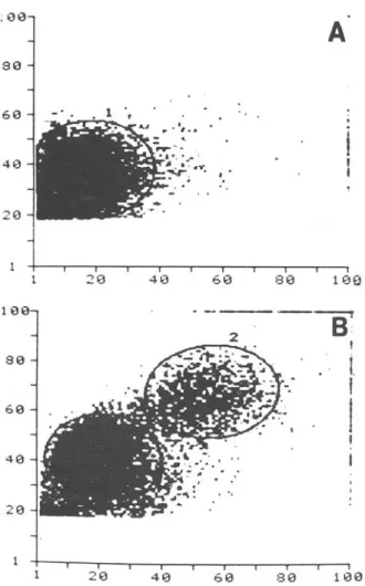
Figure 1. Laser lightscatter cytogram of 30,000 periph-eral blood mononuclear cells: (A)cells originating from a noninfected control and(B) cells from a subject with acutesymptomatic toxoplasmosis. Forward narrow-angle scatter is shown on the ordinate, with 900 scatter onthe abscissa. Analysiswith monoclonal antibodies of regions 1 and 2 showed that they consistedpredominantlyof lym-phocytes and monocytes.
the buffy coat of a freshly donated unit of blood (500 ml with acid citrate-dextrose anticoagulant) were stimulated with 50 /-Ig of phytohemagglutinin (PHA; GIBCO, Grand Island, NY)/mland 2 x 10⁵ 24-hr-old allogeneic PHA-induced PBL blasts/mlin Dulbecco's and alpha medium(l:1;GIBCO) and 2010 human type AB serum. After 36 hr the supernatants were harvested, sterile filtered, and tested on PHA-induced blasts for promotion of T cell growth and DNA synthesis.
Generation of toxoplasma-specific T cell lines.
Soluble antigen was prepared from the RH strain of
T.gondii as previously described [10]. All five
sub-jects studied were pretested in an eight-day lympho-cyte transformation assay with the antigen to deter-mine the optimal dose for maximal proliferation. In general, this dose was lO-l00/-ig/mI. PBLs, 2 x 10⁶/2
ml, from each subject were incubated for eight days with two doses of antigen (10 and 100 /-Ig/ml) in 24
-well culture plates (Costar, Cambridge, Mass). Af-ter eight days the cellswere harvested, pooled,and resuspended in fresh conditioned medium. The cells were expanded in culture plates (Costar) by renewal of the medium every three days. After two weeks in culture the cells were tested for toxoplasma anti-gen-specific proliferation. Flat-bottom 96-well plates (Micro Test II®; Falcon, Oxnard, Calif) were in-cubated with 1.5 x lOSirradiated (3,000 rad) autol-ogous PBLs in medium with 10010 type AB serum for I hr.The nonadherent cells were harvested, the wells were washed with PBS, and the recovered cells were counted. By this method the number of adher-ent cells (ACs) was calculated and found to be 20%-30010 of the original number of plated cells. ACs in the wells served as antigen-presenting cells.
The T cell lines (I05 per well in duplicate wells) were cultured for 72 hr in the presence of ACs plus 100 /-Ig of toxoplasma antigen/ml, 10 /-Ig of tetanus tox-oid (Institut Serotherapique et Vaccinal Suisse Berne, Bern, Switzerland)/ml, or ACs alone. For the last 8 hr of culture the cells were pulsed with 0.25 /-ICi of [125I]deoxyuridine per well and harvested (Titertek'" cell harvester; Flow Laboratories, Irvine, Ayrshire, Scotland).The number of cpm was deter-mined in a y counter (Packard Instrument Co., Downers Grove, Ill). Only when toxoplasma anti-gen-specificproliferation was documented was the distribution of the T cell markers T4,T8, and TQl of a line studied. The indirect immunoflouroescence method was used. T cells (106
) were reacted with the
monoclonal antibodies for 45 min in ice, washed •
leo
-.
80 810 --- -- ~B
·
Ii.
I
I
I I 613 610 413 20.
e.~~~
. .' ."-
";::.
.
.
.
~.-:e"
..
20 60 40 80 1\:10 :\:113A
so 613 413 -20patients the cells of the monocyte cluster (figure lB, region 2) were analyzed with use of monoclonal an-tibody Leu M3 (monocyte marker) and FITC-con-jugated goat antibody to mouse immunoglobulin (Becton-Dickinson).
Toxoplasma-specific T cell lines. Media.
Dulbec-co's modified Eagle's medium with addition of an-tibiotics and amino acids was used [8]. Pooled, heat-inactivated human type AB serum was added in a 10% concentration. T cell lines were cultured and expanded with use of medium conditioned with WOJo
crude human interleukin-2 prepared as described previously [9J. In brief, leukocytes(4x 10⁶/ml)from
318
and reacted with FITC-conjugated goat antibody to mouse immunoglobulin. After washing, the percent-age of fluorescent cells was determined by counting 200 cells with the immunoflouorescence microscope.
Limiting-dilution cultures. Toxoplasma-specific T cell sublines were obtained from one of the pa-tients with recent toxoplasmosis (A2) and one of the individuals with chronic infection (B2) according to previously described techniques [8]. Microtiter plates with irradiated autologous AC feeder layers were used. Toxoplasma antigen (50 JAg/ml) and medium conditioned with 5070 interleukin-2 were used in limiting-dilution cultures of 100, 10, and 1 cells per well. Only the dilution containing 10 cells per well showed a favorable cloning efficiency (13070 of wells positive) and allowed selection of sublines of both T4- and T8-enriched cells. The proliferative re-sponses to toxoplasma antigen of the T4 and T8 sub-lines were studied with use of the same protocol de-scribed above.
Production ofy interferon (IFN-y)by Toxoplas-ma-specific T cell lines and sublines. Irradiated ACs, 1
x
lOS, and 5x
105 T cells were incubatedwith 50 JAI of toxoplasma antigen/ml, purified pro-tein derivative (Institut Serotherapique et Vaccinal Suisse Berne), or PHA. After 72 hr the supernatants were harvested, filtered, and frozen. The superna-tants were tested for IFN in a CPE inhibition assay [11] with use of WISH cells (3 x 10⁴ per well). In
brief, WISH cells were incubated for 24 hr with su-pernatants titrated in threefold dilutions and then infected with Mengo virus (800 pfu). The antiviral IFN-y activity was measured as the dilution of the supernatant that prevented 50070 of the CPE. The supernatants were compared with an IFN-y standard obtained from Dr. S. Grossberg (Department of Microbiology, Medical College of Wisconsin, Mil-waukee), and the concentration was recorded in V/ml. The IFN-y in the supernatants was demon-strated to be acid labile by treatment at pH 2.5.
Statistical methods. Thettest statistics were per-formed, and the correlation coefficient was calcu-lated.
Results
Analysis of peripheral blood mononuclear cells according to size. Figure IA is the cytogram of cells from a control, noninfected individual. Figure IB, a cytograrn of cells from a subject with acute symp-tomatic toxoplasmosis, shows two cell populations
Sklenar et al.
markedly different from those of the control in-dividual with regard to laser light scatter character-istics. The region designated by 1 has been identi-fied previously as a lymphocyte cluster, and region 2 has been identified as a monocyte cluster [6]. Six of eight of our symptomatic subjects and only one of six controls showed increased numbers of cells in region 2. To evaluate cells in this cluster, the dis-tribution of cells with the markers Leu M3, T8, and T4 in both regions was studied in three subjects. Their ages were 28, 20, and 33 years, and they had been infected two, five, and six months ago. In these individuals 16.1070 ± 2.3070 cytofluorometrically screened cells were found in region 2, comprising 67070-75070 Leu M3-positive cells (monocytes), 16070-30070 T8-positive cells, and 0070-7070 T4-positive cells, depending on fluorescence intensity gating.
Region 1 (the lymphocyte cluster) was studied in two subjects with symptomatic toxoplasmosis (23 and 31 years of age, infected two months ago) and two noninfected controls (23 years of age) with use ofOKT 3,4, and 8 and antibody to HLA-DR (table 1). The number of total T cells (T3 positive) in the lymphocyte cluster ranged from 60070 to 76070. In pa-tients with symptomatic toxoplasmosis, the percent-age of T4-positive (helper marker) cells was slightly lower, and the percentage of T8-positive (cytotoxic suppressor marker) cells was slightly higher in the lymphocytes clustered in region 1, similar to the al-terations seen in region 2, and the absolute numbers of T8-positive cells were greater (see below). The number of HLA-DR-positive (B cells and mono-cytes) cells was not different in patients and controls in region 1. The numbers of cells showing the Leu 7/HNK 1 (NK/K) marker as well as the dual T8 and Leu 7 marker were increased in the patients. This observation was confirmed by additional analysis of three patients (see below).
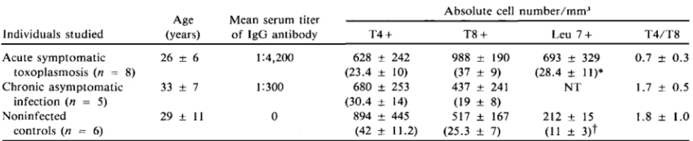
T8-, TQl-, and Leu 7-positive cells in peripheral blood ofpatients. Table 2 shows characteristics of the individuals studied and summarizes the results of peripheral T cell subsets contained in the lympho-cyte cluster analyzed by three monoclonal antibod-ies with use of flow cytofluorometry. The absolute numbers of T8-positive cells/mm" were significantly higher (P< .001) in individuals with recent toxoplas-mosis (one to six months ago) than in chronically infected individuals and in noninfected controls.
The T4/T8 cell ratio was significantly lower in sub-jects with recent toxoplasmosis compared with chronically infected and noninfected individuals(P
Immunity in Acute Symptomatic Toxoplasmosis 319
Table 1. Analysis of the lymphocyte cluster.
-Percentage of fluorescent cells
Individuals studied T3+ T4+ T8+ HLA-DR+ Leu 7+ Leu 7 + /T8 +
-- -Symptomatic toxoplasmosis 1 (8 weeks ago) 68.8 25.1 41.4 5.2 26.3 18.8 2 (10 weeks ago) 64.5 18.4 32.0 3.5 14.8 11.1 No infection 1 60.4 32.9 31.5 4.9 10.7 9.0 2 76.0 63.4 16.5 7.1 7.6 5.4 - - _ . - - .__._- - - _..- - . . - - , - - - .
-NOTE. The cells were analyzed with use of antibodies to lymphocyte surface antigens: T3 (pan T cell marker), T4 (helper cell marker), T8 (cytotoxic/suppressor cell marker), HLA-DR (B cells and monocytes), and Leu 7/HNK 1 (NK/K cell marker). Leu 7/T8-positive cells were detected by dual laser cytofluorometry for simultaneous green and red fluorescence.
<
.001 and P<
.01, respectively). This difference was not due to decreased numbers of T4-positive (helper/inducer) cells, which did not differ signifi-cantly among all three groups of individuals. In ad-dition, the number of cells positive for TQl marker (the marker of certain suppressor cell subsets) [7, 12] did not vary in the three groups. Absolute mean± SD numbers of TQl-positive cells/rum" were 643 ± 331 in recent toxoplasmosis(n = 3), 576 ± 399 in chronic infection(n
=
5), and 560 ± 231 in con-trols (n = 3).The number of Leu 7-positive (NK/K) cells in five patients with acute toxoplasmosis (mean ± SD age, 27 ± 5 years) was significantly increased compared with three noninfected controls (mean ± SD age, 32 ± 15; table 2). This increase was not due to the age of the subjects noted in the normal population [5] because ages in the two groups were comparable. Peripheral blood monocytes in acute
toxoplasmo-sis. The light scatter cytogram suggested increased numbers of monocytes in persons with acute symp-tomatic toxoplasmosis. This proposal was confirmed by analysis of the differential blood counts from all patients studied. Acutely infected patients had 631 ± 203 monocytes/rnm" (9.4070 ± 2.9070), those chronically infected had cell counts of 516 ± 248 (7.2070 ± 2.8070),and controls had cell counts of 402 ± 177(6.2070 ± 1.9070),results indicating significant monocytosis (P
<
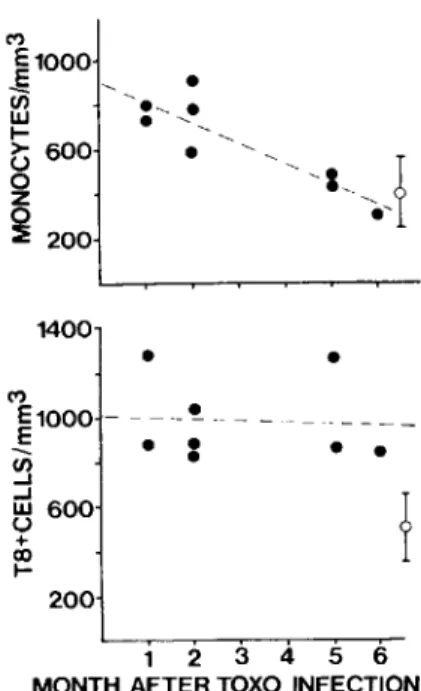
.05) in acutely infected patients. Time course of changes in monocytes and T8-positive cells during acute infection. Monocyte lev-els were increased during the first two months after infection but reached normal levels by five to six months (figure 2). There was a significant inverse correlation between the absolute number of mono-cytes and the time after infection (y= -
91.6x+
906,r
=
-.89, P< .01). In contrast, T8-positive lym-phocyte numbers were increased from the earliestTable 2. Characterization of subjects and their PBL subpopulations.
Acute symptomatic 26 ± 6 1:4,200 toxoplasmosis (n = 8) Chronic asymptomatic 33 ± 7 1:300 infection(n = 5) Noninfected 29 ± 11 0 controls(n = 6) Individuals studied Age (years)
Mean serum titer of IgG antibody
Absolute cell number/rum"
T4+ T8+ Leu 7+ T4/T8 - - - - ' . _ - - - ' - --"
- _
..__
. _ -628 ± 242 988 ± 190 693 ± 329 0.7 ± 0.3 (23.4 ± 10) (37 ± 9) (28.4 ± 11)* 680 ± 253 437 ± 241 NT 1.7 ± 0.5 (30.4 ± 14) (19 ± 8) 894 ± 445 517 ± 167 212 ± 15 1.8 ± 1.0 (42 ± 11.2)_. (25.3 ± 7) (11 ± 3)t ._ . _ - . _-NOTE. Data are mean ± 3D values (percentages as indicated). Subjects with acute symptomatic toxoplasmosis (AS) had been infected three months (range, one to six months) previously, whereas those with chronic asymptomatic infection (CA) had been infected many years previously. P values for comparisons were obtained with use of t test statistics: for T4 + , not significant for AS vs. CA and AS vs. noninfected individuals (N); for T8 +, P<.001 for AS vs. CA and AS vs. N; for Leu 7 +, P<.05 for AS vs. N; and for T4/T8, P <.001 for AS vs. CA andP<.01 for AS vs. N. Not tested = NT.
*n = 5.
320 Sklenar et al.
1 2 3 4 5 6
MONTH AFTER TOXO INFECTION
Figure 2. Alterations in numbers of the circulating monocytes and T8-positive lymphocytes in eight subjects with acute symptomatic toxoplasmosis in relation to time (months) after infection. Absolute numbers of cells/rum-of blood are shown. The mean ± SD (bars) values from six noninfected individuals are also shown(0). Chroni-cally infected patients showed the same values for counts of monocytes (516 ± 248 cells/mm") and T8-positive cells (437 ± 241 cells/rnm'). Regression analysis (by the least squares method) is depicted by the dotted lines. A sig-nificant inverse correlation was found for monocytes (r = - .89, P< .01) but not for T8-positive cells. period after infection up to six months after infec-tion and did not show a significant decline during the study period.
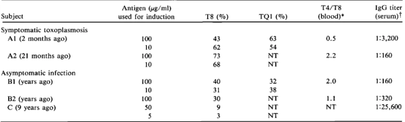
T8- and TQ1-positive subsets in Toxoplasma-specific T eel/lines. T cell lines induced from PBLs with use of soluble toxoplasma antigen that prolifer-ated specifically on restimulation with antigen and syngeneic ACs were studied with use of monoclonal antibodies and indirect immunofluorescence. T cell lines obtained from two recently infected and clini-cally ill individuals (AI, with illness two months ago, and A2, with illness 21 months ago) contained higher numbers of T8-positive (suppressor/cytotoxic) and TQI-positive (suppressor subsets) cells than lines from chronically infected individuals BI and B2, with no history of lymphadenopathy and fever (table 3). Subject AI, studied for the first time two months after onset of symptoms, had a T4/T8 cell ratio in peripheral blood of 0.5, and the T cell lines showed
increased proliferation of antigen-specific T8-posi-tive cells (62%). At five months after infection, the T4/T8 cell ratio was still low (0.6). SubjectA2, stud-ied 21 months after infection with Toxoplasma, which caused an illness of four months in duration, had a normal ratio of peripheral T4/T8 cells (2.2), but T cell lines still showed predominantly Toxoplas-ma-specific T8-positive cells (700,70) when induced in vitro by antigen. This observation was in contrast to the behavior of T cell lines obtained from the chronically infected individuals BI and B2, who had normal peripheral T cell subset ratios (l.l and 2.0) and whose lines showed lower numbers of T8-positive cells (300,70-400,70).
In addition, one individual with Hodgkin's dis-ease was studied (subject C). This subject demon-strated very low numbers ofToxoplasma-induced T8-positive cells in T cell lines and very high serum titers of IgG antibody to Toxoplasma. This pattern of re-sponse is unusual and distinct from that of subjects with recent symptomatic toxoplasmosis, who had no other known underlying disease, and that of chron-ically infected but otherwise healthy individuals.
Proliferation and IFN-y production by Toxoplas-ma-specific T eel/lines. The proliferative responses of the T cell lines obtained from subjects with re-cent toxoplasmosis and latently infected individu-als were antigen specific. Only toxoplasma antigen and ACs caused proliferation, whereas incubation of the lines with ACs alone (table 4) or with ACs and tetanus toxoid (data not shown) yielded no stim-ulation. Unseparated PBLs from three of five indi-viduals showed strong proliferation in response to tetanus toxoid (data not shown).
With use of limiting dilution, Toxoplasma-specific sublines highly enriched for one marker (either T4 or T8) were obtained (table 4). In one subject with recent toxoplasmosis (A2), SLI, depleted of T8-positive cells, showed significantly higher prolifera-tion than the parent line, which contained73070 T8-positive cells, and SL2, which had99070 T8-positive cells. Similarly, in one chronically infected individual (B2), T8-depleted SLI (00,70 T8-positive cells) prolifer-ated more than the parent line, with30070T'S-positive cells, and SL2, with only 70,70 T4-positive cells.
Two lines obtained from subject C, which con-tained very low numbers of T8 cells (9070 and 3%)l showed high 51 values (38- and 33-fold). These data suggest that an inverse correlation exists betweenthe
magnitude of proliferation of toxoplasma
anti-•
•
•
•
•
200 M • E1000 -E • •en
...J•
...J ~ 600 + co I-1400 M ~1000en
UJl-t>
600o
z~
200Immunity in Acute Symptomatic Toxoplasmosis
Table 3. P~~_~ent~~es of T8- and TQ I-positive __~ells in Toxoplasma-specific T cell lines. Antigen(j.Lg/ml)
Subject used for induction T8 (070) TQI (070)
- - - -- - - - " ..__ ._--Symptomatic toxoplasmosis A I (2 months ago) 100 43 63 10 62 54 A2 (21 months ago) 100 73 NT 10 68 NT Asymptomatic infection B1 (years ago) 100 40 32 10 31 38 B2 (years ago) 100 30 NT C (9 years ago) 50 9 NT 5 3 NT ---._---_._--_._-T4/T8 (blood)* 0.5 2.2 2.0 1.1 NT 321 IgG titer (serum)t 1:3,200 1:160 1: 160 1:320 1:25,600
NOTE. T cell lines induced with toxoplasma antigen (5-100 lig/ml) and demonstrated to proliferate specifically with the antigen were tested with monoclonal antibodies (to T8, a cytotoxic/suppressor cell marker, and to TQl, a marker of suppressor cell subsets) with use of indirect immunofluorescence.
* T4/T8 (helper/suppressor) cell ratios were measured in peripheral blood at the beginning of the T cell line induction. t The titer of IgG antibody to Toxoplasma was measured with use of indirect immunofluorescence.
gen-specific T cell lines and the number of T8-positive cells that they contain.
Table 4 shows the production of IFN-y by T cell sublines enriched for one cell type. The two sublines obtained from subject A2, with a history of prolonged disease syndrome that started 21 months ago, produced low levels of IFN-y (10 and 80 U/ml)
after antigen stimulation. None of the six T cell sub-lines obtained from an asymptomatic individual (subject B2) showed such low levels. Stimulation with the mitogen PHA of lines that produced low levels of IFN-y in response to antigen induced high levels of IFN-y. T4 cell sublines (A2 SLI and B2 SLl), one non-T4 subline (B2 SL2), and all uncharacterized
Table 4. Proliferation and IFN-y production by Toxoplasma-specific T cell lines and sublines.
Proliferation IFN-y
Subject, line or (cpm) with T8-positive cells (Vlml)after
subline (antigen dose)* ACs plus antigen ACs alone Percentage SI Antigen PHA
-A2 P (l00) 16,663 ± 1,534 3,940 ± 492 73 10 20 NT SLI (50) 71,335 ± 5,515 83 ± 32 1 859 80 500 SL2 (50) 4,631 ± 762 504 ± 106 99 9 10 700 H2 P (l00) 7,811 ± 2,107 932 ± 315 30 8 NT NT SLl (50) 10,614 ± 17 173 ± 49 0 61 700 NT SL2 (50) 5,390 ± 565 1,219 ± 98 t 4 1,400 NT SL3 (50) 15,886 ± 4,815 1,187 ± 146 NT 13 240 NT SL4 (50) 4,399 ± 157 77 ± 25 NT 57 500 NT SL5 (50) 3,220 ± 591 82 ± 7 NT 34 150 NT SL6 (50) 2,480 ± 319 330 ± 67 NT 8 500 NT C PI (50) 12,162 ± 1,499 319 ± 235 9 38 150 NT P2 (5) 16,425 ± 440 494 ± 63 3 33 1,400 NT
NOTE. Subject A2 was a patient with symptomatic toxoplasmosis, subject B2 was an asymptomatic patient with chronic tox-oplasma infection, and subject C was an asymptomatic patient with Hodgkin's disease in remission. Data are mean ± SD values as indicated. P = parent line; SL = subline, enriched by limiting dilution for one cell type; and SI = stimulation index.
* Toxoplasma antigen in lig/ml used to induce the line.
322
sublines from an asymptomatic patient (B2 SL3-6) produced IFN-y after stimulation with toxoplasma antigen. Stimulation of the sublines with purified protein derivative did not cause production of IFN-y (data not shown). These antigen-specific sublines may reflect true production of IFN-y because they do not contain significant numbers of undefined cells that could consume lymphokine [13]. Nonenriched T cell lines (i.e., mixed T cell lines) from all individ-uals studied that were induced with a high dose of toxoplasma antigen (50-100 ug/ml) demonstrated low production of IFN-y, whereas lines induced with lower antigen doses (5-10 ug/ml) produced higher amounts. This variability is very likely a result of conflicting influences on production and consump-tion of IFN-y by undefined cells. The line from sub-ject C that contained only 3070 T8-positive cells pro-duced the highest amounts of IFN-y, 1,400 V/ml (table 4), of all nonenriched lines studied.
Discussion
Observations reported here indicate that marked al-terations in antigen-specific T cell populations and in monocyte numbers occur during the first months after symptomatic infection with Toxoplasma. The alterations include high numbers of total cells posi-tive for T8, Leu 7, and both T8 and Leu 7, decreased T4/T8 cell ratios, high numbers of monocytes in pe-ripheral blood, and a high percentage of T8- and TQl-positive cells in toxoplasma antigen-induced T cell lines. The abnormalities in total peripheral blood T cell subsets were seen as late as six months, but not 20 months, after symptomatic illness, whereas high numbers of T8-positive cells in antigen-inducer T cell lines were seen as late as 21 months after fection in one individual. In contrast, chronically in-fected subjects had normal numbers of T8-positive cells, normal T4/T8 cell ratios, and fewer T8-positive cells in response to toxoplasma antigen in T cell lines. Two reports [14, 15] have shown similar changes in peripheral blood T cell subsets during toxoplas-mosis. De Wae1e et al. [14] studied one recently in-fected patient with toxoplasmosis, Luft et al. [15] studied six patients, and we now add eight more pa-tients, all of whom showed an increase in number of T8-positive cells in peripheral blood. In the pres-ent study asymptomatic patipres-ents infected chronically with Toxoplasma were also evaluated, and they showed peripheral blood T cell populations similar to those of control subjects. Luft et al. [15] studied
Sklenar et at.
four patients who were asymptomatic although re-cently infected. These individuals also showed nor-mal numbers of T8-positive cells, which distin-guished them from the symptomatic patients studied by Luft et al. [15] and by us. This single observation raises the possibility that an increase in number of T8-positive cells is a correlate of the disease state and not simply indicative of recent infection. Two of our patients provide some support for a relation between symptomatic infection and PBL abnormalities.
The illness, lymphatic toxoplasmosis, may be con-sidered an immunologic disease initiated in some in-dividuals by the infectious process. T.gondiiis simi-lar to the two viruses known to cause mononucleosis, Epstein-Barr virus and cytomegalovirus, in that both lymphadenopathy and increased numbers of T8-positive cells in the peripheral blood have been recorded [16, 17]. Similar to the cases reported here, the abnormalities in lymphocyte subsets caused by cytomegalovirus have persisted up to 10 months af-ter infection [17]. Prolonged abnormalities in lym-phocyte subsets have recently been observed during acute infections with retrovirus [18].Ithas not been possible to elucidate the reasons for the changes in T cell subsets during these infections.
This is the first study of the populations of cells seen in response to toxoplasma antigen in individu-als with recent, symptomatic, or chronic asymptom-atic infections. The increased antigen-specific selec-tion of T8-positive cells suggests that the altered ratios of total T cells are due to antigen-specific re-sponse patterns. Large numbers of T8-positive cells were generated in subjects with recent toxoplasmo-sis, unrelated to the antigen dose used. In vitro proliferation was toxoplasma antigen specific, and it tended to be highest in cell cultures with a dominance of T4-positive cells. A correlation of pre-dominance of T4-positive cells after induction with toxoplasma antigen, enhanced in vitro proliferation in response to antigen, and high serum titers of an-tibody was demonstrated in one individual (subject C) with a history of Hodgkin's disease and infec-tion withToxoplasma. The data, based on a few sub-jects, suggest the possibility that the T8-positive cells observed to be increased during toxoplasmosis may modulate the proliferation of T4-positive cells to toxoplasma antigen in vitro and the magnitude of the humoral response to Toxoplasma in vivo. Al-though antigen-specific T8-positive suppressor cells have been described [19],the mechanisms of the sup-pression are unknown [20-23]. The finding of
in-Immunity in Acute Symptomatic Toxoplasmosis
creased numbers of TQl-positive cells (a marker de-scribed on suppressor T cell subsets) [7, 12]in T cell lines induced by toxoplasma antigen from a symp-tomatic subject supports the view that T cells with a negative immunoregulatory role are induced by this antigen.
Antigen-induced T8-positive cells may also occur in individuals infected with Epstein-Barr virus, cytomegalovirus, and retrovirus, although studies of these viral infections similar to those described here have not been done. Antigen-specific immunity to herpes simplex virus type 2 has been studied in indi-viduals in various phases of infection [24].
The mechanism responsible for selection of T8-positive cells is unknown. Itdoes not appear to be dependent on antigen dose because both 10 and 100 ug/ml induced large numbers of T8-positive cells in subjects with recent toxoplasmosis. Antigen fraction-ation must be done to determine whether some toxo-plasma antigens are more likely than others to in-duce this response in vitro. Our data suggest that even after total T cell ratios are normal, antigen-specific induction still favors T8- over T4-positive cells. Long after infection, individuals appear to have the net-work for protective immunity with rapidly prolifer-ating, functioning T4-positive cells.
Enhancement of NK cell activity has been de-scribed in the mouse model [25] and in human lym-phocyte cultures [26] in response to toxoplasma antigens. In humans with acute symptomatic toxo-plasmosis, we found increased numbers of circulat-ing cells carrycirculat-ing the marker of NK and K cells (Leu 7/HNK 1). Such cells could be associated with kill-ing of parasite-infected cells via antibody-dependent cell-mediated cytotoxicity [27], or they may contrib-ute additional down regulation of the immune re-sponse because Leu 7-positive suppressor cells have been described [28]. In addition, two subjects with acute toxoplasmosis had increased numbers of cells carrying a dual marker of cytotoxic cells (T8) and of NK cells (Leu 7). Leu 7-positive cells carrying simultaneously the T8 cell marker have been de-scribed as immature bone marrow NK cells [29].
The Toxoplasma-infected subjects in this study had, in addition to the changes in lymphocyte sub-sets, increased numbers of monocytes in peripheral blood up to the second month after infection. This finding is of interest because toxoplasmosis in ani-mal models has been found to be associated with increased numbers of monocytes and macrophages at various sites[1,30, 31] (T.e.J., S.A., and P.E.,
un-323
published observation) and in degree of activation [30]. Because T cell regulation of the myelopoiesis by production of monocyte colony-stimulating fac-tors after specific antigenic stimulation of cloned T cells has been recently demonstrated [32], it is pos-sible that the increased numbers of monocytes in our symptomatic subjects were due to the described ex-pansion of T cell subsets.
We found that cloned T cells of both the T 4 and the T8 phenotypes from one symptomatic individual produced less IFN-y after stimulation with toxo-plasma antigen compared with T cells from an asymp-tomatic individual. This finding may represent (besides increased numbers of T8-positive cells in re-sponse to toxoplasma antigen in vitro) another marker that identifies individuals prone to manifest disease. This suggestion is supported by a study that showed that in the course of acute symptomatic toxo-plasmosis, one patient's cells' ability to inhibit Toxo-plasma was initially impaired [4].
Further studies are needed to determine whether these T8-positive cells are functioning as specific sup-pressor cells of cell-mediated processes such as T cell proliferation, as recently suggested by Luft et al. [33], monocyte colony stimulation [32], or specific anti-body suppression [19], or whether these cells are ex-panded cytotoxic cells competing for lymphokines. All the changes shown are indicative of immuno-logic imbalance during recent symptomatic toxoplas-mosis. The infectious agents studied so far that cause an imbalance of immunoregulatory lymphocyte sub-sets (Epstein-Barr virus, cytomegalovirus, herpes simplex virus, retroviruses, and T.gondiithave one feature in common, namely, they cause a latent, usu-ally life-long infection. These infectious agents ap-pear to be capable of inducing an antigen-specific imbalance of the immunoregulatory network lead-ing to a postinfection immune dysfunction syndrome of varying duration. In toxoplasmosis this syndrome is characterized by signs and symptoms (including lymphadenopathy, fever, and malaise) and by in-creased numbers of circulating T8-positive/Leu 7-positive cells and monocytes and by impaired production of lymphokines (IFN-y) by antigen-specific T cells. The factors that contribute to emer-gence of this syndrome in one of five
Toxoplasma-infected individuals remain undefined. References
1. Jones TC. Immunopathology of toxoplasmosis. Springer Se-min Immunopathol 1980;2:387-97
324
2. Ruskin J, Remington JS. Toxoplasmosis in the compromised host. Ann Intern Med 1976;84:193-9
3. Anderson SE Jr, Krahenbuhl JH, Remington JS. Longitudi-nal studies of lymphocyte response to toxoplasma anti-gen in humans infected with T.gondii.J Clin Lab Immunol 1979;2:293-7
4. Johnson WD Jr. Chronological development of cellular im-munity in human toxoplasmosis. Infect Immun 1981; 33:948-9
5. Abo T, Cooper MD, Balch CM. Postnatal expansion of the natural killer and killer cell population in humans identi-fied by the monoclonal HNK-l antibody. J Exp Med 1982;155:321-6
6. Hoffman RA, Kung PC, Hansen WP, Goldstein G. Simple and rapid measurement of human T lymphocytes and their subclasses in peripheral blood. Proc Natl Acad Sci USA 1980;77:4914-7
7. Reinherz EL, Morimoto C, Fitzgeral KA, Hussey RE, Daley JF, Schlossman SF. Heterogeneity of human T4+ inducer T cells defined by a monoclonal antibody that delineates two functional subpopulations. J ImmunoI1982;128:463-8 8. Schreier MH, Tees R. Long term culture and cloning of spe-cific helper T cells. In: Lefkovitz I, Pernis B, eds. Immuno-logical methods. Vol. 2. New York: Academic Press, 1981:263-75
9. Pauly JL, Russell CW, Planinsek JA, Minowada J. Studies of cultured human T lymphocytes. I.Production of the T cell growth-promoting lymphokine interleukin-2. J Im-munol Methods 1982;50:173-86
10. Jones TC, Len L, Hirsch JG. Assessment in vitro of immu-nity against Toxoplasma gondii. J Exp Med 1975;141: 466-82
11. Armstrong JA. Semi-micro, dye-binding assay for rabbit in-terferon. Applied Microbiology 1971;21:723-5
12. Morimoto C, Distaso JA, CheneyJJ,Reinherz EL, Schloss-man SF. Cellular interaction between subsets of T8 popu-lation for maximal suppression of antigen-specific anti-body response. Cell Immunol 1984;88:75-84
13. Lotzova E, Savary CA. Stimulation of NK cell cytotoxic poten-tial of normal donors by two species of recombinant al-pha interferon. J Interferon Res 1984;4:201-13 14. De Waele M, Thielemans C, Van Camp B.
Immunoregula-tory T cells in mononucleosis and toxoplasmosis [letter]. N Engl J Med 1981;305:228
15. Luft BJ, Kansas G, Engelman EG, Remington JS. Functional and quantitative alterations in T lymphocyte subpopula-tions in acute toxoplasmosis. J Infect Dis 1984;150:761-7 16. De Waele M, Thielemans C, Van Camp BKG. Characteriza-tion of immunoregulatory T cells in EBV-induced infec-tious mononucleosis by monoclonal antibodies. N Engl J Med 1981;304:460-2
17. Carney WP, Rubin RH, Hoffman RA, Hansen WP, Healey K, Hirsch MS. Analysis of T lymphocyte subsets in cytomegalovirus mononucleosis. J Immunol 1981;126: 2114-6
18. Cooper DA, Maclean P, Finlayson R, Michelmore HM, Gold J, Donovan B, Barnes TG, Brooke P, Penny R. Acute AIDS retrovirus infection. Definition of a clinical illness as-sociated with seroconversion. Lancet 1985;1:537-40
Sklenar et al.
19. Morimoto C, Reinherz EL, Todd RF, Distaso JA, Schloss-man SF. Generation of antigen-specific suppressor cells in vitro in man. J Immunol 1983;131:1209-13
20. Reinherz EL, Hussey RE, Fitzgerald K, Snow P, Terhorst C, Schlossman SF. Antibody directed at surface structure in-hibits cytolytic but not suppressor function of human T lymphocytes. Nature 1981;294:168-70
21. Moretta A, Pantaleo G, Moretta L, Mingari MC, Cerottini J-e. Quantitative assessment of the pool size and subset distribution of cytolytic T lymphocytes within human rest-ing or alloactivated peripheral blood T cell populations. J Exp Med 1983;158:571-85
22. Moretta A, Pantaleo G, Maggi E, Mingari Me. Recent ad-vances in the phenotypic and functional analysis of hu-man T lymphocytes. Semin Hematol 1984;21:257-69 23. Gunther J, Haas W, von Boehmer H. Suppression of T cell
responses through competition for T cell growth factor (in-terleukin 2). Eur J Immunol 1982;12:247-9
24. Sheridan JF, Donnenberg AD, Aurelian L, Elpern OJ. Im-munity to herpes simplex virus type 2. IV. Impaired lyrn-phokine production during recrudescence correlates with an imbalance in T lymphocyte subsets. J Immunol 1982; 129:326-31
25. Hauser WE Jr, Sharma SO, Remington lS. Augmentation of NK cell activity by soluble and particulate fractions of Toxoplasma gondii. J Immunol 1983;131:458-63 26. Sharma SO, Verhoef J, Remington JS. Enhancement of
hu-man natural killer cell activity by subcellular components ofToxoplasma gondii. Cell Immunol 1984;86:317-26 27. Herberman RB. Natural killer (NK) cells and their possible
roles in resistance against disease. Clin Immunol Rev 1981;1:1
28. Tilden AB, Abo T, Balch CM. Suppressor cell function of human granular lymphocytes identified by the HNK-l (Leu 7) monoclonal antibody. J Immunol 1983;130:1171-5 29. Abo T, Cooper MD, Balch CM. Characterization of
HNK-1+(Leu-7) human lymphocytes.I.Two distinct phenotypes of human NK cells with different cytotoxic capability. J Immunol 1982;129:1752-7
30. Krahenbuhl JL, Remington JS. The immunology of Toxo-plasma and toxoplasmosis. In: Cohen S, Warren KS, eds. Immunology of parasitic infections. 2nd ed. Oxford: Black-well, 1982:356-421
31. McLeod R, Estes RG, Mack DG, Cohen H. Immune response of mice to ingestedToxoplasma gondii: a model of toxo-plasma infection acquired by ingestion. 1 Infect Dis 1984;149:234-44
32. Griffin10,Meuer SC, Schlossman SF, Reinherz EL. T cell regulation of myelopoiesis: analysis at a clonal level. J Im-munol 1984;133:1863-8
33. Luft BJ, Remington JS. Functional abnormalities of lym-phocyte subsets in patients with acute toxoplasma infec-tion [abstract 621]. In: Program and abstracts of the 24th Interscience Conference on Antimicrobial Agents and Che-motherapy. Washington, DC: American Society for Microbiology, 1984