Lack of IL-6 augments in¯ammatory response but
decreases vascular permeability in bacterial
meningitis
Robert Paul,
1Uwe Koedel,
1Frank Winkler,
1Bernd C. Kieseier,
2Adriano Fontana,
3Manfred Kopf,
4H.-P. Hartung
2and H.-W. P®ster
11Department of Neurology, Ludwig-Maximilians-University,
Klinikum Groûhadern,2Department of Neurology,
Heinrich-Heine UniversitaÈt, Dusseldorf, Germany,3Section
of Clinical Immunology, Department of Internal Medicine, University Hospital Zurich and 4Basel Institute for
Immunology, Basel, Switzerland
Correspondence to: Hans-Walter P®ster, MD, Department of Neurology, Klinikum Groûhadern, Ludwig-Maximilians-University of Munich, Marchioninistrasse 15, 81377 Munich, Germany
E-mail: p®[email protected]
Summary
Interleukin (IL)-6 is a multifunctional cytokine with diverse actions and has been implicated in the patho-physiology of many neurological and in¯ammatory disorders. In this study, we investigated the role of IL-6 in pneumococcal meningitis. Cerebral infection in wild-type (WT) mice caused an increase in vascular permeability and intracranial pressure (ICP), which were signi®cantly reduced in IL-6±/± mice. In contrast,
meningitis in IL-6±/± mice was associated with a
signi®-cant increase in CSF white blood cell count compared with infected WT mice, indicating an enhanced in¯am-matory response. Analysis of mRNA expression in the brain showed an increase in tumour necrosis factor (TNF)-a, IL-1b, and macrophage in¯ammatory protein 2 (MIP-2) levels, but decreased expression of granulocyte±macrophage colony-stimulating factor in
infected IL-6±/± mice compared with infected WT
con-trols. Similar results were obtained when rats challenged with pneumococci were systemically treated with neutralizing anti-IL-6 antibodies, resulting in an increased pleocytosis but at the same time a reduction of vascular permeability, brain oedema formation, and ICP, which was not accompanied by a downregulation of matrix metalloproteinases. Our data indicate that IL-6 plays an important anti-in¯ammatory role in bac-terial meningitis by reducing leukocyte in®ltration but contributes to the rise in intracranial pressure by increasing blood±brain barrier (BBB) permeability. These ®ndings suggest that the migration of leukocytes across the BBB and the increase in vascular permeabil-ity are two independent processes during bacterial meningitis.
Keywords: chemokines; interleukin-6; meningitis; vascular permeability
Abbreviations: BBB = blood±brain barrier; EB = Evan's blue; GAPDH = glyceraldehyde-3-phosphate dehydrogenase; GM-CSF = granulocyte±macrophage colony-stimulating factor; i.c. = intracisternally; ICP = intracranial pressure; IL-1b = interleukin-1b; i.p. = intraperitoneally; MIP-2 = macrophage in¯ammatory protein 2; MMP = matrix metalloproteinase; PBS = phosphate-buffered saline; PMN = polymorphonuclear neutrophils; TNF-a = tumour necrosis factor a; WBC = white blood cell; WT = wild type
Introduction
Bacterial meningitis is associated with a high morbidity and mortality (Quagliarello and Scheld, 1992). The reasons for the unfavourable clinical outcome include cerebrovascular alterations, breakdown of the blood±brain barrier (BBB), formation of vasogenic brain oedema and increase in intracranial pressure (ICP) (P®ster et al., 1992). Several mediators of the pathophysiological pathway contributing to
these intracranial complications have been identi®ed, includ-ing nitric oxide (Koedel et al., 1995), reactive oxygen species (Koedel and P®ster, 1999) and matrix metalloproteinases (MMPs) (Paul et al., 1998). Moreover, experimental and clinical studies suggest that cytokines including tumour necrosis factor (TNF)-a (Nadal et al., 1989) and interleukin (IL)-1b (Quagliarello et al., 1991), and chemokines such as Brain 126 ã Guarantors of Brain 2003; all rights reserved
macrophage in¯ammatory protein 2 (MIP-2) (Diab et al., 1999), also play an important role in the pathophysiology of bacterial meningitis.
IL-6 is a multifunctional cytokine with diverse actions, e.g. regulation of in¯ammation including the induction of the acute phase reaction, immune response and cellular differ-entiation (Gadient and Otten, 1997). Increased cerebral expression of IL-6 has been demonstrated in many CNS diseases, such as HIV encephalopathy, multiple sclerosis and Alzheimer's disease (Gadient and Otten, 1997). High levels of IL-6 were also detected in the CSF of patients with viral and bacterial meningitis (Houssiau et al., 1988; Matsuzono et al., 1995). In a previous study using a rat model of pneumococcal meningitis we found elevated concentrations of IL-6 in the CSF 6 h after challenge (Koedel et al., 1996). However, the precise role of IL-6 in the pathophysiology of in¯ammatory and CNS diseases has not been elucidated. On one hand, IL-6 was reported to have bene®cial effects by reducing neuronal damage in cerebral ischaemia (Loddick et al., 1998), preventing cartilage destruction during experi-mental arthritis (van de Loo et al., 1997), and acting as an anti-in¯ammatory cytokine in endotoxemia (Xing et al., 1998). On the other hand, transgenic mice lacking the IL-6 gene were resistant to the pathophysiological alterations in experimental autoimmune encephalomyelitis (Eugster et al., 1998), and mice overexpressing IL-6 in astrocytes showed neuropathological abnormalities with a breakdown of the BBB (Brett et al., 1995), suggesting a detrimental role of IL-6 in neurological diseases associated with increased BBB permeability.
The aim of this study was to investigate the impact of IL-6 on the pathophysiology of pneumococcal meningitis using IL-6±/±mice as well as neutralizing anti-IL-6 antibodies in a
rat model. We demonstrate that during bacterial meningitis IL-6 acts as an anti-in¯ammatory cytokine by suppressing the transmigration of leukocytes into the CSF space, but plays a major role in the increase of vascular permeability, causing brain oedema and increase in ICP, indicating that these are two independent processes.
Material and methods
Mouse model of pneumococcal meningitis
All of the experiments were approved by the government of Upper Bavaria. Sixteen male and female mice were used in this study. C57BL/6 mice were purchased from Charles River (Sulzfeld, Germany). The IL-6±/± mice, backcrossed into
C57BL/6 mice, were a kind gift from Hoffmann-La Roche Ltd (Basel, Switzerland). For induction of bacterial menin-gitis the mice were short-term anesthetized with halothan (Hoechst AG, Frankfurt/M, Germany) and 15 ml 107colony
forming units/ml of Streptococcus pneumoniae strain type 3 were injected transcutaneously into the cisterna magna. Control mice received 15 ml of phosphate-buffered saline (PBS) intracisternally (i.c.), instead of pneumococci. The
mice were housed individually in cages, allowed to wake up naturally, and fed with a standard diet and water ad libitum. After 24 h the mice were anesthetized intraperitoneally (i.p.) with 100 mg/kg chloralhydrate, and the left femoral vein was cannulated for ¯uid substitution and the administration of Evan's Blue (EB) dye. The body temperature of the mice was kept constant at 37.5 6 0.5°C using a rectal thermometer-controlled heating pad. A burr hole was made at the occipital bone and a catheter was inserted into the cisterna magna for ICP monitoring and collection of CSF samples to determine CSF white blood cell (WBC) count at the end of the experiment.
Determination of BBB permeability in the
mouse model
BBB permeability was determined as described previously by Gijbels et al. (1994). Mice were injected intravenously with 0.1 ml of 1% (w/v) EB. After 1 h the brains were perfused with 10 ml of ice-cold PBS and removed. The brains were weighed and tissue samples were extracted for 3 days in formamide (5 ml/mg tissue) and the extract centrifuged for 5 min at 500 g. EB concentration in the supernantants was determined by measuring the absorbance at 650 nm. The BBB permeability index was calculated by dividing the value for each sample by the mean value of the control animals.
Mouse intracerebral injection and determination
of BBB disruption
Determination of the effect of IL-6 on cerebral vascular permeability was performed as previously described (Paul et al., 2001). Recombinant human IL-6 (Biosource, Cammarillo, CA, USA) was injected stereotactically into the left frontal lobe (1.5 mm left and 0.5 mm rostral from bregma, 2.5 mm in depth from the dura) of C57BL/6 mice. PBS was injected into the right hemisphere as a control. The animals received 100 ml of a 2% EB solution intravenously 15 min after IL-6 injection. After additional 45 min, the brains were perfused with PBS and removed. Whole-mount direct EB ¯uorescence was observed using confocal micro-scopy of fresh in®xed brains.
Detection of mouse mRNA levels by RT-PCR
Frozen brains from IL-6±/±(n = 3) and wild-type (WT) mice
(n = 3) were cut with a cryostat. Total RNA was prepared from frozen sections containing lateral ventricules and hippocampal tissue with Trizol-LS-reagent (Gibco-BRL, Gaithersburg, MD, USA). Oligo(dt)-primed cDNA was prepared from 5 mg total RNA using Superscript II reverse transcriptase (Gibco-BRL) as recommended by the manu-facturer. Speci®c primers were designed for granulocyte± macrophage colony-stimulating factor (GM-CSF) (sense 5¢-TTC CTG GGC ATT GTG GTC T-3¢; antisense 5¢-TGG ATT
CAG AGC TGG CCT GG-3¢), MIP-2 (sense 5¢-AGT TTG CCT TGA CCC TGA AGC C-3¢; antisense 5¢-TGG GTG GGA TGT AGC TAG TTC C-3¢), b-actin (sense 5¢-GGA CTC CTA TGT GGG TGA CGA GG-3¢; antisense 5¢-GGG AGA GCA ATA GCC CTC GTA AGA T-3¢) and glyceraldehyde-3-phosphate dehydrogenase (GAPDH) (sense 5¢-CAT CAC CAT CTT CCA GGA GCG-3¢; antisense 5¢-GAG GGG CCA TCC ACA GTC TTC-3¢).
Of the cDNA product, 1.0 ml (corresponding to 125 ng RNA) was used for PCR in a reaction mix (40 ml) consisting of 0.18 U Taq DNA polymerase (Amplitaq Gold; Perkin Elmer, Foster City, CA, USA), 1 mM of each primer, 0.2 mM dNTP mix, 2.5 mM MgCl2and 4.0 ml of 103 PCR buffer.
Negative controls with RNA before reverse transcription were performed to ensure cDNA ampli®cation. PCR was performed in an Eppendorf thermocycler using the following pro®le: denaturation at 94°C for 30 s, annealing at individual temperatures (61±66°C) for 30 s and elongation at 72°C for 1 min 30 s. The 25 cycles (GAPDH), 35 cycles (MIP-2) and 40 cycles (GM-CSF) used were in the linear range of ampli®cation. PCR products were seperated on a 1.7% agarose gel and stained with ethidium bromide. Photographs were scanned and analysed by densitometry. PCR was carried out in duplicate and the mean of product density was expressed relative to GAPDH and b-actin controls, respect-ively. Speci®ty of PCR products was con®rmed by DNA sequencing. The mRNA expression ratio between infected and uninfected mice was calculated by dividing the value for each sample of an infected mouse by the mean value of the uninfected mice. All experiments were performed in duplicate.
Rat model of pneumococcal meningitis
A modi®cation of a well-established rat model of pneumo-coccal meningitis was used for the experiments which was previously described in detail (Koedel and P®ster, 1997). Thirty-one adult male Wistar rats (Charles River) were used in this study. For induction of bacterial meningitis the rats were short-termed anesthetized with halothan (Hoechst AG) and 150 ml 107 colony forming units/ml of Streptococcus
pneumoniae strain type 3 were injected transcutanously into the cisterna magna. Control rats received 150 ml PBS instead of pneumococci. The rats were housed individually in cages, allowed to wake up naturally, and fed with a standard diet and water ad libitum.
Twenty-four hours after infection the rats were anesthe-tized i.p. with 100 mg/kg thiopental (Trapanal, Byk Gulden, Germany), tracheotomized and arti®cially ventilated with a small animal ventilator (Model AP-10; Effenberger, Pfaf®ng, Germany). A catheter was inserted into the left femoral artery for continuous monitoring of mean arterial blood pressure and for analysis of paCO2, paO2, and pH with a blood gas analyser
(IL 1304; Instrumentation Laboratory, Kirchheim, Germany). The left femoral vein was cannulated for ¯uid substitution and EB administration. The body temperature of the rats was
kept constant at 37.5 6 0.5°C using a rectal thermometer-controlled heating pad. A burr hole was made at the occipital bone and a catheter was inserted into the cisterna magna for continuous ICP monitoring, collection of CSF samples and determination of CSF WBC at the end of the experiment.
Determination of BBB permeability in the rat
model
For visualization of the BBB permeability during meningitis rats were injected intravenously with 1ml of 1% (w/v) EB (Sigma, Taufkirchen, Germany). After 1 h the brains were perfused with 100 ml of ice-cold PBS, removed and stored at ±80°C for further histological examinations. EB was observed under green ¯uorescence microscopy (excitation ®lter 545 nm, barrier ®lter 590 nm) and appeared red.
Rat IL-6 enyzme immunoassay
IL-6 concentration in the CSF were determined by a commercially available immunoassay kit (Laboserv, Staufenberg, Germany) that is speci®c for rat IL-6 and does not cross-react with rat IL-1b, rat IL-10 and rat TNF-a. The minimum detectable dose of rat IL-6 was 31 pg/ml. CSF samples with enzyme levels above the detection limit were appropriately dissolved in dilutent buffer.
Anti-IL-6 antibody
For neutralization of IL-6 bioactivity in vivo a polyclonal goat anti-murine IL-6 antibody (R & D Systems GmbH, Wiesbaden, Germany; <10 ng of endotoxin/mg of protein) was diluted to a concentration of 500 mg/kg body weight in 0.5 ml of PBS and injected i.p. The ability of this antibody to react with rat IL-6 and to neutralize IL-6 bioactivity was demonstrated in vivo and in vitro in previous studies (Gennari and Alexander, 1995; Shanley et al., 1997).
Rat mmunohistochemistry
Sections (10 mm thick) were ®xed with 100% ethanol. Immunohistochemistry was performed according to the instructions using the Vectastainâ ABC kit (Alexis Deutschland GmbH, GruÈnberg, Germany). A polyclonal goat anti-murine IL-6 antibody (see above) served as the primary antibody at a dilution of 20 mg/ml at 4°C overnight. Endogenous peroxidase activity was suppressed by incu-bating the sections with 0.3% H2O2in methanol for 30 min
prior to the primary antibody. The stained slides were counterstained with Mayer's hemalum solution (Merck Diagnostic, Darmstadt, Germany) and examined by light microscopy.
Competitive RT±PCR of rat MMP mRNA
For quantitation of rat MMP mRNA levels, PCR using a multi-competitior DNA standard was performed as described previously (Kieseier et al., 1999). Brie¯y, primer pairs for collagenase-3, matrilysin, gelatinase A, gelatinase B, stro-melysin-1, -2 and -3, and b-actin were used according to the sequences published elsewhere. Poly(A+) RNA was extracted
from fozen brain specimens (n = 3) and used as a template for cDNA snthesis using AMV reverse transcriptase (Promega, Madison, WI, USA). Three-fold serial dilutions of competi-tive standard cDNA were combined with a ®xed amount of sample cDNA, and PCR was performed in 50 ml reactions containing 200 mM dNTP, 50 pmol of sense and antisense MMP primers, 1 U Taq DNA polymerase (Perkin Elmer, Branchburg, NJ, USA) and 1 mCi [a-32P]dCTP (Amersham,
Braunschweig, Germany). Ten microlitres of the reaction products were eletrophoresed on a 6% polyacrylamide gel, exposed to appropiate screens, and the uptake of [a-32P]dCTP
within each individual PCR product was determined using a PhosphorImaging system (Storm 360; Molecular Dynamics, Krefeld, Germany). Levels of MMP mRNA were determined by plotting the ratio of sample cDNA to standard DNA against the standard dilution using a double logarithmic scale.
Experimental groups
Seven experimental groups were investigated: group 1, six rats were injected i.c. with PBS (controls); group 2, 11 rats were injected i.c. with pneumococci; group 3, six rats were injected i.c. with pneumococci and treated with 500 mg/kg anti-IL-6 antibody i.p. at the time of infection; group 4, eight rats were injected i.c. with pneumococci and treated with 500 mg/kg anti-IL-6 antibody i.p. 6 h after infection; group 5, three WT mice were injected i.c. with PBS (controls); group 6, ®ve WT mice were injected i.c. with pneumococci; group 7, eight IL-6±/±mice were injected i.c.with pneumococci.
Statistical analysis
All values are expressed as mean 6 SEM. Data sets of group 1 and 2, and 6 and 7 (BBB permeability index) were compared using the unpaired Student's t-test. Data sets of group 2, 3 and 4, and 5, 6 and 7 (CSF WBC count) were compared using one-way ANOVA (analysis of variance) and Scheffe's test. Differences were considered signi®cant at P < 0.05.
Results
IL-6 is an anti-in¯ammatory cytokine in
meningitis
I.c. injection of pneumococci caused a signi®cant increase of CSF-IL-6 levels in rats after 24 h (P < 0.05) compared with uninfected controls (Fig. 1A). Immunohistochemistry was performed on brain sections after pneumococcal challenge
and in controls, respectively. During meningitis IL-6 was localized predominantly in the leptomeningeal space sur-rounding pial vessels, but also in the underlying cortical parenchyma (Fig. 1C). No IL-6 could be detected in controls with this method (Fig. 1B).
Pneumococcal challenge in C57Bl/6 WT mice signi®-cantly increased CSF WBC count after 24 h (P < 0.05) compared with uninfected controls (Fig. 2A). Similar results were obtained in rats challenged i.c. with pneumococci or PBS (controls), respectively (Fig. 2B). In IL-6±/± mice, i.c.
injection of pneumococci caused a signi®cant increase in CSF WBC count, which was almost three times higher than in infected WT mice (P < 0.05) (Fig. 2A). Likewise, rats treated systemically with neutralizing antibodies against IL-6 showed an increased in¯ammatory response. Irrespective of the time of administration (15 min before or 6 h after infection), antibody treatment augmented meningitis-induced CSF pleocytosis by ~35% (not signi®cant), showing a trend to an increased in¯ammatory response (Fig. 2B). These data demonstrate that IL-6 acts as an anti-in¯ammatory cytokine in bacterial meningitis suppressing the migration of leuko-cytes into the CSF space.
Cerebral TNF-
a
, IL-1
b
and MIP-2 expression
are increased in the absence of IL-6 during
bacterial meningitis
To determine the reasons for the increased leukocyte count in the infected IL-6±/± mice we investigated the changes of
cerebral expression of TNF-a, IL-1b, GM-CSF and MIP-2 using RT±PCR 24 h after injection of pneumococci and PBS, respectively. No expression of TNF-a and IL-1b mRNA was found in uninfected WT and IL-6±/± mice (data not shown),
whereas profound differences in the basal mRNA content of GM-CSF and MIP-2 were detected between the two strains (Fig. 3A). GM-CSF expression in IL-6±/±mice was
approxi-mately twice that in WT mice, whereas MIP-2 mRNA was almost absent (Fig. 3A).
Meningitis caused the induction of cerebral mRNA expression of all investigated cytokines/chemokines in WT as well as in IL-6±/±mice after 24 h. Both, TNF-a and IL-1b
mRNA expression were increased in infected WT and IL-6±/±
mice (Fig. 3B). However, the elevation of the mRNA content was more pronounced in IL-6±/±mice compared with infected
WT mice, indicating that the lack of IL-6 promotes the expression these pro-in¯ammatory cytokines (Fig. 3B). Similar results were found among the chemokines. Since the basal gene expression of GM-CSF and MIP-2 was different in both strains, the ratio of mRNA levels in infected versus uninfected mice was calculated to assess the impact of IL-6 on mRNA expression. GM-CSF mRNA content in the brain was more than eight times higher in infected WT mice compared with WT controls, whereas in IL-6±/± mice
GM-CSF expression was increased only more than four times (Fig. 3C). These data indicate that GM-CSF mRNA
upregulation is attenuated in the absence of IL-6 during bacterial meningitis.
In contrast, cerebral MIP-2 expression was 38 times higher in infected IL-6±/±mice, compared with a two-fold increase in
infected WT mice (Fig. 3C). These results suggest that IL-6 downregulates MIP-2 mRNA expression during bacterial meningitis.
IL-6 increases BBB permeability and ICP
BBB permeability in mice was assessed by measuring the permeability index using EB as a marker for extravasation of serum proteins. Bacterial meningitis in control mice caused leakage of cerebral blood vessels as demonstrated by an
increase in the permeability index and a signi®cant increase in ICP 24 h after infection (P < 0.05) (Fig. 4A and B). In infected IL-6±/±mice, however, the rise in ICP was
signi®-cantly attenuated to almost basal levels (P < 0.05), and the
Fig. 1 IL-6 expression during bacterial meningitis. (A) I.c. injection of pneumococci signi®cantly increased IL-6 levels in the CSF of rats after 24 h. Localization of IL-6 by immunohistochemistry: (B) no IL-6 expression could be detected in controls; (C) in contrast, strong staining for IL-6 was evident in infected rats in the leptomeningeal space surrounding pial vessels (white arrows) and in the underlying cortical parenchyma (black arrows). *P < 0.05 compared with controls. Data are expressed as mean + SEM. Bar = 50 mm.
Fig. 2 IL-6 is an anti-in¯ammatory cytokine in bacterial meningitis. (A) CSF WBC count was signi®cantly higher in IL-6 de®cient mice (IL-6±/±) compared with WT mice 24 h after
infection. (B) Similar results were obtained in rats challenged i.c. with pneumococci and treated with neutralizing antibodies against IL-6. Irrespective of the time of administration (15 min before or 6 h after infection) antibody treatment augmented meningitis-induced CSF pleocytosis (not signi®cant). *P < 0.05 compared with controls;#P < 0.05 compared with infected WT. Data are
expressed as mean + SEM.
Fig. 3 Expression of TNF-a, IL-1b, GM-CSF and MIP-2 mRNA in brain specimens. (A) Differences in the basal mRNA content of GM-CSF and MIP-2 were found between uninfected WT and IL-6±/±mice. GM-CSF mRNA expression in uninfected IL-6±/±
mice was increased approximately twice compared with control WT mice, whereas MIP-2 was almost absent in IL-6±/±mice.
(B) Meningitis induced the expression of TNF-a and IL-1b mRNA in WT mice, which was further increased in IL-6±/±mice,
indicating an enhanced in¯ammatory response. No message for these cytokines was found in uninfected controls in either strains. (C) To assess the impact of IL-6 on gene expression during meningitis the ratio of mRNA content in infected versus uninfected mice was calculated. MIP-2 expression was 16-fold increased in IL-6±/±mice compared with WT mice, whereas
GM-CSF message was almost 50% reduced in infected IL-6±/±
mice compared with infected WT mice. Data are expressed as mean + SEM.
permeability index was signi®cantly decreased (P < 0.05), indicating that vascular leak was prevented in these mice (Fig. 4A and B).
These ®ndings were con®rmed in rats treated with neutralizing anti-IL-6 antibodies. BBB permeability was assessed using ¯uorescence microscopy to detect cerebral extravasation of systemically administered EB. Only minor EB leakage was detectable in the leptomeningeal space and no dye was found in the brain parenchyma, demonstrating an intact BBB in control rats (Fig. 4D). Bacterial meningitis caused exudation of EB into the subarachnoidal space and the underlying cerebral parenchyma 24 h after infection indicat-ing the disruption of the BBB (Fig. 4E). Extravasation of EB was attenuated in anti-IL-6 antibody-treated rats, showing only weak ¯uorescence in the subarachniodal space and no ¯uorescence in the brain parenchyma (Fig. 4F). Meningitis-induced breakdown of the BBB was associated with a signi®cant increase in ICP in untreated rats (P < 0.05)
(Fig. 4C). Treatment with anti-IL-6 antibodies before or 6 h after infection signi®cantly reduced the increase in ICP to the same extent (P < 0.05) (Fig. 4C).
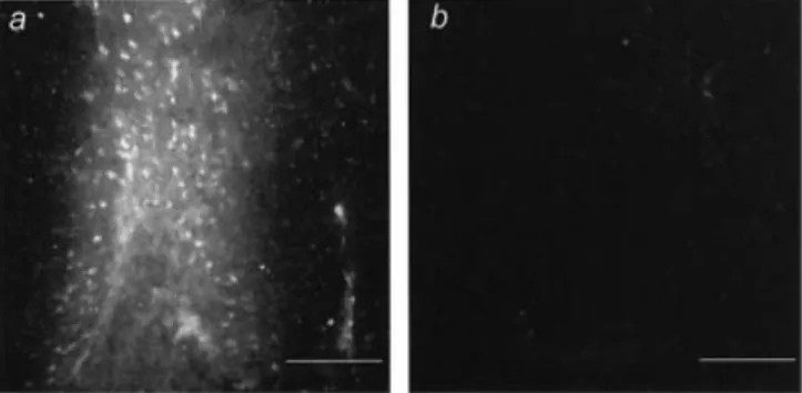
To con®rm the direct impact of IL-6 on cerebral vessel permeability, recombinant human IL-6 was injected stereo-tactically into the cortex of WT mice. Extravasation of intravenously injected EB was evident 1 h after recombinant human IL-6 application, indicating increased BBB permea-bility at the site of injection (Fig. 5A). No EB ¯uorescence could be detected in the contralateral PBS-injected control hemisphere (Fig. 5B). Together with the ®ndings described above, these results demonstrate that IL-6 plays an important role in the breakdown and sustained opening of the BBB.
Expression of MMPs is not mediated by IL-6
In previous studies we have shown that expression of MMPs, especially MMP-9, is associated with disruption of BBB in
Fig. 4 IL-6 increases BBB permeability during pneumococcal meningitis. (A) I.c. injection of pneumococci increased BBB permeability index in WT mice which was signi®cantly reduced in infected IL-6±/±mice (*P < 0.05 compared with infected WT). (B) Disruption of the
BBB in infected WT mice was associated with a signi®cant increase in ICP. However, in infected IL-6±/±mice the ICP was signi®cantly
reduced to almost normal levels (*P < 0.05 compared with WT controls;#P < 0.05 compared with infected WT mice). (C) Similar results
were obtained when rats, challenged with pneumococci, were treated with neutralizing anti-IL-6 antibodies. Irrespective of the time of administration (before or 6 h after infection), anti-IL-6 antibody treatment signi®cantly reduced the pneumococci-induced increase in ICP (*P < 0.05 compared with uninfected controls;#P < 0.05 compared with infected, untreated rats), which was accompanied by a
diminished BBB permeability as assessed by EB extravasation. (D) Only minor EB leakage was detectable in the leptomeningeal space and no dye was found in the brain parenchyma demonstrating an intact BBB in control rats. (E) Meningitis caused leakage of EB into the subarachnoidal space and the underlying cerebral parenchyma 24 h after infection. (F) Extravasation of EB was attenuated in anti-IL-6 antibody-treated rats, showing only weak ¯uorescence in the subarachniodal space and no ¯uorescence in the brain parenchyma. Data are expressed as mean + SEM. Bar = 50 mm.
bacterial meningitis (Paul et al., 1998; Kieseier et al., 1999). Therefore, we investigated the effect of IL-6 on the menin-gitis-induced expression of different MMPs in rat brains using competitive quantitative RT±PCR. Similar to the results shown in a previous study (Kieseier et al., 1999), we found constitutive expression of stromelysin-3 (MMP-11), gelatinase A (MMP-2) and matrilysin (MMP-7) at low levels, and no expression of stromelysin-2 (MMP-10). None of these MMPs was upregulated 24 h after infection (data not shown). Expression of stromelysin-1 (MMP-3), which was previously proven to be increased 6 and 9 h after infection (Kieseier et al., 1999), was no longer upregulated after 24 h (data not shown). However, cerebral mRNA expression of gelatinase B (MMP-9) (Fig. 6A) and collagenase-3 (MMP-13) (Fig. 6B) was increased in infected rats at 24 h. Pre-treatment with neutralizing anti-IL-6 antibody did not in¯uence the expres-sion of these MMPs (Fig. 6A and B) or the activity of MMP-9 as assessed by zymography (data not shown). These data indicate that IL-6 does not mediate the expression or activity of MMPs during bacterial meningitis.
Discussion
A major complication of bacterial meningitis is an increased permeability of the cerebral microvessels, resulting in vasogenic brain oedema and a rise in ICP (P®ster et al., 1999). The breakdown of the BBB was always considered to be the consequence of the invasion of leukocytes releasing cytokines, reactive oxygen species and proteases, leading to an increase in vascular permeability. Here we show that IL-6 contributes substantially to the disruption of the BBB, but at the same time acts as an anti-in¯ammatory cytokine in bacterial meningitis. These ®ndings demonstrate that the migration of leukocytes across the BBB and the increase in vascular permeability are two independent processes.
Bacterial meningitis in IL-6±/± mice caused a three-fold
increase in CSF WBC count compared with infected WT mice, which was associated with increased TNF-a, IL-1b and MIP-2 mRNA expression. TNF-a and IL-1b are well-characterized pro-in¯ammatory cytokines in bacterial men-ingitis and both can initiate meningeal in¯ammation (Ramilo et al., 1990). Our study shows that IL-6 attenuates mRNA expression of both cytokines, indicating that IL-6 acts as an anti-in¯ammatory cytokine. Similar results were found by others who documented increased TNF-a and IL-1b protein expression in IL-6±/±mice during acute in¯ammation (van der
Poll et al., 1997; Xing et al., 1998), af®rming the anti-in¯ammatory nature of IL-6. MIP-2, which parallels the action of IL-8 in humans (Broxmeyer et al., 1996), is a member of the CXC family of chemokines and a potent chemoattractant of polymorphonuclear neutrophils (PMN) (Lahrtz et al., 1998). MIP-2 was shown to be an important mediator of PMN in¯ux in many in¯ammatory diseases such as ocular bacterial infection and urinary tract infection (Hang et al., 1999; Kernacki et al., 2000). The importance of MIP-2 in attracting PMN during bacterial meningitis was demon-strated in in vitro (Spanaus et al., 1997) and in vivo (Diab et al., 1999) studies, which proved that neutralization of MIP-2 attenuated neutrophil chemotaxis. Our study is in accordance with results obtained by others (Xing et al., 1998; Hurst et al., 2001) who showed the regulatory role of IL-6 during acute in¯ammation where MIP-2 mRNA and protein expression was increased in IL-6±/±mice compared with WT
mice, resulting in an enhanced neutrophilic response. It was also shown that IL-6 can attenuate the TNF-a- and IL-1b-mediated release of IL-8 (Hurst et al., 2001), the human homologue of MIP-2, which might explain the increase of MIP-2 in infected IL-6±/± mice. The elevated number of
leukocytes cannot be accounted for by the prolongation of the lifetime of these cells, since the absence of IL-6 does not affect the percentage of apoptotic neutrophils during in¯am-mation, as shown by others (Brach et al., 1992; Xing et al., 1998). The highly reduced mRNA expression of MIP-2 in uninfected IL-6±/±compared with WT controls found in our
study also suggests that the basal expression of this pro-in¯ammatory chemokine is compensatorily downregulated to counteract the absence of the anti-in¯ammatory IL-6.
Fig. 6 Expression of MMP-9 and MMP-13 mRNA in rats treated with anti-IL-6 antibodies. Cerebral infection of rats with
pneumococci caused an increase in (A) MMP-9 and (B) MMP-13 mRNA expression compared with uninfected controls. No reduction in mRNA levels were found in infected rats pre-treated with anti-IL-6 antibodies. Data are expressed as mean + SEM. Fig. 5 Effect of IL-6 on BBB permeability. (A) Direct injection of recombinant human IL-6 into the cerebral cortex of WT mice increased vessel permeability as assessed by extravasation of systemically administered EB. (B) No EB leakage was detected in the contralateral side, which was injected with PBS. Bar = 100 mm.
On the other hand, expression of GM-CSF in infected IL-6±/± mice was also reduced compared with infected WT
mice. GM-CSF can directly act on PMN by modulating the surface expression of adhesion molecules and increasing neutrophil adherence to the endothelium (Arnaout et al., 1986). It was shown that GM-CSF can induce the expression of IL-6 in monocytes and microglia (Suzumura et al., 1996) and that vice versa IL-6 can inhibit GM-CSF gene expression (Kremlev et al., 1998), presenting a negative feedback system which might explain the fact that the basal GM-CSF mRNA level in uninfected IL-6±/±mice was more than twice that in
WT controls. Therefore, it was surprising that the increase in GM-CSF mRNA expression was attenuated in infected IL-6±/± mice, compared with infected WT mice. However,
similar results were observed in IL-6±/±mice after a focal cryo
brain injury, which showed reduced GM-CSF protein expression compared with injured WT controls (Penkowa et al., 1999). Despite the reduced GM-CSF expression in the absence of IL-6 we found higher CSF WBC counts in infected IL-6±/± mice compared with infected WT mice, con®rming
the results of a previous study which demonstrated that neutrophilic in®ltration in lungs of GM-CSF-de®cient mice is increased after pulmonary streptococcal infection (LeVine et al., 1999). It is unclear whether the increased in¯ammatory response is directly related to the reduced GM-CSF expres-sion or is an indirect effect of GM-CSF on other chemokines. For example, it was shown that MIP-2 levels are elevated in GM-CSF±/± mice with pulmonary streptococcal infection
compared with infected WT mice (LeVine et al., 1999), indicating that GM-CSF can downregulate MIP-2 expression. Therefore, we suggest that decreased GM-CSF expression in IL-6±/±mice also might have contributed to the enhanced CSF
pleocytosis during bacterial meningitis by increasing MIP-2 expression.
Despite the fact that leukocyte recruitment was enhanced in infected IL-6±/± mice and IL-6 antibody-treated rats, the
disruption of the BBB was signi®cantly reduced, suggesting that transmigration of leukocytes and increase in vascular permeability are two independent processes. In this study we demonstrated the direct impact of IL-6 on cerebral vessel permeability by injecting the cytokine directly into the brain. There are several other studies which showed that IL-6 in¯uences the integrity of the BBB. For example, IL-6 was demonstrated to reduce the transendothelial electrical resist-ance (de Vries et al., 1996) and to induce changes in the morphology and permeability of endothelial cells in an in vitro model of the BBB (Duchini et al., 1996). Moreover, the systemic administration of IL-6 increased the permeabil-ity of the BBB in rats (Saija et al., 1995), and transgenic mice overexpressing IL-6 in astrocytes showed extensive extra-vasation of horseradish peroxidase indicating an open BBB (Brett et al., 1995).
Previously, we demonstrated that MMPs, especially MMP-9, are involved in the disruption of the BBB and oedema formation during bacterial meningitis (Paul et al., 1998; Kieseier et al., 1999). Therefore, we reasoned that IL-6 might
regulate MMP expression or activity. However, in this study no reduction in the expression of MMP-9 and MMP-13 or activity of MMP-9 was found, despite the signi®cant decrease in vascular permeability in rats treated with anti-IL-6 antibodies. These results suggest that MMPs may not be directly involved in the disruption of the BBB. It seems that MMPs mediate the migration of leukocytes rather than directly disturb endothelial barrier function, and that the bene®cial effect of MMP-inhibition on brain oedema forma-tion might be secondary to its anti-in¯ammatory effect. These results are in accordance with in vitro studies showing that increased endothelial permeability is a not necessary con-sequence of PMN migration (Huang et al., 1988; Lennon et al., 1998). Our ®ndings rather suggest that IL-6 is released by migrating leukocytes and/or activated resident cells, which in turn increases cerebral vascular permeability contributing to the formation of brain oedema during bacterial meningitis. Taken together, the results of this study show that IL-6 increases BBB permeability during bacterial meningitis, causing the formation of brain oedema, but inhibits leukocyte recruitment possibly by suppressing the expression of TNF-a, IL-1b and MIP-2. Dissecting the downstream signalling pathways of IL-6 affecting leukocyte migration or vascular permeability will be helpful to better understand the pathophysiology of bacterial meningitis and other in¯amma-tory diseases.
Acknowledgements
This study was supported by a grant from the Volkswagenstiftung (to H.-W.P., grant number I/71286) and the Deutsche Forschungsgemeinschaft (to R.P., grant number Pa 749/1-1).
References
Arnaout MA, Wang EA, Clark SC, Sieff CA. Human recombinant granulocyte±macrophage colony-stimulating factor increases cell-to-cell adhesion and surface expression of adhesion-promoting glycoproteins on mature granulocytes. J Clin Invest 1986; 78: 597±601.
Brach MA, de Vos S, Gruss HJ, Herrmann F. Prolongation of survival of human polymorphonuclear neutrophils by granulocyte± macrophage colony-stimulating factor is caused by inhibition of programmed cell death. Blood 1992; 80: 2920±4.
Brett FM, Mizisin AP, Powell HC, Campbell IL. Evolution of neuropathologic abnormalities associated with blood±brain barrier breakdown in transgenic mice expressing interleukin-6 in astrocytes. J Neuropathol Exp Neurolol 1995; 54: 766±75. Broxmeyer HE, Cooper S, Cacalano G, Hague NL, Bailish E, Moore MW. Involvement of interleukin (IL) 8 receptor in negative regulation of myeloid progenitor cells in vivo: evidence from mice lacking the murine IL-8 receptor homologue. J Exp Med 1996; 84: 1825±32.
de Vries HE, Blom-Roosemalen MC, van Oosten M, de Boer AG, van Berkel TJ, Breimer DD, et al. The in¯uence of cytokines on the
integrity of the blood±brain barrier in vitro. J Neuroimmunol 1996; 64: 37±43.
Diab A, Abdalla H, Li HL, Shi FD, Zho J, Hojberg B, et al. Neutralization of macrophage in¯ammatory protein 2 (MIP-2) and MIP-1alpha attenuates neutrophil recruitment in the central nervous system during experimental bacterial meningitis. Infect Immun 1999; 67: 2590±601.
Duchini A, Govindarajan S, Santucci M, Zampi G, Hofman FM. Effects of tumor necrosis factor-alpha and interleukin-6 on ¯uid-phase permeability and ammonia diffusion in CNS-derived endothelial cells. J Investig Med 1996; 44: 474±82.
Eugster HP, Frei K, Kopf M, Lassmann H, Fontana A. IL-6-de®cient mice resist myelin oligodendrocyte glycoprotein-induced autoimmune encephalomyelitis. Eur J Immunol 1998; 28: 2178±87. Gadient RA, Otten UH. Interleukin-6 (IL-6)Ða molecule with both bene®cial and destructive potentials. Prog Neurobiol 1997; 52: 379±90.
Gennari R, Alexander JW. Anti-interleukin-6 antibody treatment improves survival during gut-derived sepsis in a time-dependent manner by enhancing host defense. Crit Care Med 1995; 23: 145±53.
Gijbels K, Galardy RE, Steinman L. Reversal of experimental autoimmune encephalomyelitis with a hydroxamate inhibitor of matrix metalloproteases. J Clin Invest 1994; 94: 2177±82. Hang L, Haraoka M, Agace WW, Lef¯er H, Burdick M, Strieter R, et al. Macrophage in¯ammatory protein-2 is required for neutrophil passage across the epithelial barrier of the infected urinary tract. J Immunol 1999; 162: 3037±44.
Houssiau FA, Bukasa K, Sindic CJ, Van Damme J, Van Snick J. Elevated levels of the 26K human hybridoma growth factor (interleukin 6) in cerebrospinal ¯uid of patients with acute infection of the central nervous system. Clin Exp Immunol 1988; 71: 320±3.
Huang AJ, Furie MB, Nicholson SC, Fischbarg J, Liebovitch LS, Sliverstein SC. Effects of human neutrophil chemotaxis across human endothelial cell monolayers on the permeability of these monolayers to ions and macromolecules. J Cell Physiol 1988; 135: 355±66.
Hurst SM, Wilkinson TS, McLoughlin RM, Jones S, Horiuchi S, Yamamoto N, et al. IL-6 and its soluble receptor orchestrate a temporal switch in the pattern of leukocyte recruitment seen during acute in¯ammation. Immunity 2001; 14: 705±14.
Kernacki KA, Barrett RP, Hobden JA, Hazlett LD. Macrophage in¯ammatory protein-2 is a mediator of polymorphonuclear neutrophil in¯ux in ocular bacterial infection. J Immunol 2000; 164: 1037±45.
Kieseier BC, Paul R, Koedel U, Seifert T, Clements JM, Gearing AJ, et al. Differential expression of matrix metalloproteinases in bacterial meningitis. Brain 1999; 122: 1579±87.
Koedel U, P®ster HW. Protective effect of the antioxidant N-acetyl-L-cysteine in pneumococcal meningitis in the rat. Neurosci Lett 1997; 225: 33±6.
Koedel U, P®ster HW. Oxidative stress in bacterial meningitis. Brain Pathol 1999; 9: 57±67.
Koedel U, Bernatowicz A, Paul R, Frei K, Fontana A, P®ster HW. Experimental pneumococcal meningitis: cerebrovascular altera-tions, brain edema, and meningeal in¯ammation are linked to the production of nitric oxide. Ann Neurol 1995; 37: 313±23. Koedel U, Bernatowicz A, Frei K, Fontana A, P®ster HW. Systemically (but not intrathecally) administered IL-10 attenuates pathophysiologic alterations in experimental pneumococcal meningitis. J Immunol 1996; 157: 5185±91.
Kremlev SG, Chapoval AI, Evans R. CSF-1 (M-CSF) enhances the in¯ammatory response of ®bronectin-primed macrophages: pathways involved in activation of the cytokine network. Nat Immun 1998; 16: 228±43.
Lahrtz F, Piali L, Spanaus KS, Seebach J, Fontana A. Chemokines and chemotaxis of leukocytes in infectious meningitis. J Neuroimmunol 1998; 85: 33±43.
Lennon PF, Taylor CT, Stahl GL, Colgan SP. Neutrophil-derived 5¢-adenosine monophosphate promotes endothelial barrier function via CD73-mediated conversion to adenosine and endothelial A2B receptor activation. J Exp Med 1998; 188: 1433±43.
LeVine AM, Reed JA, Kurak KE, Cianciolo E, Whitsett JA. GM-CSF-de®cient mice are susceptible to pulmonary group B streptococcal infection. J Clin Invest 1999; 103: 563±9.
Loddick SA, Turnbull AV, Rothwell NJ. Cerebral interleukin-6 is neuroprotective during permanent focal cerebral ischemia in the rat. J Cereb Blood Flow Metab 1998; 18: 176±9.
Matsuzono Y, Narita M, Akutsu Y, Togashi T. Interleukin-6 in cerebrospinal ¯uid of patients with central nervous system infections. Acta Paediatr 1995; 84: 879±83.
Nadal D, Leppert D, Frei K, Gallo P, Lamche H, Fontana A. Tumour necrosis factor-alpha in infectious meningitis. Arch Dis Child 1989; 64: 1274±9.
Paul R, Lorenzl S, Koedel U, Sporer B, Vogel U, Frosch M, et al. Matrix metalloproteinases contribute to the blood±brain barrier disruption during bacterial meningitis. Ann Neurol 1998; 44: 592± 600.
Paul R, Zhang ZG, Eliceiri BP, Jiang Q, Boccia AD, Zhang RL, et al. Src de®ciency or blockade of Src activity in mice provides cerebral protection following stroke. Nat Med 2001; 7: 222±7. Penkowa M, Moos T, Carrasco J, Hadberg H, Molinero A, Bluethmann H, et al. Strongly compromised in¯ammatory response to brain injury in interleukin-6-de®cient mice. Glia 1999; 25: 343±57.
P®ster HW, Borasio GD, Dirnagl U, Bauer M, Einhaupl KM. Cerebrovascular complications of bacterial meningitis in adults. Neurology 1992; 42: 1497±504.
P®ster HW, Koedel U, Paul R. Acute meningitis. Curr Infect Dis Rep 1999; 1: 153±9.
Quagliarello V, Scheld WM. Bacterial meningitis: pathogenesis, pathophysiology, and progress. New Engl J Med 1992; 327: 864± 72.
Quagliarello VJ, Wispelwey B, Long WJ, Scheld WM. Recombinant human interleukin-1 induces meningitis and blood±brain barrier injury in the rat. Characterization and
comparison with tumor necrosis factor. J Clin Invest 1991; 87: 1360±6.
Ramilo O, Saez-Llorens X, Mertsola J, Jafari H, Olsen KD, Hansen EJ, et al. Tumor necrosis factor alpha/cachectin and interleukin 1 beta initiate meningeal in¯ammation. J Exp Med 1990; 172: 497±507.
Saija A, Princi P, Lanza M, Scalese M, Aramnejad E, De Sarro A. Systemic cytokine administration can affect blood±brain barrier permeability in the rat. Life Sci 1995; 56: 775±84.
Shanley TP, Foreback JL, Remick DG, Ulich TR, Kunkel SL, Ward PA. Regulatory effects of interleukin-6 in immunoglobulin G immune-complex-induced lung injury. Am J Pathol 1997; 151: 193±203.
Spanaus KS, Nadal D, P®ster HW, Seebach J, Widmer U, Frei K, et al. C-X-C and C-C chemokines are expressed in the cerebrospinal ¯uid in bacterial meningitis and mediate chemotactic activity on peripheral blood-derived polymorphonuclear and mononuclear cells in vitro. J Immunol 1997; 158: 1956±64.
Suzumura A, Sawada M, Marunouchi T. Selective induction of interleukin-6 in mouse microglia by granulocyte±macrophage colony-stimulating factor. Brain Res 1996; 713: 192±8.
van de Loo FA, Kuiper S, van Enckevort FH, Arntz OJ, van den Berg WB. Interleukin-6 reduces cartilage destruction during experimental arthritis. A study in interleukin-6-de®cient mice. Am J Pathol 1997; 151: 177±91.
van der Poll T, Keogh CV, Guirao X, Buurman WA, Kopf M, Lowry SF. Interleukin-6 gene-de®cient mice show impaired defense against pneumococcal pneumonia. J Infect Dis 1997; 176: 439±44. Xing Z, Gauldie J, Cox G, Baumann H, Jordana M, Lei XF, et al. IL-6 is an antiin¯ammatory cytokine required for controlling local or systemic acute in¯ammatory responses. J Clin Invest 1998; 101: 311±20.
Received February 2, 2003. Revised March 12, 2003 Accepted March 20, 2003