P-gp expression in brown trout
erythrocytes: evidence of a detoxification
mechanism in fish erythrocytes
Emeline Valton
1,2,3, Christian Amblard
3, Ivan Wawrzyniak
3, Frederique Penault-Llorca
1,2& Mahchid Bamdad
1,2,3*
1Clermont Universite´ - Universite´ d’Auvergne - ERTICa– EA 4677 - Institut Universitaire de Technologie, de´partement ge´nie biolo-gique, Ensemble universitaire des Ce´zeaux, B.P. 86 - 63172 AUBIERE CEDEX France,2Clermont Universite´ Universite´ d’Auvergne -ERTICa – EA 4677- Centre Jean Perrin, 58 Rue Montalembert, BP 392 - 63011 CLERMONT-FERRAND CEDEX France,3Clermont Universite´ – Universite´ Blaise Pascal - LMGE – UMR CNRS 6023 - BP 80026- 63171 AUBIERE CEDEX France.
Blood is a site of physiological transport for a great variety of molecules, including xenobiotics. Blood cells in
aquatic vertebrates, such as fish, are directly exposed to aquatic pollution. P-gp are ubiquitous ‘‘membrane
detoxification proteins’’ implicated in the cellular efflux of various xenobiotics, such as polycyclic aromatic
hydrocarbons (PAHs), which may be pollutants. The existence of this P-gp detoxification system inducible
by benzo [a] pyrene (BaP), a highly cytotoxic PAH, was investigated in the nucleated erythrocytes of brown
trout. Western blot analysis showed the expression of a 140-kDa P-gp in trout erythrocytes. Primary
cultures of erythrocytes exposed to increasing concentrations of BaP showed no evidence of cell toxicity. Yet,
in the same BaP-treated erythrocytes, P-gp expression increased significantly in a dose-dependent manner.
Brown trout P-gp erythrocytes act as membrane defence mechanism against the pollutant, a property that
can be exploited for future biomarker development to monitor water quality.
T
he bloodstream is a hub for the transport and inter-reaction of a variety of physiological molecules (oxygen,
nutrients, hormones and waste) and also for the accumulation of xenobiotics (drugs and/or pollutants).
Whatever the type of pollution (acute or chronic), the various pollutants are either found directly in the
blood or accumulated in organs, tissues and cells before being gradually released into the circulation. Erythrocytes
are the major cellular component of blood. In mammals, including man, mature erythrocytes are not nucleated,
unlike fish, where they are
1. Among fish, Salmonids are integrative organisms with regard to environmental
conditions and water quality. They are ‘‘bioaccumulation’’ organisms
2. Brown trout (Salmo trutta fario) provide a
ubiquitous and highly sensitive index of water quality. Furthermore, the brown trout is not only native to rivers
but may also be fish-farmed.
P-gp, the well-known ‘‘Multidrug Resistance’’ (MDR) protein encoded by the MDR1 or ABCB1 gene, have
been described as membrane transporters involved in resistance to chemotherapy in cancer
3. In reality, P-gp
belongs to the evolutionarily conserved family of the ATP-binding cassette (ABC) proteins found in practically all
living organisms, whether prokaryotic
4or eukaryotic
3,5–13. P-gp acts as xenobiotics efflux pump transporting
various substrates out of cells. It recognizes various molecules with different structures and properties, including
pollutants such as polycyclic aromatic hydrocarbons (PAHs) (e.g. benzo-a-pyrene (BaP)), heavy metals (e.g.
cadmium), pesticides (e.g. diazinon, DDT), insecticides (e.g. chlorpyrifos) and various pharmaceutical drugs,
such as chemotherapeutic drugs (e.g. Vinca alkaloids, actinomycin D, taxol, antracyclines), calcium channel
blockers (e.g. verapamil)
3,5,6,11,12,14–22. Due to this property, P-gp is involved in the natural defence mechanisms of
cells, tissues and organs, and forms an essential part of the innate defence system known as ‘‘chemo-immunity’’
3.
Our previous research in both laboratory and field has focused on the development of environmental
bio-markers for PAH pollution based on MDR1/P-gp expression, using the freshwater ciliated protozoan
Tetrahymena pyriformis for water pollution and the vinegar fly Drosophila melanogaster for air pollution. In
these two models, P-gp expression was clearly induced in a dose-dependent manner, after exposure to different
concentrations of various classes of PAHs
5,6,11,12.
In the present work, we aimed to analyze P-gp expression in the erythrocytes of brown trout and their response
to BaP, a PAH model, in order to develop an aquatic biomarker.
SUBJECT AREAS:
PROTEIN TRANSPORT ENVIRONMENTAL BIOTECHNOLOGY TRANSPORTERS PROTEIN TRANSPORTReceived
22 July 2013
Accepted
15 November 2013
Published
5 December 2013
Correspondence and requests for materials should be addressed to M.B. (Mahchid. Bamdad@udamail.fr)*Current address: EA 4677 ERTICa : Equipe de Recherche sur les Traitements Individualise´s des Cancers – Site IUT, Institut Universitaire de Technologie de Clermont-Fd – Universite´ d’Auvergne, De´partement Ge´nie Biologique, Complexe Universitaire des Ce´zeaux, B.P. 86, 63172 Aubie`re Cedex.
Results
Expression of P-gp in brown trout erythrocytes.
The expression of
P-gp in erythrocytes was analyzed from a dry pellet of brown trout
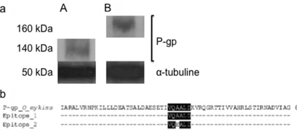
erythrocytes by Western blotting (Fig. 1, and Supplementary Fig. 1a,
and b). The results showed that the monoclonal anti-P-gp antibody
C219 clearly recognized a protein having a molecular weight (MW) of
140 kDa (Fig. 1a). As expected, in the brown trout liver extract used
as the positive control, a P-gp of 160 kDa was labelled
23. A second
band of about 75 kDa was also labelled by C219 antibody in trout
erythrocytes. Moreover, the sequence alignment of rainbow trout
P-gp available in Genbank with the ‘‘VQEALD’’ and ‘‘VQAALD’’
epitopes was recognized by C219 antibody
24was then checked via
CLUSTALW. The results confirmed that the VQAALD epitope,
recognized by C219 monoclonal antibody, was present in trout
P-gp (Fig. 1b). The expression of P-P-gp in the brown trout erythrocytes
quantified under these conditions was 0.84 6 0.05 a.u.
Development of primary culture of brown trout erythrocytes.
For
the development of primary culture of erythrocytes, three culture
media were used: i.e. IMDM, MEM and L-15, supplemented with
10% fetal calf serum, 1% penicillin/streptomycin, 1% fungizone and
1% L-glutamine, under different temperature conditions (11uC,
15uC, 18uC or 21uC) with or without shaking. One million cells
per mL were seeded in each medium, and the rate of cell growth
was assessed over time. For the three culture media tested, the best
temperature and stirring conditions for maintaining trout
erythro-cytes in primary culture were 21uC and 45 rpm (data not shown).
These conditions were thus used to track erythrocyte growth rates in
the three tested media in both the short term (24 h) and longer term
(5 days). The results presented in Figure 2 show that in all three
media, cell growth rates remained relatively stable over 5 h, i.e. the
short term, with a yield close to 1. In the longer term (1 to 5 days), the
growth rate remained close to 1 in L-15 and IMDM media, but fell
slightly in the MEM medium. These results suggest that L-15 and
IMDM media may be used for the primary culture of brown trout
erythrocytes. However, a comparison of cell morphology and cell
membrane shape in primary cultures of L-15 and IMDM media
showed that erythrocytes in the L-15 medium were less damaged
and kept their cell membranes intact (data not shown). Thus, the
L-15 medium was chosen for further experiments.
BaP toxicity analysis on trout erythrocytes.
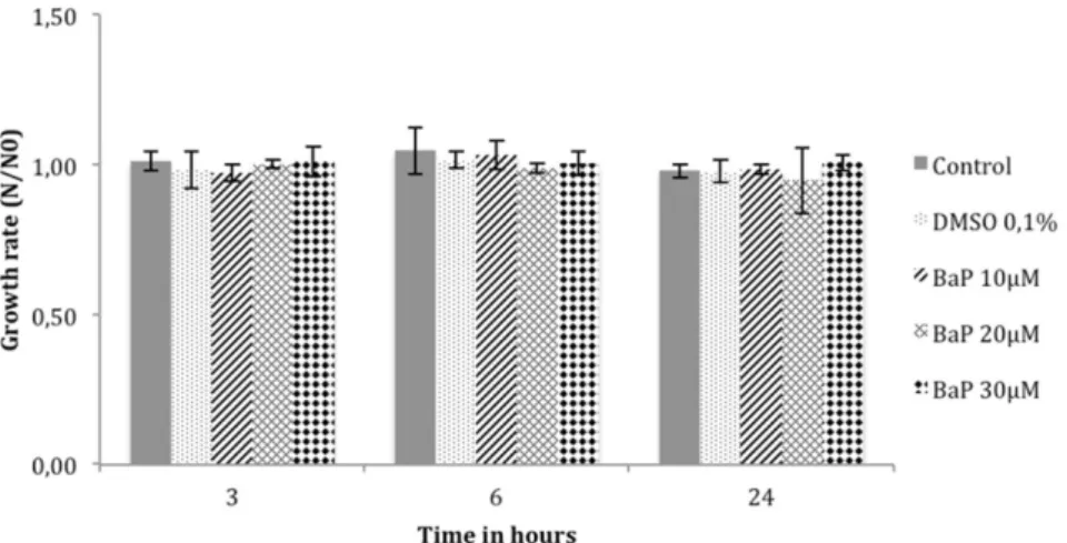
The toxicity of 10, 20
and 30 mM BaP on cultured trout erythrocytes was then studied, by
cells counts over time using a Z-2 cell and particle counter. The rate
of cell numbers was expressed as N/N0, where N and N0 are cell
numbers at time T and T0 (beginning of experiment or after addition
of DMSO or BaP). The results showed that in the presence of BaP at
10, 20 and 30 mM, the number of cells remained stable over time
without signs of BaP toxicity (Fig. 3).
P-gp expression in trout erythrocytes after treatment with
benzo[a]pyrene.
Using Western blot analysis, the expression levels
of P-gp in primary culture were measured for 3, 6 and 24 h in the
presence of increasing concentrations of BaP (10, 20 and 30 mM)
(Fig. 4). Control cultures (untreated cells and in 0.1% DMSO) were
performed in parallel. P-gp expression levels in untreated cells and in
extract from (A) a dry pellet of brown trout erythrocytes or (B) liver. P-gp weighed 140 kDa in A and 160 kDa in B. The monoclonal anti-tubulin antibody DM1A labelled a tubulin protein presented a molecular weight of about 50 kDa, used as internal control. The samples derive from the same experiment and gels were processed in parallel. The full-length blot and Coomassie blue gel are presented in Supplementary Figures 1a and 1b. (b). Sequence alignment of Oncorhynchus mykiss P-gp with the two different epitopes (VQAALD and VQEALD) recognized by C219 antibody. The alignment was performed using ClustalW. Conserved residues and similar residues are shaded in black and grey, respectively.
Figure 2
|
Growth of brown trout erythrocytes in primary culture over time. Cells counts were followed over time using a Beckman Coulter Z-2 Counter. N: number of cells at time T. N0: number of cells at time T0 (beginning of experiment).0.1% DMSO remained stable over 24 h (0.16 6 0.01 and 0.19 6
0.06 a.u. at 24 h, respectively). There was no significant difference
between controls. 0.1% DMSO had no effect on P-gp expression.
The addition of 10 mM BaP to the cell cultures did not change the
expression rate of P-gp for 6 h, which remained close to the control
values (0.12 6 0.02 a.u.). In these cells, P-gp rate only increased after
24 h of BaP exposure (0.41 6 0.05 a.u.). Conversely, in the presence
of both 20 and 30 mM BaP, P-gp expression was clearly induced
during the experiment. Indeed, in the presence of 20 mM BaP,
P-gp expression increased steadily over time to reach 0.44 6 0.1, 0.55 6
0.1 and 0.70 6 0.1 a.u. after 3, 6 and 24 h, respectively. In the
presence of 30 mM BaP, P-gp remained high, at 0.65 6 0.01, 0.42
6
0.08 and 0.51 6 0.01 a.u. after 3, 6 and 24 h, respectively.
Discussion
Blood delivers necessary substances such as nutrients and oxygen to
cells and transports metabolic waste. Blood is also a site of passage
and distribution of all xenobiotics (drugs and/or pollutants) which
enter the organism. In aquatic vertebrates, such as fish, blood cells are
directly exposed to the contamination. Therefore, they provide an
excellent tool for water pollution detection.
In cells, P-gp is a ubiquitous membrane detoxification protein
involved in the defence mechanisms against xenobiotics. It
recog-nizes a large class of xenobiotics, from different drugs
3to various
pollutants, such as PAHs
5,6,11,12,25. The latter are highly ubiquitous
environmental contaminants and include toxic compounds with
cytotoxic, genotoxic and carcinogenic properties
26–28. It has already
been demonstrated that P-gp expression may be induced after
expo-sure to PAHs in different cells models and/or organisms, suggesting
that P-gp may be used as a potential biomarker of environmental
pollution
5,11,12,25,29.
In this context, our work focused on P-gp expression in
erythro-cytes using the Salmonidae brown trout as a model since it can live
in most rivers. Firstly, P-gp expression was studied directly in
dry pellets of cells using Western blot analysis. The monoclonal
antibody anti-P-gp C219 clearly marked a protein of 140 kDa. As
Figure 3
|
BaP toxicity analysis on brown trout erythrocytes. Erythrocytes in primary culture were treated with 10, 20 and 30 mM of BaP for 3, 6 and 24 h. Cell growth was analyzed by cell counting on the Beckman Coulter Z-2 Counter. N: number of cells at time T. N0: number of cells at time T0 (beginning of experiment).Figure 4
|
P-gp expression in brown trout erythrocytes in the presence of BaP. Brown trout erythrocytes were exposed to 10, 20 and 30 mM of BaP. After 3 h, 6 h and 24 h of treatment, the cells were recovered to perform protein extraction. Following extraction, Western blotting was performed and protein expression quantified using Quantity One software (BioradH). Statistical analysis was performed by Student’s t-test. ‘‘a’’ indicate a significant difference (p , 0.05) in P-gp expression between BaP treated samples and controls.band of unknown identity was also reported in trout liver and
cul-tured hepatocytes by the same antibody
10.
Secondly, for trout P-gp erythrocytes induction study, we
developed the best set of conditions for in vitro culture, such as type
of culture medium, cell density, temperature and stirring. The results
showed that it was possible to achieve primary cultures of trout
erythrocytes over time. Indeed, as trout erythrocytes are nucleated,
it should be possible to maintain them in culture for several days. The
best conditions for in vitro culture were Leibovitz’s medium (L-15)
supplemented with 10% fetal bovine serum, 1%
penicillin/strep-tomycin, 1% fungizone and 1% L-glutamine, at a temperature of
21uC, with stirring at 45 rpm. The development of primary blood
cell cultures in the trout model offers further scope for adaptation to
other fish models or to species possessing nucleated erythrocytes.
This in vitro culture model may be used for future cytotoxicity tests
on various xenobiotics and/or for other applications linked tied to a
fish health model. After starting red blood cell culture in vitro, it was
necessary to analyze the response of P-gp to PAHs. For this purpose,
we chose to test the action of BaP, which is a cytotoxic, genotoxic and
carcinogenic model pollutant in the PAHs class. It has been found in
air, water and soil
32,33. Moreover, BaP, like several other PAHs, has
been shown to induce P-gp in other biological models of aquatic and
atmospheric pollution
5,11,12. BaP toxicity at 10, 20 and 30 mM was
first tested by analyzing red blood cell numbers in primary culture
over time. No toxicity was detected at these concentrations, probably
due to the detoxification action of P-gp in the erythrocytes. Similar
responses have already been detected in the ciliated protozoan model
Tetrahymena pyriformis and in SL2 Drosophila melanogaster cell
culture in the presence of similar concentrations of BaP and other
PAHs
5,12,21.
This hypothesis of detoxifying action by P-gp was then checked by
monitoring P-gp expression over time in primary cultures of
ery-throcytes, after treatment with increased concentration of BaP.
Under these conditions, a clear dose-dependent induction of P-gp
expression was detected in trout erythrocytes. With 10 mM BaP,
P-gp expression increased only after 24 h of treatment with a 2-fold
higher ratio than in the controls. The P-gp induction was clearly
noted with 20 and 30 mM BaP from 3 h to 24 h, with a mean ratio
3.6-fold higher than controls. These results suggest that P-gp is
involved in the mechanisms of cellular defence against BaP pollutant.
Similar findings have already been described in other models
5,6,11,12.
All these results suggest that trout erythrocytes can be used to
develop biomarkers to determine water pollution by PAHs.
Indeed, such a development offers two lines of interest. First, the
blood being a fluid tissue circulating throughout the body, this
biomarker may reflect the overall state of fish contamination, with
possible applications in the agri-food sector. It may also reflect the
state of water contamination and thus have applications in the field
of environmental monitoring. Experiments to develop a P-gp
bio-marker are currently being tested on brown trout in a natural
environment.
Red blood cell culture development.The red blood cell culture was achieved using three culture media: Iscove’s Modified Dulbecco’s Medium (IMDM), Minimum Essential Medium (MEM) and Leibovitz’s medium (L-15) (GibcoH, Invitrogen). Erythrocytes were seeded in 25-ml flasks at one million cells per mL in each culture medium, enabling development of optimal culture conditions.
Cell counts.Cells in cultures were counted electronically using a Beckman CoulterTM
Z-2 cell and particle counter. The results were expressed as N/N0, where N corresponded to cell number at time T and N0, to cell number at T0 (beginning of experiment or after addition of DMSO or BaP).
BaP solubilization and exposure.BaP was dissolved in dimethyl sulfoxide (DMSO) as a stock solution at 5 mM. For each experiment, one million cells/mL were seeded in 10 mL of culture medium. The cell cultures were then exposed to concentrations of 10 mM, 20 mM and 30 mM BaP. For each BaP concentration studied, the required volume of BaP was removed from the BaP stock solution. This was then supplemented with the appropriate volume of DMSO, so that the final DMSO concentration remained constant i.e. 0.1%, in all analyzed cell cultures. In parallel, two control cultures were carried out: (i) untreated cells and (ii) primary culture of erythrocytes treated with 0.1% DMSO.
MDR expression studies by Western blot analysis.For each experiment (red blood cell pellets and primary cell cultures), the erythrocytes were collected by a centrifugation at 800 G for 10 min at 4uC. The cells were then treated with 1 ml of lysis buffer (20 mM tris-HCl, 2 mM EDTA, 2 mM EGTA, 6 mM b-mercaptoethanol, and 0.1% protease inhibitor cocktail, pH 7.5). For protein extraction, this sample was sonicated (5 3 5 s at 4uC) and protein concentrations were determined using a ‘‘Coomassie Plus – The Better Bradford Assay’’ Kit (Thermo ScientificH). For the P-gp positive control, a trout liver extract treated under the same conditions was also used23. Western blot analysis was then carried out according to Vache´ et al., 200612.
Briefly, for each analysis, extracts were diluted to 100 mg.ml21protein with sample
buffer (1.25 M tris-HCl, 20% sodium dodecyl sulfate, 20% glycerol, 2 M
dithiothreitol, 0.5% bromophenol blue, pH 6.8). For these experiments, the primary monoclonal antibody anti-P-gp C219 (15500, CalbiochemH), the mouse monoclonal antitubulin antibody DM1A (155000, SigmaH) and the secondary antibody, a horseradish-peroxidase (HRP)-coupled goat anti-mouse (154000, PromegaH) were used. Immune complexes were visualized by chemiluminescence (ECL 1 Western blotting detection reagent, AmershamH) according to the manufacturer’s specifications. The intensities of P-gp and control tubulin bands were analyzed by densitometry using Quantity-One software (BioRadH). For each sample, P-gp expression was determined by calculating the ratio of the density of each MDR protein over the density of the tubulin control. This ratio was expressed in arbitrary units (a.u.).
Statistical analysis.Data were expressed as means 6 confidence intervals of n independent experiments. Each experiment was performed at least in triplicate and then statistically compared using ANOVA and a Student’s t-test. The comparison was applied on each culture condition and on control groups versus BaP-treated groups and P , 0.05 was considered statistically significant.
1. Soldatov, A. Red blood system in fish: peculiarities of its organisation and function. Zh Evol Biokhim Fiziol 41, 217–223 (2005).
2. Mortensen, A. S., Letcher, R. J., Cangialosi, M. V., Chu, S. & Arukwe, A. Tissue bioaccumulation patterns, xenobiotic biotransformation and steroid hormone levels in Atlantic salmon (Salmo salar) fed a diet containing perfluoroactane sulfonic or perfluorooctane carboxylic acids. Chemosphere 83, 1035–1044, DOI:10.1016/j.chemosphere.2011.01.067 (2011).
3. Sarkadi, B., Homolya, L., Szakacs, G. & Varadi, A. Human multidrug resistance ABCB and ABCG transporters: participation in a chemoimmunity defense system. Physiol Rev 86, 1179–1236, DOI:10.1152/physrev.00037.2005 (2006).
4. Gerlach, J. H. et al. Homology between P-glycoprotein and a bacterial haemolysin transport protein suggests a model for multidrug resistance. Nature 324, 485–489, DOI:10.1038/324485a0 (1986).
5. Bamdad, M., Brousseau, P. & Denizeau, F. Identification of a multidrug resistance-like system in Tetrahymena pyriformis: evidence for a new detoxication mechanism in freshwater ciliates. FEBS Lett 456, 389–393, DOI:S0014-5793(99)00978-3 (1999).
6. Bamdad, M., Reader, S., Grolie`re, C. A., Bohatier, J. & Denizeau, F. Uptake and efflux of polycyclic aromatic hydrocarbons by ‘‘Tetrahymena pyriformis’’: Evidence for a resistance mechanism. Cytometry 28, 170–175 (1997). 7. Fischer, S. et al. Constitutive mRNA expression and protein activity levels of nine
ABC efflux transporters in seven permanent cell lines derived from different tissues of rainbow trout (Oncorhynchus mykiss). Aquat Toxicol 101, 438–446, DOI:10.1016/j.aquatox.2010.11.010 (2011).
8. Kurelec, B. The multixenobiotic resistance mechanism in aquatic organisms. Crit Rev Toxicol 22, 23–43, DOI:10.3109/10408449209145320 (1992).
9. Loncar, J., Popovic, M., Zaja, R. & Smital, T. Gene expression analysis of the ABC efflux transporters in rainbow trout (Oncorhynchus mykiss). Comp Biochem Physiol C. Toxicol Pharmacol 151, 209–215, DOI:10.1016/j.cbpc.2009.10.009 (2010).
10. Sturm, A., Cravedi, J. P. & Segner, H. Prochloraz and nonylphenol diethoxylate inhibit an mdr1-like activity in vitro, but do not alter hepatic levels of P-glycoprotein in trout exposed in vivo. Aquat Toxicol 53, 215–228 (2001). 11. Vache, C. et al. A potential genomic biomarker for the detection of polycyclic
aromatic hydrocarbon pollutants: multidrug resistance gene 49 in Drosophila melanogaster. Environ Toxicol Chem 26, 1418–1424 (2007).
12. Vache, C. et al. Drosophila melanogaster p-glycoprotein: a membrane detoxification system toward polycyclic aromatic hydrocarbon pollutants. Environ Toxicol Chem 25, 572–580 (2006).
13. Zaja, R., Klobucar, R. S. & Smital, T. Detection and functional characterization of Pgp1 (ABCB1) and MRP3 (ABCC3) efflux transporters in the PLHC-1 fish hepatoma cell line. Aquat Toxicol 81, 365–376 (2007).
14. Agarwala, S., Chen, W. & Cook, T. J. Effect of chlorpyrifos on efflux transporter gene expression and function in Caco-2 cells. Toxicol In Vitro 18, 403–409, DOI:10.1016/j.tiv.2003.12.006 (2004).
15. Bard, S. M. Multixenobiotic resistance as a cellular defense mechanism in aquatic organisms. Aquat Toxicol 48, 357–389, DOI:S0166-445X(00)00088-6 (2000). 16. Einicker-Lamas, M. et al. P-glycoprotein-like protein contributes to cadmium
resistance in Euglena gracilis. J Comp Physiol B 173, 559–564, DOI:10.1007/ s00360-003-0365-5 (2003).
17. Habibollahi, P., Ghahremani, M. H., Azizi, E. & Ostad, S. N. Multi drug resistance-1 (MDRresistance-1) expression in response to chronic diazinon exposure: an in vitro study on Caco-2 cells. Bull Environ Contam Toxicol 86, 105–109, DOI:10.1007/s00128-010-0158-y (2011).
18. Marquez B, V. B. F. ABC multidrug transporters: target for modulation of drug pharmacokinetics and drug-drug interactions. Curr Drug Targets 12, 600–620 (2011).
19. Prevodnik, A., Lilja, K. & Bollner, T. Benzo[a]pyrene up-regulates the expression of the proliferating cell nuclear antigen (PCNA) and multixenobiotic resistance polyglycoprotein (P-gp) in Baltic Sea blue mussels (Mytilus edulis L.). Comp Biochem Physiol C Toxicol Pharmacol 145, 265–274, DOI:10.1016/ j.cbpc.2006.12.014 (2007).
20. Shabbir, A. et al. Differential effects of the organochlorine pesticide DDT and its metabolite p,p9-DDE on p-glycoprotein activity and expression. Toxicol Appl Pharmacol 203, 91–98, DOI:10.1016/j.taap.2004.07.011 (2005).
21. Sugihara, N. et al. Effects of benzo(e)pyrene and benzo(a)pyrene on P-glycoprotein-mediated transport in Caco-2 cell monolayer: a comparative approach. Toxicol In Vitro 21, 827–834, DOI:10.1016/j.tiv.2007.02.005 (2007). 22. Urayama, K. Y. et al. MDR1 gene variants, indoor insecticide exposure, and the
risk of childhood acute lymphoblastic leukemia. Cancer Epidemiol Biomarkers Prev 16, 1172–1177, DOI:10.1158/1055-9965.EPI-07-0007 (2007).
23. Sturm, A., Ziemann, C., Hirsch-Ernst, K. I. & Segner, H. Expression and functional activity of P-glycoprotein in cultured hepatocytes from Oncorhynchus mykiss. Am J Physiol Regul Integr Comp Physiol 281, R1119–1126 (2001). 24. Van Den Elsen, J. M., Kuntz, D. A., Hoedemaeker, F. J. & Rose, D. R. Antibody
C219 recognizes an alpha-helical epitope on P-glycoprotein. Proc Natl Acad Sci U S A 96, 13679–13684 (1999).
25. Fardel, O., Lecureur, V., Corlu, A. & Guillouzo, A. P-glycoprotein induction in rat liver epithelial cells in response to acute 3-methylcholanthrene treatment. Biochem Pharmacol 51, 1427–1436 (1996).
26. Ikenaka, Y. et al. Effects of polycyclic aromatic hydrocarbons (PAHs) on an aquatic ecosystem: acute toxicity and community-level toxic impact tests of benzo[a]pyrene using lake zooplankton community. J Toxicol Sci 38, 131–136 (2013).
27. Song, M. K. et al. Formation of a 3,4-diol-1,2-epoxide metabolite of benz[a]anthracene with cytotoxicity and genotoxicity in a human in vitro hepatocyte culture system. Environ Toxicol Pharmacol 33, 212–225, DOI:10.1016/j.etap.2011.12.020 (2012).
28. Topinka, J. et al. DNA adducts formation and induction of apoptosis in rat liver epithelial ‘stem-like’ cells exposed to carcinogenic polycyclic aromatic hydrocarbons. Mutat Res 638, 122–132, DOI:10.1016/j.mrfmmm.2007.09.004 (2008).
29. Lampen, A., Ebert, B., Stumkat, L., Jacob, J. & Seidel, A. Induction of gene expression of xenobiotic metabolism enzymes and ABC-transport proteins by PAH and a reconstituted PAH mixture in human Caco-2 cells. Biochim Biophys Acta 1681, 38–46, DOI:10.1016/j.bbaexp.2004.09.010 (2004).
30. Germann, U. A. P-glycoprotein--a mediator of multidrug resistance in tumour cells. Eur J Cancer 32A, 927–944 (1996).
31. Beaulieu, E. et al. P-glycoprotein of blood brain barrier: cross-reactivity of Mab C219 with a 190 kDa protein in bovine and rat isolated brain capillaries. Biochim Biophys Acta 1233, 27–32 (1995).
32. Ao, J., Ruan, X., Yan, Y. & Cai, M. Distribution, sources and ecological risks of polycyclic aromatic hydrocarbons in an urban landscape river. Water Sci Technol 66, 934–941, DOI:10.2166/wst.2012.248 (2012).
33. Zhong, Y. & Zhu, L. Distribution, input pathway and soil-air exchange of polycyclic aromatic hydrocarbons in Banshan Industry Park, China. Sci Total Environ 444, 177–182, DOI:10.1016/j.scitotenv.2012.11.091 (2013).
Acknowledgments
This work was funded by grants from the Regional Council of Auvergne and European Regional Development Fund. The authors wish to express their gratitude to Fabrice Kwiatkowski for statistical analysis and helpful discussion.
Author contributions
E.V. executed the experiments. E.V. and M.B. wrote the main manuscript text. E.V. and I.W. prepared figures 1–4. All authors read and approved the final manuscript.
Additional information
Supplementary informationaccompanies this paper at http://www.nature.com/ scientificreports
Competing financial interests:The authors declare no competing financial interests. How to cite this article:Valton, E., Amblard, C., Wawrzyniak, I., Penault-Llorca, F. & Bamdad, M. P-gp expression in brown trout erythrocytes: evidence of a detoxification mechanism in fish erythrocytes. Sci. Rep. 3, 3422; DOI:10.1038/srep03422 (2013).
This work is licensed under a Creative Commons
Attribution-NonCommercial-ShareAlike 3.0 Unported license. To view a copy of this license, visit http://creativecommons.org/licenses/by-nc-sa/3.0

