Review
DNA repair nucleases
T. M. Martiaand O. Fleckb,*
Institute of Cell Biology, University of Bern, Baltzerstrasse 4, 3012 Bern (Switzerland), Fax: +41 31 631 4684, e-mail: [email protected]
aPresent address: UCSF Comprehensive Cancer Center, University of California, 2340 Sutter Street, Box 0808, San Francisco, California 94143 (USA)
bPresent address: Department of Genetics, Institute of Molecular Biology, University of Copenhagen,
Øster Farimagsgade 2 A, 1353 Copenhagen K (Denmark), Fax: +45 35 32 2113, e-mail: [email protected] Received 12 June 2003; received after revision 29 July 2003; accepted 16 September 2003
Abstract. Stability of DNA largely depends on accuracy
of repair mechanisms, which remove structural anomalies induced by exogenous and endogenous agents or intro-duced by DNA metabolism, such as replication. Most re-pair mechanisms include nucleolytic processing of DNA, where nucleases cleave a phosphodiester bond between a deoxyribose and a phosphate residue, thereby producing 5′-terminal phosphate and 3′-terminal hydroxyl groups. Exonucleases hydrolyse nucleotides from either the 5′ or 3′ end of DNA, while endonucleases incise internal sites DOI 10.1007/s00018-003-3223-4
© Birkhäuser Verlag, Basel, 2004
CMLS
Cellular and Molecular Life Sciencesof DNA. Flap endonucleases cleave DNA flap structures at or near the junction between single-stranded and dou-ble-stranded regions. DNA nucleases play a crucial role in mismatch repair, nucleotide excision repair, base excision repair and double-strand break repair. In addition, nucle-olytic repair functions are required during replication to remove misincorporated nucleotides, Okazaki fragments and 3′ tails that may be formed after repair of stalled repli-cation forks.
Introduction
DNA, the carrier of genetic information of every living or-ganism, is composed of a sugar-phosphate backbone and four organic bases. DNA is subject to cellular metabolic processes such as replication, transcription and repair. Several functions, especially during repair, require con-trolled cleavage of DNA. In 1903, Araki first reported the enzymatic breakdown of nucleic acids, and in the same year Iwanoff introduced the expression ‘nucleases’ for such enzymes [1, 2]. Today the nucleases are classified as sugar-specific nucleases, i.e. DNA nucleases and RNA nucleases, and sugar nonspecific nucleases [3]. Nucleases can be further divided into exonucleases, which hydrolyse either from the 5′ or the 3′ end of nucleic acids, and
en-donucleases, which hydrolyse internal phosphodiester bonds without the requirement of a free DNA end (fig. 1). DNA nucleases cleave a phosphodiester bond between a deoxyribose and a phosphate group. One cleavage prod-uct contains a 5′-terminal phosphate, and the second prod-uct contains a 3′-terminal hydroxyl group (fig. 1). In con-trast to DNA nucleases, AP lyase activities process DNA either by a β-elimination reaction producing a 3′-termi-nal phosphoglyceraldehyde residue (fig. 1) or, like
Es-cherichia coli MutM, by β-δ-elimination, which results in
a 3′-phosphate end [4–7]. Similarly, the dRPase activity of DNA polymerase β (Pol β), removes an abasic sugar-phosphate molecule by β-elimination [6]. Special types of nucleases are AP endonucleases, which cleave 5′ to apurinic/apyrimidinic (AP) sites (fig. 1).
This review focuses on DNA nucleases with a function in repair. The various repair processes will be briefly
de-*Corresponding author.
scribed, followed by a more detailed description of the role and substrate specificity of DNA nucleases. Repair nu-cleases of eukaryotes are listed in table 1. The substrates and products of representative DNA repair nucleases are schematically shown in figure 2. In general, DNA prod-ucts that result from nucleolytic cleavage are further processed by factors acting downstream in a pathway. This is, as a minimum, DNA synthesis and ligation, but can also be a cascade of events, as e.g. the whole process of
homologous recombinational (HR) repair of double-strand breaks (DSBs), which is initiated by resection of 5′ DNA ends. It should also be pointed out that many repair processes include redundant nuclease functions, and that a given nuclease can be involved in more than one process. For example, degradation of mismatched DNA in
E. coli can be redundantly carried out by one out of four
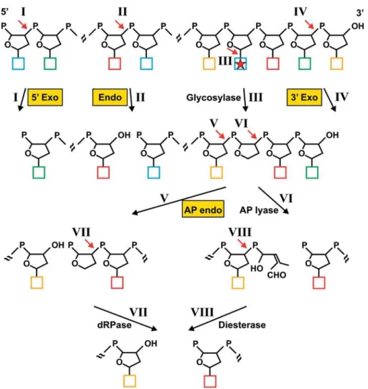
exonucleases (fig. 2 A), and to different degrees each of them is involved in other processes. Repair mechanisms Figure 1. Enzymatic cleavage of DNA. Enzymatic incisions are indicated by red arrows and roman numbers, DNA nucleases are highlighted in yellow boxes. Phosphate groups are drawn as P’s, deoxyriboses as pentagons and bases as coloured squares. The asterisk indicates a dam-aged or mismatched base, which is substrate of a base excision repair glycosylase. Only the products remaining in the DNA chain are shown. DNA nucleases cleave a phosphodiester bond between a deoxyribose and a phosphate residue in a way that one of the products contains a 5′-terminal phosphate group and the second a 3′-terminal hydroxyl group. For excision DNA exonucleases need a free 5′ DNA end (5′ Exo) or a free 3′ end (3′ Exo). Most exonucleases excise a single nucleotide from the 5′ or 3′ end of DNA (I and IV). The DNA strand can be degraded by consecutive nucleotide excisions. Some exonucleases, however, hydrolyse in an internal DNA region distant from the free DNA end (not shown). DNA endonucleases (Endo) do not require a free DNA end and cleave DNA at an internal phosphodiester bond, thereby creating a single-strand break (II). A special type of endonucleases are AP endonucleases (AP endo), which hydrolyse 5′ of an AP site (V). An AP site can be produced spontaneously or enzymatically by a DNA glycosylase, which cleaves the N-glycosylic bond between base and sugar (III). AP sites can exist with a cyclic sugar moiety (shown) or as open chain configurations (not shown) [5, 6]. An AP site can be also processed by the AP lyase activity of bifunctional DNA glycosylases, which in contrast to AP endonucleases cleave 3′ of the AP site by a β-elimination reaction, producing a fragmented sugar with a double bond (VI). Subsequently, a single nucleotide gap between termini with a 5′ phosphate and a 3′ hydroxyl group is produced by phosphodiesterase activity (VIII). The same products are formed when the 5′-ter-minal AP site created by an AP endonuclease (V) is further processed by a dRPase activity (VII).
without the need of a nuclease also exist, such as damage repair by O6-methylguanine-DNA methyltransferase, which transfers the methyl group of the damaged base to one of its own cysteine residues in a suicide reaction, and by photolyases, which split covalent bonds of ultraviolet (UV) light-induced pyrimidine dimers [8].
With respect to their substrate preference, DNA nucleases can be structure, damage or sequence specific. Structure-specific nucleases recognise intermediates of DNA repair. For example, during nucleotide excision repair (NER), en-zymatic unwinding of DNA around a lesion results in a bubblelike structure which is incised by the endonucleases XPF-ERCC1 and XPG at or near the junctions between double-stranded (ds) and single-stranded (ss) regions (fig. 2C). Damage-specific DNA repair nucleases usually have a specific- and a nonspecific binding mode. These nucleases recognise DNA in a nonspecific manner and scan along DNA to find damage. Once detected, the dam-age is bound in a specific manner, allowing the nuclease to dock at the damage with the active-site residues. Sequence-specific nucleases are rather rarely used in repair. One example is MutH of E. coli, which introduces a nick in GATC sequences when the adenine is not methy-lated [9].
The primary sequences of nucleases do not show high similarity except for some conserved residues in their catalytic sites [10]. Therefore, it is often not possible to
conclude from the primary sequence what the specific function of a nuclease could be. Based on the three-di-mensional structure that has been solved for many DNA repair nucleases, they can be classified in the following folding families: RNase H-like, resolvase-like, restriction endonuclease-like, RecJ-like, metallo-dependent phos-phatase, DNase I-like, TIM α/β barrel and His-Me finger endonuclease [10].
Long-patch mismatch repair in E. coli
DNA mismatches can arise during replication by strand slippage or false integration of nucleotides, by sponta-neous or induced base alterations, and during recombina-tion. The major defence against manifestation of premu-tagenic mismatches that arise during replication is the long-patch mismatch repair (MMR) pathway. The basic principle of MMR appears to be quite similar between prokaryotic and eukaryotic organisms, with some striking differences [11].
In E. coli, MutS binds to a mismatch, while MutH binds a hemimethylated dam (GATC) site located either 5′ or 3′ to the mismatch [11, 12]. MutS can exist as a homodimer and a homotetramer, the latter being likely the native state on mismatched DNA [13, 14]. MutL mediates complex formation of MutS and MutH and thereby enables activa-Table 1. Eukaryotic DNA repair nucleases.
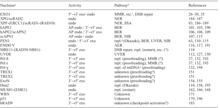
Nucleasea Activity Pathwayb References
EXO1 5′→3′ exo/ endo MMR, rec.c, DSB repair 26–30, 35
XPG/scRAD2 endo NER 184–187
XPF-(ERCC1)/scRAD1-(RAD10) endo NER, SSA 83, 186–189
hAPE1 AP endo / 3′→5′ exo BER 101, 103, 190
hAPE2/scAPN2 AP endo / 3′→5′ exo BER 106, 108, 109
scAPN1 AP endo / endo BER, NIR 107, 115
FEN1 endo / 5′→3′ exo repl.d (Okazaki), BER, UVER, NIR 63, 110–115
ENDO V endo AER 116, 117, 191
MRE11-(RAD50-NBS1) 3′→5′ exo DSB repair, repl. (restart), rec. (?) 118
UVDE endo UVER 112, 127, 130
Pol δ 3′→5′ exo repl. (proofreading), MMR (?) 37, 132, 192
Pol ε 3′→5′ exo repl. (proofreading), MMR (?) 37, 132, 193
Pol γ 3′→5′ exo repl. of mtDNAe(proofreading) 132, 194
TREX1 3′→5′ exo unknown (proofreading?) 151
TREX2 3′→5′ exo unknown (proofreading?) 151
ExoN 3′→5′ exo unknown (proofreading?) 154, 155
Dna2 endo repl. (Okazaki) 110, 158, 195
MUS81-(EME1) endo repl. (restart) 162, 166, 168
WRN 3′→5′ exo Unknown 171
p53 3′→5′ exo Unknown 179, 196
hRAD9 3′→5′ exo unknown (checkpoint activation?) 183
aAlthough different nomenclatures have been established for different species, for simplification we used uppercase letters for eukaryotic
proteins in this review. A few exceptions have been made to avoid confusion, e.g. we further used ExoN instead of EXON. Prefixes h and sc indicate proteins from human and S. cerevisiae, respectively.
bMechanisms for that nuclease function is required. c Recombination.
d Replication. e Mitochondrial DNA.
tion of the endonuclease activity of MutH, which incises the transiently nonmethylated strand. Since this strand is the newly synthesised strand, incision by MutH allows strand discrimination and, in a subsequent step, removal of the falsely incorporated nucleotide. Helicase II unwinds the DNA, and the nicked ssDNA is exonucleolytically de-graded, resulting in excision tracts of up to 1 kb [12]. De-pending on the position of the nicked dam site, either a
5′→3′ or a 3′→5′ exonuclease degrades the nicked strand towards and beyond the mismatch (fig. 2 A). Finally, the resulting gap is filled in by DNA Pol III and ligated by DNA ligase I.
Mutational analysis revealed that Tyr212of MutH is im-portant, if not the only amino acid residue that is respon-sible for verification of the DNA methylation status at
dam sites [15]. Methylation of both adenine residues in a
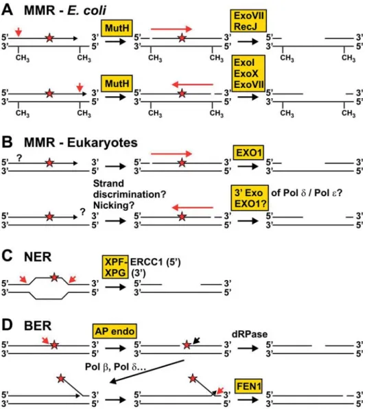
Figure 2. Substrates and products of DNA nucleases in repair and replication processes. Nucleolytic activities are shown as red arrows; nucleases are highlighted in yellow boxes. DNA damage is indicated as red asterisks. (A) MMR in E. coli. MutH incises the newly syn-thesised strand at a hemimethylated dam site when a mismatch is produced during replication and not removed by the proofreading activ-ity of the polymerase (see H). The nick can be either 5′ or 3′ to the mismatch. Exonucleolytic degradation towards and beyond the mismatch can occur by ExoVII or RecJ from the 5′ side (top line) and by ExoI, ExoX or ExoVII from the 3′ side (bottom line). (B) MMR in eukaryotes. It is not yet known how eukaryotic MMR can discriminate between the new and the template strand and whether enzymatic nicking is re-quired. Exo1 is involved in eukaryotic MMR for excision from the 5′ side (top line) and likely also has a function for excision from the 3′ side (bottom line). In addition, the 3′ exonucleolytic activities of Pol δ and ε may be involved in 3′ excision. (C) NER. After unwinding of the DNA around the damage, dual incision by XPF-ERCC1 (5′ to the damage) and XPG (3′ to the damage) occurs. (D) BER. AP sites that are produced spontaneously or by monofunctional DNA glycosylases (compare with fig. 1) are incised on the 5′ side by an AP endonuclease. The resulting 5′ abasic terminus can be processed by the dRPase activity of Pol β (short-patch BER, top line). After strand displacement, a flap structure is formed that can be cleaved by FEN1 (long-patch BER, bottom line).
dam site drastically reduces the endonuclease activity of
MutH. Similar to restriction endonucleases, structural adaptations of MutH may prevent cleavage at fully methy-lated dam sites. The Tyr212forms a hydrogen bond with the unmethylated N6atom of the adenine in the nascent strand and simultaneously makes van der Waals binding with the methyl group of the adenine of the opposite DNA strand. Binding is verified when all contacts of the bases of the
recognition sequence are properly formed. This leads to activation of the catalytic centre and subsequent cleavage of the nonmethylated nascent strand [15].
After DNA unwinding by helicase II and protection of the template strand by single-strand binding (SSB) protein, the nicked strand is degraded towards and beyond the mis-match by one out of four exonucleases [16]. Degradation occurs by the 5′→3′ exonuclease activity of either ExoVII Figure 2 (continued). (E) NIR. Alternatively to BER, oxidative damage can be directly incised by an AP endonuclease. The 5′-terminal dam-age can be released by FEN1. (F) DSB repair by HR. A DSB produced by chemical dam-agents or radiation can be repaired by HR. In the first step of HR, the 5′ ends may be endonucleolytically cleaved by MRE11-RAD50-NBS1. Note that the polarity is different to the polarity found for the MRE11 complex in vitro (see fig. 3D). (G) SSA. SSA represents a specific mode of HR in which repeats (green boxes) flank the DSB. After resection of the 5′ ends (see F), a joint molecule can be formed by pairing of the repeats. The NER endonuclease XPF-ERCC1 (RAD1-RAD10 in S. cerevisiae) can remove the nonhomologous ends by incision. (H) Proofreading during replication. Mismatches pro-duced during replication can be removed by the 3′ exonuclease activity of replicative polymerases or by associated 3′→5′ exonucleases. (I) Processing of Okazaki fragments. Okazaki fragments are displaced by DNA synthesis (the RNA part is indicated as blue box). Dna2 cleaves inside the 5′ flap, thereby removing the RNA and most of the ssDNA tail. After branch migration, a double-flap structure with a one-nu-cleotide tail on the displacing strand can be produced and cleaved by FEN1. (J) Removal of 3′ tails that can be formed after repair of stalled replication forks. MUS81-EME1 (-MMS4) incises dsDNA 5′ to the 3′ tail.
or RecJ, when the nick was introduced 5′ to the mismatch, or by the 3′→5′ exonuclease activity of ExoI, ExoX or ExoVII when the nick was introduced 3′ to the mismatch (fig. 2 A). In contrast to the other exonucleases, ExoVII possesses both 5′→3′ and 3′→5′ exonuclease activities [17]. ExoI, ExoVII and RecJ are ssDNA-specific exonu-cleases, while ExoX can also degrade dsDNA, although with lower efficiency [11].
In mutants defective in three of the four exonucleases, ExoI, ExoVII, ExoX and RecJ, repair initiated at a nick lo-cated on one site of a mismatch is strongly reduced, while repair initiated from a nick at the opposite site remains considerably efficient [17]. One exception is the exoI
exoX recJ triple mutant, which shows significant repair
from both sites, due to the presence of 5′→3′ and 3′→5′ exonuclease functions of ExoVII. Mutation rates of the various triple mutants are not increased when compared with wild type [17]. Thus, the function of one remaining exonuclease is sufficient for MMR. The exoI exoVII exoX
recJ quadruple mutant shows a moderately increased
mu-tation rate when compared with wild type, which is clearly not as strong as in a mutS mutant. However, since the mu-tation rate of the quadruple mutant is epistatic to mutS, the mutator phenotype of the exoI exoVII exoX recJ mutant is likely due to a defect in MMR [17]. It has been proposed that the weak mutator phenotype of the quadruple mutant is caused by frequent chromosome loss, which may result in underrecovery of mutation events [16].
Despite their involvement in MMR, ExoI, ExoVII, ExoX and RecJ also have a function in repair of UV-induced damage and in suppression of homologous recombination [18, 19]. The 3′→5′ exonucleases ExoI and ExoX appear to be less important for UV damage repair than the 5′→3′ exonuclease activities of RecJ and ExoVII.
Long-patch MMR repair in eukaryotes
Eukaryotic cells have multiple MutS and MutL homo-logues but obviously no MutH homologue [11]. It is cur-rently not known how the template and the nascent strand are distinguished during eukaryotic MMR. One possibil-ity is that the asymmetric sliding clamp, proliferating cel-lular nuclear antigen (PCNA), which has a function in DNA replication, is responsible for strand discrimination [20, 21]. On the other hand, nicks or free 3′ ends that are present in the nascent strand during replication may be sufficient for strand discrimination. Indeed, in vitro ex-periments showed that preferentially the strand with a nick or, even more efficiently, a short gap is degraded by eu-karyotic MMR [22]. Since the nick or gap can be located either 5′ or 3′ to the mismatch, eukaryotic MMR appears to be bidirectional, like MMR of E. coli [22, 23]. Eu-karyotic MMR is initiated by binding of a MutS-related heterodimer, which is either MutSα (MSH2-MSH6) or
MutSβ (MSH2-MSH3). MutSα and MutSβ differ in their ability to recognise base-base mismatches and small loops [11]. Subsequently, a MutL-related heterodimer sta-bilises binding of MutSα and MutSβ to the mismatched substrate. The major MutL-related dimer is MutLα (MLH1-PMS2 in humans). In some but not all eukary-otes, additional MutL-related heterodimers exist, which play a minor role in MutSβ-dependent repair of loops [11]. Binding of MutLα induces a conformational change, which may allow interaction with downstream factors. One such factor is likely the 5′→3′ exonuclease EXO1 (fig. 2B) [24–26]. EXO1 is member of the FEN1 family of structure-specific flap endonucleases, which is defined by two conserved motifs. Eukaryotic EXO1, however, is not homologous to ExoI of E. coli. Despite its role in MMR, EXO1 also has a function in recombination and DSB repair [27–30].
The Saccharomyces cerevisiae EXO1 gene was identified in a two-hybrid screen with MSH2 [25]. Compared with MMR mutants defective in MSH2 or MLH1, mutation frequencies are only slightly increased in the exo1 mutant, which, similar to the situation in E. coli, may be due to functional redundancy with other exonucleases. Mutation rates of the msh2 exo1 double mutant are in the same range as those of the msh2 single mutant, indicating that EXO1 is involved in the MMR pathway. However, since the mutator effect of exo1 is rather small, a possible addi-tive effect in the msh2 exo1 double mutant may not be de-tectable. Since MSH2 and EXO1 also have a function in recombination [27, 31], it is possible that physical inter-action between MSH2 and EXO1 reflects a role during a recombinational process and that the mutator of exo1 cells is due to a defect in an MMR-independent mechanism. Besides their dsDNA- and ssDNA-specific 5′→3′ exonu-clease activities, human and S. cerevisiae EXO1 also pos-sesses a 5′ flap-endonuclease activity, similar to the other members of the FEN1 family (fig. 3) [32, 33]. In S.
cere-visiae, site-specific mutation analysis revealed that the
ds-DNA 5′→3′ exonuclease and flap-endonuclease activities largely depend on the Asp173residue, which, however, is not required for the ssDNA 5′→3′ exonuclease activity [32]. The flap-endonuclease activity of scEXO1 does not have a preference for a specific flap structure, in contrast to RAD27 (S. cerevisiae homologue of FEN1), which preferentially cleaves a double flap with an additional one-nucleotide 3′ flap (fig. 3B) [32, 34].
Besides its 5′→3′ exonuclease activity human EXO1 also has a function in the excision step of MMR when the strand break is located 3′ to the mismatch. This function is either of a structural nature or due to a cryptic 3′→5′ ex-onuclease activity of EXO1 [35]. In crude protein extracts, excision tracts of human MMR span several hundred nucleotides from the initial strand break, reaching 90–170 bp beyond the mismatch [23]. In contrast, with purified human MutSα, MutLα and EXO1, the excision
patches extend over several thousand nucleotides from the strand break [35]. This difference may be due to control of EXO1-dependent excision by as yet unidentified MMR components.
Mouse EXO1 has a function in the repair of base mis-matches and loops with one unpaired nucleotide in both 5′ and 3′ nick-directed repair [36]. In addition, EXO1-defi-cient mouse cells exhibit instability of a mononucleotide repeat. EXO1 knockout mice show reduced survival, fre-quently develop lymphoma and are sterile due to a defect in meiosis [36].
Genetic studies with S. cerevisiae indicate that the 3′→5′ exonuclease activities of the DNA polymerases δ and ε participate in MMR (fig. 2B), besides their function in MMR-independent mutation avoidance mechanisms, e.g. proofreading during replication [37].
Very short patch repair in E. coli
In E. coli a very short patch (VSP) repair pathway exists, which recognises T/G mismatches in dcm (CCA/
TGG) methylation sites and related sequences and restores them exclusively to C:G pairs [38–40]. The Dcm methyltrans-ferase methylates the internal cytosines of both strands of
dcm sites. The resulting 5-methylcytosine can be
deami-nated to thymine, which if not repaired pairs with an
ade-nine during the next replication. The VSP repair system counteracts the formation of such C:G to T:A transitions. The sequence-specific Vsr endonuclease is the initial fac-tor of VSP repair and incises DNA immediately 5′ to the mismatched thymine. DNA Pol I performs nick trans-lation, creating a repair patch size of less than 10 nu-cleotides. Finally, DNA ligase I seals the nick.
VSP is stimulated by the MMR factors MutS and MutL, but not by MutH [38, 41]. Independent of VSP, T/G mis-matches in dcm sites can also be repaired by MMR. How-ever, this error-prone repair is largely avoided by cell cy-cle regulation, since MMR is induced during exponential growth, whereas VSP repair is most efficient during sta-tionary phase [42].
Structural analysis and biochemical data indicate that Vsr has a function similar to MutH and that both have a struc-ture similar to type II restriction endonucleases [43, 44]. Binding of Vsr to a T/G mismatch leads to insertion of amino acid side chains into the minor and major grooves of DNA, which causes bending and widening of both grooves of the DNA. Insertion of three aromatic side chains into the major groove leads to separation of the T/G mismatch from the adjacent A:T base pair [43].
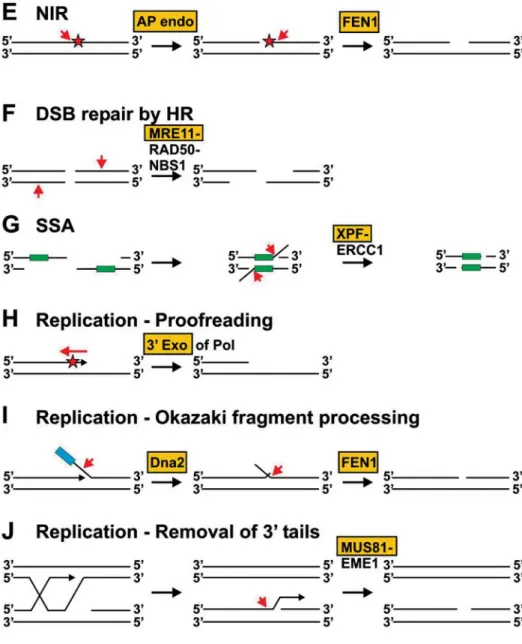
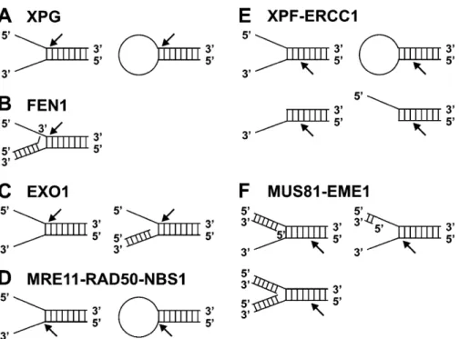
High cellular levels of Vsr are mutagenic, which may be due to interaction of Vsr with MutL, which inhibits dimer-ization of MutL [45, 46]. Vsr interacts with MutL through five C-terminal residues, which are similar to the MutL-Figure 3. DNA structures preferentially cleaved by flap endonucleases. Members of the FEN1 family incise at ssDNA/dsDNA junctions (A–C). Preferred substrate of FEN1 is a double-flap structure in which the 3′ dsDNA arm contains a one-nucleotide flap (B). The MRE11 complex preferentially cleaves 3′ ssDNA at the junction to dsDNA (D). XPF-ERCC1 incises in the dsDNA region two to eight nu-cleotides from the junction (E). MUS81-EME1 (-MMS4) preferentially incises Y structures with 5′ dsDNA tails (F). Cleavage occurs five nucleotides from the 5′ end of the 5′ dsDNA arm. The NER endonucleases XPG (A) and XPF-ERCC1 (E) and the MRE11 complex (D) can incise stem-loop structures, which are not cleaved by FEN1, EXO1 and MUS81-EME1.
interacting residues of MutH. Although MutS has some function in VSP, it cannot simultaneously bind with Vsr to a mismatch. MutS and MutL may deform the DNA around a mismatch, thereby enhancing recognition and binding of Vsr [43].
NER in E. coli
DNA photoproducts induced by UV radiation and other bulky DNA lesions, such as psoralen monoadducts and in-trastrand crosslinks, can be repaired by NER. In addition, methylated bases and AP sites, usually repaired by base excision repair (BER), and to some extent even mis-matches are ‘repaired’ by NER [47].
In E. coli, a protein complex composed of UvrA2 and UvrB binds to undamaged DNA, with gene promoters be-ing the preferred dockbe-ing sites for initial bindbe-ing [48]. The protein complex slides along the DNA in an ATP hydro-lysis-dependent manner. When a damaged base is en-countered, the molecular matchmaker UvrA2is released, resulting in a UvrB-DNA complex and bending of the DNA. UvrC recognises UvrB-DNA with high specificity, forming a stable UvrB-UvrC-DNA complex. Dual inci-sion occurs four to five nucleotides 3′ and eight nu-cleotides 5′ to the damage [49]. The catalytic sites for both incision activities are located in UvrC [50]. After incision, DNA helicase II is required for release of UvrC and for excision of the damage in a 12–13-nucleotide-long oligonucleotide. NER is completed by DNA Pol I-depen-dent repair synthesis and ligation [51]. UvrC contains a UvrBC domain, which is responsible for interaction with UvrB, and two domains that are required for damage in-cision [52, 53]. 3′ incision is catalysed by the N-terminal Uri domain and 5′ incision by the endoV domain, which contains conserved aspartate residues [53]. The C-termi-nal helix-hairpin-helix (HhH)2domain with two HhH mo-tifs is required for 5′ incision and, depending on the se-quence context of a lesion, can be also involved in 3′ incision [54]. The (HhH)2domain specifically binds to ssDNA/dsDNA junctions with a preference for bubblelike DNA structures containing at least six unpaired bases. It has been proposed that two interacting UvrC molecules bind via their (HhH)2domains to the two ssDNA/dsDNA junctions of a bubble and that the (HhH)2domains sta-bilise such DNA structures for dual incision [54, 55]. Cho (UvrC homologue), an E. coli protein with homology to the N-terminal part of UvrC, can perform 3′ incision during NER [56]. Compared with UvrC, incision by Cho occurs four nucleotides further downstream of a lesion, which could be due to a different interaction with UvrB. It has been speculated that Cho could be required for 3′ in-cision of DNA adducts that are poor substrates of UvrC [57]. Since Cho lacks 5′ incision activity, 5′ incision has still to be done by UvrC, also when Cho has made the 3′
incision. In the absence of UvrC, 3′ incision by Cho may be followed by DNA excision carried out by a 3′→5′ ex-onuclease.
NER in eukaryotes
The process of human NER has been uncovered by analy-sis of NER-defective individuals with the inherited disor-der xerodisor-derma pigmentosum (XP) [58]. Individuals af-flicted by XP show an extreme photosensitivity and exhibit a high incidence of skin cancer. During NER, a preincision complex is formed by transcription factor IIH (TFIIH), XPA, replication protein A (RPA) and the two structure-specific endonucleases XPF-ERCC1 and XPG. Two subunits of TFIIH, the ATP-dependent helicases XPB (3′→5′) and XPD (5′→3′) are responsible for opening the DNA double helix around the lesion [59]. After dual inci-sion by XPG and XPF-ERCC1, the damage is released in a 24–32-nucleotide-long oligonucleotide (fig. 2C) [60]. XPG cleaves predominantly 4-6 nucleotides 3′ to the le-sion, and XPF-ERCC1 cleaves predominantly 20–24 nu-cleotides 5′ to the lesion [60]. The resulting gap is filled in by Pol δ or ε, and the remaining nick is sealed by ligase I. NER consists of two subpathways, global genome repair (GGR) and transcription-coupled repair (TCR) [61]. A specific defect in TCR causes Cockayne syndrome (CS). Most CS patients have a mutation in CSA, significantly fewer in CSB and rarely in XPB, XPD or XPG. Patients suffering from CS are UV sensitive, but they do not show an enhanced incidence of skin cancer, which distinguishes CS from XP. XPC in complex with hHR23B and UV-DDB have a specific function in the damage recognition step of GGR, while TCR is thought to be initiated by RNA polymerase II stalled at a lesion [62].
Like EXO1 and FEN1, XPG belongs to the FEN1 family of structure-specific endonucleases. Members of the fam-ily contain an N-terminal and an internal domain with highly conserved acidic amino acid residues that are essential for nuclease activity [63]. All FEN1 family members cleave ssDNA/dsDNA junctions in the double-stranded part adjacent to the 5′ ssDNA arm. The endonu-cleases have overlapping but different substrate speci-ficity, which may reflect adaptation of their function to individual repair processes. For example, XPG can effi-ciently incise bubble or stem-loop substrates, whereas FEN1 and EXO1 only process substrates with free 5′ ssDNA ends (fig. 3) [33, 59, 64, 65].
In addition to 3′ incision during NER, XPG has a struc-tural function during NER and in the not well charac-terised transcription-dependent BER pathway [66–69]. XPG helps to open the DNA around a lesion and to sta-bilise the damage recognition complex [66]. In addition, a role for the S. cerevisiae homologue RAD2 in RNA Pol II-dependent transcription has been reported [70].
XPG is able to cleave several ssDNA/dsDNA structures with the same polarity that is required for 3′ incision dur-ing NER. Such structures are bubbles, splayed arms, stem loops and flaps (fig. 3 A) [59, 65]. Although not cleaved, XPG has a higher binding affinity for three-way junctions than for splayed arms and flap structures [65]. The 3′ and not the 5′ ssDNA arm adjacent to the dsDNA region of an ssDNA/dsDNA junction is important for binding and in-cision by XPG. The presence of at least two nucleotides of the 3′ arm is sufficient for substantial binding and cleav-age by XPG, while complete lack of the 5′ arm reduces in-cision activity only by 50%. Although the 5′ arm is not re-quired for binding and incision, the nuclease activity of XPG, but not binding to DNA, is inhibited when the 5′ arm is double stranded in the junction, indicating that ac-cessibility or flexibility of the junction is important for in-cision but not for binding of XPG [65]. Recognition and incision by XPG might have different claims on a sub-strate, which may reflect the different functions of XPG in different repair systems.
Mutated XPG can cause XP, CS or a combined XP/CS phenotype, depending on the type and position of the mu-tation [71]. Mumu-tations affecting the nuclease activity of XPG generally cause a XP phenotype, while mutations that result in truncation of the protein lead to a phenotype with symptoms from both XP and CS [71]. It is assumed that a deficiency of the NER function of XPG causes XP, whereas a deficiency of the function of XPG in transcrip-tion and in transcriptranscrip-tion-coupled BER is responsible for the characteristics of CS [69, 70, 72].
XPF and homologues in other eukaryotes (RAD1 in
S. cerevisiae) belong to a family of structure-specific
en-donucleases which includes MUS81 and the recently identified archaeal Hef endonuclease [73, 74]. XPF and MUS81 have catalytic activities but perform their function as heterodimers with ERCC1 (RAD10 in S. cerevisiae) and EME1 (MMS4 in S. cerevisiae), respectively, while Hef forms a homodimer [73–78]. XPF-ERCC1 acts as a structure-specific endonuclease which incises the DNA 5′ to the damage in a bubble structure (fig. 3E) [58, 60]. XPF-ERCC1 and RAD1-RAD10 also cleave nonhomolo-gous 3′ flap structures and have an NER-independent role in homologous recombination, during single-strand an-nealing (SSA) (fig. 2G) and in repair of interstrand crosslinks [79–83]. In addition, XPF-ERCC1, but not other NER factors, has a role in the formation of UV-in-duced chromosome exchanges, which indicates that XPF-ERCC1 functions in bypass or repair of damage dur-ing replication [84].
de Laat and co-workers extensively analysed the structure specificity of XPF-ERCC1 endonuclease activity [79]. As assumed from its function in 5′ incision during NER, XPF-ERCC1 is able to cleave stem-loop structures 5′ to the loop in dsDNA two to eight nucleotides away from the junction (fig. 3E). XPF-ERCC1 also cleaves 3′ ssDNA
arms from splayed arms and flap structures. The presence of either a 3′ or 5′ ssDNA arm is sufficient for cleavage by XPF-ERCC1. This is in contrast to XPG, which requires an at least two-nucleotide-long 3′ arm for efficient cleav-age [65].
BER
BER is responsible for repair of DNA damages that are produced by oxidation, alkylation, deamination or hy-droxylation of DNA bases. BER also processes mis-matched and fragmented bases, exocyclic adducts and cyclobutane pyrimidine dimers (CPDs) [85]. BER is ini-tiated by a damage-specific DNA glycosylase which cleaves the N-glycosylic bond between base and sugar, producing an AP site (fig. 1) [6, 86]. AP sites can also occur spontaneously by depurination and de-pyrimidination events [87]. AP sites can be further processed by BER in several ways (figs 1, 2D). An AP endonuclease can hydrolyse the DNA sugar-phosphate backbone 5′ of an AP site, producing a 3′-terminal hy-droxyl group and a 5′-dRP moiety (fig. 1) [88]. The 5′-dRP moiety can be processed during short-patch BER by the dRPase activity of Pol β, leading to a one-nu-cleotide gap between nuone-nu-cleotides with a 3′-hydroxyl and a 5′-phosphate terminus. Subsequently, a single nucleotide is inserted by Pol β, and the remaining nick is sealed either by DNA ligase I or by a complex of DNA ligase III with XRCC1 [86, 88]. Depending on the nature of the 5′-dRP moiety and the DNA substrate, the 5′-dRP moiety can be also removed by long-patch BER, which further processes the nick. Strand displacement during long-patch BER by Pol β, Pol δ and perhaps Pol ε results in incorporation of two to eight nucleotides and formation of a flap structure (fig. 2D) [86]. The flap struc-ture is cleaved by FEN1 in a PCNA-dependent reaction. As in short-patch BER, the remaining nick is either sealed by DNA ligase I or by DNA ligase III-XRCC1 [6, 86]. AP sites can also be processed by the AP lyase activity of bi-functional DNA glycosylases (fig. 1). The resulting frag-mented sugar residue is cleaved by a diesterase activity (likely contributed by an AP endonuclease), which pro-duces a one-nucleotide gap between nucleotides with 3 ′-hydroxyl and 5′-phosphate termini [6, 86]. Thus, sequen-tial cleavage by AP lyase and diesterase activities leads to the same DNA products as sequential cleavage by an AP endonuclease and a dRPase activity (fig. 1). Alternatively to the mechanisms described above, the bifunctional DNA glycosylases MutM (Fpg) and Nei of E. coli and the mam-malian Nei-like enzymes NEIL1 and NEIL2 incise AP sites by β-δ-elimination, resulting in a one-nucleotide gap between nucleotides with 3′ and 5′-phosphate ends [89–94]. Before repair can be completed by gap filling and ligation, the 3′-phosphate group has to be removed. A
third mammalian Nei-like glycosylase, NEIL3, has not yet been characterised [94, 95].
Several BER subpathways have been reconstituted in vitro with purified proteins. Short-patch BER of U/G mis-matches is achieved by UDG glycosylase, hAPE1, Pol β and DNA ligase III [96]. XRCC1 is not absolutely re-quired but enhances efficient ligation. Short-patch BER of 8-oxoG is sufficient with the bifunctional glycosylase OGG1, hAPE1, Pol β and ligase I [97]. Long-patch BER of a reduced AP site requires FEN1, which is stimulated by proliferating cell nuclear antigen (PCNA), and addi-tionally the proteins involved in short-patch BER [98]. Pol β, δ and ε can perform the DNA synthesis step [98–100]. Human APE1 (hAPE1) has a strong AP endonuclease activity and less efficient 3′→5′ exonuclease, 3′ phospho-diesterase and 3′ phosphatase activities [101]. The exo-nuclease activity of hAPE1 is influenced by reac-tion condireac-tions and by the structure of the 3′-terminal nucleotide [102, 103]. The exonuclease activity of hAPE1 preferentially removes nucleoside analogues, 3′ mismatched nucleotides and the natural dinucleotide com-pound Gp4G [102–105]. The exonuclease activity seems to have an important function as a proofreading activity for Pol β. A second ExoIII-like protein, called hAPE2, has been identified in human cells [106]. Although the criti-cal catalytic residues are conserved between hAPE1 and hAPE2, hAPE2 shows only weak AP endonuclease activ-ity.
With APN1 and APN2, two AP endonucleases have been identified in S. cerevisiae [107, 108]. APN1 belongs to the EndoIV family and APN2 to the ExoIII family. Like En-doIV, besides an AP endonuclease function, APN1 also possesses 3′-phosphatase and 3′-phosphodiesterase activ-ities, which can remove 3′-modified termini [88]. APN1 is responsible for almost all of the cellular AP endonuclease activity. APN2 possess weak AP endonuclease activity, but has strong 3′→5′ exonuclease and 3′-phosphodi-esterase activities [109]. S. cerevisiae cells defective in APN1 are moderately sensitive to the alkylating agent methyl methanesulphonate (MMS), which primarily in-troduces AP sites, and sensitivity is further increased when APN2 is additionally defective [108]. Cells defec-tive in APN1 and APN2, but not in only one of the AP en-donucleases are sensitive to the oxidising agent H2O2. Thus, APN1 and APN2 play redundant roles in repair of oxidative damage and AP sites through alternate path-ways, and APN1 is more important for repair of AP sites [108, 109].
During long-patch BER, strand displacement produces a flap structure that is processed by FEN1 (fig. 2D) [86]. FEN1 has also a function in other DNA metabolism such as in Okazaki fragment maturation during DNA replica-tion (fig. 2I), in nonhomologous end joining (NHEJ), and in some organisms in an NER-independent UV-damage excision repair (UVER) pathway [63, 110–112]. FEN1 is
a structure-specific 5′ flap endonuclease, which cuts the ssDNA arm at the junction of a branched DNA structure (fig. 3B) [113]. It is thought that FEN1 binds to a nicked end and then translocates along the ssDNA to the branch point before it performs incision [64]. Recent studies with the S. cerevisiae homologue RAD27 showed that the pre-ferred in vitro substrate is a double-flap structure that pos-sess an one-nucleotide 3′ flap (fig. 3B) [34, 114]. In ad-dition to its endonuclease activity, FEN1 also possesses a 5′→3′ exonuclease activity [63].
Nucleotide incision repair
A repair system has been identified in E. coli, S.
cere-visiae and human cells that, alternatively to BER, repairs
various types of oxidative damage [7, 115]. In this path-way, the AP endonucleases Nfo (EndoIV) of E. coli and APN1 of S. cerevisiae incise DNA 5′ to the damage, thereby producing a 3′-hydroxyl end and a 5′-phosphate end that includes the damaged nucleotide (fig. 2E). Dam-age-specific 5′ incision occurs also in human cell extracts by a not yet identified endonuclease. The 5′-terminal dam-age can be released as a mononucleotide by E. coli DNA Pol I and in the presence of PCNA by human FEN1 (fig. 2E) [115]. The biological advantage of nucleotide in-cision repair (NIR) is that it can process oxidative damage without producing AP sites and blocking 3′-ends, geno-toxic intermediates that are produced by BER. The exis-tence of NIR may explain why DNA glycosylase-deficient
E. coli and mice cells are not sensitive to reactive oxygen
species [7].
Damage-specific 3′ incision by endonuclease V
Endonuclease V of E. coli recognizes a wide spectrum of DNA damage, including AP sites, deaminated bases and mismatches [116]. Damage repair initiated by EndoV rep-resents an alternative to BER initiated by DNA glycosy-lases, and has been termed alternative excision repair (AER) pathway [116]. EndoV hydrolyses the second phosphodiester bond 3′ to a lesion, resulting in a 3′-ter-minal hydroxyl group and a 5′-terminal phosphate group adjacent to the damaged nucleotide. It has been suggested that subsequent removal of the damage is carried out by a not yet identified 3′→5′ exonuclease in a short repair patch of a few nucleotides [116]. EndoV shares signifi-cant similarities with motifs of UvrC required for catalytic activity. Sequence homologues of EndoV are present in eukaryotes, suggesting that AER represents a conserved back-up system for BER [116]. In fact, it has recently been shown that the mouse homologue mENDO V incises DNA containing a hypoxanthine residue in the same way as the bacterial enzyme, but that it has a more limited
sub-strate spectrum [117]. Like NIR, AER avoids the forma-tion of genotoxic repair intermediates which are formed during BER.
Repair of DSBs
DNA DSBs can be induced by ionising radiation, chemi-cal agents, during replication and enzymatichemi-cally during re-combination [83]. DSBs can be repaired either by HR (fig. 2F) or NHEJ and in some special cases by SSA (fig. 2G). Repair of DSBs requires the MRE11 complex, which is composed of MRE11, RAD50 and NBS1 (XRS2 in S. cerevisiae). Inactivation of the MRE11 com-plex causes pleiotrophic defects in DSB repair, replica-tion, checkpoint signalling, early meiosis, V(D)J recom-bination and telomere-length maintenance [118]. In addition, the MRE11 complex may play a role in homol-ogous recombination, although an increasing amount of data argues against this [118]. In humans, mutations in MRE11 and NBS1 cause AT-like syndrome (ATLS) and Nijmegen breakage syndrome (NBS), respectively [119, 120]. The AT-like syndrome is related to ataxia telangiec-tasia, which is caused by a defect in the checkpoint pro-tein ATM [121].
MRE11 exhibits DNA nuclease, strand-dissociation and strand-annealing activities [118]. MRE11 contains an N-terminal nuclease domain with four conserved phospho-esterase motifs. The MRE11 nuclease domain has a ds-DNA-specific 3′→5′ exonuclease activity and a 3′ flap endonuclease activity, which are structure specific [118]. The DNA-processing activity of MRE11 has a wide sub-strate spectrum which partially depends on the interaction with the other proteins of the MRE11 complex and on the accessibility of ATP [118]. The exonuclease is active on dsDNA substrates with blunt ends and with 3′ recessed ends, but not on dsDNA with 3′ overhangs. The endonu-clease cleaves 3′ ssDNA flaps in splayed-arm structures and at ssDNA/dsDNA junctions of stem-loop structures and hairpins (fig. 3D) [118]. Remarkably, the polarity of the nuclease is not compatible with the mechanistic re-quirement for 3′ overhangs during homologous recombi-national (HR) (compare fig. 2F and fig. 3D). An interest-ing model for how 3′ ssDNA tails could be created by the MRE11 complex has been suggested by Trujillo and Sung [122]. After unwinding the dsDNA by a helicase, cleavage of secondary structures, such as stem loops and hairpins in the 5′ ssDNA arm, would lead to 3′ ssDNA tails. This model, however, predicts that the 3′ ssDNA arm has to be selectively protected against incision.
In S. cerevisiae, the MRE11 complex seems to be in-volved in NHEJ [123]. This is in contrast to the situation in Schizosaccharomyces pombe and chicken cells, where the MRE11 complex plays a minor role in NHEJ, if that [124–126].
UVDE-dependent repair
UVDE (also termed UVE1) is an ATP-independent en-donuclease that has been found in S. pombe, Bacillus
sub-tilis and Neurospora crassa, but not in other species so far
studied [127–129]. UVDE is the key factor of the NER-independent UVER pathway and initiates UVER by inci-sion of a phosphodiester bond either immediately or one or two nucleotides 5′ to damage. UVDE incises at a vari-ety of lesions, including the two major photoproducts, CPDs and 6-4 photoproducts (6-4PPs), AP sites, oxidative damage, base-base mismatches and small loops, indicat-ing a more general role of UVDE in DNA repair [112]. RAD2 (the S. pombe FEN1 homologue) further processes DNA incised by UVDE through its 5′→3′ exonuclease activity [130]. The resulting gap can be filled in by Pol δ. In the absence of RAD2, DNA repair intermediates produced by UVDE are channelled into an error-prone or sometimes lethal process [112]. Factors involved in RAD2-independent UVER are EXO1, RHP18 and RHP51, which have a function in recombinational processes [112].
Repair processes during replication
Before mitosis and cell division can take place, a cell has to duplicate its genetic information with high accuracy. DNA replication is performed by a multiprotein complex called the replisome, which itself is located in so-called nuclear replication factories [131]. Key factors of the replisome are DNA polymerases, which replicate dsDNA in a semiconservative manner.
Replication is initiated at origins of replication by syn-thesis of an RNA primer by the primase activity of the Pol α-primase complex. Subsequently, the RNA primer is ex-tended to Okazaki fragments by Pol α [132]. For replica-tion elongareplica-tion, Pol α is replaced by Pol δ or Pol ε, which are able to synthesise long stretches of DNA [132, 133]. Besides replication, DNA polymerases are also involved in DNA repair, recombination and translesion synthesis [133]. Some DNA polymerases have 3′→5′ exonuclease activity, and DNA Pol I of E. coli additionally has a 5′→3′ exonuclease function. DNA polymerases that lack an in-trinsic 3′→5′ exonuclease can in principle be associated with factors exhibiting such activity. During replication, nuclease functions are required for proofreading (fig. 2H), for removal of Okazaki fragments (fig. 2I) and for cleav-age of DNA intermediates that result from recombina-tional repair of stalled or collapsed replication forks (fig. 2J).
Proofreading by 3′→5′ exonucleases
DNA Pol I of E. coli is involved in replication but shows low processivity [8]. Pol I has 3′→5′ and 5′→3′ exonu-clease activity. The 5′→3′ exonuclease activity is structure specific and cuts at ssDNA/dsDNA junctions [134]. The cut is made between the first two nucleotides of dsDNA [135]. The 5′→3′ exonuclease activity of Pol I has struc-tural homology to members of the FEN1 family [136, 137]. Pol I without 5′→3′ exonuclease activity is called a Klenow fragment.
DNA Pol III is the primary replication enzyme of E. coli and consists of 10 different subunits. The 3′→5′ exonu-clease activity is located in the ε subunit and the poly-merase activity in the α subunit [132]. The rate-limiting step in the proofreading reaction of Pol III is melting of DNA in which the 3′ terminus undergoes a conforma-tional change before the DNA becomes a substrate of the 3′→5′ exonuclease [138].
DNA Pol II has a function in error-free and error-prone translesion synthesis and exhibits 3′→5′ exonuclease activity [132, 139, 140]. After blocking of replication by DNA damage, Pol II plays a central role in restarting replication by a damage bypass mechanism.
In eukaryotic cells, DNA Pol α, Pol δ and Pol ε are es-sential for replication of nuclear DNA and Pol γ for repli-cation of mitochondrial DNA. Pol δ, Pol ε and Pol γ, but not Pol α, have a 3′→5′ exonuclease activity for proof-reading of misincorporated nucleotides [132].
Pol δ has a function in DNA synthesis during BER, NER, MMR and recombination. Mice with a homozygous defi-ciency in Pol δ proofreading activity develop cancer, with skin squamous cell carcinoma being the most common form [141]. PCNA stimulates the processivity of Pol δ, and concomitantly reduces its fidelity by promoting mis-incorporation of nucleotides and extension of synthesis at mismatched substrates [142]. Surprisingly, in vitro repli-cation fidelity of S. pombe Pol δ is one to two orders of magnitude lower than the fidelity of E. coli Pol III [143]. While the misinsertion rate of the two enzymes is nearly the same, the proofreading activity of S. pombe Pol δ appears to be very inefficient. The authors assumed that additional cofactors may be required for efficient proof-reading by S. pombe Pol δ [143].
Similar to Pol δ, Pol ε is likely involved in DNA synthe-sis during BER, NER, MMR and recombination. In con-trast to Pol δ, processivity of Pol ε is not stimulated by PCNA [133]. Pol ε of S. cerevisiae consists of four sub-units with a proposed 1:1:1:1 stoichiometry [144]. The proofreading activity of Pol ε is located in the N-terminal region of the catalytic subunit POL2 [132]. The proof-reading activity of Pol ε efficiently removes mismatched nucleotides during DNA replication, but is slowed down when dsDNA with matched nucleotides is encountered [145, 146].
The mitochondrial Pol γ is a member of the nuclease fam-ily A, in contrast to Pol α, β and ε, which belong to the nu-clease family B [132]. In S. cerevisiae, mutation of one of the conserved residues of the exonuclease domain of Pol γ leads to a drastic decrease in 3′→5′ exonuclease activ-ity and to an increased mutation frequency in mitochon-drial DNA [147, 148]. However, the residual activity is sufficient to maintain enough mitochondrial DNA mole-cules for correct segregation into the daughter cells [147]. Pol α, Pol β and the translesion polymerases lack intrin-sic proofreading activity [149]. Several 3′→5′ exonucle-ases have been identified which may substitute for the missing proofreading function of some of the poly-merases. TREX1, exhibiting the major 3′→5′ exonuclease activity in human cells, processes ssDNA and dsDNA [150]. TREX2 is a TREX1 homologue with an overall amino acid identity of 44%, and an 80% identity in the Exo motifs (I, II, IIIε), which are conserved in exonucle-ases from phages to humans [151]. In an in vitro recon-stituted DNA repair system, containing Pol β, DNA ligase III and XRCC1, the TREX1 proofreading activity is re-quired for rejoining of an ssDNA break containing a mis-match at the 3′ end [152]. These data indicate that TREX1 can substitute for the missing proofreading activ-ity of Pol β. TREX1 and TREX2 excise 3′ unpaired nu-cleotides from dsDNA more efficiently than nunu-cleotides from dsDNA with blunt ends and from ssDNA [153]. Besides TREX1 and TREX2, 3′→5′ exonuclease activi-ties have been also discovered for WRN, p53, hRAD9 and MRE11 [149]. However, the 3′→5′ exonuclease of TREX1 and TREX2 appears to be about 1000-fold more active than that of the other proteins [153].
Human ExoN possesses 3′→5′ exonuclease activity which excises nucleotides from ssDNA and dsDNA in a nonprocessive way [154]. Fidelity and elongation from mismatched base pairs by Pol α are increased in the pres-ence of ExoN [155].
Processing Okazaki fragments
Synthesis of Okazaki fragments is initiated by the primase activity of the Pol α-primase complex, which synthesises an RNA primer of 8–12 nucleotides in length [156]. The RNA primer is further extended by 20–30 DNA nu-cleotides by Pol α. In a reaction requiring replication fac-tor C (RFC) and PCNA, Pol α is replaced by Pol δ. When Pol δ encounters the next downstream Okazaki fragment, strand displacement synthesis occurs. PCNA stabilises the strand displacement reaction in a way that Pol δ does not dissociate from DNA at RNA-DNA sites [157]. On the other hand, RPA ensures that strand displacement is lim-ited to about 30 nucleotides and regulates the sequential function of Dna2 and FEN1 in removing the whole Okazaki fragment [157, 158]. Binding of RPA to the 5′
ssDNA flap protects the structure from FEN1 binding and cleavage [158]. Dna2 cuts the flap just 3′ to the end of the RNA-DNA primer (fig. 2I) synthesised by Pol α and thereby releases Dna2 and RPA [156, 158]. The remaining 5–7-nucleotide flap, which is too short for RPA binding, is subsequently cleaved by FEN1 (fig. 2I) or alternatively by other nucleases, such as EXO1 or RNase H [158]. The cleavage reaction results in a nicked duplex which can be sealed by ligase I [156].
Repair of DNA structures formed after replication fork arrest
Various causes lead to DNA replication fork arrest. For example, inhibition of ribonucleotide reductase leads to depletion of the dNTP pool and thereby pauses replication [159]. In addition, DNA damage, such as chemical DNA crosslinks and strand breaks as well as DNA-protein com-plexes formed during cellular metabolism, can induce replication arrest [160]. Stalling of replication forks can lead to formation of abnormal DNA structures. The ab-normal DNA structures induce a checkpoint response, and subsequent cell cycle arrest allows time for repair before replication is reinitiated [161]. In a recent study in yeast, it has been suggested that the abnormal DNA structure formed during replication is ssDNA and not a DSB [162]. This ssDNA can be repaired either by recombination or nonrecombinational gap filling. In contrast to the situation in yeast, stalling of replication in human cells leads to the formation of DSBs [163].
MUS81 was first identified in S. cerevisiae as a protein which interacts with the recombination protein RAD54 [164]. Shortly afterwards, the MUS81 homologue of
S. pombe was identified [165]. spMUS81 interacts with
the checkpoint kinase CDS1, which has a function in sur-vival of cells confronted with replicational stress. MUS81 from S. cerevisiae, S. pombe and humans has en-donuclease activity [75, 166, 167]. Enen-donuclease activity of S. pombe and S. cerevisiae MUS81 is performed in complex with EME1 and MMS4, respectively, which show limited homology to each other [77]. Human MUS81 interacts with hEME1, which is more homolo-gous to EME1 of S. pombe than to MMS4 of S. cerevisiae. A second protein, hEME2, exhibits some homology to hEME1, which is mainly restricted to the C-terminal part of hEME1 [77].
Initial studies indicated that the MUS81 complex plays a role in cleavage of stalled replication forks and in resolu-tion of Holliday juncresolu-tions [75, 166, 167]. However, more recent studies gave new insights into the function of the MUS81 complex. Splayed-arm structures, which are effi-ciently cleaved by RAD1-RAD10 and XPF-ERCC1 (fig. 3E), are poorly processed by scMUS81-MMS4 and hMUS81-hEME1 [77, 168]. Notably, MUS81 complexes
poorly incise Holliday junctions [77, 166, 169]. 3′ flap structures are efficiently cleaved by scMUS81-MMS4. Importantly, incision occurs five nucleotides upstream of a 5′ end and thus requires a 5′ terminus near the junction (fig. 3F) [168]. It was further shown that hMUS81-hEME1 cleaves replication fork structures with two dsDNA arms [77].
A model for cleavage by the MUS81 complex has been proposed in which a stalled or collapsed replication fork is processed by synthesis-dependent strand annealing (SDSA) (fig. 2J), requiring the functions of recombina-tional repair proteins of the RAD52 epistasis group [162, 168]. After displacement and reannealing of the invading strand, a 3′ flap structure is produced that can be cleaved by the MUS81 complex (fig. 2J). Alternatively, the TOP3-SGS1 helicase can complete reannealing of the 3′ tail, thereby producing a 5′ flap structure, which may be processed by FEN1. That cleavage by the MUS81 com-plex occurs downstream and not upstream of recombina-tional repair is supported by the finding that synthetic lethality of a sgs1 mus81 double mutant of S. cerevisiae is suppressed by additional mutation of rad54 [162]. Inacti-vation of both pathways that deal with 3′ tails leads to ac-cumulation of lethal HR intermediates, which in the
sgs1 mus81 rad54 triple mutant can be channelled into the
nonrecombinational gap-filling process. In addition, it is unlikely that the MUS81 complex directly cleaves stalled replication forks since the presence of extensive stretches of ssDNA can be assumed. Such structures are likely not processed by the MUS81 complex, since cleavage re-quires a 5′ end near the junction (fig. 3F) [168].
3′→5′ exonuclease activity of WRN, p53 and hRAD9
The Werner syndrome is a disease of premature ageing and is caused by mutations in WRN [170]. The WRN pro-tein belongs to the family of the RecQ-like helicases, which includes E. coli RecQ, S. cerevisiae SGS1 and hu-man RECQL and BLM [170]. In contrast to the other he-licases, WRN also has 3′→5′ exonuclease activity which is able to degrade dsDNA [171]. The 3′→5′ exonuclease activity of WRN is more efficient on dsDNA with a 5′ overhang than on dsDNA with a 3′ overhang or blunt-ended dsDNA [172]. WRN removes a terminal mis-matched nucleotide just as well as terminal mis-matched nu-cleotides. WRN is able to initiate degradation from gapped or nicked dsDNA, a substrate preference similar to that of E. coli ExoIII [172].
WRN interacts with FEN1 and EXO1 and increases their nuclease activities [170, 173, 174]. WRN also interacts with the Ku70/80 heterodimer, which is required for repair of DSBs by NHEJ [175, 176]. In complex with Ku70/80, WRN efficiently digests dsDNA with either a 3′ or 5′ overhang and blunt-ended dsDNA.
The tumour suppressor protein p53 functions to maintain the genomic integrity of mammalian cells by playing a role in cell-cycle control, apoptosis and DNA repair [177]. p53 has sequence-specific DNA binding activity, which is required for transactivation functions, and 3′→5′ exonu-clease activity, which may be involved in various aspects of DNA repair.
More than 50% of all human cancers have a mutation in p53, frequently in the conserved central part, which is re-sponsible for DNA binding and 3′→5′ exonuclease activ-ity [178]. The major substrate of the 3′→5′ exonuclease activity of p53 is ssDNA and dsDNA with mismatched nucleotides, while matched nucleotides effectively retard excision [179]. p53 enhances the replication fidelity of Pol α, but not of Pol ε or E. coli Pol I [179]. In addition, p53 interacts with hAPE1 and Pol β, stabilises the inter-action of Pol β with AP sites and stimulates the BER of uracil in DNA [180].
In S. pombe, six so-called checkpoint Rad proteins were identified which have a function in the early DNA damage checkpoint response [181]. It was shown by immunoprecipitation that a highly modified form of human Rad9 (hRad9) forms a complex with hRad1 and hHus1 (9-1-1 complex) [182]. The three proteins form a heterotrimeric checkpoint clamp complex (CCC), which is likely loaded by a checkpoint-specific clamp loading factor onto damaged sites of DNA [161]. CCC is phos-phorylated in a damage-dependent manner, but it is not clear whether this is necessary for downstream signalling. Surprisingly, hRad9 has 3′→5′ exonuclease activity, which is located at its N-terminal part [183]. It has been suggested that hRad9 exonuclease activity has a func-tion in primary DNA damage processing which may be important for the DNA damage checkpoint re-sponse [183].
Conclusions
With a few exceptions, DNA repair mechanisms require the function of one or more nucleases to ensure removal of lesions from DNA and thus integrity of the genetic information. Several nucleases have endonuclease and exonuclease activity and are implicated in multiple path-ways. A defect in a repair pathway can result in ac-cumulation of mutations, cancer, inherited diseases and cell death. However, only a few human disorders are cor-related with a defect in a DNA repair nuclease. This may be in part due to functional redundancy of DNA nucleases within a given pathway and to repair of the same type of lesion by multiple repair mechanisms. In recent years, new DNA nuclease activities and DNA repair pathways have been discovered which provide further evidence for such back-up functions. Clearly, more studies are neces-sary to further understand the functions of classical and
novel repair nucleases and pathways and their intercon-nections.
Acknowledgement. This work was supported by the Swiss National
Science Foundation, grant 31–58840.99.
1 Araki T. (1903) Enzymatic decomposition of nucleic acids. Z. Physiol. Chem. 38: 84–92
2 Iwanoff L. (1903) Fermentative decomposition of thymo-nu-cleic acid by fungi. Z. Physiol. Chem. 39: 31–37
3 Rangarajan E. S. and Shankar V. (2001) Sugar non-specific endonucleases. FEMS Microbiol. Rev. 25: 583–613
4 Bailly V. and Verly W. G. (1989) AP endonucleases and AP lyases. Nucleic Acids Res. 17: 3617–3618
5 Doetsch P. W. and Cunningham R. P. (1990) The enzymology of apurinic/apyrimidinic endonucleases. Mutat. Res. 236: 173–201
6 Dogliotti E., Fortini P., Pascucci B. and Parlanti E. (2001) The mechanism of switching among multiple BER pathways. Prog. Nucleic Acid Res. Mol. Biol. 68: 3–27
7 Gros L., Saparbaev M. K. and Laval J. (2002) Enzymology of the repair of free radicals-induced DNA damage. Oncogene 21: 8905–8925
8 Friedberg E. C., Walker G. C. and Siede W. (1995) DNA Repair and Mutagenesis, ASM Press, Washington, DC 9 Welsh K. M., Lu A. L., Clark S. and Modrich P. (1987)
Isola-tion and characterizaIsola-tion of the Escherichia coli mutH gene product. J. Biol. Chem. 262: 15624–15629
10 Nishino T. and Morikawa K. (2002) Structure and function of nucleases in DNA repair: shape, grip and blade of the DNA scissors. Oncogene 21: 9022–9032
11 Marti T. M., Kunz C. and Fleck O. (2002) DNA mismatch repair and mutation avoidance pathways. J. Cell. Physiol. 191: 28–41
12 Modrich P. (1991) Mechanisms and biological effects of mismatch repair. Annu. Rev. Genet. 25: 229–253
13 Takamatsu S., Kato R. and Kuramitsu S. (1996) Mismatch DNA recognition protein from an extremely thermophilic bac-terium, Thermus thermophilus HB8. Nucleic Acids Res. 24: 640–647
14 Bjornson K. P., Blackwell L. J., Sage H., Baitinger C., Allen D. and Modrich P. (2003) Assembly and molecular activities of the MutS tetramer. J. Biol. Chem. 278: 3467–34673 15 Friedhoff P., Thomas E. and Pingoud A. (2003) Tyr212: a key
residue involved in strand discrimination by the DNA mis-match repair endonuclease MutH. J. Mol. Biol. 325: 285–297 16 Burdett V., Baitinger C., Viswanathan M., Lovett S. T. and
Modrich P. (2001) In vivo requirement for RecJ, ExoVII, ExoI and ExoX in methyl-directed mismatch repair. Proc. Natl. Acad. Sci. USA 98: 6765–6770
17 Viswanathan M., Burdett V., Baitinger C., Modrich P. and Lovett S. T. (2001) Redundant exonuclease involvement in
Escherichia coli methyl-directed mismatch repair. J. Biol.
Chem. 276: 31053–31058
18 Viswanathan M. and Lovett S. T. (1999) Exonuclease X of
Escherichia coli. A novel 3′-5′ DNase and Dnaq
super-family member involved in DNA repair. J. Biol. Chem. 274: 30094–30100
19 Feschenko V. V., Rajman L. A. and Lovett S. T. (2003) Stabi-lization of perfect and imperfect tandem repeats by single-strand DNA exonucleases. Proc. Natl. Acad Sci. USA. 100: 1134–1139
20 Johnson R. E., Kovvali G. K., Guzder S. N., Amin N. S., Holm C., Habraken Y. et al. (1996) Evidence for involvement of yeast proliferating cell nuclear antigen in DNA mismatch repair. J. Biol. Chem. 271: 27987–27990
21 Umar A., Buermeyer A. B., Simon J. A., Thomas D. C., Clark A. B., Liskay R. M. et al. (1996) Requirement for PCNA in DNA mismatch repair at a step preceding DNA resynthesis. Cell 87: 65–73
22 Iams K., Larson E. D. and Drummond J. T. (2002) DNA tem-plate requirements for human mismatch repair in vitro. J. Biol. Chem. 277: 30805–30814
23 Fang W. H. and Modrich P. (1993) Human strand-specific mis-match repair occurs by a bidirectional mechanism similar to that of the bacterial reaction. J. Biol. Chem. 268: 11838–11844 24 Szankasi P. and Smith G. R. (1995) A role for exonuclease I
from S. pombe in mutation avoidance and mismatch correc-tion. Science 267: 1166–1169
25 Tishkoff D. X., Boerger A. L., Bertrand P., Filosi N., Gaida G. M., Kane M. F. et al. (1997) Identification and characterization of Saccharomyces cerevisiae EXO1, a gene encoding an exonuclease that interacts with MSH2. Proc. Natl. Acad. Sci. USA 94: 7487–7492
26 Tishkoff D. X., Amin N. S., Viars C. S., Arden K. C. and Kolodner R. D. (1998) Identification of a human gene encod-ing a homologue of Saccharomyces cerevisiae EXO1, an exonuclease implicated in mismatch repair and recombination. Cancer Res. 58: 5027–5031
27 Fiorentini P., Huang K. N., Tishkoff D. X., Kolodner R. D. and Symington L. S. (1997) Exonuclease I of Saccharomyces
cere-visiae functions in mitotic recombination in vivo and in vitro.
Mol. Cell. Biol. 17: 2764–2773
28 Tsubouchi H. and Ogawa H. (2000) Exo1 roles for repair of DNA double-strand breaks and meiotic crossing over in
Saccharomyces cerevisiae. Mol. Biol. Cell 11: 2221–2233
29 Khazanehdari K. A. and Borts R. H. (2000) EXO1 and MSH4 differentially affect crossing-over and segregation. Chromosoma 109: 94–102
30 Kirkpatrick D. T., Ferguson J. R., Petes T. D. and Symington L. S. (2000) Decreased meiotic intergenic recombination and increased meiosis I nondisjunction in exo1 mutants of
Saccharomyces cerevisiae. Genetics 156: 1549–1557
31 Sugawara N., Paques F., Colaiacovo M. and Haber J. E. (1997) Role of Saccharomyces cerevisiae Msh2 and Msh3 repair proteins in double-strand break-induced recombination. Proc. Natl. Acad. Sci. USA 94: 9214–9219
32 Tran P. T, Erdeniz N., Dudley S. and Liskay R. M. (2002) Characterization of nuclease-dependent functions of Exo1p in
Saccharomyces cerevisiae. DNA Repair (Amst.) 1: 895–912
33 Lee B. I. and Wilson D. M. III (1999) The RAD2 domain of human exonuclease 1 exhibits 5′ to 3′ exonuclease and flap structure-specific endonuclease activities. J. Biol. Chem. 274: 37763–37769
34 Kao H. I., Henricksen L. A., Liu Y. and Bambara R. A. (2002) Cleavage specificity of Saccharomyces cerevisiae flap en-donuclease 1 suggests a double-flap structure as the cellular substrate. J. Biol. Chem. 277: 14379–14389
35 Genschel J., Bazemore L. R. and Modrich P. (2002) Human exonuclease I is required for 5′ and 3′ mismatch repair. J. Biol. Chem. 277: 13302–13311
36 Wei K., Clark A. B., Wong E., Kane M. F., Mazur D. J., Parris T. et al. (2003) Inactivation of Exonuclease 1 in mice results in DNA mismatch repair defects, increased cancer susceptibility, and male and female sterility. Genes Dev. 17: 603–614 37 Tran H. T., Gordenin D. A. and Resnick M. A. (1999) The
3′→5′ exonucleases of DNA polymerases delta and epsilon and the 5′→3′ exonuclease Exo1 have major roles in postrepli-cation mutation avoidance in Saccharomyces cerevisiae. Mol. Cell. Biol. 19: 2000–2007
38 Zell R. and Fritz H. J. (1987) DNA mismatch-repair in
Escherichia coli counteracting the hydrolytic deamination of
5-methyl-cytosine residues. EMBO J. 6: 1809–1815 39 Hennecke F., Kolmar H., Brundl K. and Fritz H. J. (1991)
The vsr gene product of E. coli K-12 is a strand- and
sequence-specific DNA mismatch endonuclease. Nature 353: 776–778
40 Gonzalez-Nicieza R., Turner D. P. and Connolly B. A. (2001) DNA binding and cleavage selectivity of the Escherichia coli DNA G:T-mismatch endonuclease (vsr protein). J. Mol. Biol. 310: 501–508
41 Lieb M. (1987) Bacterial genes mutL, mutS and dcm partici-pate in repair of mismatches at 5-methylcytosine sites. J. Bac-teriol. 169: 5241–5246
42 Bhagwat A. S. and Lieb M. (2002) Cooperation and competi-tion in mismatch repair: very short-patch repair and methyl-directed mismatch repair in Escherichia coli. Mol. Microbiol. 44: 1421–1428
43 Tsutakawa S. E., Jingami H. and Morikawa K. (1999) Recog-nition of a TG mismatch: the crystal structure of very short patch repair endonuclease in complex with a DNA duplex. Cell 99: 615–623
44 Ban C. and Yang W. (1998) Structural basis for MutH activa-tion in E.coli mismatch repair and relaactiva-tionship of MutH to re-striction endonucleases. EMBO J. 17: 1526–1534
45 Macintyre G., Doiron K. M. and Cupples C. G. (1997) The Vsr endonuclease of Escherichia coli: an efficient DNA repair enzyme and a potent mutagen. J. Bacteriol. 179: 6048–6052
46 Mansour C. A., Doiron K. M. and Cupples C. G. (2001) Characterization of functional interactions among the
Escherichia coli mismatch repair proteins using a bacterial
two-hybrid assay. Mutat. Res. 485: 331–338
47 Huang J. C., Hsu D. S., Kazantsev A. and Sancar A. (1994) Substrate spectrum of human excinuclease: repair of abasic sites, methylated bases, mismatches and bulky adducts. Proc. Natl. Acad. Sci. USA 91: 12213–12217
48 Orren D. K. and Sancar A. (1989) The (A)BC excinuclease of
Escherichia coli has only the UvrB and UvrC subunits in the
incision complex. Proc. Natl. Acad. Sci. USA 86: 5237–5241 49 Lin J. J. and Sancar A. (1992) Active site of (A)BC
excinucle-ase. I. Evidence for 5′ incision by UvrC through a catalytic site involving Asp399, Asp438, Asp466 and His538 residues. J. Biol. Chem. 267: 17688–17692
50 Verhoeven E. E., van Kesteren M., Moolenaar G. F., Visse R. and Goosen N. (2000) Catalytic sites for 3′ and 5′ incision of
Escherichia coli nucleotide excision repair are both located in
UvrC. J. Biol. Chem. 275: 5120–5123
51 Orren D. K., Selby C. P., Hearst J. E. and Sancar A. (1992) Post-incision steps of nucleotide excision repair in
Es-cherichia coli. Disassembly of the UvrBC-DNA complex by
helicase II and DNA polymerase I. J. Biol. Chem. 267: 780–788
52 Moolenaar G. F., Franken K. L., van de Putte P. and Goosen N. (1997) Function of the homologous regions of the
Es-cherichia coli DNA excision repair proteins UvrB and UvrC in
stabilization of the UvrBC-DNA complex and in 3′-incision. Mutat Res. 385: 195–203
53 Aravind L., Walker D. R. and Koonin E. V. (1999) Conserved domains in DNA repair proteins and evolution of repair sys-tems. Nucleic Acids Res. 27: 1223–1242
54 Verhoeven E. E., van Kesteren M., Turner J. J., van der Marel G. A., van Boom J. H., Moolenaar G. F. et al. (2002) The C-terminal region of Escherichia coli UvrC contributes to the flexibility of the UvrABC nucleotide excision repair system. Nucleic Acids Res. 30: 2492–2500
55 Singh S., Folkers G. E., Bonvin A. M., Boelens R., Wechsel-berger R., Niztayev A. et al. (2002) Solution structure and DNA-binding properties of the C-terminal domain of UvrC from E.coli. EMBO J. 21: 6257–6266
56 Moolenaar G. F., van Rossum-Fikkert S., van Kesteren M. and Goosen N. (2002) Cho, a second endonuclease involved in
Escherichia coli nucleotide excision repair. Proc. Natl. Acad.