HAL Id: hal-01789010
https://hal.archives-ouvertes.fr/hal-01789010
Submitted on 14 May 2018HAL is a multi-disciplinary open access
archive for the deposit and dissemination of sci-entific research documents, whether they are pub-lished or not. The documents may come from teaching and research institutions in France or abroad, or from public or private research centers.
L’archive ouverte pluridisciplinaire HAL, est destinée au dépôt et à la diffusion de documents scientifiques de niveau recherche, publiés ou non, émanant des établissements d’enseignement et de recherche français ou étrangers, des laboratoires publics ou privés.
Sponge community of the western Black Sea shallow
water caves: diversity and spatial distribution
Alexander Ereskovsky, Oleg Kovtun, Konstantin Pronin, Apostol Apostolov,
Dirk Erpenbeck, Viatcheslav Ivanenko
To cite this version:
Alexander Ereskovsky, Oleg Kovtun, Konstantin Pronin, Apostol Apostolov, Dirk Erpenbeck, et al.. Sponge community of the western Black Sea shallow water caves: diversity and spatial distribution. PeerJ, PeerJ, 2018, 6, pp.e4596. �10.7717/peerj.4596�. �hal-01789010�
Submitted 22 December 2017 Accepted 20 March 2018 Published 8 May 2018 Corresponding author Alexander Ereskovsky, alexander.ereskovsky@imbe.fr Academic editor Joseph Pawlik
Additional Information and Declarations can be found on page 23
DOI 10.7717/peerj.4596
Copyright 2018 Ereskovsky et al.
Distributed under
Creative Commons CC-BY 4.0
OPEN ACCESS
Sponge community of the western Black
Sea shallow water caves: diversity and
spatial distribution
Alexander Ereskovsky1,2, Oleg A. Kovtun3, Konstantin K. Pronin4,
Apostol Apostolov5, Dirk Erpenbeck6and Viatcheslav Ivanenko7
1Institut Méditerranéen de Biodiversité et d’Ecologie Marine et Continentale (IMBE), Aix Marseille University, CNRS, IRD, Avignon Université, Marseille, France
2Department of Embryology, Faculty of Biology, Saint-Petersburg State University, Saint-Petersburg, Russia 3Hydrobiology and General Ecology Department, Marine Research Station, Odessa National I. I. Mechnikov
University, Odessa, Ukraine
4Physical and Marine Geology Department, Odessa National I. I. Mechnikov University, Odessa, Ukraine 5Burgas University, Burgas, Bulgaria
6Department of Earth and Environmental Sciences & GeoBio-Center, Ludwig-Maximilians-Universität München, Munich, Germany
7Department of Invertebrate Zoology, Biological Faculty, Moscow State University, Moscow, Russia
ABSTRACT
Marine caves possess unique biocoenotic and ecological characteristics. Sessile benthic species such as sponges associated with cave habitats typically show a marked zonation from the cave entrance towards the end of the cave. We describe three semi-submerged karstic caves of 50 to 83 m length and 936 to 2,291 m3volume from the poorly explored cavernicolous fauna of North-East Bulgaria. We surveyed sponge diversity and spatial variability. Eight demosponge species were identified based on morphological and molecular data, of which six are known from the adjacent open sea waters of the Black Sea. Two species, Protosuberites denhartogi van Soest & de Kluijver, 2003 and
Halichondria bowerbankiBurton, 1930, are reported from the Black Sea for the first
time. The spatial sponge distribution inside the caves is in general similar, but shows some differences in species composition and distribution depending on cave relief and hydrodynamics. The species composition of sponges of Bulgarian caves is found to be different from Crimean caves. An updated checklist of the Black Sea sponges is provided.
SubjectsBiodiversity, Marine Biology, Zoology
Keywords Bulgaria, The Black Sea, Porifera, Marine caves, Checklist, Karst cave habitat, Extreme environment, Inventory, Cave-dwelling fauna
INTRODUCTION
During the last decades it has repeatedly been shown that the environmental conditions in submarine caves produce a rich and diversified biota, and bear several faunistic peculiarities including ‘relict’ species (Hart, Manning & Iliffe, 1986;Ohtsuka, Hanamura & Kase, 2002;Manconi, Serusi & Pisera, 2006;Manconi et al., 2009; Manconi & Serusi, 2008; Iliffe & Kornicker, 2009;Pisera & Vacelet, 2010), bathyal forms (Harmelin, 1997;
Vacelet, 1996;Glover et al., 2010;Janssen, Chevaldonné & Martínez Arbizu, 2013), and truly troglobial taxa (Pansini, 1996;Bussotti et al., 2006;Chevaldonné & Lejeusne, 2003;Lejeusne
& Chevaldonné, 2005). These faunas are composed of species able to tolerate the cave conditions and possess adaptive colonization strategies. Moreover, for meiofauna (Todaro et al., 2006;Janssen, Chevaldonné & Martínez Arbizu, 2013) as well as for macrofauna, the marine caves represent hotspots of biodiversity and endemicity (Gerovasileiou &
Voultsiadou, 2012). Submerged or partially submerged sea caves are protected by the EU
Habitats Directive as a distinct habitat type (UNEP-MAP-RAC/SPA, 2008;Giakoumi et al., 2013).
In contrast to submerged marine caves, semi-submerged caves are characterized by higher hydrodynamics, possess insenvironmental conditions on short and long term, and are rarely oligotrophic (Corriero et al., 2000). Light and water-movement variations are related to the peculiar cave characteristics in terms of morphology, dimensions, exposure, and depth. In the shallow semi-submerged, the rapid decrement of light intensity may not correspond to a similar decreasing trend of water-movement. It has been shown that more illuminated sections in caves and shallow and/or semi-submerged caves are more susceptible to marine biological invasions than darker sections (Gerovasileiou et al., 2016;
Dimarchopoulou, Gerovasileiou & Voultsiadou, 2018).
Sponges (Phylum Porifera) represent one of the most abundant animal phyla in many benthic ecosystems (Bell & Smith, 2004) and can constitute the dominant sessile organism group in marine cave environments (Sarà, 1962;Corriero et al., 2000;Richter et al., 2001;Bell, 2002;Gerovasileiou & Voultsiadou, 2012;Gerovasileiou & Voultsiadou, 2016;Gerovasileiou et al., 2016;Radolović et al., 2015;Ereskovsky, Kovtun & Pronin, 2016). The sponge fauna of marine semi-submerged caves has been investigated predominantly in the Mediterranean Sea (Sarà, 1958;Sarà, 1961a;Sarà, 1961b;Laborel & Vacelet, 1959;
Labate, 1964;Vacelet, 1976;Vacelet, 1996;Pansini & Pronzato, 1982;Corriero et al., 1997;
Corriero et al., 2000;Corriero, 1989;Knittweis et al., 2015;Dimarchopoulou, Gerovasileiou & Voultsiadou, 2018), although a few studies were performed in Ireland (Bell, 2002), in the Caribbean area (Macintyre et al., 1982;Hart, Manning & Iliffe, 1986;Kobluk & Van Soest, 1989), and the Black Sea (Ereskovsky, Kovtun & Pronin, 2016). Here, it was clearly demonstrated that superficial caves do not resemble submerged caves in terms of benthos diversity and community structure. As submarine caves (submerged and semi-submerged) are subject to severe temperature alterations attributed to global climate change (Bianchi, Morri & Russo, 2003), as well as to biological invasions (Gerovasileiou et al., 2016) and anthropogenic impacts (Nepote et al., 2017), knowledge on the Black Sea sponge diversity is vital. Submarine caves represent poorly resilient ecosystems (Harmelin, 1980;Lejeusne & Chevaldonné, 2006), therefore, understanding their recovery capacity after disturbances is mandatory for conservation.
Our knowledge on the biodiversity of the Black Sea sponges is scarce; data describing the sponge diversity in its caves is practically non-existent, except for one publication on Crimean cave sponges (Ereskovsky, Kovtun & Pronin, 2016).
The objectives of the present study are the description of three shallow water semi-submerged marine caves explored in the Bulgarian Black Sea coast, including the assessment of their sponge diversity and distribution as groundwork for the environmental management of coastal zones in the Black Sea.
Figure 1 Location of the investigated caves in the North-East Bulgaria (Black Sea).
Full-size DOI: 10.7717/peerj.4596/fig-1
MATERIAL & METHODS
The research was carried out in 2016 in three semi-submerged marine caves (Budova, Tulenova and Temnata dupka) located in Dobrich province of Bulgaria and at the territory of North-Tyulenovo speleological area of the Eastern-Dobrudzha karst region (Fig. 1). The primary host rock of the caves is thickly layered or massive limestone ranging in color from light grey to white.
Cave descriptions
Geomorphological descriptions of the caves were conducted using classic geological and hydrobiological methods including three-dimensional mapping (Pronin et al., 2013;
Ereskovsky, Kovtun & Pronin, 2016). The coordinates of the cave entrances were determined using a Garmin 12XL GPS. Depths were measured with a diving depth gauge (Mares M2) to an accuracy of 0.1 m. Underwater tunnels, domes and crevices were measured using a reinforced-plastic ruler. Underwater topographic mapping was conducted by the following technique: length and azimuth were assessed between survey pegs placed near the marine cave entrance; then an azimuthal survey was done using an underwater compass and the survey pegs; cross-sections of each cave were drawn using survey pegs. Photographs were taken with a Nikon P6000 digital camera.
Specimen sampling
Visual exploration and sampling were conducted in 2016 at depths varying from the intertidal zone to 7 m by SCUBA and snorkeling. Sponges were photographed in situ with a 5 cm scale bar and an inventory number. Depth and water temperature were recorded by photographing depth gauges at the time of each sampling. Samples were stored in 96% ethanol. The skeletal architecture was studied by light microscopy using thick, polished sections obtained by embedding a fragment of the specimen in Araldite R
followed by sectioning with a low speed saw using a diamond wafering blade and wet-ground on polishing discs. Spicule measurements were performed on 25 spicules. All surveys of the caves were digitally recorded using a Sony 3CCD camcorder.
Additional collection of Dysidea fragilis (Montagu, 1818) (BsBul16-9-047/GW30179/6 and BsBul16-9-048/GW30180/6) was realized at the South-East of Bulgaria coast: in Agalina cape, rocks, canyon (42◦22043–6300N–27◦43023–8300E) at the depth 4–9 m.
DNA extraction, PCR and sequencing
For molecular taxonomic support of the morphological results we sequenced two markers frequently used for molecular biodiversity studies of sponges, 50region of the mitochondrial cytochrome oxidase subunit 1 (in the following referred to as CO1) and the C-Region of the nuclear large ribosomal subunit (in the following referred to as 28S) (see e.g.,Erpenbeck
et al., 2016). DNA was extracted with the QIAmp mini Kit (Qiagen, Hilden, Germany).
The CO1 fragment was amplified using the primers dgLCO1490 (GGT CAA CAA ATC ATA AAG AYA TYG G) and dgHCO2198 (TAA ACT TCAG GGT GAC CAA ARA AYC
A) (Meyer & Paulay, 2005). For 28S the primers 28S-C2-fwd (GAA AAG AAC TTT GRA
RAG AGA GT) and 28S-D2-rev (TCC GTG TTT CAA GAC GGG) were used (Chombard, Boury-Esnault & Tillier, 1998). The 25µL PCR mix consisted of 5µL 5x green GoTaq R
PCR Buffer (Promega Corp, Madison, WI, USA), 4µL 25 mM MgCl2(Promega Corp,
Madison, WI, USA), 2µL 10 mM dNTPs, 2µL BSA (100µg/ml), 1µL each primer (5
µM), 7.8µL water, 0.2µL GoTaq R
DNA polymerase (5 u/µl) (Promega Corp, Madison, WI, USA) and 1–2µL DNA template. The PCR regime comprised an initial denaturation phase of 94◦C for 3 min followed by 35 cycles of 30 s denaturation at 94◦C, 15 s annealing (45◦C for CO1; 51◦C for 28S), 60 s elongation at 72◦C each and a final elongation at 72◦C for 7 min. PCR products were purified with freeze squeeze extractions out of the agarose gels (Tautz & Renz, 1983) before cycle sequencing using the BigDye-Terminator Mix v3.1 (Applied Biosystems). Both strands of the template were sequenced on an ABI 3,730 automated sequencer. Raw sequences were basecalled, trimmed and assembled in CodonCode Aligner v 3.7.1.1 (http://www.codoncode.com). Sequences are deposited in NCBI Genbank under accession numbersMH157894–MH157912, and with location data, specimen photos (including in situ photos) and additional morphological information in the form of thin section photos and spicule preparations in the Sponge Barcoding Database (SBD,http://www.spongebarcoding.org,Wörheide & Erpenbeck, 2007), accession numbers SBD #1758–1776.
Phylogenetic analyses were performed after incorporation of all available 28S and CO1 respectively sequences as currently published in NCBI Genbank and aligned in the
Sponge Genetree Server (Erpenbeck et al., 2008). Here, secondary structure information as suggested earlier (Erpenbeck et al., 2007) has been implemented where feasible. Phylogenetic reconstructions were performed with RAxML 7.2.8 as implemented in GENEIOUS 8.1.6 (http://www.geneious.com,Kearse et al., 2012) under the GTRCAT model and 100 rapid bootstrap replicates.
Data collection and taxonomic updating
The scientific literature concerning sponges and benthic invertebrates of the Black Sea was reviewed and all biodiversity data were incorporated into a single annotated Porifera species list along with spatial information. The basis for this was the checklist from previous work (Ereskovsky, Kovtun & Pronin, 2016), which was substantially supplemented.
RESULTS
All caves investigated are semi-submerged and abrasion karst formed by dense layered light-gray limestone.
Budova cave
The cave (PB-105/532) is a semi-submerged cavity with entrance coordinates 43◦30018–800N and 28◦35035–600E (Table 1,Fig. 2). Its lower part is flooded with sea-water to a depth of 4.5 m in the entrance decreasing to 1 m towards the end. In the entrance the ceiling is 8 m above the water surface. Maximum width of the cave observed in the entrance is 8.4 m. A big boulder is located at the entrance and a smaller one is in the siphon. The siphon has a sharp rise and further rises gently in the S-W direction above the sea level. The roof of the cave is uneven. The walls have uneven relief with numerous smooth protrusions. The limestone in the walls is corroded. The wall is smoother under water, forming niches along the bottom. The bottom of the cave from the entrance to siphon is almost even and covered with boulders. In the farthest part of the Southern branch, the bottom is rocky. The Northern branch is covered with sand and small well-rounded limestone pebbles. The entire bottom of the cave, except for small areas at the very end, is flooded by seawater.
The cave is hardly accessible when the Eastern and Southern winds blow due to strong wave action. In the inner reaches of the cave wave action is less, allowing for abundant incrustation of aquatic organisms in the wall niches and the small branches of the cave. Illumination is very limited in most parts of the cave, except for the part near the small entrance where the visibility extends to about 10–15 m. However, under the water surface, where diffused light penetrates further, the entrance to the cave can be seen from a distance of 70–80 m in good weather conditions. The room most distant from the entrance is totally dark.
Sponges
The species Dysidea incrustans (Schmidt, 1862) dominant from the cave entrance as far as section A–A1 (up to 10–15 m deep), on rocky walls and large rocks at the bottom. These sponges are generally bright blue in color with uneven, convex surface, especially towards the illumination. The sponge surface is reticulated and conulose with the conules
Figure 2 Budova Cave in the North-East Bulgaria, Black Sea.
Full-size DOI: 10.7717/peerj.4596/fig-2
Table 1 Main characteristics of investigated underwater caves from North-East Bulgaria.
Caves Submerged Semi-submerged Length (m) Area (m2) Volume (m3) Depth (m)
Budova No Yes 50.2 240.2 936.4 3.5 to 1
Tulenova No Yes 83 692 2,150 4 to 1.2
Temnata dupka No Yes 73.7 344.5 2,291 3 to 0.5
1–3 mm high and 3–5 mm apart. Oscula with transparent apical part are scattered. In most cases they are flat crust in shape, with sizes varying from 5 to 15 cm in diameter with a thickness of 1 cm (Figs. 3A,3B;Video S1). The skeleton network is irregular with meshes of 200–600µm in diameter formed by ascending primary fibers (70–90 µm in diameter, cored with foreign material), and secondary fibers (5–30µm in diameter, lacking inclusions) (Fig. 4A). This species also inhabits the large rocks at the bottom of the cave. In the dimly lighted zones of the cave between sections A–A1 and B–B1, D. incrustans occurs less frequently and can display a flattened habitus and pale violet color.
A similar distribution pattern is exhibited by Protosuberites denhartogi van Soest, de Kluijver, 2003, an encrusting and mustard yellow in color sponge (Figs. 3A,3D;Video S1), which covers not only the rocky surface but harbors colonies of different barnacles species. The sponge surface is even, smooth and slightly hispid. Consistency is compact. Ectosomal skeleton is lacking special architecture, with short tylostyles, oriented parallel to the surface. At the surface the spicule bundles usually support projecting spicule tufts. The choanosomal skeleton consists of tylostyles arranged perpendicularly to the substrate, with the heads directed to the substrate (Fig. 4B). In thicker specimens, basal spicules are overlain by spicule bundles that anastomose and may run either perpendicular or parallel
Figure 3 Sponges from the Budova Cave. (A) Dysidea incrustans and Protosuberites denhartogi at Cave
entrance. (B) Dysidea incrustans in situ blue morph (cave entrance). (C) Dysidea incrustans in situ pale morph in middle part of the Cave. (D) Protosuberites denhartogi in situ. (E) Clathria (Microciona)
cleis-tochela in situ; (F) Halichondria bowerbanki in situ; (G) Haliclona sp. 1 in situ; (H) Haliclona sp. 2 in situ; (I) Pione cf. vastifica. Photographs by Oleg A. Kovtun.
Full-size DOI: 10.7717/peerj.4596/fig-3
to the substrate. Overall spicule density is high. The megascleres are tylostyles, considerably variable but overlapping in size, without distinct size categories, 110–258.7–456 × 4–6.3– 11µm. Microscleres are absent.
In this part of the cave the surface of P. denhartogi can reach more than 40 cm2with not more than 1-3 mm thickness. In the middle part of the cave the surface of P. denhartogi is reduced to 2–5 cm2.
Another common species in the entrance area is Clathria (Microciona) cleistochela (Topsent, 1925) as a thin, orange crust up to 20–30 cm in diameter (Fig. 3D;Video S1). Its ectosomal skeleton is composed of monactinal auxiliary spicules in one or two categories forming sparse, paratangential structures. The choanosomal skeletal tracts are usually enclosed within spongin fibers and embedded megascleres and are erect on basal layer. The skeleton of the sponge consists of megascleres in form of ectosomal subtylostyles (175–320µm long) and choanosomal subtylostyles of two-dimensional categories: large (up to 475µm long) and small (up to 175µm long); and microscleres in form of numerous isochelae (13–15µm in length) and toxas of the same size category (about 150µm in
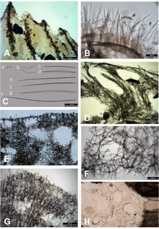
Figure 4 Skeleton and spicular complement of Bulgarian cave sponges. (A) Dysidea incrustans. (B)
Pro-tosuberites denhartogi. (C) Spicules of Clathria (Microciona) cleistochela: a, isochela; b, toxas; c,d, small choanosomal subtylostyles; e,f, large choanosomal subtylostyles; g,h, ectosomal subtylostyles. (D) Skeleton of Halichondria bowerbanki and skeleton. (E) Skeleton of Haliclona sp. 1. (F) Skeleton of Haliclona sp. 2. (G) Skeleton of Haliclona sp. 3. (H) Spicules of Pione cf. vastifica. Photographs by Alexander Ereskovsky.
length) (Fig. 4C). In the middle part of the cave the surface of this species is reduced to 3–7 cm in diameter, where it is covered by numerous Spirorbis polychaetes.
In the middle part of the cave beyond section B–B1, where the marked narrowing of the entrance leads to a drastic reduction of incoming light, the bottom part of the wall is encrusted by the green-gray species Halichondria bowerbanki Burton, 1930 (Fig. 3E;
Video S1). This sponge is moderately firm and compressible in consistence. The oscules are highly variable in size and position. The ectosomal skeleton is tangential with spicules arranged disorderly or in tight bundles, leaving open spaces between spicule bundles. The choanosomal skeleton is largely confused. It consists of oxea tracts running toward the surface and anastomose at intervals (Fig. 4D). These tracts contain between 6 and 12 spicules. The spicules are oxea megascleres only: long, thin, straight or gently curved at the midpoint and varying considerably in size from 135 to 390µm long and 3.7 to 13.1µm wide.
In the same cave zone there is a rare Haliclona (referred to as Haliclona sp. 1 hereafter), a flat sponge of grey-blue color (Fig. 3F;Video S1) with diameters of 7–10 cm, and with small outstretched oscular chimneys of about 5–8 mm. The sponge surface is even and smooth, slightly hispid due to projecting spicules. The ectosomal skeleton is a rather regular, tangential, unispicular, with isotropic reticulation; the choanosomal skeleton is a regular, unispicular, paucispicular reticulation (Fig. 4E). The spicules are exclusively oxeas of uniform shape and size: 64–100 × 2–4µm. No microscleres are present.
The inner, dark parts of the cave are inhabited by small oval and pale blue D. incrustans (Fig. 3C), and a different, large encrusting Haliclona species (referred to as Haliclona sp. 2). Haliclona sp. 2 has ivory color with long, fine outgrowths at the surface (Fig. 3G;
Video S1). The sponge surface is hispid due to projecting spicules. The ectosomal and choanosomal skeletons are tangential. Primary multispicular and secondary unispicular tracts have a regular, triangular reticulation (Fig. 4F). The spicules constitute exclusively megascleres oxeas of uniform shape and size: 150–190 × 6.5–9µm. In this dark part of the cave numerous small crusts (1–3 cm in diameter) of P. denhartogi are present. Finally in the terminal and totally obscured post-syphon zone small individuals of P. denhartogi and big specimens of Haliclona sp. 2 with outgrowths were observed.
The walls of the cave have been perforated by the boring sponges Pione cf. vastifica (Hancock, 1849) (Fig. 3H). The choanosomal skeleton of P. cf. vastifica consists of randomly arranged microspined oxeas and scattered tylostyles and oxeas, sometimes arranged in bundles. The superficial skeleton is organized by spirasters parallel arranged to the surface (Fig. 4H). The skeleton of the papillae is composed by tylostyles (160–340µm long and 3–9µm in diameter) and spiraster microscleres. The spirasters are 8–25µm long and 1–2µm in diameter, straight or with a bent shaft and truncated ends and small spines. Additional microscleres are microspined oxeas with slightly bent shaft of 60–140µm length and 2–6µm in diameter.
The vertical distribution pattern of sponges in the post-syphon area is typical for an open-water area: the largest number of sponges is found in the lower and middle parts of the cliffs, while the upper 2–3 m of rock is almost free of sponges due to the strong hydrodynamic effects of storm activity in this zone.
Tulenova cave
This cave (PB-105/550) has the principal entrance coordinates 43◦29040–5200N and 28◦35005–5800E (Table 1, Fig. 5). The Southern part of the cave is situated from the entrance to A–A1. Then the main part turns almost 90◦. Large caverns and niches, about 0.5 m high, are present in the Southern part of the cave. The surfaces of the walls are uneven, with an angular, flat roof. On the South side at a depth of about 0.5 m a smooth rocky ledge is present. The wall between the ledge and the flat roof of the cave is very uneven, sloping, with large niches and ledges. The parts North of A–A1, on which the mainsail narrows, and the connection to the Eastern part are similar in nature. After the restriction on C the cave is expanding rapidly and reaches its maximum width of 18 m with rounded and smooth outlines. Its bottom is covered with sand and gently rises above sea level. The water depth at the input reaches 3.5 m. With growing distance from the cave entrance water depth gradually decreases. Most of the bottom of the cave is flooded. Almost the entire bottom of the cave halts limestone boulders. Sometimes there are small areas of rock bottom—smooth ledges located at a depth of 0.5 m. On the far side of the cave there is a large sand and pebble beach with detritus and a height of 1 m.
Sponges
The sponge distribution in the entrance zone of the cave is different from that of the rocks outside the cave entrance. The entrance of the cave is completely exposed to waves and light, so the cave per se begins with section A–A1, where the distribution of sponges is typical of dark areas. At the entrance the small pale-blue sponge Dysidea incrustans (Fig. 6A;
Video S2) is reduced in number. However, Protosuberites denhartogi (Fig. 6C;Video S2) constitute the dominant species in this part of the cave with a projective cover about 60–70%. The sponges overgrow Spirorbis polychaetes, barnacles, and mussels. Behind section A–A1, the Eastern side of the cave differs from the Western in the abundance of epibionts. The Eastern wall of the cave, rich in different niches and cavities, is more densely populated by sponges. Up to section C–C1 P. denhartogi is the dominant sponge species, but only in the upper and middle part of the cave. From the middle to the depth part, a flat white-gray Haliclona sp. 2 (Fig. 6F;Video S2) is more abundant in niches and recesses as well as on the mussel shells. In relatively hydrological calm zones some individuals of Haliclona sp. 2 have long outgrowths beginning from the peripheral part of oscular tubes. D. incrustans is quite numerous in the deep zone of the cave; the individuals of this species have rater small dimensions and pale-blue color and inhabit the shells of numerous mussels. Specimens of Haliclona sp. 1 are found in the same zone. This species has small size (2–5 cm), and white color (Fig. 6E;Video S2). There is also a third Haliclona species,
Haliclonasp. 3, with a characteristic violet-blue colour (Fig. 4G;Video S2). The sponge surface is slightly hispid due to projecting ends of choanosomal tracts in varied positions. The choanosomal skeleton is regular, consists of primary multispicular (3–4 spicules) and secondary unispicular tracts form triangular meshes (Fig. 6G). Spicules include only slender and fusiform oxeas: 120–145 × 3.7–5µm and no microscleres.
The rare species Halichondria bowerbanki (Fig. 6D;Video S2) is also found in this zone. This sponge has distinct dermal membrane and chains of oscula at the surface. In addition
Figure 5 Tulenova Cave in the North-East Bulgaria, Black Sea.
Figure 6 Sponges from the Tulenova Cave. (A) Dysidea incrustans in situ blue morph at Cave entrance.
(B) Clathria (Microciona) cleistochela in situ (arrow); (C) Protosuberites denhartogi in situ. (D)
Halichon-dria bowerbanki in situ; (E) Haliclona sp. 1 in situ; (F) Haliclona sp. 2 with outgrowths (arrow) in situ; (G)
Haliclonasp. 3 in situ. Photographs by Oleg A. Kovtun.
Figure 7 Temnata dupka Cave in the North-East Bulgaria, Black Sea.
Full-size DOI: 10.7717/peerj.4596/fig-7
the encrusting sponge Clathria cleistochela (Fig. 6B;Video S2) is found on vertical walls side. This species species has small size and is rare in Tulenova Cave. The rocks in the niches are also perforated by the boring sponge Pione cf. vastifica.
Temnata dupka cave
The cave (PB-105/496) entrance is located at 43◦3001500N, and 28◦35032–1600E (Table 1,
Fig. 7). This cave is a large wave-cut niche with trapezoidal shape. On the far side of the cave, there are several branches/niches. Two of them, in the terminal wall, have small dimensions. The other two branches, extending in the North-East and South-West directions, are comparatively large—the South-West branch has a length of about 30 m. The bottom falls slightly, but then is raised again to a dead end. The height from the water surface to the arch of the dome is 2.5 m with water depths of 1.5 m. The North-East branch length of 16 m is very different. Its water depth at the end of branch reaches 3.5 m. The cross section is rectangular only at the beginning of the branch, and at the middle part it is close to triangular. The surfaces of the walls are more angular, although projections are smoothed. The roof is uneven. The maximum height of the cave at the entrance is 15 m (of which 3 m are flooded). Basically, the cave height comprises about 7–9 m and a maximum
width of 18.5 m. The bottom of the main part of the cave is almost flat, slightly rising in the end of cave and covered with boulders, pebbles and sand. The entire bottom is flooded. The water depth in the entrance parts is up to 3 m, in the North-Eastern branch about 2 m, in the South-West branch maximum depth is 1.4 m, in the end 0.5 m. During storms the access to the cave is difficult.
Sponges
Numerous blue colored Dysidea incrustans specimens inhabit the entrance area of the cave, as well as the surrounding rocks (Fig. 8A:Video S3). Similarly to other caves, Protosuberites
denhartogiis one of dominant species in this area with a projective cover about 30–40%
(Fig. 8C;Video S3). In the middle part of the cave individuals of D. incrustans are small in the beginning, very thin, of whitish color, and very numerous (Fig. 8B;Video S3). The number of this species decreases in the North-Eastern outgrowth of the cave. Here, they often grow on mussels. In this area there are many big individuals of Haliclona sp. 1 with violet-whitish color and relatively big oscula. The sponges have various shapes from oval to irregular (dimension ranging from 3 × 5 cm to 5 × 10 cm) (Fig. 8E;Video S3). In the North-Eastern outgrowth of the cave large, encrusting Halichondria bowerbanki specimens are found (Fig. 8D;Video S3), while on vertical walls in the middle part, several
P. denhartogi and Clathria cleistochela (Fig. 8C;Video S3) specimens can be observed. Encrusting Haliclona sp. 3 of blue-gray color with prominent oscular chimneys up to 8 mm (Fig. 8G;Video S3) was rarely found. In the Western outgrowth of the cave there are many small blue D. incrustans, rare small P. denhartogi and encrusting Haliclona sp. 2 of ivory colour. Pione cf. vastifica perforates the surface of rocks and the shells of old mollusks.
Molecular results
DNA of the relevant sponge species has successfully been extracted, amplified and 28S and CO1 sequenced. The final trees comprised 1,108 (28S) and 2,531 taxa (CO1) respectively. Relevant excerpts of the phylogenetic trees are displayed in Figs. 9A–9D. The samples fall into the four orders Haplosclerida, Dictyoceratida (Dysideidae), Suberitida, and Poecilosclerida (Microcionidae) as expected. Protosuberites denhartogi results in a distinct monophyletic clade with other conspecifics. Halichondria bowerbanki is identical to a previously published H. bowerbanki from the North Sea (HQ379241). For Dysidea spp.,
Haliclonaspp. and Clathria cleistochela, no conspecific, or identical 28S counterpart was found.
DISCUSSION
Geological and morphological features of the caves
In spite of a high number of underwater caves in northern and western parts of the Black Sea (Pronin, 2010;Pronin, 2011) biological investigations were conducted up to now only for the Bulgarian coast and the Crimean Tarkhankut Peninsula (Kovtun & Makarov, 2011;
Petrjachev & Kovtun, 2011;Vorobyova et al., 2012;Ereskovsky, Kovtun & Pronin, 2016); this work). These two areas are similar in their geological structure, despite their separation of about 400 km. The morphological structure of their coastlines is also similar, displaying
Figure 8 Sponges from the Temnata dupka Cave. (A) Dysidea incrustans in situ blue morph at Cave
en-trance. (B) Dysidea incrustans in situ pale morph middle part of the Cave. (C) Clathria (Microciona)
cleis-tochela(C) and Protosuberites denhartogi (P) in situ. (D) Halichondria bowerbanki in situ; (E) Haliclona sp. 1 in situ; (F) Haliclona sp. 2 in situ; (G) Haliclona sp. 3 in situ. Photographs by Oleg A. Kovtun.
99 82 100 98 94 99 100 78 100 99 99 100 100 Spongionella sp KC869493 Dysidea frondosa KC869598 Dysidea etheria KC869555 Dysidea sp KC869532 Dysidea sp KC869550 Dysidea arenaria KC869568
Dysidea fragilis GW30179, BsBul16-9-047 Dysidea fragilis GW30180, BsBul16-9-048 Lamellodysidea herbacea KC869535
Demospongiae sp GW26952 KU060327
Dysidea incrustans GW30164, BsBul16-2-008 Dysidea incrustans GW30165, BsBul16-2-009 Dysidea incrustans GW30162, BsBul16-2-007
Demospongiae sp GW2343 KU060310 Dictyoceratida KC869610 Demospongiae sp GW2347 KU060313 Demospongiae sp GW2341 KU060308 Dysidea sp KC869454 Dysidea sp KC869464
Dysidea incrustans GW30171, BsBul16-4-023 Euryspongia lobata KC869651
Demospongiae sp GW6036 KU060537
Dysidea incrustans GW30168, BsBul16-1-014 Dysidea incrustans GW30170, BsBul16-4-023
Demospongiae sp GW5892 KU060503 Demospongiae sp GW5847 KU060494 Demospongiae sp GW5868 KU060499 Demospongiae sp GW26940 KU060324 0.4 96 77 Echinoclathria dichotoma KC869501 Clathria reinwardti KC869449 Clathria sp. DQ299249 Clathria eccentrica KC869597 Echinochalina sp KC869603
Clathria cleistochela GW30167, BsBul16-2-010 Antho inconstans HQ379399 Antho sp. KC869629 Protosuberites mereui KX601213 80 100 100 100 100 91 96 92 100 92 99 99 95 Ciocalypta penicillus HQ379240 Protosuberites ectyoninus KX601205
Caulospongia biflabellata AY626318 & AY626341 Protosuberites incrustans KX601206
Protosuberites mereui KX601212 Terpios granulosa HQ379250
Pseudosuberites sp AY626317 & AY561913
Halichondria bowerbanki GW30169, BsBul16-1-015 Halichondria bowerbanki HQ379241
Halichondria panicea AF062607 Halichondria panicea HQ379242 Halichondria melanadocia KC869508 Hymeniacidon heliophila KC869620 Pseudosuberites sp. AY561917 Demospongiae sp. GW3535 KU060467 Hymeniacidon kitchingi HQ379243 Ulosa stuposa HQ379227 Demospongiae sp. GW26943 KU060325 Demospongiae sp. GW26927 KU060319 Terpios aploos KC869465 Demospongiae sp GW3375 KU060417 Protosuberites denhartogi KX601207 Protosuberites denhartogi KX601210 Protosuberites denhartogi KX601209
Protosuberites denhartogi GW30163, BsBul16-1-004
Protosuberites denhartogi KX601211 Protosuberites denhartogi KX601208 95 76 92 100 100 100 93 90 62 100 73 100 100 100 Neopetrosia carbonaria KC869628
Amphimedon queenslandica EF654518
Haliclona sp. 2 GW30172, BsBul16-5-024 Haliclona vansoesti KC869631 Haliclona sp. 2 GW30173, BsBul16-5-027 Demospongiae sp GW3387 KU060423 Demospongiae sp GW3336 KU060406 Haliclona sp. 2 GW30144 (Ukraine) Haliclona sp. 2 GW30143 (Ukraine) Haliclona sp. 3 GW30174, BsBul16-5-028 Demospongiae sp GW3337 KU060407 Haliclona sp. 1 GW30177, BsBul16-8-043 Haliclona sp. 1 GW30182, BsBul16-10-051 Demospongiae sp GW2342 KU060309 Demospongiae sp GW2346 KU060312 Haliclona sp. 3 GW30166, BsBul16-2-009/1 Haliclona sp. 3 GW30175, BsBul16-5-029 Demospongiae sp GW3404 KU060429 Demospongiae sp GW3400 KU060428 Demospongiae sp GW3340 KU060408 Demospongiae sp GW3360 KU060410 Demospongiae sp GW3543 KU060471 Demospongiae sp GW3528 KU060465 Demospongiae sp GW6140 KU060547 Demospongiae sp GW3364 KU060413 Demospongiae sp GW3416 KU060435
A
B
C
D
Figure 9 Excerpts from the Maximum Likelihood phylogenetic reconstructions of in total 1,108 se-quences in 28S rDNA (C-region) for species collected in the Bulgarian caves. (A) Halichondria
bower-bankiand Protosuberites denhartogi; (B) Dysidea spp.; (C) Clathria cleistochela; (D) Dysidea spp. Numbers on branches indicate bootstrap support>70. Taxon names in bold are specimens newly sequenced and re-ferred to in this work. Numbers following taxon names indicate either Genbank accession numbers or col-lection numbers of the Bavarian State colcol-lections for Paleontology and Geology ([SNSB-BSPG.]GWxxxxx) plus field collection numbers (BsBulxxxx). The scale bar indicates substitutions per site for all figures.
Table 2 Comparison of shallow-water semi-submerged caves of Tarkhankut (Crimea) and North-East Bulgaria. Crimea Length (m) Depth (m) Area (m2) Width of the enter (m) Sponge species number Bulgaria Length (m) Depth (m) Area (m2) Width of the enter (m) Sponge species number Love 101 4–1.2 1,168 27 7 Tulenova 83 4–1.2 692 19 8 PK-356 24 3–0.8 130 7 4 Temnata dupka 74 3–0.5 345 19 8 PK-324 9 2.5–1 40 4 4 Budova 50 3.5–1 240 8 7
steep banks of 15-40 m height, vertically extending into the sea. Below sea level we find cliffs of up to 6 m before the bottom becomes shallow.
The Bulgarian sites are nearly straight, stretching from North to South, and the entrances to the caves are exposed to the East (Pronin et al., 2013). Due to the straightness of the banks, these areas very often undergo strong wave disturbances. The coastline of the Tarkhankut is oriented East-West, and the entrances to the caves are exposed to the South (Kovtun & Pronin, 2011a;Kovtun & Pronin, 2011b;Pronin, 2011).
The sea coastal cliffs in the Bulgarian and Crimean regions are composed of medium-Sarmatian limestones with identical structures of its layers. Their largest caves, including those mentioned in this paper, are formed in the lower part of the coastal cliffs in dense monolithic or thick-layered limestone. Consequently, the shallow-water semi-submerged caves located on these two shores are morphologically similar.
The shallow semi-submerged caves of Crimea are different in their morphometric parameters (Ereskovsky, Kovtun & Pronin, 2016). Their length differ by factor 10 and the area by factor 29 (from 9 m in the cave PK-324, to 101 m in the cave ‘‘Love’’) (Table 2). The Bulgarian caves studied in this work are more homogeneous, their differences are smaller, their length differ by factor 1.7, and the area differs in 2.7 times (caves of Budova and Tulenova) (Table 2).
Sponge fauna
The analysis of the sponge material reveals that the shallow-water caves in Bulgaria differ in species diversity from caves in Crimea (caves: Love, PK 356, PK 324) (Ereskovsky, Kovtun & Pronin, 2016). In particular, Geodia stellosaCzerniavsky, 1880ordinary inhabitant of the caves of Tarkhankut is absent from the Bulgarian caves, while Halichondria bowerbanki often found in Bulgaria, was not reported from caves of Crimea (Table 3). At the same time, the caves of Bulgaria are more diverse in Haliclona species: seven morphotypes, representing at least three molecularly distinguishable species, are found (Fig. 9). Our observations show that some species in Bulgarian caves reach larger size (e.g., Halichondria
bowerbanki, Dysidea incrustans, Protosuberites denhartogi).
For all three investigated caves sponge distribution is similar in the entrance zone. The dominant species here are D. incrustans and P. denhartogi that cover not only rocky surface, but also the clusters of mussels and different barnacles species. Both sponge species are encrusting a the surface of more than 40 cm2. D. incrustans is generally bright blue in color, like D. fragilis in the entrance of the caves in Crimea (Ereskovsky, Kovtun & Pronin, 2016). However, at the entrance of Tulenova cave, D. incrustans specimens are small and reduced
Table 3 Taxonomical composition and distribution of Black Sea sponge fauna. Numbers refer to the references given at the bottom of this table. Sponge taxon Sponge distribution in Black Sea
Romania Bulgaria Turkey Georgia Russia Ukraine NW
Crimea Crimea caves Class Calcarea
Subclass Calcaronea Order Leucosolenida
Family Sycettidae
Sycon ciliatum(Fabricius, 1780) 1, 16 1 2 3 4, 26
Sycon setosumSchmidt, 1862 1 2 5
Sycon tubaLendenfeld, 1891 2
Syconcf. vigilans Sarà & Gaino, 1971 28 Unpubl
Class Demospongiae Order Tetractinellida
Family Geodiidae
Geodia stellosaCzerniavsky, 1880 6 6 6 5, 9
Stellettasp. 29
Order Suberitida
Family Suberitidae
Suberites domuncula(Olivi, 1792) 1 1 2 9 8, 9 9, 10
Suberites carnosus(Johnston, 1842) 1, 6 6 2 6 4 4
Protosuberites brevispinus(de Laubenfels, 1951)
1, 25
Protosuberites denhartogivan Soest & de Kluijver, 2003
27a
Protosuberites mereuiManconi, 2016 Unpubl Unpubl Unpubl
Protosuberites prototypusSwartschewsky, 1905
1, 6 9 8 8, 10 7
Family Halichondriidae
Halichondria panicea(Pallas, 1766) 1 1, 22 6 9 8 8
Halichondria bowerbankiBurton, 1930 27a
Halichondria(Halichondria) foraminosa (Czerniavsky, 1880)
9
Halichondria(Halichondria) longispicula (Czerniavsky, 1880)
9
Halichondria(Halichondria) pontica (Czerniavsky, 1880)
9 9 10
Hymeniacidon luxurians(Lieberkühn, 1859)
10
Hymeniacidon perlevis(Montagu, 1818) 18, 22 4 4 4 8
Order Clionaida
Family Clionaidae
Pionecf. vastifica (Hancock, 1849) 1, 6 1, 27a 9 11 9, 10 7
Cliona lobataHancock, 1849 1, 6
Table 3(continued)
Sponge taxon Sponge distribution in Black Sea Romania Bulgaria Turkey Georgia Russia Ukraine
NW
Crimea Crimea caves Order Poecilosclerida
Family Mycalidae
Mycale(Aegogropila) contarenii (Lieberkühn, 1859)
1 8 9 8 4, 9
Mycale(Aegogropila) dubia (Czerniavsky, 1880)
9
Mycale(Aegogropila) syrinx (Schmidt, 1862)
16 1 3 4 4, 26
Mycale stepanovii(Czerniavsky, 1880) 9
Mycale lobimana(Czerniavsky, 1880) 9
Mycale jophon(Swartschewsky, 1905) 10
Mycale muscoides(Czerniavsky, 1880) 8 8, 9, 10
Mycale(Mycale) simplex (Czerniavsky, 1880)
9 Family Myxillidae
Myxilla(Myxilla) swartschewskii Burton, 1930
1 4 6 4
Family Tedaniidae
Tedania(Tedania) anhelans (Olivi, 1792) 1 1 2 9
Family Coelosphaeridae
Lissodendoryx(Lissodendoryx) variisclera (Swartschewsky, 1905)
1, 6 24 6 8, 9
Family Crellidae
Crella(Crella) elegans (Schmidt, 1862) 10, 26
Crella(Yvesia) gracilis (Alander, 1942) 1 4 6 8
Family Hymedesmiidae
Hymedesmia(Stylopus) coriacea (Fristedt, 1885)
8 8 8, 10
Hymedesmia(Hymedesmia) pansa Bowerbank, 1882
17
Hymedesmia(Hymedesmia) veneta (Schmidt, 1862)
10 Family Microcionidae
Clathria(Microciona) cleistochela (Topsent, 1925)
12 1, 27a 13
Antho(Antho) involvens (Schmidt, 1864) 9
Order Haplosclerida
Family Chalinidae
Chalinula limbata(Montagu, 1818) 1
Chalinula renieroidesSchmidt, 1868 17
Haliclona alba(Schmidt, 1862) 1 2 9 9
Haliclona albapontica(Czerniavsky, 1880)
9
Haliclona boutschinskii(Kudelin, 1910) 14
Haliclona cribrosa(Czerniavsky, 1880) 9
Table 3(continued)
Sponge taxon Sponge distribution in Black Sea Romania Bulgaria Turkey Georgia Russia Ukraine
NW
Crimea Crimea caves
Haliclona cylindrigera(Czerniavsky, 1880)
9
Haliclona densa(Lendenfeld, 1887) 1, 6 1, 6 10
Haliclona flavescens(Topsent, 1893) 1 1, 19, 25 4 8 7
Haliclona foraminosa(Czerniavsky, 1880) 9 9
Haliclona(Halichoclona) fulva (Topsent, 1893)
17
Haliclona gracilis(Miklucho-Maclay, 1870)
1, 6 3 6 8
Haliclona inflatavar. taurica (Czerniavsky, 1880)
9, 10
Haliclona informisvar. taurica (Czerniavsky, 1880)
1 9 9, 10, 26
Haliclona irregularis(Czerniavsky, 1880) 9 10
Haliclona odessana(Kudelin, 1910) 14
Haliclona palmata(sensu Lieberkühn, 1859)
9 9, 10
Haliclona nigricans(Czerniavsky, 1880) 9
Haliclona pontica(Czerniavsky, 1880) 8 9
Haliclona(Rhizoniera) rosea (Bowerbank, 1866)
17
Haliclona schmidti(Czerniavsky, 1880) 9
Haliclona transitans(Czerniavsky, 1880) 9
Haliclona tubulifera(Swartschewsky, 1905)
1 10
Haliclona(Gellius) angulata (Bowerbank, 1866)
6 6 15 15
Haliclona(Haliclona) simulans (Johnston, 1842)
1 6 26
Haliclona(Reniera) aquaeductus var.
tau-rica(Czerniavsky, 1880)
1, 6 1, 6 2 6 10
Haliclona(Reniera) cinerea (Grant, 1826) 1, 6 1, 6 6 6 5
Haliclona(Reniera) cratera (Schmidt, 1862)
6 2
Haliclona(Rhizoniera) grossa (Schmidt, 1864)
6 2 10
Haliclona(Soestella) implexa (Schmidt, 1868) 1, 6 1 11 5 Haliclonasp.1 27a Haliclonasp.2 27a Haliclonasp.3 27a Haliclonasp.4(2) 7 Family Petrosiidae
Petrosia(Petrosia) clavata (Esper, 1794) 26
Petrosia coriaceaSwartschewsky, 1905 10
Table 3(continued)
Sponge taxon Sponge distribution in Black Sea Romania Bulgaria Turkey Georgia Russia Ukraine
NW
Crimea Crimea caves
Petrosia ficiformis(Poiret, 1789) 1 1 11 11 11
Petrosia(Petrosia) intermedia (Czerniavsky, 1880)
9 9, 10
Family Phloeodictyidae
Oceanapia ascidia(Schmidt, 1870) 6 6 5, 10
Order Dictyoceratida
Family Dysideidae
Dysidea fragilis(Montagu, 1818) 1,6 1, 20, 21,
23, 25
2 6 8, 26 7
Dysidea elegansvar. pontica (Czerniavsky, 1880)
9 6 4
Dysidea avara(Schmidt, 1862) 9 9
Dysidea incrustans(Schmidt, 1862) 27a 9 9
Dysidea pallescens(Schmidt, 1862) 9
Family Irciniidae
Ircinia variabilis(Schmidt, 1862) 17
Family Spongiidae
Spongia(Spongia) officinalis Linnaeus, 1759
6 2
Order Chondrillida
Family Halisarcidae
Halisarca dujardiniJohnston, 1842 2 9 9
Notes.
1,Gomoiu & Skolka, 1998; 2,Topaloğlu & Evcen, 2014; 3,Terentiev, 1998; 4,Kaminskaya, 1968; 5,Kiseleva & Kostenko, 2004; 6,Bačescu, Muller & Gomoiu, 1971; 7,Ereskovsky, Kovtun & Pronin, 2016; 8,Kaminskaya, 1961; 9,Czerniavsky, 1880; 10,Swartschewsky, 1905; 11,Kaminskaya, 1967; 12,Skolka & Gomoiu, 2004; 13,Ereskovsky & Kovtun, 2013; 14, Kudelin, 1910; 15,Kaminskaya, 1966; 16,Begun, Teacă & Gomoiu, 2010; 17,Evcen et al., 2016; 18,Christie et al., 1994; 19,Elenkov, Popov & Andreev, 1999; 20,Christie et al., 1992; 21,De Rosa et al., 2000; 22,Elenkov, Popov & Andreev, 1999; 23,Elenkov et al., 1994; 24,Elenkov et al., 1996; 25,Laubenfels, 1951; 26,Galtsoff, 1923; 27, this work; 28, F Azevedo, pers. comm., 2017; 29, P Cardenas, pers. comm., 2017.
aSponges from the caves of Bulgaria.
in number, because this zone is completely exposed to the waves and sunlight. In the same zone of Budova cave, numerous small individuals of Clathria cleistochela develop.
In the middle part of the caves, a relatively hydrological calm zone with drastic reduction of incoming light, other sponge species with more thickened and soft body contribute to the diversity, like Haliclona spp. 1–3, and Halichondria bowerbanki. For Haliclona spp. small outstretched oscular chimneys, and long, fine outgrowths are found at the surface. The latter is similar to hydrological calm zones of Crimea underwater caves (Ereskovsky, Kovtun & Pronin, 2016). In this zone D. incrustans and P. denhartogi become small and very thin and their abundance decreases towards the inner part of caves.
So far, 87 species belonging to the classes Demospongiae and Calcarea (Table 3) are described from the Black Sea. This number is low number compared to the Mediterranean Sea, which harbors more than 650 species from all four poriferan classes (Pansini & Longo, 2003). For the coastal zone of Bulgaria some 37 species have been reported, which constitute 42.5% of the known Black Sea sponge fauna. Of these, only eight sponge species (21.6%) inhabit the shallow-water semi-submerged caves (Table 4). The use of combine genetic
Table 4 Sponges from marine caves from the North-East of Bulgaria and their spatial distribution in relation to the entrance to the cave.
No Sponges Budova Tulenova Temnata dupka
1 Dysidea incrustans(Schmidt, 1862) 0–37 m 0–33 m 0–6 m
2 Protosuberites denhartogivan Soest, de Kluijver, 2003 0–42 m 0–33 m 0–70 m
3 Clathria (Microciona) cleistochela(Topsent, 1925) 0–22 m 22–25 m 9–22 m
4 Pionecf vastifica (Hancock, 1849) 6–35 m 22–33 m 22–70 m
5 Halichondria bowerbankiBurton, 1930 20–30 m 22–33 m 22–28 m
6 Haliclonasp. 1 20–28 m 25–33 m 22–28 m
7 Haliclonasp. 2 31–42 m 22–33 m 22–70 m
8 Haliclonasp. 3 No 25 m 12–15 m
and morphology analyses allowed us for the first time for the Black Sea to identify two species: Protosuberites denhartogi and Halichondria bowerbanki.
In general, semi-submerged caves host a characteristic lower abundance of sponge diversity in comparison to fully submerged marine caves. This is rather a consequence of higher hydrodynamics and instable environmental conditions than oligotrophy, which is rare in this type of caves. Sponge species composition in caves of Bulgaria as well as Crimea is different from those studied in the Mediterranean (Pouliquen, 1972;Balduzzi et al., 1989;
Harmelin & Vacelet, 1997; Arko-Pijevac et al., 2001; Gerovasileiou & Voultsiadou, 2012;
Gerovasileiou & Voultsiadou, 2016;Manconi et al., 2013), probably due to the geographic isolation of the Black Sea and the differences in the hydro-chemical parameters of the milieu.
The study of sponges from caves of the Black Sea is just at its beginning. At present, only two areas have been surveyed: the West of Crimea and the North-East of Bulgaria. In total 15 species (17.2% from all Black Sea sponges) were reported from caves, of which only
Pionecf vastifica and Clathria cheilochela inhabit the caves of both regions. This percentage is rather small compared to the 311 species (45.7% of the Mediterranean poriferans), representing all four sponge classes, which have been recorded in Mediterranean marine caves (Gerovasileiou & Voultsiadou, 2012).
The present study of underwater cave sponge assemblages in the Black Sea fills regional knowledge gaps for a habitat of special conservation interest. The results of our study highlight the need for (1) further study of the Black Sea underwater caves, and (2) a deep revision of the sponge fauna present in this sea.
ACKNOWLEDGEMENTS
AE acknowledges Christian Marschal, Mediterranean Institute of Marine and Terrestrial Biodiversity and Ecology (IMBE), Marseille, for his technical help. Fieldwork and dive in Bulgarie was possible thanks to Damian Damyanov. DE thanks Gabriele Büttner, Simone Schätzle and Gert Wörheide, LMU Munich, for support in various aspects of the molecular investigations. We acknowledge also Fernanda Azevedo, Universidade Federal
do Rio de Janeiro, Brazil and Paco Cardenas, Department of Medicinal Chemistry, Uppsala University, Sweden for help in sponge species determination.
ADDITIONAL INFORMATION AND DECLARATIONS
Funding
Fieldwork and morphological laboratory work were conducted with support from the Russian Foundation for Basic Research (#15-29-02601 and #15-04-07554, correspondingly); illustrative work and processing of the paper were supported by the Russian Science Foundation (#14-50-00029). Dirk Erpenbeck acknowledges funding by Lehre@LMU. This work also is a contribution to Labex OT-Med (n◦ ANR-11-LABX-0061) and has received funding from Excellence Initiative of Aix-Marseille University— A*MIDEX, a French ‘‘Investissements d’Avenir’’ program. The funders had no role in study design, data collection and analysis, decision to publish, or preparation of the manuscript.
Grant Disclosures
The following grant information was disclosed by the authors:
Russian Foundation for Basic Research: #15-29-02601 and #15-04-07554. Russian Science Foundation: #14-50-00029.
Lehre@LMU.
Labex OT-Med: ANR-11-LABX-0061.
Excellence Initiative of Aix-Marseille University—A*MIDEX.
Competing Interests
The authors declare there are no competing interests.
Author Contributions
• Alexander Ereskovsky and Dirk Erpenbeck conceived and designed the experiments, performed the experiments, analyzed the data, contributed reagents/materials/analysis tools, prepared figures and/or tables, authored or reviewed drafts of the paper, approved the final draft.
• Oleg A. Kovtun conceived and designed the experiments, performed the experiments, analyzed the data, contributed reagents/materials/analysis tools, prepared figures and/or tables, photos and video supplemental files preparation.
• Konstantin K. Pronin conceived and designed the experiments, performed the experiments, contributed reagents/materials/analysis tools, prepared figures and/or tables.
• Apostol Apostolov analyzed the data, authored or reviewed drafts of the paper. • Viatcheslav Ivanenko conceived and designed the experiments, analyzed the data,
contributed reagents/materials/analysis tools, authored or reviewed drafts of the paper, approved the final draft.
Data Availability
The following information was supplied regarding data availability:
Sequences are deposited in NCBI Genbank under accession numbersMH157894–
MH157912, and with location data, specimen photos (including in situ photos) and additional morphological information in the form of thin section photos and spicule preparations in the Sponge Barcoding Database (SBD,http://www.spongebarcoding.org,
Wörheide & Erpenbeck, 2007), accession numbers SBD #1758–1776.
Supplemental Information
Supplemental information for this article can be found online athttp://dx.doi.org/10.7717/ peerj.4596#supplemental-information.
REFERENCES
Arko-Pijevac M, Benac C, Kovacic M, Kirincic M. 2001. A submarine cave at the Island
of Krk (North Adriatic Sea). Natura Croatica 10:163–184.
Bačescu M, Muller G, Gomoiu M-T. 1971. Cercetari de ecologie bentala in Marea Neagra—analiza cantitativa si comparata a faunel bentale Pontice. Vol. 4. Bucuresti: Romanian Academy of Sciences Publications, 1–352.
Balduzzi A, Bianchi CN, Boero F, Cattaneo Vietti R, Pansini M, Sarà M. 1989. The
suspension-feeder communities of a Mediterranean Sea cave. Scientia Marina
53:387–395.
Begun T, Teacă A, Gomoiu MT. 2010. State of macrobenthos within Modiolus
phase-olinusbiocoenosis from romanian Black Sea continental shelf. Geo-Eco-Marina
16:5–18.
Bell JJ. 2002. The sponge community in a semi-submerged temperate sea cave: density,
diversity and richness. Marine Ecology 23:297–311.
Bell JJ, Smith D. 2004. Ecology of sponge assemblages (Porifera) in the
Waka-tobi region, south-east Sulawesi, Indonesia: richness and abundance.
Jour-nal of the Marine Biological Association of the United Kingdom84:581–591
DOI 10.1017/S0025315404009580h.
Bianchi CN, Morri C, Russo GF. 2003. Deplezione trofica. In: Cicogna F, Bianchi CN,
Ferrari G, Forti P, eds. Grotte marine: cinquant’anni di ricerca in Italia. Roma: Ministero dell’Ambiente e della Tutela del Territorio, 297–305.
Bussotti S, Terlizzi A, Fraschetti S, Belmonte G, Boero F. 2006. Spatial and temporal
variability of sessile benthos in shallow Mediterranean marine caves. Marine Ecology
Progress Series325:109–119DOI 10.3354/meps325109.
Chevaldonné P, Lejeusne C. 2003. Regional warming-induced species shift in north-west
Mediterranean marine caves. Ecology Letters 6:371–379
DOI 10.1046/j.1461-0248.2003.00439.x.
Chombard C, Boury-Esnault N, Tillier S. 1998. Reassessment of homology of
mor-phological characters in tetractinellid sponges based on molecular data. Systematic
Christie WW, Brechany EY, Marekov IN, Stefanov KL, Andreev SN. 1994. The fatty
acids of the sponge Hymeniacidon sanguinea from the Black Sea. Comparative
Biochemistry and Physiology 109B:245–252.
Christie WW, Brechany EY, Stefanov KL, Popov S. 1992. The fatty acids of the sponge
Dysidea fragilisfrom the Black Sea. Lipids 27:640–644DOI 10.1007/BF02536125.
Corriero G. 1989. Primi dati sul popolamento di Poriferi delle grotte superficiali
dell’isola di Ustica. Nova Thalassia 10:585–588.
Corriero G, Scalera Liaci L, Gristina M, Riggio S, Mercurio M. 1997. Composizione
tas-sonomica e distribuzione della fauna a Poriferi e Briozoi in una grotta semisommersa della Riserva Naturale Marina Isola di Ustica. Biologie Marine Mediterraneen 4:34–43.
Corriero G, Scalera Liaci L, Ruggiero D, Pansini M. 2000. The sponge
commu-nity of a semi-submerged Mediterranean cave. Marine Ecology 21:85–96
DOI 10.1046/j.1439-0485.2000.00655.x.
Czerniavsky V. 1880. Spongiae littorales Pontis Euxini et Maris Caspii. Bulletin de la Société impériale des naturalistes de Moscou54:88–128.
De Rosa S, De Giulio A, Tommonaro G, Popov S, Kujumgiev A. 2000. A
beta-Amino acid containing tripeptide from a pseudomonas-alteromonas bacterium associated with a Black Sea sponge. Journal of Natural Products 63:1454–1455
DOI 10.1021/np0002132.
Dimarchopoulou D, Gerovasileiou V, Voultsiadou E. 2018. Spatial variability of
sessile benthos in a semi-submerged marine cave of a remote Aegean Island (eastern Mediterranean Sea). Regional Studies in Marine Science 17:102–111
DOI 10.1016/j.rsma.2017.11.015.
Elenkov I, Milkova T, Andreev S, Popov S. 1994. Sterol composition and biosynthesis
in the Black Sea sponge Dysidea fragilis. Comparative Biochemistry and Physiology
107B:547–551.
Elenkov I, Popov S, Andreev S. 1999. Sterol composition of the Black Sea sponges
Hymeniacidon sanguinea(Grant) and Halichondria panicea (Pallas). Zeitschrift für
Naturforschung 54:972–976.
Elenkov I, Stefanov KL, Andreev SN, Popov SS, Christie WW, Robertson GW, Shep-herd T. 1996. Sterol composition of the Black Sea sponge Lissodendoryx variisclera.
Comparative Biochemistry and Physiology115B:287–289.
Ereskovsky AV, Kovtun OA. 2013. Ukraine’s new invasive sponge species, Clathria
(Microciona) cleistochela (Topsent, 1925) (Porifera: Demospongiae, Poeciloscle-rida, Microcionidae) from the submarine caves of western Crimea (Black Sea, Tarkhankut). Marine Ecology Journal 12:104 (In Russian).
Ereskovsky AV, Kovtun OA, Pronin KK. 2016. Marine cave biota of the Tarkhankut
Peninsula (Black Sea, Crimea), with emphasis on sponge taxonomic composition, spatial distribution and ecological particularities. Journal of the Marine Biological
Association of the United Kingdom96:391–406 DOI 10.1017/S0025315415001071.
Erpenbeck D, Nichols SA, Voigt O, Dohrmann M, Degnan BM, Hooper JNA, Wörheide G. 2007. Phylogenetic analyses under secondary structure-specific substitution
models outperform traditional approaches: case studies with diploblast LSU. Journal
of Molecular Evolution64:543–557DOI 10.1007/s00239-006-0146-3.
Erpenbeck D, Voigt O, Al-Aidaroos AM, Berumen ML, Büttner G, Catania D, Guirguis AN, Paulay G, Schätzle S, Wörheide G. 2016. Molecular
biodi-versity of Red Sea demosponges. Marine Pollution Bulletin 105:507–514
DOI 10.1016/j.marpolbul.2015.12.004.
Erpenbeck D, Voigt O, Gültas M, Wörheide G. 2008. The sponge genetree server—
providing a phylogenetic backbone for poriferan evolutionary studies. Zootaxa
1936:58–60.
Evcen A, Çinar ME, Zengin M, Süer S, Rüzgar M. 2016. New records of five sponge
species (Porifera) for the Black Sea. Zootaxa 4103:267–275
DOI 10.11646/zootaxa.4103.3.5.
Galtsoff PS. 1923. The amoeboid movement of dissociated sponge cells. Biological
Bulletin45:153–161DOI 10.2307/1536625.
Gerovasileiou V, Voultsiadou E. 2012. Marine caves of the Mediterranean Sea: a sponge
biodiversity reservoir within a biodiversity hotspot. PLOS ONE 7(7):e39873
DOI 10.1371/journal.pone.0039873.
Gerovasileiou V, Voultsiadou E. 2016. Sponge diversity gradients in marine caves of
the eastern Mediterranean. Journal of the Marine Biological Association of the United
Kingdom92:407–416.
Gerovasileiou V, Voultsiadou E, Issaris Y, Zenetos A. 2016. Alien biodiversity in
Mediterranean marine caves. Marine Ecology 37:239–256DOI 10.1111/maec.12268.
Giakoumi S, Sini M, Gerovasileiou V, Mazor T, Beher J, Possingham HP, Abdulla A, Çinar ME, Dendrinos P, Gucu AC, Karamanlidis AA, Rodic P, Panayotidis P, Taskin E, Jaklin A, Voultsiadou E, Webster C, Zenetos A, Katsanevakis S. 2013.
Ecoregion-based conservation planning in the Mediterranean: dealing with large-scale heterogeneity. PLOS ONE 8(10):e76449DOI 10.1371/journal.pone.0076449.
Glover AG, Gooday AJ, Bailey DM, Billett DSM, Chevaldonné P, Cola¸co A, Copley J, Cuvelier D, Desbruyères D, Kalogeropoulou V, Klages M, Lampadariou N, Lejeusne C, Mestre NC, Paterson GL, Perez T, Ruhl H, Sarrazin J, Soltwedel T, Soto EH, Thatje S, Tselepides A, Van Gaever S, Vanreusel A. 2010. Temporal change in
deep-sea benthic ecosystems: a review of the evidence from recent time-series stud-ies. Advance in Marine Biology 58:1–95 DOI 10.1016/B978-0-12-381015-1.00001-0.
Gomoiu MT, Skolka M. 1998. Evaluation of marine and coastal biological diversity at
the Romanian littoral—a workbook for the Black Sea ecological diversity. Analele
Universitatii ‘‘Ovidius’’ Constanta. Universitatea ‘‘Ovidius’’ Constanta2(anul 2):1–167.
Harmelin JG. 1980. Etablissement des communautés de substrats durs en milieu obscur.
Résulta de Biologia marina y Oceanografia10:29–52.
Harmelin JG. 1997. Diversity of bryozoans in a Mediterranean sublitoral cave with
bathyal-like conditions: role of dispersal processes and local factors. Marine Ecology
Harmelin JG, Vacelet J. 1997. Clues to deep-sea biodiversity in a nearshore cave. Vie et
Milieu47:351–354.
Hart CW, Manning RB, Iliffe TM. 1986. Mesozoic relicts in marine caves of Bermuda. Stygologia2:156–166.
Iliffe TM, Kornicker LS. 2009. Worldwide diving discoveries of living fossil animals from
the depths of anchialine and marine caves. Smithsonian Contributions to the Marine
Sciences38:269–280.
Janssen A, Chevaldonné P, Martínez Arbizu P. 2013. Meiobenthic copepod fauna of a
marine cave (NW Mediterranean) closely resembles that of deep-sea communities.
Marine Ecology Progress Series479:99–113DOI 10.3354/meps10207.
Kaminskaya LD. 1961. New facts concerning the fauna of Black Sea sponges. Dopovidi
Akademii Nauk ukraini RSR8:1091–1093.
Kaminskaya LD. 1966. An addition to the sponge fauna of the Black Sea. Zoologicheskii
Zhurnal65:928–929.
Kaminskaya LD. 1967. Some patterns of distribution of species of sponges in the Black
Sea. Zoologicheskii Zhurnal 64:327–329.
Kaminskaya LD. 1968. Klass gubok—Porifera. In: Vodjanitskij VV, ed. Opredelitel fauny Chernogo I Azovskogo morey ( A key to Black Sea and Azov Sea fauna). 1. Kiev: Naukova dumka, 35–55 (In Russian).
Kearse M, Moir R, Wilson A, Stones-Havas S, Cheung M, Sturrock S, Buxton S, Cooper A, Markowitz S, Duran C, Thierer T, Ashton B, Meintjes P, Drummond A. 2012. Geneious basic: an integrated and extendable desktop software platform
for the organization and analysis of sequence data. Bioinformatics 28:1647–1649
DOI 10.1093/bioinformatics/bts199.
Kiseleva GA, Kostenko NS. 2004. Phylum Porifera. In: Kudrjavtsev VB, ed. Karadag: proceedings of the conference on hydrobiologivcal investigations 2. Sonat: Simpheropol, 322–323 (In Russian).
Knittweis L, Chevaldonné P, Ereskovsky A, Schembri PJ, Borg JA. 2015. A preliminary
survey of marine cave habitats in the Maltese Islands. Xjenza Online 3:153–164
DOI 10.7423/XJENZA.2015.2.07.
Kobluk DR, Van Soest RWM. 1989. Cavity-dwelling sponges in a southern Caribbean
coral reef and their paleontological implications. Bulletin of Marine Science
44:1207–1235.
Kovtun OA, Makarov YN. 2011. The features of biology and morphology of the rare
Black Sea shrimp Palaemon serratus Pennant, 1777 (Decapoda: Palaemonidae) from karst caves and underwater caves Tarkhankut Peninsula (Western Crimea). Morskoj
ecologicheskij zhurnal10:26–32 (In Russian).
Kovtun OA, Pronin KK. 2011a. Marine caves of Ukrainian coast (problems, history of
study, biology). In: Sirenko IN, ed. Second international scientific and practical
con-ference Speleology and speleology. Naberejnie Chelni: Naberejnie Chelni University Press, 33–38 (In Russian).
Kovtun OA, Pronin KK. 2011b. Morphological and biological features of underwater
caves Tarzanka (Tarhankut Peninsula, Maliy Atlesh). Speleology and Karstology
6:53–66.
Kudelin N. 1910. Zur Frage Spongien des Schwarzen Meeres. Zapiski Novorossijskogo Obshestva Estestvoispitatelej35:1–40.
Labate M. 1964. Poriferi di grotta superficiale del litorale adriatico pugliese. Annali del Pontificio Istituto superiore di Scienze e Lettere ‘‘Santa Chiara’’ di Napoli14:319–342. Laborel J, Vacelet J. 1959. Les grottes sous-marines obscures en Méditerranée.
Comptes-Rendus de l’Académie des Sciences de Paris248:2619–2626.
Laubenfels MW de. 1951. A collection of sponges from the Black Sea. Archiv fur
Hydrobiologie45:213–216.
Lejeusne C, Chevaldonné P. 2005. Population structure and life history of Hemimysis
margalefi(Crustacea: Mysidacea), a ‘thermophilic’ cave-dwelling species benefiting
from the warming of the NW Mediterranean. Marine Ecology Progress Series
287:189–199DOI 10.3354/meps287189.
Lejeusne C, Chevaldonné P. 2006. Brooding crustaceans in a highly fragmented habitat:
the genetic structure of Mediterranean marine cave-dwelling mysid populations.
Molecular Ecology 15:4123–4140DOI 10.1111/j.1365-294X.2006.03101.x.
Macintyre IG, Rützler K, Norris JN, Fauchald K. 1982. A submarine cave near
Colum-bus Cay, Belize: a bizarre cryptic habitat. In: Rützler K, Macintyre IG, eds. The
Atlantic Barrier Reef Ecosystem at Carrie Bow Cay, Belize, I structure and communities. Vol. 12. Washington, D.C.: Smithsonian Contributions to the Marine Sciences, 127–142.
Manconi R, Cadeddu B, Ledda F, Pronzato R. 2013. An overview of the Mediterranean
cave-dwelling horny sponges (Porifera, Demospongiae). ZooKeys 281:1–68
DOI 10.3897/zookeys.281.4171.
Manconi R, Ledda FD, Serusi A, Corso G, Stocchino GA. 2009. Sponges of marine
caves: notes on the status of the Mediterranean palaeoendemic Petrobiona massiliana (Porifera: Calcarea: Lithonida) with new records from Sardinia. Italian Journal of
Zoology76:306–315DOI 10.1080/11250000802629471.
Manconi R, Serusi A. 2008. Rare sponges from marine caves: discovery of
Neophris-sospongia nananov. sp. (Demospongiae, Corallistidae) from Sardinia with an
annotated checklist of Mediterranean lithistids. ZooKeys 4:71–87
DOI 10.3897/zookeys.4.39.
Manconi R, Serusi A, Pisera A. 2006. Mediterranean ‘Lithistids’ sponges: discovery
of Aciculites mediterranea sp. n. (Porifera, Demospongiae) in dark marine caves of Sardinia. Journal of the Marine Biological Association of the United Kingdom
86:691–698DOI 10.1017/S0025315406013580.
Meyer CP, Paulay G. 2005. DNA barcoding: error rates based on comprehensive
sampling. PLOS Biology 3:e422DOI 10.1371/journal.pbio.0030422.
Nepote E, Bianchi CN, Morri C, Ferrari M, Montefalcone M. 2017. Impact of a harbour
construction on the benthic community of two shallow marine caves. Marine