Cognitive signals for brain–machine interfaces in posterior
parietal cortex include continuous 3D trajectory commands
The MIT Faculty has made this article openly available.
Please share
how this access benefits you. Your story matters.
Citation
Hauschild, M. et al. “Cognitive Signals for Brain-machine Interfaces
in Posterior Parietal Cortex Include Continuous 3D Trajectory
Commands.” Proceedings of the National Academy of Sciences
109.42 (2012): 17075–17080. ©2013 National Academy of Sciences
As Published
http://dx.doi.org/10.1073/pnas.1215092109
Publisher
National Academy of Sciences (U.S.)
Version
Final published version
Citable link
http://hdl.handle.net/1721.1/78850
Terms of Use
Article is made available in accordance with the publisher's
policy and may be subject to US copyright law. Please refer to the
publisher's site for terms of use.
Cognitive signals for brain
–machine interfaces in
posterior parietal cortex include continuous 3D
trajectory commands
Markus Hauschild
a, Grant H. Mulliken
b, Igor Fineman
c, Gerald E. Loeb
d, and Richard A. Andersen
a,1aDivision of Biology, California Institute of Technology, Pasadena, CA 91125;bBrain and Cognitive Sciences Department and McGovern Institute for Brain
Research, Massachusetts Institute of Technology, Cambridge, MA 02139;cDepartment of Neurosurgery, Huntington Memorial Hospital, Pasadena, CA 91105;
anddDepartment of Biomedical Engineering, University of Southern California, Los Angeles, CA 90089
Contributed by Richard A. Andersen, September 4, 2012 (sent for review June 23, 2012)
Cortical neural prosthetics extract command signals from the brain with the goal to restore function in paralyzed or amputated patients. Continuous control signals can be extracted from the motor cortical areas, whereas neural activity from posterior parietal cortex (PPC) can be used to decode cognitive variables related to the goals of movement. Because typical activities of daily living comprise both continuous control tasks such as reaching, and tasks benefiting from discrete control such as typing on a keyboard, availability of both signals simultaneously would promise significant increases in performance and versatility. Here, we show that PPC can provide 3D hand trajectory information under natural conditions that would be encountered for prosthetic applications, thus allowing simulta-neous extraction of continuous and discrete signals without re-quiring multisite surgical implants. We found that limb movements can be decoded robustly and with high accuracy from a small population of neural units under free gaze in a complex 3D point-to-point reaching task. Both animals’ brain-control performance improved rapidly with practice, resulting in faster target acquisition and increasing accuracy. Thesefindings disprove the notion that the motor cortical areas are the only candidate areas for continuous prosthetic command signals and, rather, suggests that PPC can pro-vide equally useful trajectory signals in addition to discrete, cogni-tive variables. Hybrid use of continuous and discrete signals from PPC may enable a new generation of neural prostheses providing superior performance and additionalflexibility in addressing in-dividual patient needs.
cognitive neural prosthetic
|
parietal reach region|
area 5D
ifferent cortical areas have been identified as sources for
cortical prosthetics to assist subjects with paralysis or
am-putation (1–13). Motor cortex can provide continuous control
of trajectories (3–5, 11–13), which is consistent with its normal
function of sending commands directly to the movement
gener-ating circuits of the spinal cord. More cognitive variables related
to reach goals have been extracted from the parietal reach region
(PRR) and area 5d in posterior parietal cortex (PPC) (7, 14, 15).
There are several advantages of these cognitive variables for
pros-thetic applications: (i) decodes of goals are very fast, in the order
of 100 ms, and can assist in typing applications (7); (ii) at least
two sequential goals can be represented in PRR, and this feature
can augment typing and sequential limb movements (16); (iii) goal
and trajectory information, when combined, provide better
decod-ing of trajectories than trajectory information alone (17); (iv)
bilateral arm movements to a goal are represented and can assist
in decoding bimanual behaviors from a single hemisphere (18);
and (v) the anterior intraparietal area (AIP) of PPC represents
grasp shape, which may reduce the number of cells needed to
decode grasping (19).
If PPC also encodes trajectories, then its repertoire of uses for
prosthetics control would be further expanded. Deficits in online
control of movement trajectories found in clinical studies, for
in-stance, difficulty in trajectory correction during movement (20–22),
indicate that PPC is an important site for continuous control of
movement, suggesting that movement parameters can be decoded
in PPC. Moreover, recent studies show that, under very constrained
laboratory conditions of stereotyped movements (2D center-out
movements) and with the gaze
fixed, trajectory information can
be decoded from PPC neurons (17, 23). However, there has been
no demonstration that PPC can be used for the more demanding
conditions required for neural prosthetic applications that include
3D reaches from varying beginning and end points with gaze free.
The ability to use PPC for everyday prosthetics applications,
for both trajectory and goal decoding, is also an open question
given the
findings that reach targets, particularly in PRR, are coded
primarily in eye coordinates (24–26). With gaze free, decoding
would, in principle, be much more inaccurate than with gaze
fixed. Thus, in the current experiments, we tested whether PPC
could provide trajectory information in the presence of natural
eye movements and under generally more realistic conditions,
in-cluding sequences of point-to-point-movements in a 3D workspace.
To investigate the feasibility of extracting prosthetic command
signals from PPC, we simultaneously recorded ensembles of
single-and multiunit spiking activity from area 5d single-and PRR (Fig. 1D
and
Fig. S1
) in two rhesus monkeys while they performed
rea-ches. First, each monkey used his hand to steer a cursor (reach
control) in a 3D virtual reality (VR) environment (Fig. 1
A and B
and
Movie S1
). We constructed and evaluated linear ridge
re-gression (27) and Kalman
filter (28) decode models for offline
reconstruction of cursor movement from the concurrently
recor-ded neural activity (Fig. 1C). The reach sessions were followed
by brain-control sessions where VR-cursor movement was driven
by neural activity instead of hand movement to test whether the
previously identified decode model would be suitable for direct
cortical control of a prosthetic.
Results
Offline Reconstruction.
Twenty-nine reach-control sessions were
analyzed in monkey R and 33 in monkey G. The offline
recon-struction performance was quantified using the coefficient of
determination,
R
2, for the best day (Table 1) and the average
over all recording days (Table 2). Despite free gaze, the decode
model operating in a screen-centered reference frame captured
the key features of 3D hand movement (Fig. 2 and
Fig. S2
) with
best-day position reconstruction performance
R
2= 0.68/0.62
(monkey R/G) and average (over all recording days) position
reconstruction performance
R
2= 0.61/0.52 (monkey R/G.). The
Kalman
filter provided position estimates significantly more
ac-curate than the ridge
filter estimates (P < 10
−8for monkey R;
P < 10
−9for monkey G; two-sided sign test).
Author contributions: M.H., G.H.M., G.E.L., and R.A.A. designed research; M.H. performed research; I.F. performed surgical procedures; M.H. analyzed data; and M.H., G.H.M., and R.A.A. wrote the paper.
The authors declare no conflict of interest.
Freely available online through the PNAS open access option.
1To whom correspondence should be addressed. E-mail: [email protected]. This article contains supporting information online atwww.pnas.org/lookup/suppl/doi:10.
1073/pnas.1215092109/-/DCSupplemental.
NEUROSCI
To assess how well trajectories could be reconstructed from
PPC neural ensembles of different sizes, we constructed neuron
dropping curves (
Fig. S3
). They show that the position-decoding
performance for a neural ensemble of a particular size is very
similar between the two animals, although differences in decoding
accuracy for velocity and acceleration exist. The neuron-dropping
curves also reveal that the reported decoding performance (Tables
1 and 2) is better in monkey R primarily because more neural
units were available.
Neural units in PRR are known to respond to visual stimuli
(29), which could presumably impair trajectory reconstruction
performance, particularly during the onset of high-contrast visual
target cues. We, therefore, compared our decoding results with
the performance obtained from reconstruction of the same sets
of reaches, but after elimination of all visual cue onset phases,
and found that the difference in decoding performance was small
in both monkeys (
SI Results
).
Furthermore, the optimal lag time (OLT), representing the
temporal offset of movement vs. neural population activity where
R
2tuning was maximal (Table 2), showed that neural population
activity led movement execution on average by
∼80 ms in both
monkeys, despite strong known proprioceptive and visual sensory
inputs to PPC.
In summary, these offline reconstruction results suggest that
(i) PPC populations of neurons allow accurate reconstruction of
3D trajectories under free gaze in a stationary reference frame;
(ii) the decoded signal is insensitive to visual perturbations; and
(iii) the neural signal leading the movement represents the animals’
intention to move rather than a sensory correlate of movement,
thus qualifying it as a potential prosthetic control signal.
Brain Control.
Twenty-five reach sessions were followed by
control sessions in monkey R and 15 in monkey G. In the
brain-control task, VR-cursor movement was driven by neural activity
instead of hand movement to test whether the previously
iden-tified decode model would be suitable for direct cortical control
of a prosthetic. Both animals performed the brain-control task
successfully (
Movie S2
). They frequently acquired targets
rap-idly, performing mostly straight reaches directed toward the goal
from the initiation of the movement (Fig. 3A), but a number of
reaches required adjustments to correct for initially erroneous
trajectories (Fig. 3B and
Fig. S4
). Such visual feedback–driven
error correction frequently resulted in successful target
acqui-sition. Over time, behavioral performance improved with
practice. During 19/10 (monkey R/G) ridge decode sessions, the
success rate increased significantly from 29.63% on the first day
to a maximum of 77.78% on day 17 [regression line slope
m =
1.48; 95% confidence interval (CI): 0.72/2.23 (lower/upper
bounds)] in monkey R and from 37.04% to 85.19% on day 10
(m = 4.24; 95% CI: 2.08/6.40) in monkey G, while always remaining
significantly above chance level (Fig. 4). The mean time each
monkey required to acquire a target successfully decreased
sig-nificantly from 2.18 to 1.54 s (m = −0.033; 95% CI: −0.052/
−0.014) in monkey R and from 1.31 to 1.13 s (not significant) in
monkey G, whereas trajectory straightness, quantifying the
goal-directedness of the brain-control trajectories, improved (m =
0.041; 95% CI: 0.026/0.057 in monkey R; not significant in
monkey G) (Fig. 4A). To benchmark brain-control task
pro-ficiency, we compared time-to-target and trajectory straightness
in monkey R (where both variables improved significantly over
time) to the same-day performances achieved under
hand-control (
Fig. S5
). The comparison highlights that (i) increasing
Stereoscopic image projection from overhead monitor
Mirror 3D shutter glasses 3D hand tracker z y x
...
Spike sorting Trajectory decoding ...A
B
D
C
Time 1 df hand-trajectory Hand-control neural and kinematic data sample Neuron 1: spikes and binned activity Neuron 2
Time Target
appears
Reaction time & reach execution
Target acquired
Target appears
Reaction time & reach execution Target 1 Target appears ... Target acquired Target acquired Juice reward Target 2
Reach sequence (6-8 point-to-point reaches)
Target 3, ... , Target 8 M1
10 mm
PRR/area 5d
Fig. 1. Behavioral paradigm. (A) In daily recording sessions, each monkey guided a cursor in a 3D VR display to a reach target. The monkeyfirst used his hand to control cursor movement (reach control). Then he steered the cursor using cortical activity (brain control) translated to cursor movement by the decode model identified from the preceding reach-control phase. (B) Timeline of the reach task. Reaches were performed in sequences of six or eight targets. The monkey was rewarded with juice after having completed a sequence of reaches. In brain-control mode, the monkey was rewarded after suc-cessful acquisition of single targets. (C) Single df tra-jectory sample, spike trains, and processed spike bins recorded simultaneously during the reaching task. (D) Unlike previous approaches targeting the mo-tor areas, here, continuous control signals were extracted from PPC. Electrodes were implanted in PRR in the intraparietal sulcus (yellow marker in the coronal MRI slice) and area 5d on the cortical surface.
Table 1. Single best-day offline reconstruction performance (mean ± SD) for ridge and Kalman filter
Monkey
(no. of neural units)
Kalmanfilter, R2 Ridgefilter, R2
x/y/z combined Single-best df x/y/z combined
Position Velocity Acceleration Position Position
R (70) 0.68± 0.03 0.59± 0.02 0.33± 0.02 0.77± 0.02 0.45± 0.05
G (55) 0.62± 0.04 0.36± 0.03 0.11± 0.02 0.76± 0.01 0.46± 0.05
performance is specific to the brain-control phase of the
ex-periment and therefore cannot be explained by generally
im-proved VR-task proficiency; and (ii) over time, brain-control
performance approaches hand-control performance. Success rate,
time to target, and trajectory straightness also showed steady
improvement during 6/5 (monkey R/G) Kalman
filter
brain-control sessions. In monkey R, the success rate saturated at
100% after four sessions, and in monkey G, performance
re-covered from initially 44% to a maximum of 63% success rate
despite availability of only a few neural units from aging array
implants (Fig. 4B).
Monkey R was required not to move his limb while controlling
cursor movement during a set of nine separate sessions to test
brain-control in the absence of proprioceptive feedback
modu-lating PPC activity. The monkey was not accustomed to the
electromyographic (EMG) recording equipment attached to his
arm to monitor muscle activation; therefore, movements
per-formed while wearing the equipment (Fig. 3D) were less smooth
and targets were acquired more slowly than under regular
con-ditions under both hand-control (e.g., Fig. 2A vs. 3D) and brain
control (e.g., Fig. 3
A and B vs. 3C). Despite this limitation,
monkey R reached up to 66.67% brain-control success rate
(chance performance 23.67
± 1.49%) in the absence of
detect-able limb movement (Fig. 3
C and D and
Movie S3
). This result
suggests that somatosensory feedback is not necessary to
gen-erate control signals in PPC, which will be important for clinical
applications in patients who typically have sensory, as well as
mo-tor, deficits. The algorithm was trained during actual reaching
movements, presumably accompanied by proprioceptive feedback,
whereas the brain-control results were obtained in absence of limb
movement, thus generating a mismatch between the decoding
model and the inputs it expected based on the data on which it was
trained. Results may, therefore, be even better when algorithms
are trained in the absence of proprioception from the limb, such as
in prosthetic patients for whom algorithms will need to be trained
using neural activity during imagined movements.
Discussion
The results of this study show that complex, 3D point-to-point
movement trajectories can be decoded from PPC under free gaze
and that PPC-based brain–machine interfaces (BMIs) for
con-tinuous neural control of 3D manipulators are feasible.
Prior PPC studies reported substantially lower performance
(R
2below 0.3) in two free-gaze 2D decoding studies (2, 5). These
low values may reflect the small number of electrodes implanted
in the one study (2), whereas in the other study (5), very good
grasp-decoding performance was reported, suggesting that the
actual targeted site of the PPC was more involved with grasp.
R
2results comparable to those reported previously by our group in a
highly constrained 2D center-out PPC decoding study (17)
sug-gest that the removal of behavioral constraints such as eye
fixation
and increased task complexity does not impair the usefulness of
PPC signals for prosthetic applications. Furthermore, the
decod-ing algorithms operated continuously, requirdecod-ing neither
reinitiali-zation at the beginning of a trial or a sequence nor elimination of
visual cue onset responses (29), thus generalizing previous
findings (17) to a realistic, unconstrained 3D prosthetic limb control
scenario without compromising decoding accuracy.
R
2decoding performances reported for M1 have ranged from 0.3
to 0.7 (2, 3, 5), and, thus, the PPC offline decoding results appear to
be on par with M1 performance. Brain-control performance,
com-monly quantified by success rates, appears to be similar to the results
reported in a motor cortex–based 3D brain-control study by Taylor
et al. (4) (
SI Results
). Notwithstanding caution in consideration of
methodological differences, these results suggest that achievable
brain-control performance is comparable to motor cortex.
Table 2. Average (across all sessions) offline reconstruction performance (mean ± SD) for ridge and Kalman filter
Monkey
(no. of neural units)
Kalmanfilter Ridgefilter
R2x/y/z combined OLT (ms) R2x/y/z combined
Position Velocity Acceleration Position Position
R (65.86± 6.89) 0.61± 0.06 0.58± 0.05 0.34± 0.07 82.35± 40.18 0.45± 0.05 G (64.29± 15.02) 0.52± 0.06 0.24± 0.05 0.07± 0.02 79.63± 39.43 0.36± 0.06
The reported performance was achieved using all neural units (single- and multiunit activity) recorded from area 5d and PRR combined. Because 75% of the implanted electrodes were designed for surface recordings, the neural ensembles reported contained more surface (area 5d) neural units than neural units from the deeper structures (PRR).
A
B
C
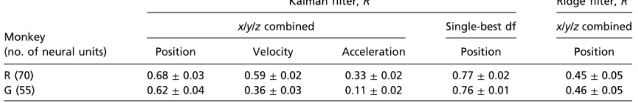
Fig. 2. Offline Kalman filter 3D-trajectory reconstruction. PPC populations of neurons allow the decoding of position, velocity, and acceleration profiles with high accuracy in a free gaze point-to-point reaching task. (A) Position reconstruction (black) of a previously recorded sequence of reaches (red) to eight targets (blue).●, discrete reconstruction points resulting from the 90-ms sampling rate used. (B and C ) Velocity reconstruction (B) and acceleration reconstruction (C) for the same sequence.
NEUROSCI
At
first glance, it is surprising to find that PPC encodes a
trajec-tory because it is motor cortex and not PPC that sends movement
commands directly to the spinal cord. However, computational
models of motor control, as well as lesions to patients and
recordings from animals, suggest that PPC signals represent state
estimates of ongoing movement whereas M1 signals carry motor
commands (20–23, 30–32). Thus, the signals from PPC and M1,
although serving different purposes in the brain, are equally
suitable for decoding trajectories.
Previous research suggested that neurons in PRR rely primarily
on gaze-centered reference frames to represent reach goals (24)
and that area 5d neurons use simultaneous gaze- and limb-centered
target representations (25). Thus, it appears to be
counterintui-tive that ongoing movement can be decoded from populations of
neurons in a stationary body-centered reference frame, especially
in the presence of changing hand–eye coordination patterns. The
finding that free gaze does not limit decodability raises the
pos-sibility that PPC relies on a limb or body-centered reference frame.
Many of the recordings were made from area 5d, and recent
results show that a majority of cells in area 5d codes reaches in
limb-centered coordinates (33). Another possibility is that
tra-jectories and goals are encoded in different coordinate frames, with
hand trajectory representations being affected little by eye
move-ments, whereas reach targets are. This latter possibility is analogous
to the medial superior temporal area (MST) encoding visual
sig-nals in eye coordinates and vestibular sigsig-nals in head coordinates
(34). A third possibility is that spatial representations depend on
the context of the task and, although being more gaze-centered
during gaze
fixation (23, 24), could be mostly limb-centered during
gaze free, thus always being in the coordinate frame that is most
pertinent at the current stage of the task (35). Additional studies
will be needed to distinguish between these or other explanations.
These
findings, strongly suggesting that continuous prosthetic
command signals from PPC are on par with continuous signals
extracted from the motor areas, have implications for future
approaches to BMIs. Their performance may be enhanced by
simultaneous extraction of complementary continuous trajectory
signals and a variety of high-level goal signals simultaneously,
without requiring surgery and implantation of additional recording
devices in other brain areas.
This wide array of control signals in PPC is perhaps indicative
of its being a bridge between sensory and motor areas and, thereby,
providing a broad pallet of sensorimotor variables.
Materials and Methods
General Methods. Two rhesus monkeys were used in this study. All experiments were performed in compliance with the guidelines of the Caltech Institutional Animal Care and Use Committee and the National Institutes of Health Guide for the Care and Use of Laboratory Animals. Chronic recording electrode arrays (Floating Microelectrode Arrays; MicroProbes) (36) were implanted stereo-taxically using magnetic resonance imaging (MRI) to guide the implantation. Four arrays with 32 recording electrodes each were placed in the medial bank of the intraparietal sulcus (IPS), a portion of PRR, and area 5d (Fig. 1D andFig. S1). The differentially recorded neural signals from all electrically intact elec-trodes were band-passfiltered (154 Hz to 8.8 kHz), analog to digital converted (40-kHz sampling rate), spike-sorted using window-discriminator spike-sorting (Multichannel Acquisition Processor; Plexon), and stored to hard disk. The neural activity used for offline and online decoding included well-isolated single units and multiunit activity from all electrodes (Fig. S1). All neural units from the cortical surface (area 5d) and from the PRR (medial bank of the intraparietal sulcus) were processed identically and grouped to create the neural ensemble. The spike sorting was adjusted on a daily basis to capture changes in the neural activity available from the recording electrodes. The total number of neural units in the neural ensemble, therefore,fluctuated be-tween days (Tables 1 and 2). All experiments were conducted in a VR en-vironment providing closed-loop, real-time visual feedback (SI Materials and Methods).
The monkey performed reaches by steering his cursor to the target using hand movement during the reach-control phase and using cortical activity during the brain-control phase. The manual reaches were performed in sequences of eight or six, after which the animal received afluid reward (Fig. 1B), whereas individual reaches were rewarded in brain-control mode. Each sequence started with the presentation of one target chosen pseudorandomly from the pool of 27 possible target locations. The monkey had 10 s to move his cursor to the target in the reach-control task and 4 s (monkey G) or 8 s (monkey R) in the brain-control task. After successful target acquisition, the target extinguished, and the next target appeared at a different location, chosen from the pool of the 26 remaining targets, and so on. A reach was successful if the animal kept the center of the hand cursor within<20 mm of the center of the target for a minimum of 300 ms (reach-control, both monkeys) and<30 mm for 90 ms (brain-control, monkey G) or<30 mm, 180 ms (brain-control, monkey R). Brain-control accuracy requirements were less stringent for animal G than for animal R because an early version of the array implant used in monkey G provided fewer neural channels than the later, revised version of the array implant in monkey R, thus making it harder for monkey G to meet the same accuracy requirements.
General Decoding Methods. The spike events were collected in 90ms non-overlapping bins, separately for each neural unit (Fig. 1C). Thefiring rates were then standardized byfirst subtracting the neurons’ mean firing rates and then dividing by their SDs. Neural and kinematic data starting from the appearance of thefirst reach target in a sequence until completion of the last reach in the same sequence were isolated for further processing, whereas recordings from in between sequences (reward and resting phase) were discarded. A total of 216 reaches, i.e., 27 reach sequences consisting of 8 reaches or 36 sequences consisting of 6 reaches, were used for decoding algorithm identification and validation for both ridge and Kalman filter. The sequences recorded during the reach-control segment were shuffled. Eighty percent of the shuffled data were used for training and 20% were used for validation. The shuffling, training, and validation procedure was repeated 100 times to obtain a mean± SD offline reconstruction per-formance. Velocity and acceleration signals for Kalmanfilter algorithm
A
B
C
D
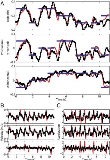
-0.1 -0.1 Position (m) x y z EMG activity Biceps Triceps Deltoid Trapezius 0.1 0 0.1 0 0.4 0.3 0.2 Time (s) 1s Time (s) 1s -0.1 -0.1 0.1 0 0.1 0 0.4 0.3 0.2 Time (s) Time (s) 1 0 1 2 0 1 0 1 2 3 4 5 0 Position (m) x y zFig. 3. Online 3D brain-control. (A) Samples of direct brain-control trajecto-ries resulting in target acquisition without requiring correction (reach target: blue; brain-control trajectory: black). (B) Samples of brain-control trajectories resulting in target acquisition after correction for initially wrong direction. (C) Brain-control trajectories in the absence of limb movement verified by the lack of visible EMG activity (lower four graphs) recorded simultaneously from biceps, triceps, deltoid, and trapezius. (D) For comparison: same-ses-sion reach-control showing characteristic EMG bursts on all four channels.
training were obtained through numerical differentiation after con-volving the position trajectory with a Gaussian kernel (σ = 12 ms) for smoothing.
Offline Ridge Filter. The linear regression ridge model (17, 27) reconstructed instantaneous 3D cursor position as a function of the standardizedfiring rates r(t) of N simultaneously recorded neural units. Each sample of the behavioral state vector, x(t), was modeled as a function of the vector of ensemblefiring rates measured for four successive 90-ms bins. Only the four causal bins immediately preceding the movement were used; i.e., thefiring rates used in conjunction with the behavioral state x(t) were centered at (t− 315 ms), (t − 225 ms), (t − 135 ms), and (t − 45 ms). An estimate of the 3D cursor position,^xðkÞ, was constructed as a linear combination of the ensemble offiring rates, r, sampled at the four leading binning intervals according to
^xðkÞ = β0+
XN j
βjrjðkÞ + εðkÞ; [1]
where k denotes the discretized 90-ms time steps,ε represents the ob-servational error, and N represents the total number of neural inputs each incorporating four successive bins.β, representing the regression coef-ficients, was determined using linear ridge regression (SI Materials and Methods).
Offline Kalman Filter. The discrete Kalman filter implementation (17, 28, 37) estimated the current state of the movement, including velocity and ac-celeration, in all three degrees of freedom from single causal 90-ms bins of firing rates. Two equations govern the recursive reconstruction of the hand kinematics from thefiring rates: an observation equation that modeled the firing rates (observation) as a function of the state of the cursor, xk, and a
process equation that propagated the state of the cursor forward in time as a function of only the most recent state, xk−1. Both models were assumed
to be linear stochastic functions, with additive Gaussian white noise: xk= Akxk−1+ Buk−1+ wk−1ðprocess equationÞ; [2]
rk= Hkxk+ vkðobservation equationÞ; [3]
The control term, u, was assumed to be unidentified and was, therefore, set to zero in our model, excluding B from the process model.
One simplifying assumption was that the process noise (w∈ ℜ9×1),
ob-servation noise (v∈ ℜ9×1), transition matrix (A∈ ℜ9×9), and the observation
matrix (H∈ ℜN×9) werefixed in time, thus simplifying Eqs. 2 and 3 to
xk= Axk−1+ w; [4] rk= Hxk+ v; [5]
where A and H were identified using least squares regression.
To estimate the state of the cursor, at each time-step k, the process model produced an a priori estimate,^x−k, which was then updated with
measure-ment data to form an a posteriori state estimate,^xk. More specifically, the a
priori estimate was linearly combined with the difference between the output of the observation model and the actual neural measurement (i.e., the neural innovation) using an optimal scaling factor, the Kalman gain, Kk, to produce
an a posteriori estimate of the state of the cursor:
^xk= ^x−k+ KkðRk− H^x−kÞ; [6]
minimizing the a posteriori estimation error.
The entire two-step discrete estimation process of a priori time update and subsequent a posteriori measurement update was iterated recursively to generate an estimate of the state of the cursor at each time step in the trajectory. Both the Kalman gain, Kk, and the estimation error covariance
matrix, Pk, have been shown to converge rapidly, decaying exponentially,
in<1.5 s (17), and then to remain stable throughout the decoded segment. Brain Control. The identified decoding models (ridge filter, Kalman filter) were used to guide cursor movement during the brain-control phase of the experiment, allowing the animal to use cortical signals instead of hand movement to guide the cursor. Cursor position was updated every 90 ms and visualized continuously, without reinitialization, throughout the brain-control session. To assess behavioral performance, daily success rates were computed. Although both animals typically performed brain-control reaches to all 27 targets multiple times, the success rate for the most successful set of 27 reaches was reported in Fig. 2. The average success rate over all trials in a session was typically biased (lower) because it frequently included sets of targets where the monkey chose to rest instead of attempting to perform a brain-control reach, making the best set of 27 brain-control reaches the more appropriate measure to assess success rates.
To calculate the chance levels for success rates,firingratebinsamplesforagiven neural unit recorded during brain control were shuffled randomly, effectively preserving each neural unit’s mean firing rate but breaking its temporal structure. Chance trajectories were then generated by simulation, iteratively applying the actual decoder to the shuffled ensemble of firing rates to generate a series of pseudocursor positions. The criteria used during actual brain-control trials were applied to these pseudocursor positions to detect successful target acquisition by
A
B
Fig. 4. Learning brain-control. Im-proving behavioral performance in both monkeys for consecutive brain-control days shows that the monkeys learned to use PPC spike activity driving the decoding algorithm to direct cursor movement. Top graphs show daily success rates and chance performance± SD (gray band) for ridge (A) and Kalmanfilter decode (B). Middle graphs show time-to-target for successful reaches. Bottom graphs show trajectory straightness. The trajectory straightness describes the ratio of the shortest (straight) distance from initial cursor location to target location and the actual distance the cursor traveled during the target acquisition, i.e., increasing straightness values indicate more direct trajectories. The trajectory straightness was normalized for first-day performance.
NEUROSCI
chance. This procedure was repeated 50 times to obtain a distribution of chance performances for each session, from which a mean and SD were derived.
The time-to-target reported quantifies the average duration of all successful reaches in a session, measured from target cue appearance to successful target acquisition.
The trajectory straightness was assessed by calculating the ratio of tra-jectory lengths: the shortest possible (straight) tratra-jectory to acquire a target and the actual distance the cursor traveled. Trajectories were analyzed from when the target cue appeared (initial cursor position) until detection of successful target acquisition (final cursor position). The trajectory straightness results, reported as daily averages for all reaches completed successfully, were normalized forfirst day performance.
Because PPC receives projections from S1 (40, 41) that carry proprioceptive signals, it is unclear whether the movement representation decoded from PPC persists when proprioceptive feedback from the limb is compromised. This was
tested by (i) mechanically immobilizing the limb during the brain-control de-code session and (ii) monitoring the EMG activity of the muscle groups typically involved in reaching movements in monkey R. EMG recordings were made via small percutaneous hook electrodes (paired hook-wire electrode, 30 mm× 27 gauge; VIASYS Healthcare). Recordings were taken simultaneously from the deltoid, trapezius, biceps, and triceps muscles. To verify proper placement and function of the EMG electrodes, recordings were taken before and after the brain control session during a series of reach sequences where the monkey was required to move his limb to control cursor movement.
ACKNOWLEDGMENTS. We thank I. Kagan for performing the MRI scans, K. Pejsa for animal care, and V. Shcherbatyuk and T. Yao for technical and administra-tive assistance. This work was supported by the Defense Advanced Research Projects Agency, the National Eye Institute of the National Institutes of Health, the Boswell Foundation and an Alfred E. Mann doctoral fellowship to M.H.
1. Kennedy PR, Bakay RA, Moore MM, Adams K, Goldwaithe J (2000) Direct control of a computer from the human central nervous system. IEEE Trans Rehabil Eng 8: 198–202.
2. Wessberg J, et al. (2000) Real-time prediction of hand trajectory by ensembles of cortical neurons in primates. Nature 408:361–365.
3. Serruya MD, Hatsopoulos NG, Paninski L, Fellows MR, Donoghue JP (2002) Instant neural control of a movement signal. Nature 416:141–142.
4. Taylor DM, Tillery SI, Schwartz AB (2002) Direct cortical control of 3D neuroprosthetic devices. Science 296:1829–1832.
5. Carmena JM, et al. (2003) Learning to control a brain-machine interface for reaching and grasping by primates. PLoS Biol 1:E42.
6. Shenoy KV, et al. (2003) Neural prosthetic control signals from plan activity. Neuro-report 14:591–596.
7. Musallam S, Corneil BD, Greger B, Scherberger H, Andersen RA (2004) Cognitive control signals for neural prosthetics. Science 305:258–262.
8. Patil PG, Carmena JM, Nicolelis MA, Turner DA (2004) Ensemble recordings of human subcortical neurons as a source of motor control signals for a brain-machine interface. Neurosurgery 55:27–35.
9. Wolpaw JR, McFarland DJ (2004) Control of a two-dimensional movement signal by a noninvasive brain-computer interface in humans. Proc Natl Acad Sci USA 101: 17849–17854.
10. Hochberg LR, et al. (2006) Neuronal ensemble control of prosthetic devices by a hu-man with tetraplegia. Nature 442:164–171.
11. Santhanam G, Ryu SI, Yu BM, Afshar A, Shenoy KV (2006) A high-performance brain-computer interface. Nature 442:195–198.
12. Velliste M, Perel S, Spalding MC, Whitford AS, Schwartz AB (2008) Cortical control of a prosthetic arm for self-feeding. Nature 453:1098–1101.
13. Hochberg LR, et al. (2012) Reach and grasp by people with tetraplegia using a neu-rally controlled robotic arm. Nature 485:372–375.
14. Hwang EJ, Andersen RA (2009) Brain control of movement execution onset using local field potentials in posterior parietal cortex. J Neurosci 29:14363–14370.
15. Hwang EJ, Andersen RA (2010) Cognitively driven brain machine control using neural signals in the parietal reach region. Conf Proc IEEE Eng Med Biol Soc 2010:3329–3332. 16. Baldauf D, Cui H, Andersen RA (2008) The posterior parietal cortex encodes in parallel
both goals for double-reach sequences. J Neurosci 28:10081–10089.
17. Mulliken GH, Musallam S, Andersen RA (2008) Decoding trajectories from posterior parietal cortex ensembles. J Neurosci 28:12913–12926.
18. Chang SW, Dickinson AR, Snyder LH (2008) Limb-specific representation for reaching in the posterior parietal cortex. J Neurosci 28:6128–6140.
19. Baumann MA, Fluet MC, Scherberger H (2009) Context-specific grasp movement representation in the macaque anterior intraparietal area. J Neurosci 29:6436–6448. 20. Wolpert DM, Goodbody SJ, Husain M (1998) Maintaining internal representations:
The role of the human superior parietal lobe. Nat Neurosci 1:529–533.
21. Desmurget M, et al. (1999) Role of the posterior parietal cortex in updating reaching movements to a visual target. Nat Neurosci 2:563–567.
22. Sirigu A, et al. (1996) The mental representation of hand movements after parietal cortex damage. Science 273:1564–1568.
23. Mulliken GH, Musallam S, Andersen RA (2008) Forward estimation of movement state in posterior parietal cortex. Proc Natl Acad Sci USA 105:8170–8177.
24. Batista AP, Buneo CA, Snyder LH, Andersen RA (1999) Reach plans in eye-centered coordinates. Science 285:257–260.
25. Buneo CA, Jarvis MR, Batista AP, Andersen RA (2002) Direct visuomotor trans-formations for reaching. Nature 416:632–636.
26. Pesaran B, Nelson MJ, Andersen RA (2006) Dorsal premotor neurons encode the relative position of the hand, eye, and goal during reach planning. Neuron 51: 125–134.
27. Hoerl AE, Kennard RW (1970) Ridge regression: Biased estimation for nonorthogonal problems. Technometrics 12:55–67.
28. Kalman RE (1960) A new approach to linearfiltering and prediction problems. Trans ASME Ser D J Basic Eng 82:35–45.
29. Hwang EJ, Andersen RA (2011) Effects of visual stimulation on LFPs, spikes, and LFP-spike relations in PRR. J Neurophysiol 105:1850–1860.
30. Shadmehr R, Krakauer JW (2008) A computational neuroanatomy for motor control. Exp Brain Res 185:359–381.
31. Sirigu A, et al. (1995) Congruent unilateral impairments for real and imagined hand movements. Neuroreport 6:997–1001.
32. Kalaska JF, Caminiti R, Georgopoulos AP (1983) Cortical mechanisms related to the di-rection of two-dimensional arm movements: Relations in parietal area 5 and comparison with motor cortex. Exp Brain Res 51:247–260.
33. Bremner LR, Andersen RA (2012) Coding of the reach vector in parietal area 5d. Neuron 75:342–351.
34. Fetsch CR, Wang S, Gu Y, Deangelis GC, Angelaki DE (2007) Spatial refer-ence frames of visual, vestibular, and multimodal heading signals in the dorsal subdivision of the medial superior temporal area. J Neurosci 27:700– 712.
35. Bremner LR, Andersen RA (2012) Evolution of reference frames in area 5d during a reaching task. Soc Neurosci 78:07.
36. Musallam S, Bak MJ, Troyk PR, Andersen RA (2007) Afloating metal microelectrode array for chronic implantation. J Neurosci Methods 160:122–127.
37. Wu W, et al. (2003) Neural decoding of cursor motion using a Kalmanfilter. Adv Neural Info Process Syst 15:133–140.
38. Ashe J, Georgopoulos AP (1994) Movement parameters and neural activity in motor cortex and area 5. Cereb Cortex 4:590–600.
39. Averbeck BB, Chafee MV, Crowe DA, Georgopoulos AP (2005) Parietal representation of hand velocity in a copy task. J Neurophysiol 93:508–518.
40. Jones EG, Powell TP (1970) An anatomical study of converging sensory pathways within the cerebral cortex of the monkey. Brain 93:793–820.
41. Jones EG, Coulter JD, Hendry SH (1978) Intracortical connectivity of architectonic fields in the somatic sensory, motor and parietal cortex of monkeys. J Comp Neurol 181:291–347.