ORIGINAL ARTICLE
Does bladder wall thickness decrease when obstruction is resolved?
Annette Kuhn&Sonja Brandner&Peter Kuhn&Dudley Robinson&Luigi Raio
Received: 19 June 2011 / Accepted: 24 December 2011 / Published online: 17 January 2012 # The International Urogynecological Association 2012
Abstract
Introduction and hypothesis The aim of the current study was to determine if sonographic bladder wall thickness diminishes after symptomatic obstruction is resolved in female patients after stress incontinence surgery.
Methods Between December 2008 and December 2010, 62 female patients with symptomatic bladder outlet obstruction, as defined by Blaivas, who had undergone prior surgery for urinary stress incontinence were included in the study. The patients’ history was taken and symptoms were noted. Patients underwent gynaecological examination, and multichannel urodynamic assessment was performed. Vaginal sonographic assessment of the bladder wall thickness (BWT) was per-formed before and after urethrolysis.
Results 62 patients were included in this study, 55 of whom had undergone suburethral sling insertion and seven had Burch colposuspension. Postoperatively, BWT decreased significantly from 9.1 mm±2.1 to 7.6 mm±2.2 (p<0.0001). In seven patients, obstruction was still unresolved postopera-tively; of these, two had undergone a retropubic sling insertion and two had a Burch colposuspension. An ROC curve analysis showed a significant positive association between residual
urine and persistent obstruction before surgery (AUC 0.76, 95%CI 0.58–0.94; p<0.05).
Conclusions If obstruction is resolved, bladder wall thick-ness decreases. Preoperatively elevated residual urine may increase the risk of persistent obstruction after urethrolysis. Keywords Sonographic bladder wall thickness .
Urodynamic obstruction . Urethrolysis . Surgery for urodynamic stress incontinence
Introduction
In patients with urinary incontinence, clinical assessment including history and physical examination has been shown to be an ineffective method of assessing women with lower urinary tract dysfunction, therefore the bladder has been described as an“unreliable witness” [1,2]. Urodynamic assess-ment of these incontinent patients may be costly, time consum-ing, invasive, and sometimes inaccurate [3–5]. Measurement of bladder wall thickness using transvaginal ultrasound has pre-viously been shown to discriminate between women with confirmed detrusor overactivity and those with urodynamic stress incontinence [6].
It has been reported that the bladder wall thickness of women with detrusor overactivity is greater than in asymp-tomatic women [7]. The assessment of bladder wall thick-ness allows an indirect measurement of the detrusor muscle thickness and provides an indication of the likelihood for DO. The utility of ultrasound imaging of the bladder wall is becoming increasingly important to those interested in urogy-necologic diagnosis and treatment. The assessment of bladder wall thickness (BWT) is considered a useful additional tool when investigating women with lower urinary tract symptoms (LUTS) [5].
A. Kuhn (*)
:
S. Brandner:
L. RaioUrogynaecology, Department of Gynaecology, University Hospital and University of Bern, Effingerstrasse 102, CH 3010 Bern, Switzerland e-mail: [email protected] P. Kuhn Effingerzentrum Bern, Effingerstr. 45, Bern, Switzerland D. Robinson King’s College, London, UK
Various techniques have been suggested to date. A recent study has suggested that BWT, as measured by an automated device, correlates well with results from a conventional hand-held device when both use the abdominal approach [8].
Previous studies have advocated the transvaginal approach using transvaginal probes [5,6] while others prefer perineal or introital sonography [9].
Our group has recently shown that the vaginal measure-ment of BWT is the most reliable [10] compared to abdom-inal or perineal measurements.
Voiding dysfunction has been estimated to occur in 2.5– 24% of patients following surgery to correct stress urinary incontinence [11]. The rates of bladder outlet obstruction are reportedly 5–20% following the Marshall–Marchetti–Krantz procedure [12,13], 4–7% after the Burch colposuspension, [14,15] and 5–7% after needle suspension [16].
With the tension-free vaginal tape (TVT), previous studies have shown that postoperative outlet obstruction occurs with an incidence of 4.9–10% [17]. To date, no data are available investigating sonographic bladder wall thickness in obstructed patients after the obstruction is resolved. The aim of the current study was to determine if BWT diminishes after obstruction is resolved in female patients after they have undergone stress incontinence surgery.
Methods
Between December 2008 and December 2010, all patients with symptomatic bladder outlet obstruction, as defined by Blaivas [18], who had undergone prior surgery for urinary stress incon-tinence and agreed to participate were included in this study.
Blaivas [18] defines obstruction in a nomogram as the relation of the free urinary flow (ml/sec) to the micturition pressure pdet assessed during pressure-flow studies.
Ethical consent was obtained from the local ethics commit-tee (KEK Bern), and patients gave consent to participate.
The patients’ history was taken, and symptoms were noted. Patients underwent gynaecological examination, and namic assessment was performed using multichannel urody-namic systems according to ICS recommendations [6]. The patients were in the sitting position for the urodynamic studies [19].
Cystometry was performed in the sitting position with the patient in the 45-degree upright position with a six French microtip transducer which was introduced into the bladder for intravesical pressure measurement, and a water perfused bal-loon catheter was introduced into the rectum for intraabdomi-nal pressure measurement. The bladder was filled at a rate of 20 m/min with saline solution at 37°. Filling was continued until the patient experienced a strong desire to void. A cough stress test was performed at every hundred millilitres of filling. At bladder capacity, pressure-flow studies were performed.
Ultrasound was performed using the Aloka SSD-1400 (Aloka ® Co Ltd, Japan) with the vaginal 5 MHz probe UST-984-5. Ultrasound examination was undertaken with the patient in the supine position with a residual of less than 50 ml, as previously described [5], applying light pressure. All measurements were made at maximum magnification with the bladder wall being measured perpendicular to the luminar surface of the bladder in the anterior bladder wall, trigone and dome as described by Robinson [5].
All ultrasound measurements were performed by a single investigator who saw the patients as part of their consulta-tion in the urogynecological outpatient clinic.
Surgical management
Preoperatively, all patients received a single-dose broad spectrum I.V. antibiotic.
After suburethral sling, the patient was placed in the lithotomy position, and a transurethral catheter was inserted. After midline colpotomy, the tape was identified and cut at the midline. If tape identification was difficult, an 8 Fr Hegar dilator was inserted into the urethra using an appro-priate amount of lubricant. The Hegar dilator was then withdrawn with slight pressure on the dorsal urethra and the sling could then be identified by resistance difference after the site of the sling was passed, and the sling was incised until the resistance was gone and the Polypropylene was completely cut.
Vaginal epithelium was closed with interrupted vertical mattress sutures in a single layer using Vicryl® 2–0. Patients with obstruction after Burch colposuspension underwent laparoscopic urethrolysis with removal of the colposuspen-sion stitches, mobilisation of the urethra, and interposition of the omentum.
Patients were followed-up at a median of 11 weeks (range 9–15 weeks) after sling incision or urethrolysis, with urody-namic testing and sonographic bladder wall thickness measurement.
Statistical analysis was performed with GraphPad Prism version 5.0 for Windows (GraphPad Software, San Diego CA, USA). Analysis of variance was used to compare continuous variables. Post hoc multiple comparisons were performed using the Dunn test or Kruskal–Wallis test.
Results
62 patients could be included in the study, 55 of whom had undergone suburethral sling insertion and seven had Burch colposuspension. The time interval between initial inconti-nence surgery and a diagnosis of obstruction was a median of 14 months (range 3–89 months).
The slings were a classic retropubic TVT (Ethicon Women’s Health and Urology, Somerville NJ 08876–0151, US) in 30 cases, a transobturator tape (TVT-O; Ethicon, see above, inside-out technique) in seven, transobturator Monarch sling (outside-in technique; AMS Minnetonka, Minnesota 55343, US) in 14 and TVT Secur short slings (Ethicon, see above) in four women. Table1shows demographic data and menopause status.
Patients’ symptoms were overactive bladder (OAB) in 42 cases, voiding difficulties in 18, and two patients had both. Figure1shows BWT before urethrolysis and at follow-up.
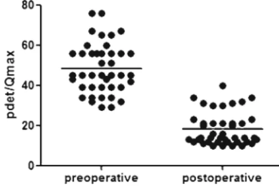
BWT decreased significantly postoperatively from 9.1 mm± 2.1 to 7.6 mm±2.2 (p<0.0001; paired t test). Figure2shows the voiding pressure pdet/Qmax that decreased significantly post-operatively from 48.3 cm H2O±12.5 to 18.4 cm H2O±8.2; p< 0.0001(paired t test]).
In seven patients, obstruction was not resolved postoper-atively whereas two had a retropubic sling insertion and two Burch had colposuspension.
Figures3and4sub-analyse the voiding pressure pre- and postoperatively in patients with resolution of obstruction and those with persistent obstruction.
Preoperatively, the voiding pressure was not statistically different between the groups (p00.35, paired t test), although there was postoperative significance (p<0.001; paired t test). Figure5shows BWT in these cases without resolution of obstruction. BWT did not decrease significantly (9.6 mm± 1.4 vs. 9.8 mm±1.6; p00.09 paired t test).
Preoperative residual urine was significantly lower in the group of patients with successful urethrolysis (median 34 ml, range 9–900 ml) compared to patients with unsuccessful urethrolysis (median 110 ml, range 20–190 ml; p<0.05; Fig.6).
Twenty-nine patients (46.7%) became stress inconti-nent after urethrolysis, requiring further therapy (conser-vative therapy involving physiotherapy, pessaries and drug therapy n013; bulking n08; adjustable sling n05; retropubic sling n03).
Discussion and conclusion
Voiding dysfunction is a known complication of surgery to correct stress urinary incontinence since continence surgery is thought to work by causing some level of obstruction [20, 21]. To our knowledge, this is the first study comparing urodynamically obstructed symptomatic patients pre- and postoperatively after urethrolysis.
In obstruction, detrusor hypertrophy may occur and this may be reflected by an increased BWT. Evidence from Abrams [22] suggests that the development of bladder tra-beculation may be dependent upon detrusor overactivity rather than bladder outlet obstruction. However, detrusor hypertrophy is characterised by increased collagen deposi-tion and myohypertrophy at the microscopic level and by reduction of cell junctions, abundant protrusion junctions and ultraclose abutments at the ultrastructural level; the latter two features are characteristics of bladder outlet ob-struction [23]. Although urodynamic pressure-flow studies are expensive, they are generally considered the gold stan-dard for diagnosing bladder outlet obstruction [24,25]. We found an excellent correlation between micturition pressure and BWT as well as a highly significant correlation between BWT and urodynamic diagnoses in obstructed patients, which may lead to the conclusion that BWT may be used for these diagnoses instead of pressure-flow studies. A
Table 1 Demographic data and
menopause status Age (years) (median, range) BMI (median, range) Parity (median, range) Menopause status
59 (35–91) 29.5 (19–32) 2.5 (0–6) 90premenopausal
140perimenopausal
390postmenopausal
similar suggestion has been made by Oelke [26] who mea-sured detrusor wall thickness in men and concluded that measuring the thickness of the bladder wall can be used as screening test to detect bladder outlet obstruction.
Interestingly, patients with a successful urethrolysis had significantly less preoperative residual than those with unsuc-cessful urethrolysis. It is possible that a preoperatively elevated residual could be used as marker for successful urethrolysis.
Although the number of patients is small, this could be a useful clinical finding in clinics or hospitals where access to urodynamics is limited. This issue is being evaluated in follow-up studies.
Despite the suggestive label“tension-free vaginal tape”, 55 patients in the current study with urodynamic obstruction after suburethral tape insertion were identified, which means that obstruction does occur in women even in tension-free procedures.
Seventy percent of our patients with OAB had been treated with anticholinergics, one patient even had Botox injection. Although pharmacologic treatment was not suc-cessful, treating the mechanical obstruction by cutting the tape suburethrally was effective in 90% of our patients. This means that, in patients with outlet obstruction and OAB symptoms post TVT, it might be better to remove the
mechanical obstruction rather than administer other treat-ments. Urodynamically, the obstruction resolved, free flow increased, and BWT decreased significantlly in patients who were successfully treated for obstruction.
In patients whom obstruction persisted due to bladder wall thickness, values were similar to those before urethrolysis, which means that BWT may be a sensitive marker.
Segal [27] suggested urethral dilatation in cases of iatro-genic obstruction secondary to pubovaginal sling using a Walther sound for varying levels of obstruction in 28 women. However, there are concerns about the potentially traumatic nature of dilatation which could induce scarring of the urethra. The cutting of slings instead of a formal urethrolysis may limit morbidity, potential soft tissue and nerve injury and fibrosis from surgical dissection.
Patients with obstruction often present with a variety of LUTS, and diagnosis often remains challenging. Since OAB symptoms are often initially managed by a combination of anticholinergics and behavioural therapy, bladder outlet ob-struction is often underreported and definitive therapy may be delayed [28] as in the current study: the median time between initial incontinence surgery and diagnosis was 14 months.
A weakness of the study is that we did not measure the pressure applied during vaginal ultrasound. Pressure and movement on the tissue from the probe could significantly alter the results, therefore we were careful to avoid excessive movements during the examination.
Fig. 3 pdet/Qmax preoperatively in patient with resolution and persistent obstruction
Fig. 4 pdet/Qmax postoperatively in patients with resolution and persistent obstruction
Fig. 5 BWT in patients with persistent obstruction
Despite attempts to develop standardised urodynamic crite-ria for female bladder outlet obstruction, none are universally accepted. We applied the definition as described by Blaivas [18], because we have been using this nomogram for the past ten years both clinically and scientifically, and we consider it useful and easy to understand in a teaching setting.
Another weakness of the study is that the investigator was not blinded to the patients’ symptoms as ultrasound measurement was part of the general urogynaecologic con-sultation; however, at follow-up, the sonographer (AK) was blinded to the urodyamic results as urodynamics were per-formed in a neighbouring room and the readings were done after ultrasound had occurred. Despite this, there is still a potential for bias by knowing the patients’ history and development of symptoms.
Some publications suggest a concomitant stress inconti-nence procedure after urethrolysis has been achieved. In our study, the incidence of stress incontinence after urethrolysis was 47%. We do not advocate adding stress incontinence surgery at the time of urethrolysis because the rate of recurrent stress incontinence is very variable between 0 and 50% [29–31] and we are unable to predict in whom this will occur. Additionally, if concomitant continence surgery is performed and the patient continues to have voiding problems, it is unclear whether urethrolysis failed or the continence proce-dure is obstructive.
We recently showed the correlation of urodynamics and BWT [32]; the current study adds information to the validity and correlation of BWT and urodynamics in patients who are obstructed. Ultrasound is particularly useful in gynaecology and obstetrics, and it is also widely available in urology, easy to apply, and, when compared to urodynamics, it is less invasive. We cannot determine with certainty if bladder wall thickness is a result of OAB or if it is due to obstruction as BWT is increased in OAB patients [32].
Future studies will confirm if BWT is entirely reversible and if this correlates with symptoms; long-term follow-up is needed to answer these questions.
Conflicts of interest None.
Ethical approval Ethical approval was given by the Kantonale
Ethikkommission Bern (KEK), President: Prof. Niklaus Tüller. Regis-tration number is E 01-04-10 and this study belongs to the quality control rules of our institution.
References
1. Jarvis GH, Hall S, Stamp S, Millar DR, Johnson A (1980) An assessment of urodynamic examination in incontinent women. Br J
Obstet Gynaecol 87:893–896
2. Bates CP, Whiteside CG, Turner-Warwick R (1970) Synchronous cine-pressure-flow-cystourethrography with special reference to
stress and urge incontinence. Br J Urol 42:714–723
3. Glazener CM, Lapitan MC (2002) Urodynamic investigations for management of urinary incontinence in adults. Cochrane Database Syst Rev 3:CD 003195
4. Gorton E, Stanton SL (2000) Ambulatory urodynamics: do they help clinical management? BJOG 107:316–319
5. Robinson D, Anders L, Cardozo L, Bidmenad J, Toozs-Hobson P, Khullar V (2002) Can ultrasound replace ambulatory urodynamics when investigating women with irritative urinary symptoms?
BJOG 109:145–148
6. Abrams P, Cardozo L, Fall M, Griffiths D, Rosier P, Ulmsten U, van Kerrebroek P, Victor A, Wein A (2002) The standardisation of terminology of lower urinary tract function: report from the stand-ardisation sub-committee of the international continence society.
Neurourol Urodyn 21:167–178
7. Khullar V, Salvatore S, Cardozo LD, Kelleher C, Bourne TH (1994) A novel technique for measuring bladder wall thickness in women
using transvaginal ultrasound. Ultrasound Obstet Gynecol 4:220–
223
8. Salvatore S, Khullar V, Anders K, Cardozo LD (1998) Re-ducing artefacts in ambulatory urodynamics. BrJ Urol 81:211– 214
9. Oelke M, Mamoulakis C, Ubbink DT, de la Rosette JJ, Wijkstra H (2009) Manual versus automatic bladder wall thickness measurements: a method comparison study. World J Urol
27(6):747–753
10. Kuhn A, Bank S, Robinson D, Klimek M, Kuhn P, Raio L (2010) How should bladder wall thickness be measured? a comparison of vaginal, perineal and abdominal ultrasound. Neurourol Urodyn 29
(8):1393–1396
11. Dorflinger A, Monga A (2001) Voiding dysfunction. Curr Opin
Obstet Gynecol 13:507–512
12. Zimmern PE, Hadley HR, Leach GE, Raz S (1987) Female urethral
obstruction after Marshall–Marchetti–Kranz operation. J Urol
138:517–520
13. McDuffie RW, Litin RB, Blundon KE (2010) Urethrovesical
sus-pension Marshall–Marchetti–Krantz Experience with 204 cases.
Am J Surg 141:297–298
14. Akpinal H, Cetinel B, Demirksen O (2008) Long-term results in Burch colposuspension. Int J Urol 7:119–125
15. Ward KL, Hilton P, Browning J (2000) A randomized trial of colposuspension and tension-free vaginal tape for primary stress
incontinence. Neurourol Urodyn 19:386–388
16. Holschneider CH, Solh S, Lebhertz TB, Montz FJ (1994) The modified Pereyra procedure in recurrent stress urinary
inconti-nence: a 15-year review. Obstet Gynecol 83:573–578
17. Karram MM, Segal JL, Vassallo BJ, Kleeman SD (2003) Compli-cations and untoward effects of the tension-free vaginal tape
pro-cedure. Obstet Gynecol 101:929–932
18. Blaivas JG, Groutz A (2000) Bladder outlet normogram for women
with lower urinary tract symptomatology. Neurourol Urodyn 19:553–
564
19. Al-Hayek S, Belal M, Abrams P (2008) Does the patient’s position
influence the detection of detrusor overactivity? Neurourol Urodynam
27:279–286
20. Bump RC, Hurt WG, Elser DM, Theofrastous JP, Addison WA, Fantl JA (1999) Understanding lower urinary tract function in women soon after bladder neck surgery. Neurourol Urodyn 18:629–637
21. Klutke JJ, Klutke CG, Bergman G, Elia G (1999) Urodynamic changes in voiding after anti-incontinence surgery: an insight into
the mechanism of cure. Urology 54:1003–1007
22. Abrams P, Roylance J, Feneley RC (1976) Excretion urography in
23. Elbadawi A, Yalla SV, Resnick NM (1993) Structural basis of geriatric
voiding dysfunction IV bladder outlet obstruction. J Urol 150:1681–1695
24. Belal M, Abrams P (2006) Noninvasive methods of diagnosing bladder outlet obstruction in men. Part I: Nonurodynamic approach. J Urol 176:22–28
25. McConnel JD (1994) Why pressure flow studies should be optional and not mandatory for evaluating men with benign prostate hyper-plasia. Urology 44:156–158
26. Oelke M, Höfner K, Wiese B, Grünewald V, Jonas U (2002) Increase in detrusor wall thickness indicates bladder outlet obstruction in men.
World J Urol 19(6):443–452
27. Segal J, Steele AC, Vassallo BJ, Kleeman S, Silva AW, Pauls R, Walsh P, Karram M (2006) Various surgical approaches to treat voiding
dysfunc-tion following anti-incontinence surgery. Int Urogynecol J 17:372–377
28. Starkman JS, Duffy JW, Wolter CE, Kaufman MR, Scarpero HM, Dmochowski R (2008) The evolution of obstruction-induced
overactive bladder symptoms following urethrolysis for female
bladder outlet obstruction. J Urol 179:1018–1023
29. Odorica R, Rodriguez AR, Coste-Delvecchio F, Hoffman M, Lockhart J (2008) Disabling conditions with slings for managing stress urinary incontinence. BJUI 102:333–336
30. Starkman JS, Duffy JW, Wolter CE, Kaufman MR, Scarpero HM, Dmochowski RR (2008) The evolution of obstruction-induced overactive bladder symptoms following urethrolysis for female bladder outlet obstruction. J Urol 179:1018–1023
31. McCrey R, Appell R (2007) Transvaginal urethrolysis for obstruc-tion after anti-incontinence surgery. Int J Urogynecol J Pelvic
Floor Dysfunct 18(6):627–633
32. Kuhn A, Genoud S, Robinson D, Herrmann G, Guenthert AR, Brandner S, Raio L (2010) Sonographic transvaginal bladder wall thickness: does the measurement discriminate between urodynamic