HAL Id: hal-02434783
https://hal.archives-ouvertes.fr/hal-02434783
Submitted on 14 Apr 2020HAL is a multi-disciplinary open access archive for the deposit and dissemination of sci-entific research documents, whether they are pub-lished or not. The documents may come from teaching and research institutions in France or abroad, or from public or private research centers.
L’archive ouverte pluridisciplinaire HAL, est destinée au dépôt et à la diffusion de documents scientifiques de niveau recherche, publiés ou non, émanant des établissements d’enseignement et de recherche français ou étrangers, des laboratoires publics ou privés.
Light-induced crystallization-driven formation of
hierarchically ordered superhydrophobic sol-gel coatings
Lingli Ni, Cheng Zhu, Shizhong Zhang, Peng Cai, Aissam Airoudj, Laurent
Vonna, Samar Hajjar-Garreau, Abraham Chemtob
To cite this version:
Lingli Ni, Cheng Zhu, Shizhong Zhang, Peng Cai, Aissam Airoudj, et al.. Light-induced crystallization-driven formation of hierarchically ordered superhydrophobic sol-gel coatings. Progress in Organic Coatings, Elsevier, 2019, 135, pp.255-262. �10.1016/j.porgcoat.2019.05.045�. �hal-02434783�
Light-Induced Crystallization-Driven Formation of Hierarchically
1
Ordered Superhydrophobic Sol-Gel Coatings
2
Lingli Ni, †, * Cheng Zhu, † Shizhong Zhang, † Peng Cai, †, * Aissam Airoudj, ‡, § Laurent Vonna, ‡, §
3
Samar Hajjar-Garreau, ‡, § and Abraham Chemtob ‡, §, *
4
5
† Key Laboratory for Palygorskite Science and Applied Technology of Jiangsu Province, 6
College of Chemical Engineering, Huaiyin Institute of Technology, 223003 Huaian, People’s
7
Republic of China
8
‡ Université de Haute-Alsace, CNRS, IS2M UMR7361, F-68100 Mulhouse, France 9
§
Université de Strasbourg, France
10 11
12
*Corresponding authors:
13
Dr. Lingli Ni; E-mail: linglini@hyit.edu.cn; Tel: +86 517 8355 9056; Fax: +86 517 8355
14
9056;
15
Dr. Peng Cai; E-mail: caipeng16@hyit.edu.cn; Tel: +86 517 8355 9619; Fax: +86 517 8355
16
9056;
17
Dr. Abraham Chemtob; E-mail: abraham.chemtob@uha.fr; Tel: +33 3 8960 8799; Fax: +33 3
18
8933 5017;
19
2
Abstract
21
Nano/micro structures are crucial for superhydrophobic surfaces, but rare are methods able to
22
generate such dual structuring in a single step using a single precursor. We show that the
23
visible light-controlled self-assembly of bis-silylated alkane precursors can create in one-step
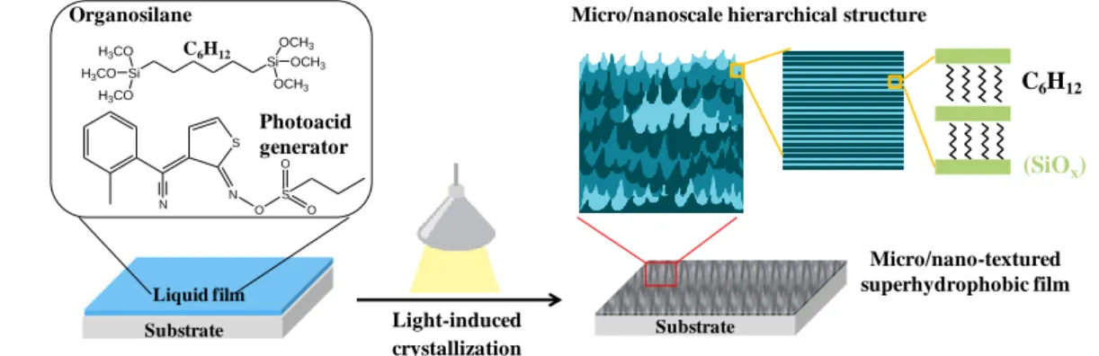
24
organosilica coatings exhibiting two levels of hierarchy: lamellae at nanoscale, and plate-like
25
crystals at microscale. Crystallization rate can be precisely controlled by alkylene bridging
26
group and irradiance. This allowed us to achieve superhydrophobic properties by the creation
27
of a dense and uniform network of nanostructured microcrystals with high surface roughness.
28
The obtained coatings displayed chemical stability, good thermal resistance and high
29
mechanical strength through the cross-linked siloxane structure, which is essential for
30
applications.
31
32
Keywords: superhydrophobic; silsesquioxane; photo sol-gel; crystallization
33
34
Introduction
35
Superhydrophobic surfaces combining water contact angles higher than 150º and low
36
contact angle hysteresis have attracted extensive interest from both an economic and an
37
academic perspective [1-3]. High water repellency properties can ensure further developments
38
in many advanced materials with broad potential application [1, 4], such as self-cleaning
39
surfaces [5-7], anti-corrosive coatings [8-11], drag reduction [12-14], or microfluidic devices [15].
40
It is now well established that a combination of low-surface-energy material and hierarchical
41
micro/nanoscopic structure is a key feature to fabricate superhydrophobic surfaces [16, 17].
Inspired by lotus leaf [18], many different approaches have been developed to achieve both
43
properties in a single coating. Lithography [19], plasma or chemical etching [20], controlled
44
crystallization [21, 22], phase separation phenomena [23], electrochemical deposition [24],
45
chemical vapor deposition [25], and self-assembly [26-28] are the main ones. Despite the
46
diversity of synthetic methodologies, there are still many obstacles to a widespread use of
47
superhydrophobic materials, including multiple steps, use of aggressive chemical reagents, or
48
complex instrumentations [29]. In this context, a facile, cost-effective and environmentally
49
friendly method to superhydrophobic coatings would be highly desirable [6].
50
In response to this major challenge, we report herein a superhydrophobic sol-gel coating
51
produced in a single-step from a single precursor. As depicted in Figure 1, the photoinduced
52
condensation of a α,ω-bis-silylated alkane was able to create a superhydrophobic hierarchical
53
structure driven by the self-assembly of the organosilica network. Conventional sol-gel
54
process based on specific organosilane [30-33] or polyhedral oligomeric silsesquioxane [34,
55
35] precursors have long been recognized as a major method for the preparation of
56
superhydrophobic coatings. By contrast, the development of a radiation-mediated sol-gel
57
process has received little attention so far. This in spite of the fact that photopolymerization is
58
considered today as one of the most eco-efficient organic coating technologies [36]. The few
59
studies reporting a light-driven route to superhydrophobic coatings involved volatile organic
60
solvents either to ensure deposition [37, 38] or to induce roughness by phase separation [39]
61
or simple evaporation [40]. In addition, their green credentials were undermined by the use of
62
ultraviolet (UV) radiation and perfluorinated chemicals, both having potential health effects.
63
By contrast, our methodology is based on a solvent-free sol-gel photopolymerization of
4
1,6-bis(trimethoxysilyl)hexane ((H3CO)3Si-(CH2)6-Si(OCH3)3, BC6TMS) performed at 65
ambient temperature, low irradiance (< 1 mW cm-2) and using a 405 nm light-emitting diode
66
(LED). A visible radiation promotes a safer environment, while the use of LEDs offer many
67
technical advantages compared to conventional mercury arcs [41]. While alkoxysilane sol-gel
68
photopolymerization is already a well established approach to design nanostructured
69
organosilica coatings [42, 43], it has been harnessed as a way to create a hierarchically
70
ordered micro/nanostructure which is ideal for achieving superhydrophobicity. By precise
71
energetic dosage of irradiation, crystallization conditions can be finely tuned to yield in a
72
single-step a silsesquioxane coating ((OH)3-xOxSi-(CH2)6-SiOx(OH)3-x, BC6SQ) composed of 73
crystalline nanolayers assembling in microcrystals with high surface roughness. In this work,
74
the conditions to create a superhydrophobic surface from such hierarchically ordered structure
75
have been investigated. The challenge lies in the capacity to control template-free
76
organosilane self-assembly, microcrystals growth and surface energy of the hybrid coating.
77
78
Figure 1. Schematic illustration showing the visible light-induced crystallization process of a
79
bis-silylated alkane precursor forming a micro/nano structured organosilicate coating.
80 81 82 Organosilane C6H12 Photoacid generator Liquid film Substrate Light-induced crystallization (SiOx) C6H12
Micro/nanoscale hierarchical structure
Substrate S O N S O O N Micro/nano-textured superhydrophobic film Si Si OCH3 OCH3 OCH3 H3CO H3CO H3CO
Experimental section
83
Chemicals
84
1,2-Bis(trimethoxysilyl)ethane (95 %) and 1,6-bis(trimethoxysilyl)hexane (95 %) were
85
supplied by Gelest. Trimethylsiloxy teminated poly(dimethyl siloxane) (PDMS, 3.0 cSt, 550
86
g/mol) was purchased from Fluochem. Trimethoxysilane and 1,9-decadiene were purchased
87
from Aladdin and TCI chemicals (Shanghai), respectively. Photoacid generator Irgacure
88
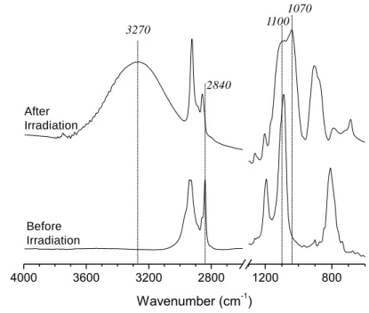
PAG103 was provided by BASF. All of the chemicals were used as received.
89
Synthesis of 1,10-bis(trimethoxysilyl)decane
90
1,10-Bis(trimethoxysilyl)decane was prepared according to the previous literature procedure
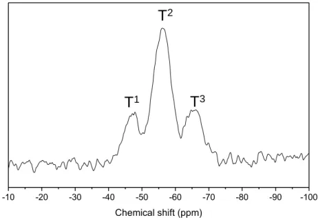
91
[44]. Typically, 6.9 g of 1,9-decadiene (0.05 mol), 12.3g of trimethoxysilane (0.1 mol) and 0.35
92
ml of chloroplatinic acid in isopropanol (2.73 × 10⁻³ M) were mixed together by magnetic
93
stirring, then the mixture was heated to 100 ºC and refluxed for 6 hours under nitrogen
94
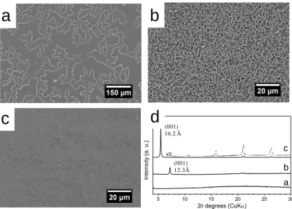
atmosphere. After distillation of the reaction mixture under vacuum, 6.7 g (35 % yield) of
95
1,10-bis(trimethoxysilyl)decane was collected. 1H NMR (400 MHz, CDCl3): δ (ppm) 0.58 (t, 4 96
H), 1.17–1.6 (m, 34 H), 3.7 (q, 12 H); 13C NMR (100 MHz, CDCl3): δ (ppm) 10.3, 18.3, 22.7, 97
29.2, 29.5, 33.2, 58.2.
98
Preparation of hierarchically ordered coating
99
In a typical procedure, photoacid generator (PAG103, 0.5 % wt.) was dissolved in a mixture
100
of 1,6-bis(trimethoxysilyl)hexane and PDMS (75/25 wt. %) to form a photolatent solution in
101
the absence of light. Then the resultant formulation was deposited on a silicon wafer or glass
102
substrate by spin coating (1000 rpm, 20 s) to produce a liquid coating. UV-curing was
103
performed at room temperature under the light of a LED lamp (Shenzhen xianghe, 20 W) with
6
a controlled irradiance from 20 to 0.1 mW cm-2. The samples were irradiated 1800 s to yield
105
solid silsesquioxane hybrid coatings (~ 4 µm). During irradiation, the relative humidity (RH)
106
was maintained between 50 and 55 %.
107
Characterization
108
Infrared spectra were recorded with a spectrophotometer equipped with a MCT detector. The
109
resolution of the infrared spectra was 2 cm-1. X-ray diffraction patterns (XRD) were obtained
110
on a Bruker D8-Discover diffractometer with fixed slits using Cu/Kα radiation (λ = 1.5418 Å)
111
and θ-2θ mounting. Before analysis, coatings on silicon wafers were directly deposed on a
112
plastic sample holder. Data were collected between 1 and 30 ° 2θ degrees (XRD) with a
113
scanning step of 0.01° s-1. Morphologies of the samples were characterized by scanning
114
electron microscopy (SEM, Hitachi S3000N microscope working at 30 kV). The samples being
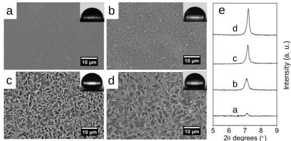
115
non-conductive, they were metalized with gold (15 nm thickness). Water contact angles were
116
measured by sessile drop experiments on a DSA25 contact angle goniometer (Krüss GmbH,
117
Germany) with 6 μL deionized water droplet. Atomic Force Microscopy (AFM) measurements
118
were carried out in a Bruker Multimode IV, with a Nanoscope V controller and an E “vertical”
119
scanner, by the Peak Force Quantitative Nanomechanical Mapping (PF-QNM, Bruker) method.
120
PF-QNM is a contact AFM mode, based on the force-volume method. In this method, force
121
distance curves are collected by nanoindentation of the sample in a point-by-point mode.
122
During measurement, the maximum (peak force) is controlled at each pixel to obtain
123
force-distance curves which are then used as feedback signal. In this method, the loading and
124
unloading force-distance curves are collected at a frequency of 2 kHz at each position within
125
the mapped area of the specimen. In parallel to topography images, information on material
elasticity (Young’s modulus), tip-to-surface adhesion were obtained. All the experiments were
127
carried out in air and at room temperature. 5 µm × 5 µm (256 × 256 pixels at 0.5 Hz) were taken
128
at three different areas on the sample surface. To get relevant results, the cantilever and the tip
129
geometry are taking into account in the PF-QNM measurements. Thus, a calibration procedure
130
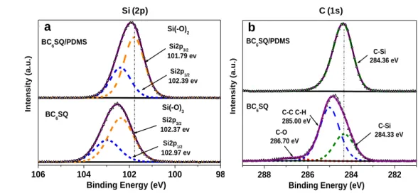
was first followed. All quantitative measurements were carried out with RTESPA-300
131
cantilever (Bruker, USA) with a spring constant of 40 N m-1 and resonance frequency of 300
132
kHz, a width of 40 µm and a length of 125 µm. Thanks to the Sader method [45] (using the
133
length, the width, the resonance frequency and the quality factor of the cantilever) the actual
134
spring constant was determined and found to be around 42 N m-1. Then, the deflection
135
sensitivity (around 33 nm V-1) was measured on a sapphire surface. Tip radius was calibrated
136
against a polystyrene standard provided by Bruker. The measured value of the tip radius was 30
137
nm. The Poisson’s ratio was assumed to be equal to 0.3. For all experiments, samples were
138
previously (at least half a day before) fixed on a sample holder with a double-sided tape. X-ray
139
photoelectron spectroscopy (XPS) analyses were performed with a VG Scienta SES 2002
140
spectrometer equipped with a monochromatized Al(Kα) X-ray source (1486.6 eV), a
141
hemispherical analyzer and an electron gun to compensate the charging effect. The high
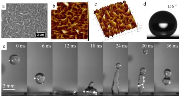
142
resolution spectra and wide scan were recorded with pass energy of 100 eV and 500 eV
143
respectively. The analyzed zone had a surface of 24 mm2. The decomposition of the spectra into
144
different components was performed with Gaussian-Lorentzian, after having subtracted a
145
Shirley-type background. The mean composition of the sample surface expressed in atomic
146
percentages was determined using integrated peak areas of each component, and taking into
147
account the transmission factor of the spectrometer, means free path, and sensibility factors of
8
each atom. The bouncing of the droplets was recorded with a high-speed camera (4M180-CL
149
from IO Industries Inc., London, ON, Canada) at a frame rate of 500 fps. The impact velocities
150
were calculated from the five last images before the contact between the droplet and the surface.
151
We used a microsyringe with a tip geometry allowing to deliver water droplets with a diameter
152
of 2.7 mm ± 0.2 mm. The height of the syringe tip was increased in order to reach high impact
153
velocities. With these droplet diameters, the highest impact velocities considered in this work
154
were around than 1.2 m /s-1 ± 0.1 m·s-1. Beyond these velocities, fragmentation of the droplets
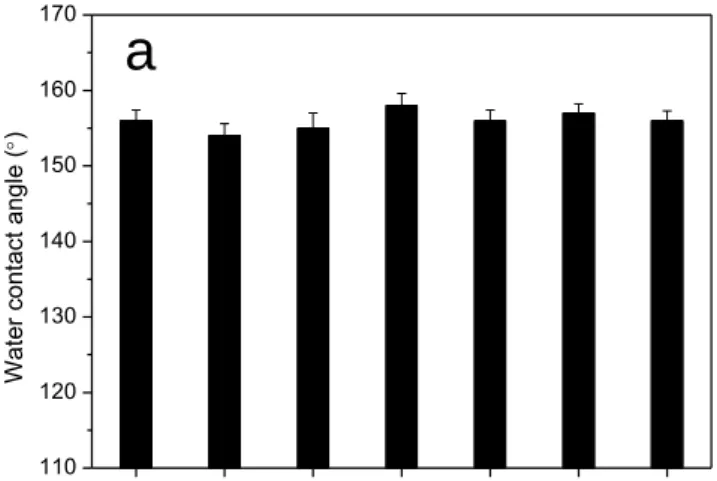
155
was observed. As for the sessile drop experiments, we used fresh deionized water. The abrasion
156
resistance of the superhydrophobic surface was evaluated by a homemade Taber abrasion
157
equipment. The test specimen was placed on the abrasion tester. A 100 g load was then placed
158
on top of the abrader wheel (1500-mesh sandpaper) and allowed to spin for a specified number
159
of revolutions at a speed of 3 cm·s-1. The contact area between the abrader wheel and the
160
underlying superhydrophobic coating was 4.8 cm2. Water CA and sliding angle of test
161
specimen were measured after different number of abrasion cycles.
162
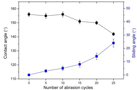
163
Results and discussion
164
The synthesis of bridged silsesquioxane coating BC6SQ proceeds through an 165
acid-catalyzed sol-gel photopolymerization in presence of a commercial photoacid generator
166
(PAG, (5-propylsulfonyl-oxyimino-5H-thio-phen-2-ylidene)-(2-methylphenyl) acetonitrile). Upon
167
exposure to visible light, n-propyl sulfonic acid is liberated, acting as a catalyst for
168
hydrolyzing methoxysilyl (Si-OCH3) functions into Si-OH, and for their subsequent 169
condensation into siloxane (Si-O-Si) bonds. This series of reactions was triggered by
irradiating a BC6TMS/PAG coating (1/0.005 wt%, thickness: 4 µm) with a 405 nm LED lamp at 171
low irradiance (0.5 mW cm-2) during 30 min. The efficiency of the photo sol-gel process was
172
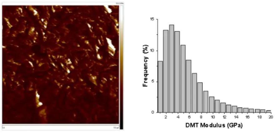
supported by FTIR spectroscopy (Figure 2) through the disappearance of the CH3 symmetric 173
stretch of SiO-CH3 groups at 2840 cm -1
and the concomitant emergence of a broad band at
174
3000-3400 cm-1, indicative of the silanol formation. The presence of asymmetric stretching
175
bands of siloxane bonds at 1100 and 1070 cm-1 proved the formation of the
176
polysilsesquioxane network. It was further evidenced by 29Si SPE-MAS NMR spectroscopy
177
(see spectrum in Figure S1 in supporting information) which revealed T1 (-49 ppm,
178
C-Si(OR)2(O-Si), 15.9 %), T 2
(-58 ppm, C-Si(OR) (O-Si)2, 59.4 %) and T 3
(-68 ppm,
179
C-Si(O-Si)3, 24.7 %) siloxane species. 180
181
Figure 2. FTIR spectra of BC6TMS-derived coating before and after the sol-gel 182
photopolymerization (I = 0.5 mW cm-2, RH = 50 %, T = 25 ºC).
183
184
Scanning electronic microscopy (SEM) was used to assess the possible surface
185 4000 3600 3200 2800 1200 800 Wavenumber (cm-1) 3270 1070 2840 1100 Before Irradiation After Irradiation
10
microstructuration. Figure 3 displays a representative image for BC6SQ as well as two other 186
images obtained with bridged precursors exhibiting other chain lengths
187
((CH3O)3SiCnH2nSi(OCH3)3, n = 2 and 10). BC2SQ (Figure 3a) containing the shortest 188
bridging group formed a surface locally uneven and rough, resulting only in a moderate water
189
contact angle (CA) of 78.0° ± 0.9°. Upon increasing the chain length, BC6SQ (Figure 3b) 190
revealed a uniform, highly rough surface, driving a much higher CA (129.5° ± 1.1°). In contrast,
191
BC10SQ (Figure 3c) exhibited only local features protruding from the plane, leading to a sharp 192
decrease of CA (85.5° ± 0.9°). Further investigation of the hybrid coatings’ nanostructure was
193
performed by X-ray diffraction (XRD). As shown in Figure 3d, the coating derived from the
194
ethylene precursor (trace a) was mainly amorphous. In contrast, increasing the alkylene chain
195
length to C6 (trace b) led to a single sharp diffraction signal, indicative of a long range ordered 196
sample. The onset of a highly ordered nanostructure was even more evident with the BC10SQ 197
coating whose XRD pattern (trace c) displayed 5 reflection peaks from (001) to (005)
198
suggestive of a lamellar mesostructure. In the latter two cases, the (001) peaks at 12.3 Å (b) and
199
16.2 Å (c) were consistent with an interlamellar distance for structures based on alternating
200
stacks of CnH2n bridging groups connected to a single siloxy chain. As expected in organosilane 201
self-assembly, a higher ordering is driven by longer alkylene bridging groups because of
202
stronger Van der Waals interactions [46].
203
205
Figure 3. SEM micrographs of BCnSQ samples derived from BCnTMS precursors 206
[(CH3O)3SiCnH2nSi(OCH3)3, n = 2 (a), 6 (b), 10 (c)]. XRD patterns of the three 207
photopolymerized BCnSQ hybrid coatings (d). I = 0.5 mW cm -2
, 30 min irradiation.
208 209
In addition, the microstructures observed by SEM may be assigned to polycrystals formed
210
by the assembly of dense and regular arrays of nanolamellae. This assumption is supported by
211
images of polarized optical microscopy confirming that the three samples (Figure S2) exhibit
212
birefringence (anisotropy of refraction) that is characteristic of their crystalline structure.
213
Although the microcrystals stem from the growth of lamellar nanocrystals, their density and
214
uniformity were strongly dependent on the chain length. Compared to other precursors,
215
BC6SQ was unique for its ability to form a homogeneous hierarchical surface through a 216
crystallization process expressing at both nano- and microscale levels. Consequently, in the
217
view to provide superhydrophobic properties, C6TMS was preferentially used in the rest of 218
this study. Next efforts were focused on utilization of light, in particular irradiance, to
219
precisely control the morphology of the BC6SQ surface. The objective was to optimize 220
c
b
a
x5 a b c In te n s it y (a . u .) 5 10 15 20 25 30 2 degrees (CuK)d
(001) 16.2 Å (001) 12.3Å12
crystallization conditions in a way to create hierarchically ordered coating displaying a
221
uniform and highly rough spiky surface.
222
223
Figure 4. SEM micrographs of BC6SQ prepared at different irradiances: 20 (a), 2 (b), 0.5 (c), 224
0.1 (d) (unit: mW cm-2, 30 min irradiation). The corresponding (001) XRD peak of each
225
sample is shown on the right side (e).
226
227
Both nano- and microstructures are expected to be influenced by the crystallization time,
228
which is dependent on the condensation rate [47-48]. One key advantage of a
229
radiation-mediated process is that a modulation of irradiance represents an efficient means to
230
control this condensation kinetics. Figure 4a-d show SEM images of BC6SQ prepared at 231
different irradiances (0.1 - 20 mW cm-2). A smooth surface with limited hydrophobic
232
properties was obtained (Figure 4a, CA = 82.0° ± 0.7°) at 20 mW cm-2. In accordance with
233
this result, the corresponding XRD pattern (Figure 4e) featured only a low intense (001) peak
234
because condensation reactions were presumably too fast to favor self-assembly. Upon
235
decreasing irradiance to 2 mW cm-2, microcrystals appeared at the coating surface, resulting
236
in an increased hydrophobicity (Figure 4b, CA = 95.0 ° ± 0.9°). Consistently, there was a
237 20 mw 2 mw
a
b
c
d
5 6 7 8 9 2 degrees () In te n s it y ( a . u .) a b c de
sharpening and intensity increase of the (001) reflections, suggesting a higher level of
238
ordering (Figure 4e). At 0.5 mW cm-2, the level of ordering still increased, which translated in
239
sharper reflection peaks. Concomitantly, the size and number of flat and edgy plates on the
240
coating surface increased, resulting in a larger CA value (Figure 4c, CA = 129.5° ± 1.1°). At
241
0.1 mW cm-2, the XRD pattern followed the same trend, but the water contact angle fell
242
markedly (Figure 4d, CA = 112.0° ± 1.2°), likely as a result of the collapse of the poorly
243
condensed microstructure. Therefore, an irradiance 0.5 mW cm-2 (Figure 4c) seemed the best
244
trade-off. Under these conditions, crystallization time was slow enough to drive a high level
245
of organization, which proved to be beneficial for a higher crystallinity and the emergence of
246
well-defined nano- and micro-structures. Slow condensation is assumed to increase the
247
nucleation rate and the density of flakes at the surface contributing to increase roughness. In
248
these conditions, the inhomogeneity and size of the pores increase, contributing to the
249
increase of water CA. Nevertheless, the present system was not yet able to achieve
250
superhydrophic properties. One possible reason is a too high surface energy of the bridged
251
silsesquioxane structure.
252
253
Figure 5. Si (2p) (a) and C (1s) (b) core level XPS spectra of BC6SQ and BC6SQ/PDMS. 254 106 104 102 100 98 (A) Si2p3/2 102.37 ev Si2p1/2 102.39 ev Si2p1/2 102.97 ev Si(2p) Inte nsi ty (a. u.) BC 6SQ BC 6SQ/PDMS Si(-O)3 Si(-O)2 Si2p3/2 101.79 ev
Binding Energy (eV)
288 286 284 282 (B) C-O 286.70 eV C-C C-H 285.00 eV C-Si 284.33 eV BC 6SQ/PDMS BC 6SQ Inte nsi ty (a. u.) C1s C-Si 284.36 eV
Binding Energy (eV)
a b
C (1s) Si (2p)
14
In order to increase the water CA, a small amount of trimethylsilyl terminated
255
poly(dimethylsiloxane) (PDMS, 550 g/mol) was added. The goal was to coat the BC6PSQ 256
microstructure with a material having a lower surface energy than alkyl bridged
257
silsesquioxane while keeping the advantage of a one-step procedure. The analysis of the XRD
258
pattern confirmed that this additive did not significantly alter the nanoscale organization
259
(Figure S3), with only a slight broadening of the XRD signal. In order to assess the surface
260
migration ability of PDMS chains, X-ray photoelectron spectroscopy (XPS) was performed
261
with BC6SQ/PDMS and pure BC6SQ (Figure 5). In both cases, the Si2p peak (Figure 5a) 262
displayed two components, Si2p3/2 and Si2p1/2 with a spin-orbit coupling of 0.6 eV. However, 263
the position of these two features was distinct depending on the sample, suggesting a different
264
Si environment. As expected, the binding energies in BC6SQ (102.37 eV, 102.97 eV) were in 265
agreement with the presence of T siloxane species RSi(OH)x(OSi)3-x [49]. By contrast, the 266
positions in BC6SQ/PDMS, (101.79 eV, 102.39 eV) were consistent with a majority of D 267
siloxane units R2Si(OSi)2 [49] and correspond well to literature values for PDMS [50]. As 268
shown in Figure 5b, the C1s peak of pure BC6SQ could be fitted into three peaks assigned at 269
C-O (286.70 eV), C-C and C-H (285.00 eV) and C-Si (284.33 eV). Conversely, the C1s peak
270
of BC6SQ/PDMS was deconvoluted only in a single peak centered at 284.36 eV assigned to 271
C-Si bonds [50], which is the only C-containing bonds in the PDMS used. Therefore, XPS
272
survey of Si and C areas both supported the formation of a PDMS-rich surface region in
273
BC6SQ/PDMS. 274
275
Figure 6. (a) SEM micrographs of the surface of BC6SQ/PDMS coating obtained by visible 276
light induced polymerization (0.5 mW cm-2, 30 min irradiation). Inset: shape of water droplet
277
on BC6PSQ/PDMS coating surface); (b) AFM height image (5 µm × 5 µm); (c) AFM 3D 278
topography of BC6PSQ/PDMS coating surface; (d) High-speed images sequence of a water 279
droplet hitting the BC6PSQ/PDMS coating at an impact velocity of 1.2 m·s -1 ± 0.1 m·s-1 280 (falling height of 5 cm). 281 282
In addition, SEM observation (Figure 6a) revealed that microcrystals of BC6SQ/PDMS 283
still formed a homogeneous array of flat plates exhibiting sharp edges, giving the
284
microstructure a shape similar to sand rose rocks. AFM characterization (Figure 6b-c)
285
confirmed such morphology presenting a high roughness (arithmetic average roughness Ra = 286
98 ± 10 nm). The crystals were shaped like petals or flat plates, growing up to around 1 µm.
287
Remarkably, this surface showed high superhydrophobicity. The repellency was such that it
288
was extremely difficult to measure an equilibrium contact angle or a contact angle hysteresis,
289
the water droplets just rolling off the surface. While scanning the surface with the water
16
droplet, it was possible however to find some anchoring points corresponding to local
291
heterogeneities in the coating. In this case, the water droplet contact angle was 156° (Figure
292
6d). Additionally, the CA hysteresis was found to be zero, as expected from a surface on
293
which water droplets could not adhere. The bouncing drop technique was used to further
294
characterize the resistance of the surface texture to wetting. The bouncing of water droplets
295
on BC6SQ/PDMS surface was observed using a high speed video camera, with increasing 296
falling heights. It was not possible even for the highest falling height (just before
297
fragmentation of the droplet) to produce the forced wetting of the surface texture, the droplet
298
still detaching from the substrate after the first impact, and bouncing on the surface (see
299
Figure 6e). Such a water repellency and pressure resistant superhydrophobic surface is
300
remarkable and might be particularly relevant for underwater applications.
301
In order to evaluate the environmental stability and solvent resistance properties of this
302
superhydrophobic surface, the influence of exposure to organic solvents, hot water and
303
aqueous solutions displaying a range of pH was examined (Figure 7). Remarkably, the
304
superhydrophobicity of BC6PSQ/PDMS coating was preserved after immersion during 30 305
min in different solvents: ethanol, acetone, toluene, chloroform, octane or boiling water
306
(Figure 7a). In all instances, for droplets adhering locally on some defects, the CAs were still
307
extremely high (> 150°), and no contact angle hysteresis could be measured (Δθ = 0°). Even
308
after being exposed to air for 3 months, the surface retained its water-repellent property.
309
Furthermore, the superhydrophobicity of BC6PSQ/PDMS coating was maintained even after 310
24 h immersion in acid/alkali solution with pH from 1 to 13 (Figure 7b).
312
Figure 7. (a) The solvent-resistance of the BC6PSQ/PDMS superhydrophobic coating upon 313
treatment with various solvents during 30 min; (b) Variation of water CA for BC6PSQ/PDMS 314
superhydrophobic coating after immersion in aqueous solutions with different pH values for
315
24 h.
316
The mechanical durability of the BC6PSQ/PDMS superhydrophobic coating was also 317
assessed using an abrasion wear test [51]. The methodology of the abrasion test involved
318
rubbing the BC6PSQ/PDMS coating surface against a rotating abrader wheel (see 319
characterization section for details). The variations of the water CA and sliding angle (SA)
320
values along with abrasion cycles are shown in Figure 8. Interestingly, the surface of
321
BC6PSQ/PDMS coating maintained superhydrophobicity (≥ 150 °) even after 20 abrasion 322 1 3 5 7 9 11 13 110 120 130 140 150 160 170 Wat er con tact an gl e ( ) pH
before ethanol acetone toluene chloroform octane hot water
110 120 130 140 150 160 170 Wat er con tact an gl e ( )
a
b
18
cycles. Although the SA values increased with the number of abrasion cycles, it is only 14 °
323
after 20 cycles of abrasions. Such results indicated the mechanical stability of the hybrid
324
coating surface.
325
326
Figure 8. Water CA and sliding angle of the BC6PSQ/PDMS superhydrophobic coating 327
surface as a function of abrasion cycles with a pressure of ~2.4 kPa.
328
329
Such coating robustness was attributed to siloxane cross-linked structure as well as the
330
high modulus value provided by the crystalline structure. In agreement with these two
331
hypotheses, 29Si solid state NMR spectroscopy (Figure S4) revealed a relatively high degree
332
of condensation (72 %). Additionally, the Derjaguin–Müller–Toporov (DMT) fit model was
333
used to estimate the elastic modulus using the unloading parts of the AFM force curves. The
334
average values of the DMT elastic modulus of BC6SQ/PDMS surface was 3.2 ± 1.2 GPa 335 (Figure 9). 336 0 5 10 15 20 25 110 120 130 140 150 160 C on tact an gl e ( )
Number of abrasion cycles
0 10 20 30 40 50 S lidi ng an gl e ( )
337
Figure 9. Representative maps DMT modulus and corresponding histogram of the
338
BC6SQ/PDMS sample obtained with PeakForce QNM. 339
340
Conclusion
341
We have demonstrated a new strategy based on light-induced crystallization to fabrication of
342
hybrid superhydrophobic coatings for the first time. Of particular interest is that the described
343
light-induced crystallization process proceeds in bulk without any solvent and water, thus
344
obviating not only the insolubility problem of precursors but also the environmental issue.
345
The one-step sol-gel polymerization of bridged precursor BC6TMS is catalyzed by in situ 346
light irradiation generated photoacid of n-propane sulfonic acid that formed
347
polysilsesquioxane hybrid coating shows micro/nanoscale hierarchical structure.
348
Template-free organosilane self-assembly accounts for formation of crystalline nanolayers
349
assembling in flat plates microcrystals. With sufficient roughness simply tuned by irradiation
350
intensity, the micro/nanotextured surfaces result in high water contact angles (≥156°) and no
351
contact angle hysteresis could be measured (Δθ = 0°). Furthermore, the cross-linked
352
silsesquioxane hybrid coatings are mechanically robust (20 cycles abrasion and with a DMT
353
elastic modulus value of 3.2 ± 1.2 GPa) as well as chemically (various organic solvents,
354
acid/alkali solution) and thermally resistant (even for boiling water). Thus, the present
355
strategy has potential applications for superhydrophobic coatings, in particular for large-area,
356
temperature or even UV light sensitive substrates.
357 358
20
Supporting Information. 29Si solid state SPE-MAS NMR spectra of the visible LED cured
359
BC6SQ hybrid coatings (Figure S1), polarized optical microscopy images of the BCnSQ 360
hybrid coatings (Figure S2), XRD patterns of the visible LED cured hybrid coatings (Figure
361
S3), 29Si solid state SPE-MAS NMR spectrum of the visible LED cured BC6SQ/PDMS hybrid 362 coatings (Figure S4). 363 364 Acknowledgements 365
The authors would like to thank the financial support from the National Natural Science
366
Foundation of China (No. 51503072), Natural Science Foundation of Jiangsu Province (No.
367
BK20150419) and Six Talent Peaks Project in Jiangsu Province (No. 2016XCL010,
368 2017KTHY007). 369 370 References 371
[1] C. Peng, Z. Chen, M.K. Tiwari, All-organic superhydrophobic coatings with
372
mechanochemical robustness and liquid impalement resistance, Nat. Mater. 17 (2018)
373
355-360.
374
[2] B. Su, Y. Tian, L. Jiang, Bioinspired Interfaces with Superwettability: From Materials to
375
Chemistry, J. Am. Chem. Soc. 138 (2016) 1727-1748.
376
[3] S. Wang, K. Liu, X. Yao, L. Jiang, Bioinspired Surfaces with Superwettability: New
377
Insight on Theory, Design, and Applications, Chem. Rev. 115 (2015) 8230-8293.
378
[4] B. Bhushan, Y.C. Jung, Natural and biomimetic artificial surfaces for superhydrophobicity,
379
self-cleaning, low adhesion, and drag reduction, Prog. Mater. Sci. 56 (2011) 1-108.
[5] R. Blossey, Self-cleaning surfaces — virtual realities, Nat. Mater., 2 (2003) 301.
381
[6] Y. Lu, S. Sathasivam, J. Song, C.R. Crick, C.J. Carmalt, I.P. Parkin, Robust self-cleaning
382
surfaces that function when exposed to either air or oil, Science 347 (2015) 1132-1135.
383
[7] K.M. Wisdom, J.A. Watson, X. Qu, F. Liu, G.S. Watson, C.H. Chen, Self-cleaning of
384
superhydrophobic surfaces by self-propelled jumping condensate, Proc. Natl Acad. Sci. 110
385
(2013) 7992-7997.
386
[8] F. Zhang, L. Zhao, H. Chen, S. Xu, D.G. Evans, X. Duan, Corrosion Resistance of
387
Superhydrophobic Layered Double Hydroxide Films on Aluminum, Angew. Chem. Int. Ed.
388
47 (2008) 2466-2469.
389
[9] P.M. Barkhudarov, P.B. Shah, E.B. Watkins, D.A. Doshi, C.J. Brinker, J. Majewski,
390
Corrosion inhibition using superhydrophobic films, Corros. Sci. 50 (2008) 897-902.
391
[10] K. Liu, M. Zhang, J. Zhai, J. Wang, L. Jiang, Bioinspired construction of Mg-Li alloys
392
surfaces with stable superhydrophobicity and improved corrosion resistance, Appl. Phys. Lett.
393
92 (2008) 183103.
394
[11] X. Zhou, J. Kong, J. Sun, H. Li, C. He, Stable Superhydrophobic Porous Coatings from
395
Hybrid ABC Triblock Copolymers and Their Anticorrosive Performance, ACS Appl. Mater.
396
Interfaces 9 (2017) 30056-30063.
397
[12] J. Ou, B. Perot, J.P. Rothstein, Laminar drag reduction in microchannels using
398
ultrahydrophobic surfaces, Phys. Fluids 16 (2004) 4635-4643.
399
[13] S.T. Yohe, Y.L. Colson, M.W. Grinstaff, Superhydrophobic Materials for Tunable Drug
400
Release: Using Displacement of Air To Control Delivery Rates, J. Am. Chem. Soc. 134 (2012)
401
2016-2019.
22
[14] P. Ball, Engineering Shark skin and other solutions, Nature 400 (1999) 507-509.
403
[15] F. Mumm, A.T.J. van Helvoort, P. Sikorski, Easy Route to Superhydrophobic
404
Copper-Based Wire-Guided Droplet Microfluidic Systems, ACS Nano 3 (2009) 2647-2652.
405
[16] L. Jiang, R. Wang, B. Yang, T.J. Li, D.A. Tryk, A. Fujishima, K. Hashimoto, D.B. Zhu,
406
Binary cooperative complementary nanoscale interfacial materials, Pure Appl. Chem. 72
407
(2000) 83-90.
408
[17] L. Feng, S. Li, Y. Li, H. Li, L. Zhang, J. Zhai, Y. Song, B. Liu, L. Jiang, D. Zhu,
409
Super-Hydrophobic Surfaces: From Natural to Artificial, Adv. Mater. 14 (2002) 1857-1860.
410
[18] W. Barthlott, C. Neinhuis, Purity of the sacred lotus, or escape from contamination in
411
biological surfaces, Planta 202 (1997) 1-8.
412
[19] G. Azimi, R. Dhiman, H.M. Kwon, A.T. Paxson, K.K. Varanasi, Hydrophobicity of
413
rare-earth oxide ceramics, Nat. Mater. 12 (2013) 315.
414
[20] Y. Liu, L. Moevius, X. Xu, T. Qian, J.M. Yeomans, Z. Wang, Pancake bouncing on
415
superhydrophobic surfaces, Nat. Phys. 10 (2014) 515.
416
[21] H.Y. Erbil, A.L. Demirel, Y. Avci, O. Mert, Transformation of a Simple Plastic into a
417
Superhydrophobic Surface, Science 299 (2003) 1377-1380.
418
[22] J.T. Han, Xu, K. Cho, Diverse Access to Artificial Superhydrophobic Surfaces Using
419
Block Copolymers, Langmuir 21 (2005) 6662-6665.
420
[23] Z. Sun, B. Liu, S. Huang, J. Wu, Q. Zhang, Facile fabrication of superhydrophobic
421
coating based on polysiloxane emulsion, Prog. Org. Coat. 102 (2017) 131-137.
422
[24] F. Shi, Z. Wang, X. Zhang, Combining a Layer-by-Layer Assembling Technique with
423
Electrochemical Deposition of Gold Aggregates to Mimic the Legs of Water Striders, Adv.
Mater. 17 (2005) 1005-1009.
425
[25] T. Sun, G. Wang, H. Liu, L. Feng, L. Jiang, D. Zhu, Control over the Wettability of an
426
Aligned Carbon Nanotube Film, J. Am. Chem. Soc. 125 (2003) 14996-14997.
427
[26] Q. Ke, G. Li, Y. Liu, T. He, X.-M. Li, Formation of Superhydrophobic Polymerized
428
n-Octadecylsiloxane Nanosheets, Langmuir 26 (2009) 3579-3584.
429
[27] J.T. Han, D.H. Lee, C.Y. Ryu, K. Cho, Fabrication of Superhydrophobic Surface from a
430
Supramolecular Organosilane with Quadruple Hydrogen Bonding, J. Am. Chem. Soc. 126
431
(2004) 4796-4797.
432
[28] J. Genzer, K. Efimenko, Creating Long-Lived Superhydrophobic Polymer Surfaces
433
Through Mechanically Assembled Monolayers, Science 290 (2000) 2130-2133.
434
[29] L. Li, G. Zhang, Z. Su, One-Step Assembly of Phytic Acid Metal Complexes for
435
Superhydrophilic Coatings, Angew. Chem. 128 (2016) 9239-9242.
436
[30]R. Taurino, E. Fabbri, M. Messori, F. Pilati, D. Pospiech, A. Synytska, Facile preparation
437
of superhydrophobic coatings by sol–gel processes, J. Colloid Interf. Sci. 325 (2008) 149-156.
438
[31] M. A. Aegerter, R. Almeida, A. Soutar, K. Tadanaga, H. Yang, and T. Watanabe, Coatings
439
made by sol–gel and chemical nanotechnology. J. Sol-Gel Sci. Technol. 47 (2008) 203-236.
440
[32] J. Li, Z. Zhao, Y. Zhang, B. Xiang, X. Tang, and H. She, Facile fabrication of
441
superhydrophobic silica coatings with excellent corrosion resistance and liquid marbles. J.
442
Sol-Gel Sci. Technol. 80 (2016) 208-214.
443
[33] D. Lin, X. Zeng, H. Li, X. Lai, T. Wu, One-pot fabrication of superhydrophobic and
444
flame-retardant coatings on cotton fabrics via sol-gel reaction, J. Sol-Gel Sci. Technol. 533
445
(2019) 198-206.
24
[34] T.M. Schutzius, I.S. Bayer, G.M. Jursich, A. Das, C.M. Megaridis, Superhydrophobic-
447
superhydrophilic binary micropatterns by localized thermal treatment of polyhedral
448
oligomeric silsesquioxane (POSS)-silica films, Nanoscale 4 (2012) 5378-5385.
449
[35] Y. Jin, P. Wang, K. Hou, Y. Lin, L. Li, S. Xu, J. Cheng, X. Wen, P. Pi, Superhydrophobic
450
porous surface fabricated via phase separation between polyhedral oligomeric silsesquioxane-
451
based block copolymer and polyethylene glycol, Thin Solid Films 649 (2018) 210-218.
452
[36] A. Javadi, H.S. Mehr, M. Sobani, M.D. Soucek, Cure-on-command technology: A review
453
of the current state of the art, Prog. Org. Coat. 100 (2016) 2-31.
454
[37] B.J. Sparks, E.T. Hoff, L. Xiong, J.T. Goetz, D.L. Patton, Superhydrophobic Hybrid
455
Inorganic-Organic Thiol-ene Surfaces Fabricated via Spray-Deposition and
456
Photopolymerization, ACS Appl. Mater. Interfaces 5 (2013) 1811-1817.
457
[38] S. Qiang, K. Chen, Y. Yin, C. Wang, Robust UV-cured superhydrophobic cotton fabric
458
surfaces with self-healing ability, Mater. Design 116 (2017) 395-402.
459
[39] S. Kato, A. Sato, Micro/nanotextured polymer coatings fabricated by UV curing-induced
460
phase separation: creation of superhydrophobic surfaces, J. Mater. Chem. 22 (2012)
461
8613-8621.
462
[40] Y. Liu, Q. Wang, X. Zhu, F. Yang, M.Y. Akram, J. Nie, Preparation of superhydrophobic
463
surface via one-step photopolymerization, Mater. Lett. 190 (2017) 48-51.
464
[41] C. Dietlin, S. Schweizer, P. Xiao, J. Zhang, F. Morlet-Savary, B. Graff, J.P. Fouassier, J.
465
Lalevee, Photopolymerization upon LEDs: new photoinitiating systems and strategies, Polym.
466
Chem. 6 (2015) 3895-3912.
467
[42] A. Chemtob, L. Ni, C. Croutxé-Barghorn, A. Demarest, J. Brendlé, L. Vidal, S. Rigolet,
Self-Organized Poly(n-octadecylsilsesquioxane) Films via Sol-Gel Photopolymerization,
469
Langmuir 27 (2011) 12621-12629.
470
[43] L. Ni, M. Wu, F. Chen, I. Deroche, A. Chemtob, Ordering minimalist bridged
471
polysilsesquioxane films under visible LED light irradiation, Soft Mater. 15 (2017) 196-204.
472
[44] H.W. Oviatt, K.J. Shea, J.H. Small, Alkylene-bridged silsesquioxane sol-gel synthesis
473
and xerogel characterization. Molecular requirements for porosity, Chem. Mater. 5 (1993)
474
943-950.
475
[45] J.E. Sader, J.W.M. Chon, P. Mulvaney, Calibration of rectangular atomic force
476
microscope cantilevers, Rev. Sci. Instrum. 70 (1999) 3967-3969.
477
[46] J.L. Bantignies, L. Vellutini, D. Maurin, P. Hermet, P. Dieudonne, M. Wong Chi Man, J.R.
478
Bartlett, C. Bied, J.L. Sauvajol, J.J.E. Moreau, Insights into the Self-Directed Structuring of
479
Hybrid Organic-Inorganic Silicas through Infrared Studies, J. Phys. Chem. B 110 (2006)
480
15797-15802.
481
[47] L. Ni, A. Chemtob, C. Croutxé-Barghorn, J. Brendlé, L. Vidal, S. Rigolet, Kinetics,
482
Thermodynamics, and Dynamics in Organosilane Self-Assembly, J. Phys. Chem. C 116 (2012)
483
24320-24330.
484
[48] L. Gránásy, T. Pusztai, T. Börzsönyi, J.A. Warren, J.F. Douglas, A general mechanism of
485
polycrystalline growth, Nat. Mater. 3 (2004) 645.
486
[49] M.R. Alexander, R.D. Short, F.R. Jones, W. Michaeli, C.J. Blomfield, A study of
487
HMDSO/O2 plasma deposits using a high-sensitivity and -energy resolution XPS instrument: 488
curve fitting of the Si 2p core level, Appl. Surf. Sci. 137 (1999) 179-183.
489
[50] G.Beamson, D.Briggs, High Resolution XPS of Organic polymers. The Scienta ESCA
26
300, Database., John Wiley & Sons. 1992.
491
[51] A. Milionis, E. Lotha, I.S. Bayer, Recent advances in the mechanical durability of
492
superhydrophobic materials, Adv. Colloid Interface. 229 (2016) 57-79.
Electronic Supplementary Information (ESI)
494
Light-Induced Crystallization-Driven Formation of Hierarchically
495
Ordered Superhydrophobic Sol-Gel Coatings
496
Lingli Ni, †, * Cheng Zhu, † Shizhong Zhang, † Peng Cai, †, * Aissam Airoudj, ‡, § Laurent Vonna, ‡, §
497
Samar Hajjar-Garreau, ‡, § and Abraham Chemtob ‡, §, *
498
499
† Key Laboratory for Palygorskite Science and Applied Technology of Jiangsu Province, 500
College of Chemical Engineering, Huaiyin Institute of Technology, 223003 Huaian, People’s
501
Republic of China
502 ‡
Université de Haute-Alsace, CNRS, IS2M UMR7361, F-68100 Mulhouse, France
503 §
Université de Strasbourg, France
504 505 506
*Corresponding authors:
507
Dr. Lingli Ni; E-mail: linglini@hyit.edu.cn; Tel: +86 517 8355 9056; Fax: +86 517 8355
508
9056;
509
Dr. Peng Cai; E-mail: caipeng16@hyit.edu.cn; Tel: +86 517 8355 9619; Fax: +86 517 8355
510
9056;
511
Dr. Abraham Chemtob; E-mail: abraham.chemtob@uha.fr; Tel: +33 3 8960 8799; Fax: +33 3
512
8933 5017;
513 514 515
28
516
Figure S1. 29Si solid state SPE-MAS NMR spectra of the visible LED cured BC6SQ hybrid 517
coatings derived from BC6TMS precursor (I = 0.5 mW/cm 2 , RH = 50 %, T = 25 ºC). 518 519 520 -10 -20 -30 -40 -50 -60 -70 -80 -90 -100 Chemical shift (ppm)
T
1T
2T
3521
Figure S2. POM micrographs of the BCnSQ hybrid coatings: (a) BC10SQ, (b) BC6SQ, and (c) 522 BC2SQ (I = 0.5 mW/cm 2 , RH = 50 %, T = 25 ºC). 523 524
a
b
c
30
525
Figure S3. XRD patterns of the visible LED cured hybrid coatings: (a) BC6SQ; (b) 526 BC6SQ/PDMS (I = 0.5 mW/cm 2 , RH = 50 %, T = 25 ºC). 527 528 5 10 15 20 25 30 2 degrees ()
a
b
529
Figure S4. 29Si solid state SPE-MAS NMR spectrum of the visible LED cured BC6SQ/PDMS 530 hybrid coatings (I = 0.5 mW/cm2, RH = 50 %, T = 25 ºC). 531 532 533 534 -10 -20 -30 -40 -50 -60 -70 -80 -90 -100 Chemical shift (ppm)