HAL Id: hal-00320498
https://hal.archives-ouvertes.fr/hal-00320498
Submitted on 11 Sep 2008
HAL is a multi-disciplinary open access archive for the deposit and dissemination of sci-entific research documents, whether they are pub-lished or not. The documents may come from teaching and research institutions in France or abroad, or from public or private research centers.
L’archive ouverte pluridisciplinaire HAL, est destinée au dépôt et à la diffusion de documents scientifiques de niveau recherche, publiés ou non, émanant des établissements d’enseignement et de recherche français ou étrangers, des laboratoires publics ou privés.
Pierre Bongrand
To cite this version:
Pierre Bongrand. LIGAND-RECEPTOR INTERACTIONS. Reports on Progress in Physics, IOP Publishing, 1999, 62, pp.921-968. �hal-00320498�
this unedited manuscript was published in
Reports on Progress in Physics 62:921-968 (1999)
LIGAND-RECEPTOR INTERACTIONS
Pierre Bongrand
Laboratoire d'Immunologie - INSERM U 387, Hôpital de Sainte-Marguerite BP 29 13274 Marseille Cedex 09 FRANCE
Email : [email protected]
(Received 4 novembre 1998, in final form 7 january 1999)
Abstract
The formation and dissociation of specific noncovalent interactions between a variety of macromolecules play a crucial role in the function of biological systems. During the last few years, three main lines of research led to a dramatic improvement of our understanding of these important phenomena. First, combination of genetic engineering and X ray cristallography made available a simultaneous knowledg of the precise structure and affinity of series or related ligand-receptor systems differing by a few well-defined atoms. Second, improvement of computer power and simulation techniques allowed extended exploration of the interaction of realistic macromolecules. Third, simultaneous development of a variety of techniques based on atomic force microscopy, hydrodynamic flow, biomembrane probes, optical tweezers, magnetic fields or flexible transducers yielded direct experimental information of the behavior of single ligand receptor bonds. At the same time, investigation of well defined cellular models raised the interest of biologists to the kinetic and mechanical properties of cell membrane receptors.
The aim of this review is to give a description of these advances that benefitted from a largely multidisciplinar approach.
Contents 1. Introduction
1.1 - Aim and scope of the review.
1.2 - Basic description of molecular associations. 1.2.1 - Thermodynamics of binding. 1.2.2 - Kinetics of molecular association.
1.3 - Typical thermodynamic and kinetic properties of ligand-receptor association. 1.4 - Inability of the conventional framework to account for biological phenomena.
1.5 - Models for relating cell adhesive behaviour to molecular properties of their surface receptors. 2 – New information on the structure-function relationship in ligand-receptor interaction
2.1 - General features of protein-ligand complexes.
2.2. - Information obtained by studying series of mutant molecules.
3 – Experimental study of ligand-receptor interaction at the single molecule level. 3.1 - Lifetime and mechanical strengh of ligand-receptor bonds.
3.2 - Direct determination of energy-distance relationship. 3.3 - Direct determination of the rate of bond formation. 4 – Theoretical analysis of ligand-receptor interaciton
4.1 - Intermolecular forces.
4. 1. 1 - Conventional description of intermolecular forces. 4.1.2 - Accurate quantitative study of interaction energies. 4.1.3 - Some remarks on the so-called hydrophobic bond.
4.2 - Link between intermolecular forces and thermodynamic or kinetic reaction parameters. 4.2.1 - Association rate.
4.2.2 - Dissociation rate. 4.2.3 - Affinity constant. 5. Conclusion
1. INTRODUCTION.
1.1 - Aim and scope of the review.
The structure and functions of living cells are critically dependent on the formation and termination of associations between an impressive number of biomolecules. Thus, the cell shape is determined by the organization of a multimolecular scaffold called the cytoskeleton that is made of several tens of protein species whose specific interactions regulate mechanical and topological properties (Pollard, 1994 ; Richelme et al., 1996). The migration of different cell populations through living organisms is dependent on the continuous formation and dissociation of specific bonds between adhesion molecules borne by cells and surrounding tissues. The behavioral response of cells to external stimuli such as adhesive interactions or soluble mediators involves the triggering of a cascade of activation of messenger molecules that will become able to bind to specific receptors scattered through the cells (Bongrand and Malissen, 1998). Thus it is not surprising that Creighton (1993), as quoted by Northrup and Erickson (1992), wrote in his well known treatise on proteins that "the biological functions of proteins almost invariably depend on their direct physical interaction with other molecules".
During the sixties and seventies, a considerable amount of information was obtained on the characterization of many biomolecules with a binding capacity. Many authors reported on the experimental determination of conventional interaction parameters such as affinity constants or kinetic rates of bond formation and dissociation. Much theoretical work was done to achieve correct interpretation of these parameters (Page and Jencks, 1971 ; DeLisi, 1980) and relate them to structural properties of receptors and ligands combined with current knowledge of intermolecular forces (Fersht, 1977 ; Creighton, 1983).
During the following years, at least five major advances gave a new impetus to the study of ligand-receptor interaction :
i) continuous progress in the field of cristallography and biochemistry made available the structure of many ligand-receptor complexes with angström resolution.
ii) Adequate use of site directed mutagenesis allowed to assess the contribution of individual aminoacids to the binding affinity and specificity of protein receptors.
iii) The continuous increase of computer power allowed to take full advantage of the simulation techniques developed during the fifties and develop new procedures. These techniques yielded valuable information on the behaviour of realistic macromolecular systems.
iv) Continous progress in cell biology made it clear that the conventional description of ligand-receptor interaction (through equilibrium and kinetic constants) was insufficient to account for all of phenomena driven by interactions between surface-bound receptors subjected to mechanical stress and imposed displacement.
v) During the last few years, a variety of experimental methods developed by physicists and biologists allowed direct monitoring of ligand-receptor interaction at the single molecule level.
The aim of the present paper is to present an overview of the present situation. Indeed, this opens new research opportunities to physicists that may be willing either to use a physical approach to solve biological problems or to take advantage of biological systems to test physical concepts.
First, we shall briefly provide a general background that may not be familiar to all readers. Then we shall sequentially review recent advances on structural properties of some ligand-receptor couples, new information of the behavior of individual binding molecules, and new theoretical analyses assisted with computer simulations. In each case, we shall present a few examples selected on a quite arbitrary basis rather that aiming at some unattainable completeness. Most examples will refer to proteins, in view of the importance of this class of molecules as well as the author's preference. 1.2 - Basic description of molecular associations.
As described in standard textbooks of biochemistry, ligand-receptor interactions might seem a straightforward process liable to fairly simple description. When two molecular species A and B with
mutual affinity are mixed in a solution, a time-dependent association between these molecules is expected to occur following the simple equation :
A B AB
koff kon
+ ← → (1)
where the kinetic constants kon and koff account for the forward and reverse reaction according to the following equation :
d[AB]/dt = kon [A][B] - koff [AB] (2)
here, the square brackets stand for the concentration of any molecular species, usually expressed in mole/liter. It is readily found by solving equation (2) that, whatever the initial conditions, the system will tend to an equilibrium state following the well-known Guldberg-Waage (or mass action) law. When applied to reactions in solution, this is usually written :
[A]eq[B]eq/[AB]eq = koff/kon = Kd = 1/Ka (3)
where "eq" is meant to recall that we are dealing with equilibrium concentrations, Ka is called the affinity constant (in liter/mole) and Kd is called the dissociation constant (in mole/liter).
These simple equations might be considered as a starting point for two main lines of development.
1.2.1 - Thermodynamics of binding.
As pointed out by Williams (1991), "the concept of affinity dominated most thinking about complex biological reactions for many years". Indeed, a major goal consisted of establishing a relationship between the affinity constant and molecular structure of biomolecules. In addition to their conceptual interest, these investigations might be expected to facilitate the design of active drugs or artificial enzymes. In order to fulfil this program, the thermodynamical basis of equation (3) must be discussed (see e.g. Hill, 1960 ; Sommerfeld, 1964a). Provided the concentrations of reagents A, B and AB are low enough, we may write the following relationship :
[AB]eq/{[A]eq[B]eq} = [AB]°/{[A]°[B]°} exp(-∆F°/RT) (4)
here, the superscript ° stands for "standard conditions", this usually corresponds to an hypothetical solution of a given species with 1 molar concentration and absence of interaction between these molecules (this amounts to assume that the perfect gas approximation remains valid for 1 molar concentration, see Hill, 1960 ; Gilson et al., 1997).
The quantity ∆F° is the standard (Helmoltz) free energy of the reaction, this is the variation of free energy caused by combining one mole of A with one mole of B to obtain one mole of complex in an infinite reservoir where A, B and AB are in standard conditions. Finally, R is simply the perfect gas constant (i.e. 8.31 J/°K/mole) and T is the absolute temperature. Since concentrations are equal to 1 mole/litre under standard conditions, equations (3) and (4) may be used to write :
Ka = exp(-∆F°/RT) (5)
There is a problem with this expression, since the right hand side is dimensionless. Thus, the correct equation (4) should be used when affinity constants are calculated ab initio from basic principles.
Now, ∆F° may be written as the sum of two contributions (Jencks, 1981) :
i) the association between (A) and (B) results in the loss of some degrees of freedom (or only the replacement of free translations and rotations with vibrations). The corresponding contribution to ∆F° may be denominated as a "connection term" noted ∆F°c following Jencks (1981).
ii) The intrinsic contribution of the formation of molecular bonds ∆Fi. Ligand-receptor association involves the formation and dissociation of numerous bonds involving reagents A and B and solvent molecules. This may include internal changes of the structure of interacting molecules. More details will be given in the last section of this review.
Note also that in many cases the free enthalpy or Gibbs free energy, G=E+PV-TS, is used instead of Helmoltz free energy, and the enthalpy H=E+PV is used instead of the energy E, when equilibria are studied under constant pressure rather than with constant volume. The product PV of pressure and volume is however quite low in aqueous solution.
A consequence of equation (5) is van't Hoff equation :
dln(Ka)/dT = ∆E/RT2 (6)
(see Weber, 1996, for a discussion of some problems that are often overlooked). Thus, the energy and entropy changes involved in the reaction may be determined by studying the temperature dependence of the affinity constant. Also, the enthalpy may be studied with microcalorimetry.
1.2.2 - Kinetics of molecular association.
Since life works under nonequilibrium conditions, it was warranted to study the kinetics of association between biomolecules. A first point consisted of splitting reaction (1) into the following two steps : A B A B AB d d r r + − − + − + ← → ← → (7)
the first step is the formation of a so-called encounter complex between A and B as a consequence of diffusion. The second step is bond formation. While a theory elaborated by von Smoluchowski (1917) at the beginning of the century is considered as a sound basis for the determination of the rate of molecular encounter, much work was recently devoted to the second step. Further, a simple link between equations (1) and (7) is provided by the widely used (but not-so-easy to prove) steady-state approximation (see e.g. Cantor and Schimmel, 1980 ; deLisi, 1980). Assuming that the concentration of the encounter complex A-B is stationary (i.e. d[A-B]/dt=0), one readily obtains :
kon = d+r+/(d-+r+) ; koff = d-r-/(d-+r+) (8)
A notable interest of this concept is that it allowed an extension of the conventional formalism to the domain of surface-attached molecules. This was achieved by George Bell (1978) who elaborated a theoretical framework to account for receptor-mediated cell adhesion. Two major points may be mentioned :
First, in order to study the kinetics of bond formation, Bell separated ligand-receptor association into a diffusion and a reaction phase (equation 7). Further, he suggested that the reaction rate was identical for free and bound molecules, whereas the kinetic constants for the diffusion phase were obtained through a standard Smoluchowski approach, replacing 3-dimensional diffusion with 2-dimensional displacement in the plane of the membrane. Finally, he made use of the steady-state approximation to obtain quantitative estimates for the rate of bond formation between receptor-bearing cells. The limitation of this approach is that it did not account for possible variations of membrane to membrane distance. Thus, it was not suitable to estimate the formation of the first few bonds following cell-to-cell encounter.
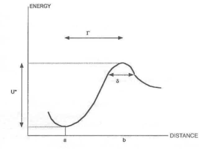
Second, a major point emphasized by Bell was that the rate of bond dissociation should be dependent on applied forces. He suggested the following empirical formula :
r-(F) = r-(0) exp(γF/kT) = r- exp(F/F°) (9)
where F is the applied force, k is Boltzmann's constant, T is the absolute temperature and γ is a parameter that should be close to the interaction range of ligand-receptor bonds. Bell estimated γ at
about 0.5 nm. Although this formula was inspired by experimental data obtained on the rupture of macroscopic material samples (Zurkhov, 1965), it may be somewhat justified with standard theories of reaction rates (see below). Equation (9) proved quite useful since i) it emphasized that bond rupture is a stochastic event, that may occur in absence of distractive force, and ii) it provided an estimate for the force required to substantially enhance the rate of bond formation : using Bell's estimate, kT/γ is of order of 10 pN. As will be described below, these concepts were subjected to extensive experimental check during the last few years, and recently some theoretical attempts were done to relate these experiments to results from statistical mechanics.
A thermodynamic approach to the effect of stress on intermolecular association was followed by Dembo and colleagues a few years later (Bell et al., 1984 ; Dembo et al., 1988). Modeling ligands and receptors as Hookean springs (i.e. springs elongating proportionally to the applied force), it is concluded that subjecting a molecular link of length L to a force F will result in a length increase F/κ and energy increase F2/2κ, yielding an equilibrium constant :
K(F) = K(0) exp(- F2/2κ kT) (9)
where κ is the spring constant. Dembo et al. (1988) further reasoned that there was no thermodynamic necessity implying that bond dissociation rate be increased by a distractive force, and they introduced the concept of catch-bonds, whose lifetime would be increase by applied force, in contrast to slip-bonds, whose life time should be decreased by disruptive forces, in accordance with intuitive prediction.
Now, in order to provide a quantitative feeling for the parameters we defined, we shall describe several representative examples.
1.3 - Typical thermodynamic and kinetic properties of ligand-receptor association.
A prominent example is constituted by antibody molecules that were first obtained by injecting animals with foreign substances called antigens. This procedure induced the synthesis of molecules with a selective capacity to bind antigens used for stimulation. These antibody molecules shared remarkable structural properties allowing them to be included in a family of blood proteins called immunoglobulins. The most abundant immunoglobulins belong to a subtype called immunoglobulin G or IgG. These molecule were observed with electron microscopy by Valentine and Greene (1967) : they appeared as Y-shape assemblies of three rods (about 50 Å length and 40 Å thickness) joined in a fairly flexible region. Each IgG molecule is endowed with two identical antigen-specific binding sites. A typical binding site may be viewed as a cleft of variable depth (5-10 Å), 15-20 Å length and about 10 Å width (Richards et al., 1977), as determined with X Ray cristallography. Antibodies may bind molecules as small as a dinitrophenol group, or large proteins or polysaccharides. The binding sites may involve 5-6 aminoacids or hexose residues (Kabat, 1968).
In a typical series of 21 compilated antigen-antibody couples (Steward, 1977), the affinity constant ranged between 104 and 1010 M-1, although values as high as 1012-1013 were reported by others (Voss, 1993). Association rates displayed relatively restricted variation, ranging between 8× 10⁶ and 1.8×10⁸ M-1s-1, whereas the dissociation rate varied from 3.4×10⁻⁴s-1 to 6000s-1, thus leading to the common view that antigen-antibody reactions are diffusion-limited, and affinity differences are due to differences in dissociation rates.
In another study, Wurmser et al. (1972) measured the thermodynamic properties of some antibodies specific for protein antigens (albumin or insulin) or carbohydrates (blood group antigens). The affinity constant ranged between 8×10³ and 6×10⁸ M-1. The reaction enthalpy and entropy changes ranged respectively between 0 and - 16 kcal/mole and - 35 and 24 cal/mole/°K. Note that the interpretation of older data on antigen-antibody reactions might be somewhat hampered by the heterogeneity of antibody samples. This difficulty was raised by the advent of monoclonal antibody technology.
The range of affinity constants spanned by antibodies is representative of results obtained with other biomolecules. Thus, Lollo et al. (1993) estimated at 107 M-1 the affinity of solubilized forms of
LFA-1, a cell membrane receptor allowing strong association with ICAM-1, which is another cell surface adhesion molecule. The affinity displayed 100 fold increase upon cell activation : affinity changes related to modification of receptor conformation are indeed a well-known mechanism for the regulation of cell interactions (Pierres et al. 1998a). Lower affinity constants ranging between 105 and 106 M-1 were measured on solubilized receptors involved in transient adhesive interactions, such as lymphocyte CD2 (van der Merwe et al., 1993). Conversely, the binding system with highest known affinity is the interaction between avidin or streptavidin (these are proteins of about 60,000 molecular weight) and the small molecule biotin. The affinity constant is of order of 1014-1015 M-1 (e.g. Miyamoto & Kollman, 1993). Note that the recent development of surface plasmon resonance based technology proved an incentive to study the equilibrium and kinetic properties of a number of binding systems (Szabo et al., 1995). Also, Sturtevant (1977) reported a compilation of entropy and heat capacity changes associated to a number of ligand-receptor associations, mainly enzyme-substrate binding : ∆S ranged between -90 and +34 cal/mole/°K.
1.4 - Inability of the conventional framework to account for biological phenomena.
The theoretical framework we described in § 1.2.1 is suitable to account for the behaviour of free molecules. However, cell function is often regulated by interactions involving bound receptors and ligands. In order to illustrate the problems encountered by cell biologists, we shall describe four representative models that recently attracted the interest of many investigators. Then we shall rapidly sketch some theoretical attempts that might be useful to relate cell behaviour to the interaction of individual ligand and receptor molecules.
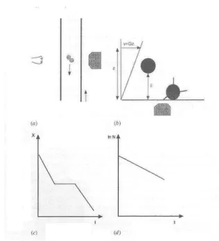
Leukocyte rolling. The experiments performed in vivo by von Andrian et al. (1991) and in vitro by Lawrence and Springer (1991) to study the phenomenon of leukocyte rolling certainly played an important role in emphasizing the importance of the lifetime and force dependence of interactions between bound ligands. A major role of white blood cells (i.e. leukocytes) is to patrol throughout living organisms in order to eliminate potentially harmful agents such as pathogens or damaged cells. Thus, if a given tissue is invaded by infectious microorganisms, specific signals will be generated, resulting in the exit of leukocytes from blood towards the site of aggression (Figure 1). Intravital microscopy revealed the basic features of this process, called diapedesis. Leukocytes that are moving with a velocity of several hundreds of micrometers per second first exhibit a spectacular velocity decrease (by a factor of one hundred) and seem to roll along the walls of blood vessels (these walls are made of so-called endothelial cells). Second, rolling cells stop completely. In a third step, they exhibit dramatic deformations allowing them to pass through transient gaps appearing between neighbouring endothelial cells, and they reach peripheral tissues.
The molecular basis of this phenomenon was essentially elucidated by Lawrence and Springer (1991) who reconstituted the main features of leukocyte-endothelium interaction in a laminar flow chamber. These authors showed that rolling and arrest were mediated by two separate classes of molecular interactions : leukocytes are endowed with a variety of receptors, including members of the
Figure 1. Adhesive interactions between white blood cells and vessel walls.When the endothelial cells lining blood
vessel walls are activated by an aggressive stimulus such as infection or trauma, they rapidly express selectin receptors. White cells that are flowing with a typical velocity of several hundreds of micrometers per second (0) are then tethered through selectin ligands borne by their membranes (1). Then they begin rolling with 50-100 fold decreased velocity (2) due to the rapid formation and dissociation of selectin-ligand bonds. The following step is a firm adhesion resulting in complete cell arrest (3) due to an interaction between integrin receptors (on white cells) and ligands such as ICAM-1 on endothelial cells. The white cell passage between endothelial cells towards peripheral tissues (4 & 5) also involves the rupture of adhesive interactions between endothelial cells.
so-called selectin and integrin families. Rolling is mediated by transient interactions between selectins and their ligands. Bonds can be formed when cells move with high velocity (i.e. several hundreds of micrometers per second, as stated above), they can stand the strong shearing forces generated by blood flow (the wall shear rate is of order of several hundreds of second-1). However, they are unable to maintain cells immobile, even if shearing forces are dramatically decreased. In contrast, the interaction between leukocyte integrin receptors and their ligand on endothelial cells may lead to a complete stop. However, integrin/ligand bond do not appear when cells move at physiological velocity in blood flow. A few years later, Patel et al. (1995) made a clever use of genetic engineering techniques to demonstrate that the remarkable property of selectins to form bonds in presence of high shearing forces was abolished when the length of these molecules was decreased without altering the binding site. Also, experimental studies performed on isolated blood cell receptors suggest that the affinity constant of both integrins (Lollo et al., 1993) and selectins (Nicholson et al., 1998) is of the order of 106-107 liter/mole. These experiments strongly suggest that the behaviour of adhesion molecules is not entirely accounted for by their affinity : other properties such as kinetic rates but also binding strength and molecular length may be of importance to regulate their function. This situation could not be ascribed to some complex function of living cells, since rolling could be reproduced either with cells that had been made inert by suitable fixatives (Lawrence and Springer, 1993) or artificial particles that had been coupled with selectins and made to roll along artificial surfaces coated with selectin ligands (Brunk and Hammer, 1997).
Cell deformability. Many cell functions are dependent on their capacity to undergo active or passive deformation (Richelme et al., 1996). Much work was devoted to the study of cell mechanical properties. Thus, accurate information on cell viscoelastic properties was obtained by monitoring the deformation of individual cells subjected to controlled aspiration into small micropipettes of a few micrometer diameter (Schmid-Schöenbein et al., 1981 ; Evans and Kukan, 1984). A major issue would be to relate these parameters to the molecular properties of the three-dimensional scaffold called the cytoskeleton. The in vitro demonstration by Sato et al. (1987) that the mechanical properties of reconstituted (and simplified) models of the cytoskeleton were dependent on deformation rate suggests that the cell deformability might depend on the lifetime and force dependence of associations between immobilized cytoskeletal components.
Cell migration. An important property of living cells is their ability to migrate when they are deposited on suitable surfaces (and possibly stimulated by soluble mediators). As reviewed by Stossel (1993), cell displacement involves the forward emission of a lamellipodium that will adhere to the surface, with subsequent contraction and detachment of the rear part of the cell. Thus, motility is dependent on continuous attachment and detachment. This qualitative concept was made quantitative by Palecek et al. (1997) who studied the migration speed of different cell populations expressing integrin receptors and deposited on surfaces bearing integrin ligands. These authors used different ways of manipulating adhesiveness by varying the surface density of integrin ligands (through standard coupling procedures) as well as cell membrane density and activity of integrins (using genetic engineering techniques). Then, they measured the mechanical strengh of cell-to-surface adhesion by subjecting bound cells to hydrodynamic flow and determining the force required for detachment. Further, they measured cell migration velocity and demontrated a quantitative relationship between the above two parameters. Velocity was maximal for some intermediate value of adhesiveness. This report further emphasizes the physiological importance of the mechanical strength of ligand-receptor bonds.
Redistribution of adhesion receptors in contact areas. Recent experimental advances raised the interest of the biological community in the dimension-dependence of affinity constants. It is now well known that most cell membrane molecules may display free lateral diffusion on the cell surface. It is thus not surprising, on a simple thermodynamical basis (Bell et al., 1984), that receptor-mediated adhesion between two cells often results in concentration of binding molecules in the contact area (Kupfer and Singer, 1986). Previous attemps at quantifying this phenomenon (McCloskey and Poo, 1986 ; André et al., 1990) were dramatically improved by Dustin et al.(1996) who deposited cells expressing the CD2 adhesion receptor on supported lipid layers where they incorporated fluorescent derivatives of CD2 ligand (called LFA-3) : Cell to surface encounter resulted in the formation of a
contact area where fluorescent molecules were gradually concentrated. This contact area could be visualized with optical techniques such as interference reflexion microscopy (Curtis, 1994). Further work by Dustin (1997) provided a formal proof that concentrated LFA-3 molecules displayed reversible interaction with cell surface receptors, thus supporting the relevance of the concept of two-dimensional binding equilibrium.
The few selected examples we described show that there is a need for quantitative models to account for the relationship between biological phenomena and ligand receptor interactions involving attached molecules. We shall now describe some selected attempts aimed at i) obtaining a workable description of ligand-receptor interaction and ii) using this framework to interpret experimental data. 1.5 - Models for relating cell adhesive behaviour to molecular properties of their surface receptors.
Several authors developed quantitative models to relate measurable cell features to the quantitative properties of membrane molecules. Thus, Bell et al. (1984) considered the equilibrium shape of cells bound by specific adhesion receptors. They assumed that the equilibrium contact area ensured minimization of the free energy contributed by i) adhesion molecules diffusing in the plane of cell membranes, with bond formation restricted to the contact area, ii) repulsion between nondiffusible repulsive elements corresponding to bulky macromolecules known to occur on cell surfaces, and iii) stretching of binding molecules to alleviate repulsion. Their model could fit quantitative experimental data on actual adhesion models reported by Capo et al. (1982), but the numerical values of repulsive force and spring constant of cell-cell bridges were fitted parameters. Also, the mechanical properties of the membrane and possibility of active cell deformation were neglected.
A dynamical model was elaborated by Hammer and Lauffenburger (1987) to account for the rate of bond formation between receptor-bearing cells and ligand-coated surfaces. This model proved a suitable framework to account for a number of experimental findings, but it included many unknown parameters such as the contact area, density and accessibility of adhesion receptors or kinetic properties of these receptors. Further models were elaborated to account for specific phenomena such as the aforementioned rolling process (Hammer and Apte, 1992).
Mechanical approaches were elaborated to account for the statics (Evans, 1985 a&b) and kinetics (Dembo et al., 1988) of separation between a cell and a surface. Interestingly, it was demonstrated that adhesion mediated by a few specific bonds could behave as an irreversible process, in accordance with experimental studies (Evans 1985b), and realistic values of fitted parameters might account for some experimental features of the rolling phenomenon (Dembo et al., 1988 ; Atherton and Born, 1972 & 1973). Later experimental studies performed on the detachment of model particles bound to artificial surfaces through anchored adhesion molecules (Kuo and Lauffenburger, 1993) were interpreted within the framework of these models and led to the experimental finding of a linear relationship between the binding strengh and affinity of ligand/receptor bonds.
In conclusion the models we briefly described show that there is a need for accurate knowledge of the behavior of anchored ligand and receptors. However, there are too many unknown features to allow an accurate derivation of ligand receptor properties from experimental studies performed on cell-size objects. Further, theoretical knowledge was markedly insufficient to yield accurate prediction of these parameters. We shall now describe three main lines of research that shed a new light on ligand-receptor interaction : i) the development of genetic engineering led to unprecedented accuracy in the understanding of correlations between structural and functional properties of biological receptors. ii) New methodologies allowed direct investigation of ligand-receptor association at the single molecule level, and iii) continuous progress of computer simulation allowed more detailed understanding of the behaviour of complex objects such as protein molecules embedded in aquous electrolyte solution.
2 - NEW INFORMATION ON STRUCTURE-FUNCTION RELATIONSHIP IN LIGAND-RECEPTOR INTERACTION.
As previously mentioned, we shall essentially focus on reactions involving proteins, in order to prevent excessive dispersion. The following questions may be considered :
- Is there a preferred kind of interaction (e.g. hydrogen bonds, hydrophobic interaction, salt-links) that is mostly used by proteins to achieve binding affinity and specificity ?
- Is binding affinity contributed by a few strong interactions or many weak bonds scattered on an important area ?
- Does binding require important conformational changes of interacting molecules or may these be considered as rigid ?
- Is there an accurate fit between interacting molecules or are there wide gaps filled with solvent in protein-protein interface ?
In order to address these questions, we shall first describe some properties of representative ligand-receptor complexes that were studied with X ray cristallography. Then we shall review some information obtained by mutagenesis experiments.
2.1 - General features of protein-ligand complexes.
The first important parameter may be the contact area. The precise definition of this parameter may not be as straightforward as it might first seem. Great simplification was brought by the concept of solvent accessible surface area (Lee and Richards, 1971 ; Richards and Richmond, 1978). This is the geometrical locus of the center of a spherical probe (considered to represent a solvent molecule, the usual radius is 1.4 Å) remaining in contact with the protein surface. The surface area that is buried in the protein-ligand interface after association provides a convenient measure of the extent of interaction. Recently, Jones and Thornton (1996) studied 59 protein complexes whose cristallographic structure was recorded in the Brookhaven protein database. The reduction of the accessible surface area generated by complex formation ranged between several hundreds and several thousands of squared angstroms. Further, the authors compared the frequency of occurrence of different aminoacid residues in the interface and on the remaining part of the protein surface. A notable conclusion whas that the frequency of nearly all hydrophobic residues was higher in the contact area. Also, they estimated the mean number of hydrogen bonds between reagent surfaces : this was of order of one per 100 Å2. Now, we shall present a few selected examples.
The structure of a complex made between lysozyme and a monoclonal antibody (called D1.3) was studied with 2.8 Å resolution (Amit et al., 1986 ; Mariuzza et al., 1987). The affinity constant was 4.5×10⁷ Mole-1. Sixteen aminoacid residues of the lysozyme surface made tight contacts with 17 residues on the antibody combining site. There was a notable complementarity between surfaces, since protrusions occurring on a molecule were matched by depressions on the opposed surface. Twelve hydrogen bonds were identified between surfaces. About 11 % of the lysozyme accessible area (i.e. 748 Å2) was buried during the interaction, together with 690 Å2 on the antibody surface. A later study performed with 1.6 Å resolution led to the conclusion that the cristallographic structure of lysozyme was identical in free and bound molecules. Similarly, no gross difference was found between the conformation the lysozyme-bound monoclonal antibody and free immunoglobulin molecules. The authors concluded that no drastic conformational change was detected in different proteins upon ligand binding. Several years later, the same model was used to compare the conformation of free and bound antibody D1.3 with 1.8 Å resolution (Bhat et al., 1994). This resolution allowed unambiguous localization of water molecules. Twenty three water molecules were bound to the free antibody site, and 48 were localized in the antigen-antibody interface, acting as bridghes between protein surfaces. This was consistent with the experimental finding (obtained with calorimetric studies) that the antigen-antibody association resulted in a net entropy decrease, in contrast with hydrophobic interactions that are supposed to increase entropy as a result of a release of solvent molecules (see § 4.1).
Low affinity interactions (in the ten micromolar range) were also investigated. Garboczi et al. (1996) studied the complex between a particular human T-cell receptor (a specific receptor for foreign structures expressed by a subpopulation of lymphocytes) and its natural antigen, a complex between an oligopeptide of viral origin and major-histocompatibility molecule HLA-A2. The solvent accessible surface area that was buried on the T cell receptor on binding was 1,011 Å2. Twenty hydrogen bonds
were identified among a total of 46 interatomic contacts (defined as interatomic distances lower than 4 Å). No gross conformational changes was induced on interacting molecules by complex formation. In another study, Gao et al. (1997) studied the interaction between lymphocyte CD8 molecule and HLA-A2. The total buried accessible area on CD8 was 947 Å2. Interactions were considered as mainly electrostatic since 80 % of atoms were polar in contact regions. Eighteen hydrogen bonds were identified between interacting proteins.The authors suggested that the relatively low affinity value was related to the high number of polar residues in the interface.
Finally, Weber et al. (1989) explored the structural origin of the high affinity (1015 Mole-1) interaction between the protein avidin and the small biotin molecule. They concluded that binding involved a high number of hydrogen bonds and a conformational change of the protein burying the biotin in a pocked that was closed with a surface loop of avidin.
2.2. - Information obtained by studying series of mutant molecules.
As will be detailed below, the difficulty of achieving a quantitative understanding of ligand-receptor association is that the binding free energy represents only a minimal fraction (say a few percent) of the total conformation energy of interacting molecules. Thus, much information could be obtained by comparing series of molecule differing by a few or even a single aminoacid, with might give accurate information on the contribution of a few or even a single molecular interaction to the total binding energy. We give a few selected examples.
The relative importance of electrostatic and hydrophobic interaction was studied by exploring the high affinity interaction between thrombin, a coagulation factor, and hirudin, a 65 aminoacid polypeptide found in medicinal leech. Stone et al. (1989) measured the affinity constant and reaction rate of four mutants obtained by replacing one to four negatively charges glutamic acid residues with glutamine (which amounts to replacing a terminal COO- group with CONH2). Interaction parameters were measured both in a solution of high ionic force (resulting in efficient screening of electrostatic interactions) and at low electrolyte concentration. The authors tentatively separated the contributions of ionic and non-ionic interactions considered as additive components of the standard free enthalpy of reaction. They found that the non-ionic component ∆G°nio displayed a similar value of about -15 kcal/mole while the ionic component algebraically increased from -6.9 kcal/mole to -2.1 kcal/mole upon sequential removal of four negative charges. Further cristallographic study demontrated that hirudin bound thrombin at sites both close and distant to the active site (Grutter et al., 1990).
The study of immunological recognition provided many opportunities to demonstrate that the replacement of a few aminoacids in a protein could markedly change binding properties. Thus, while aforementioned monoclonal antibody D1.3 bound hen egg lysozyme with high affinity, no detectable binding was measured on lysozymes from other animal species differing by only 3 or 4 aminoacids (Mariuzza et al., 1987). Further, Chacko et al. (1995) reported a study made on the interaction between lysozyme and a monoclonal antibody (HyHEL-5) : the interface region in the complex contained 23 lysozyme and 28 antibody aminoacid residues. The replacement of a single (positively charged) arginine with a lysine (of similar charge) resulted in the introduction of a water molecule in the interface and concomitant 103-fold reduction of the binding affinity. A similarly exquisite specificity was reported in another model : so-called natural killer cells are endowed with receptors for histocompatibility molecules. These receptors are able to discriminate between histocompatibility molecules from different individuals. The simple exchange of two neighbouring aminoacids (a methionine and a lysine) was sufficient to exchange the specificity of a receptor (Winter and Long, 1997).
Several reports gave some information on the possible functional importance of minimal conformational changes related to complex formation. In a study performed on the lysozyme/antibody model, Hawkins et al. (1993) studied the contribution of residues of the D1.3 antibody to hen egg lysozyme binding. Interestingly, they obtained a mutated molecule with fivefold affinity increase, while none of the altered residues was located in the contact interface. Similarly, Wedemayer et al. (1997) compared the structure of complexes involving antidodies differing by a few residues : A 30,000 fold affinity increased could be achieved by mutations located at distance (more than 15 Å) from the binding site. These mutations seemed to act by stabilizing the conformation displayed by the antibody during binding. When Tulip et al. (1992) studied the cristallographic structures of mutant
neuraminidase-antibody complexes, they reported that single sequence changes in some of the neuraminidase residues in the binding site markedly reduced affinity. However, in some cases a sequence change could be accomodated by a structural modification of the conformation of a few residues in the complex (e.g a 2.9 angström shift or a rotation of 150°).
Clackson and Wells (1995) reported a remarkable study on the high affinity (3×10⁹M-1) interaction between human growth hormone (hGH) and a fragment of the hGH receptor bearing the binding site. X ray cristallographic studies revealed that about 30 residues were involved in the interaction on each protein.Controled mutagenesis was used to replace systematically each of these residues with an alanine, which is a relatively small amino-acid (CH3CH2CH(NH2)COOH) whose introduction is supposed to remove possible electrostatic or hydrogen bond without adding bulky groups that might reduce affinity through steric repulsion. The authors found that only 9 substitutions (out of 33) on the hGH receptor resulted in marked affinity reduction (between 1 and 4.5 kcal/mole). They suggested that in some cases the desolvation energy and lateral chain reorganization associated to complex formation balanced the energy gain due to bond formation. They also suggested that water molecules might fill gaps between imperfectly matching regions of proteins and form interactions that were fairly isoenergetic with those found in free proteins. They concluded that the dominant importance of a few interactions would facilitate the design of low size synthetic ligand for medical purpose.
Recently, Vallone et al. (1998) used analytical ultracentrifugation to study the association of hemoglobin subunits. They determined the affinity changes generated by simple or double mutation. Also, they took advantage of molecular modeling to determine the accessible surface area variations resulting from these mutations. They concluded that the free energy of burying hydrophobic residues in a protein-protein interface was about -15 cal/mole/Å2. They also suggested that the contribution of polar interaction to the affinity was low. This conclusion is in line with another report from Davis et al. (1998) who prepared a series of mutant CD2 molecules and studied their low affinity (≈ micromolar) interaction with CD48 ligand. They exhaustively mutated residues located in the CD2-CD48 interface. They concluded that three fairly hydrophobic residus (leucine, phenylalanine and tyrosine) were dominant contributors to the binding energy. Since the affinity constant was independent of the ionic stength, they concluded that the binding free energy was mainly accounted for by hydrophobic interactions, while electrostatic forces might contribute the specificity of the interaction.
In conclusion, Accurate structural information, is now available at the atomic level on the structure of protein-protein interfaces. Recently, additional data obtained by systematic mutagenesis experiments led to the view that a few interactions might account for most of binding energy. Probably the steric repulsion generated by a single residue or a few unfavorable electrostatic interaction might suffice to prevent binding, thus accounting for the remarkable specificity of many receptors. Available data form a firm basis to test refined theoretical models of protein association. These will be described in the last section of this review.
3 - EXPERIMENTAL STUDY OF LIGAND-RECEPTOR INTERACTION AT THE SINGLE MOLECULE LEVEL.
While conventional lines of research on ligand-receptor interaction were followed during the last few years, and indeed methodological advances such as the use of surface plasmon resonance gave a new impetus to the experimental determination of binding rates, a qualitative change in this field was brought by the development of several experimental methods bringing direct information on individual interactions between surface-bound molecules. The interest of this approach is that data interpretation is greatly facilitated when single bonds are monitored, since there is no need to account for the mechanical properties of surfaces and the geometrical arrangement of bonds. Experimental results allowed to check conventional theories with unexpected accuracy. Now, we shall describe these techniques together with selected experimental results. The significance of reported data will be discussed in the final part fo this review. We shall first describe studies made on the determination of bond lifetime and mechanical strength. Then whe shall describe the rather scanty information available on the kinetics of bond formation between surface-attached molecules.
3.1 - Lifetime and mechanical strengh of ligand-receptor bonds.
During the last ten years, at least three different methods (Bongrand et al., 1994) became available to monitor the rupture of individual ligand-receptor bonds subjected to distractive forces in the piconewton range. It is now apparent that noncovalent associations between biomolecules can be rapidly ruptured with forces ranging between a few tens and hundreds of piconewtons. Part of the approaches listed below were described in a recent review (Pierres et al., 1998b).
- Use of hydrodynamic flow : a particle of radius a bound to another particle or a macroscopic surface in a fluid of viscosity µ with a locally varying flow of shear rate G is subjected to a distractive force of order of µa2G. If bonds are ruptured, particles will then depart with a velocity of order of aG (Figure 2). Thus, if we consider a cell-size particle of 10 µm radius in a fluid of 0.001 Pa.second viscosity such as water, a shear rate of 10s-1 may generate an hydrodynamic drag of order of 0.1 piconewton and relative velocity of 100 µm/s. Therefore, the mere observation of the particle with a conventional microscope may in principle allow a detailed examination of single bond rupture with a time resolution of a few tens of milliseconds.
The pioneering studies were performed by Tha et al. (1986) with the so-called traveling microtube apparatus (Figure 2) : they prepared highly spherical red blood cells by exposure to hypoosmotic treatment prior to fixation. These particles were then coated with low amounts of antibodies in order to allow them to bind to each other with a a few or even single bonds. Suspensions were driven through a vertical capillary tube that was mounted on a mobile stage, under continuous monitoring with a microscope whose optical axis was perpendicular to the tube. The stage velocity was adjusted to achieve exact compensation of the displacement of individual particles that could thus be followed for a fairly long period of time. The authors followed the motion of individual doublets that underwent rotation with a sequence of compressive and disruptive hydrodynamic forces. The distribution of force intensities at the moment of rupture could thus be accurately calculated, yielding an estimate of about 24 piconewtons for the binding strengh of the weakest doublets. A comparable estimate of 20 pN was later obtained with an improved methodology based on a cone-and-plate rheoscope allowing rapid variation of the shearing forces (Tees et al., 1993 ; Goldsmith et al., 1994). In later studies, computer simulations were used to extract quantitative estimates from the natural rate of bond dissociation and mechanical strength of bonds. The latter parameter was obtained assuming
Figure 2. Studying individual ligand-receptor bonds with hydrodynhamic flow. The traveling microtube technique
developed by H. Goldsmith (A) consists of sending particle suspensions through a vertical capillary tube mounted on a moving stage. Particles are coated with adhesion receptors resulting in doublet formation, and individual doublets are monitored with a microscope (with horizontal axis). The low doublet velocity (typically ∼ 25 µm/s) is accurately balanced by the stage motion, thus allowing prolonged observation of the doublet that remains fixed with respect to the microscope field. The hydrodynamic forces exerted on the doublet at the moment of rupture are accurately calculated.
The laminar flow chamber (B) is a parallelepipedic cavity whose floor is monitored with a standard inverted
microscope (the objective O is shown). Receptor bearing particles are driven along ligand-coated surfaces with a wall shear rate typically ranging between a few and 100 s-1. The force on a bond maintaining a particle arrested may be calculated if the wall shear rate and bond length are known. The distance δ between a flowing sphere and the surface may be derived from the sphere velocity with nanometer accuracy. A typical trajectory is shown when the sphere velocity is plotted versus time (C). Periods of displacement with fairly constant velocity are intersperesed with arrests (horizontal segment) of various duration. The rate of bond dissociation can be obtained by determining the distribution of arrest durations and plotting the number of particles remaining bound at any time t after arrest versus t (D).
exponential increase of the bond dissociation rate with respect to force (according to Bell's model as displayed in equation 9, the dissociation rate in presence of a disruptive force F is r- exp[F/F°]). The dissociation rate of bonds formed between a polysaccharide antigen (blood group B) and specific antibody was 0.04 s-1 with a force parameter F° of 35 pN (Tees et al., 1996). Similarly, the interaction between immunoglobulin G and protein G, a natural immunoglobulin ligand of bacterial origin, displayed a dissociation rate of 0.006 s-1 and force parameter of 11 pN (Kwong et al., 1996).
Other studies were performed on the separation of cells or particles from surfaces in parallel-plate flow chambers. Kaplanski et al. (1993) monitored the motion of human white blood cells along a surface coated with activated endothelial cells in presence of a very low shear rate (5.25 s-1). The hydrodynamic drag exerted on cells interacting with the surface was less that 5 pN. A single bond should thus be sufficient to maintain cells under arrest. Cells indeed displayed numerous transient stops whose duration could be fitted to a theoretical curve obtained with respective values of 0.75 s-1 and 0.50 s-1 for the rates of bond formation and dissociation in the region of cell-suface contact after the formation of the first bond. Inhibition experiments suggested that observed interactions were mainly due to association between E-selectin receptors expressed by endothelial cells and their ligand on white cells. These conclusions were supported by a report from Alon et al. (1995) who studied the motion of white blood cells along surfaces coated with various densities of purified P-selectin, a molecule closely related to E-selectin. The shear rate ranged between 20 and 110 s-1. Cells displayed intervals of rapid displacement interspersed with tethering events of various duration. Modeling cells as spheres with transient flattening in contact area, the authors calculated the force exerted on binding molecules and they fitted experimental distributions of arrest durations with Bell's equation. The estimated dissociation rate was 0.95 s-1 and the force parameter F° was 120 pN. The authors suggested that the low lifetime and high mechanical resistance of selectin-mediated bonds were a prerequisite to support the rolling phenomenon (see § 1.4 and figure 1). In a later study, Pierres et al. (1996) studied the motion of spherical particles bearing recombinant binding sites of CD48 along CD2-derivatized surfaces. The CD2-CD48 adhesion system is supposed to mediate transient associations between T lymphocytes that scan the surface of different cell populations in order to detect possible abnormalities (Bongrand and Malissen, 1998). The choice of spherical particles allowed the use of a computer-assisted tracking device yielding the coordinates of the centroid of particle images with 20 millisecond and 20 nm accuracy. Also, the force exerted on individual bonds could be accurately calculated. The rates of bond formation and dissociation between surfaces and particles bound by at least one bond were 39.6 s-1 and 7.8 s-1 respectively. Bell's force parameter F° was 32 pN.
While the flow chamber proved a very convenient tool for studying transient interactions, it was interesting to study the behaviour of molecules supposed to mediate durable bonds. Thus, Pierres et al. (1995) studied the interaction between rabbit immunoglobulin and spherical particle coated with mouse antibodies specific for these rabbit proteins. First, particles were incubated with fluorescent rabbit immunoglobulins, then washed and assayed for fluorescence content after different periods of time. Mean particle fluorescence exhibited minimal decrease after 3h or even 24h incubation, suggesting that binding was durable. Then spheres were driven along ligand-bearing surfaces in the flow chamber with a wall shear rate ranging between 11 and 72 s-1. Interestingly, the distribution of arrest durations could be fitted to a two-step interaction model according to the following equation :
A
B
AB
AB
kd k k+
← → ← → − +(
)
1(
)
2 (10)The dissociation rate of the intermediate complex was determined under low dilution conditions (about 3.5 molecules/µm2 on beads and 6,200 molecules /µm2 on the chamber floor). The dissociation rate of the intermediate state (AB)1 was 0.9 s-1, with a force parameter of 53 pN. The dissociation rate k- was of order of 0.01 s-1 or less and could not be determined accurately. The stabilization rate k+ was about 0.3 s-1 without any significant dependence on the shear rate, as expected. These results show that the concept of a single bound state is only an approximation that may be insufficient to account for the quantitative features of the initial interaction between a moving receptor-coated particle and ligand-bearing surface. It was indeed already known that ligand-receptor association might behave as a multi-step process (Beeson and McConnell, 1994). The above experiment show that this complexity may
actually influence particle-to-surface adhesion. In the flow chamber, the relative velocity of the floor and the surface of a sphere is of order of 50 % of the bead velocity, and the velocity of a spherical particle close to the surface is about half the product of the wall shear rate and sphere radius (Goldman et al., 1967 ; see below for more details). Assuming that the length of a ligand-receptor couple is of order of 40 nm, the time available for bond formation between a particle of 1.4 µm radius (Pierres et al., 1995) and the surface in presence of a wall shear rate of 20 s-1 is about 6 ms. Thus, provided the detection apparatus is rapid enough, the presented methodology may yield quantitative information on the details of interactions between bound molecules in the millisecond range.
More recently, Pierres et al. (1998c) used the flow chamber technology to study the high affinity interaction between biotin and streptavidin. The lifetime of interactions between biotinylated surfaces and streptavidin-coated spheres was of order of several seconds, i.e. 5-50 fold higher than previously determined on selectin/ligand or CD2/CD48 models. Further, this lifetime was not decreased when the wall shear rate was increased from 10 to 40 s-1. However, it was concluded that the flow chamber was not well suited to the study of strong interactions, since i) the chamber floor was rapidly filled with definitively attached particles, which made it difficult to follow a sufficient number of trajectories in a single experiment, and ii) it was somewhat difficult to obtain a reliable distribution of arrest durations, since the computer-assisted apparatus was not adapted to the monitoring of very long arrests.
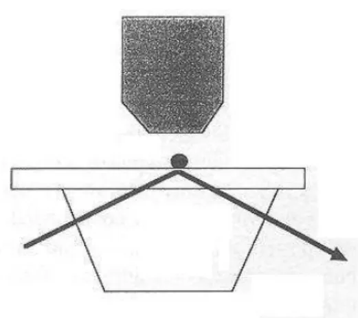
- Use of soft vesicles as tranducers : this approach was pioneered by Evans et al. (1991). As recalled on Figure 3, the principle consists of approaching with two micropipettes (mounted on micromanipulators) cells or lipid vesicles derivatized with suitable receptor and ligand molecules. After allowing bonds to form, a pipette is pulled out under microscopic control. The applied force results in vesicle deformation, and a spherical shape is recovered when the last bond is ruptured. Thus, the experimental result is the unbinding force rather than the bond lifetime. As emphasized by the authors, the interest of this procedure is that the surface tension of the vesicles may be varied in a wide range by controlling the sucking pressure applied through pipettes. Vesicles can indeed be subjected to a distractive force ranging between less than 1 and 100 piconewtons.
The method was used to study the interaction between red blood cells that were cross-linked by a low density of antibodies or lectins (i.e. molecules with an affinity for some cell surface sugars). Since the density of binding molecules was low enough that only a limited fraction of cell-cell encounters resulted in adhesion, it was suggested that attachment was mediated by a few or even a single bond. Surprisingly, the detachment force was of order of 10-20 pN for all tested bridging molecules. The authors suggested that applied forces might uproot membrane receptors rather than rupture ligand-receptor bonds. The vesicle methodology was later improved (Evans et al., 1994 & 1995) by chemically coupling microscopic latex beads to vesicles, and using a piezoelectric transducer to achieve optimal control of pipette position. Finally, an interferometric technique allowed to resolve the bead distance to a flat surface with 5 nm accuracy. Evans et al. (1995) could thus study sphere to surface interactions with piconewton sensitivity. They reported a study of biotin-streptavidin association (Merkel et al., 1995) : when bonds were subjected to a slowly increasing force (100 pN/s), the unbinding force was about 50 pN. This value was 4-5 fold lower than measured with atomic force microscopy (see below), allowing the authors to emphasize the dependence of the unbinding force on the loading rate.
More recently, Chesla et al. (1998) reported fairly accurate determination of bond lifetime with a clever modification of the micropipette technology. The basic idea was to take advantage of a piezoelectric-driven pipette to generate multiple collisions of controlled frequency and duration between immunoglobulin-coated red cells and cells expressing immunoglobulin receptors. This allowed accurate determination of the adhesion probability versus contact duration. The authors emphasized that their methods allowed accurate determination of zero-force association and dissociation rates since the force merely served to provide a signal to the observer. Further, their apparatus certainly allowed piconewton sensitivity. The dissociation rate of bonds formed between human immunoglobulin G receptor and its ligand was 0.37 s-1. Since contact duration could be accurately determined, this method might in principle allow to detect possible intermediate binding states.
- Atomic force microscopy : The principle (Figure 4) consists of moving a ligand-coated surface towards a receptor-bearing tip of a few nanometer thickness mounted on a very soft cantilever (a typical spring constant is about 100 mN/m). The surface is then pulled out, resulting in continuous increase of the distractive force, with continuous monitoring of the cantilever deformation. The rupture of the last bond between surfaces results in a sharp jump of the cantilever, allowing experimental measurement of the so-called unbinding force. The cantilever position may be monitored with angström accuracy with optical techniques. The limit set by thermal fluctuations to the force sensitivity is about (kT/λ)1/2, where λ is the spring constant. The reported force sensitivity is of order of 10 pN in liquid medium (Erlandsson and Olsson, 1994 ; Florin et al., 1994 ; Ros et al., 1998). A final point of interest that was noted by several authors (Florin et al., 1994 ; Hinterdorfer et al., 1996) is that interacting molecules do not seem to be altered by the adhesion/rupture cycle, which allows to perform hundreds of cycles on a given position of the microscope tip. Another point is that efficient bond formation may require that the length of adhesion molecules be increased with a chemical spacer (Hinterdorfer et al., 1996) or that one of interacting surfaces be sufficiently deformable (Florin et al., 1994).
Figure 3. Studying individual ligand-receptor bonds with biomembrane probes. The study of adhesive interactions with
soft vesicles (red cells or artificial liposomes) was pioneered by E. Evans. A typical experiment (A) consists of using micropipettes to push against each other two vesicles bearing low amounts of receptors and ligands. It may be convenient to use a rigid sphere (e.g. a fixed red cell) and a soft vesicle whose surface tension is accurately controlled by adapting the sucking pressure. When vesicles are progressively separated after contact under microscopic control and video-recording, the deformation of the soft vesicle may be analyzed in order to calculate the force. A refinement of this technique (B) consists of mounting a micropipette on a piezoelectric device (to achieve better control of position) and chemically coupling a small sphere on the vesicle. The distance between this small sphere and a plane surface can be determined with high accuracy by interferometric techniques.
Figure 4. Studying individual ligand-receptor bonds with an atomic force microscope. The study of specific
biomolecule interactions with atomic force microscopy was pioneered by Moy et al. (1994) and Lee et al. (1994). As shown on Fig. 4A, the piezo-driven surface bearing ligand molecules (L) is subjected to repeated cycles of
approach/retraction from the tip derivatized with receptors (R). The tip is mounted on a soft cantilever whose deformation is determined with better than nanometer resolution by optical monitoring. The unbinding force during retraction may be measured by determining the length of the jump (vertical segment 4) occurring during retraction. Actual curves may be more complicated than the very simplified drawing displayed on Fig 4B due to nonspecific forces and occurrence of multiple bonds.
While Hoh et al. (1992) may have detected hydrogen bonds with atomic force microscopy, the application of this apparatus to the study of ligand-receptor interactions was pioneered by Florin et al. (1994) and Lee et al. (1994). The first model studied was the avidin-biotin interaction. A tip was coated with biotinylated albumin and made to interact with soft agarose beads bearing streptavidin binding sites (Florin et al., 1994). Using force scan mode, multiple approach-retract cycles were performed and hundreds of unbinding events could be visualized as sharp jumps of the cantilever (Figure 4). The distribution of unnbinding forces displayed quantized peaks that appeared as multiple of 160 ± 20 pN. This was considered as representative of the detachement force of a single bond. Further, when biotin was replaced with iminotiotin, an analog with 25,000 fold lower affinity to streptavidin, the unit separation force was reduced to 85 ± 15 pN. Interestingly, the authors later determined the affinity constant (i.e. interaction free energy ∆G°), reaction enthalpy ∆H° (using microcalorimetry) and unbinding force between avidin or the related bacterial streptavidin and biotin or iminobiotin and desthiobiotin analogs (Moy et al., 1994) : they found that the unbinding force was proportional to ∆H°, which led them to define an "effective rupture length" as the ratio between ∆H° and the unbinding force, yielding a value of 0.9-1 nm. The proportionality between ∆H° and the unbinding force was confirmed in a later report by Chilkoti et al (1995). The physical significance of interaction parameters will be discussed in the last section of this review.
In other studies, Boland and Ratner (1995) studied the interaction between surfaces coated with adenine and thymine. These basic components of nucleic acids are supposed to bind to each other through two hydrogen bonds. The histogram of frequency of unbinding force suggested the occurrence of quantized peaks ascribed to the separation of individual adenine-thymine pairs. The force was 54 pN.
In another study, Danmer et al. (1995) studied the interaction forces involving a proteoglycan from a marine sponge (this was a large molecule made of a protein core and long polysaccharide chains with a multi-arm structure that is supposed to contribute cell-cell adhesion). Detachment curves suggested an intermolecular force of about 400 pN resulting from of about ten individual interactions of 40 pN each.
In a very interesting study, Nakajima et al. (1997) studied the interaction events between a single molecule of heavy meromyosin (an actin-binding fragment of the actin-binding motor protein myosin) and actin. They estimated at 11.9 milliseconds the half life of a single bond subjected to a disruptive force of 14.8 pN (this was obtained by dividing twice the standard deviation of the unbinding force by the loading rate, i.e. the time derivative of the applied force). Using Bell's formula and taking as a zero-force lifetime a value obtained by Marston (1982) on free molecules (5-100 s), they estimated at 1.7 pN the force parameter F° of the actin-myosin interaction.
In a later study, Vinckier et al. (1998) studied the interaction between Groel, a bacterial chaperone protein (i.e. a protein supposed to stabilize partially unfolded proteins during the early stages of biosynthetic pathways) and several substrates. As expected, interaction forces were greater with unfolded proteins than with native forms. Thus, unbinding forces were 420 pN and 770 pN for native and denatured citrate synthetase enzyme. Further, this force was reduced to 230 pN and 320 pN respectively in presence of ATP that is supposed to modulate GroEL state. Interestingly, the force increased from 440 pN to 620 pN when the cycle frequency was reduced from 1 Hz to 0.1 Hz, while the author reported a decrease of unbinding force when the frequency was higher than 32 Hz. This both emphasized the dependence of unbinding force on loading rate and possible occurrence of weaker intermediate states that might be detected after short contact (i.e. high frequency).
Several studies were devoted to antigen-antibody interactions : Hinterdorfer et al. (1996) reported an unbinding force of 240 pN between human albumin and specific antibodies. Interestingly, they estimated at 1.8 ms the bond lifetime in presence of the disrupting force (this was the ratio between twice the standard deviation 48 pN of unbinding force and the loading rate of 14 nN/s). The natural lifetime of bonds formed between free albumin and antibody was 1500s, i.e. 800,000 fold higher. The force parameter F° was thus 18 pN. Note that the time available for bond formation was