HAL Id: hal-00089404
https://hal.archives-ouvertes.fr/hal-00089404
Submitted on 18 Feb 2007
HAL is a multi-disciplinary open access
archive for the deposit and dissemination of sci-entific research documents, whether they are pub-lished or not. The documents may come from teaching and research institutions in France or abroad, or from public or private research centers.
L’archive ouverte pluridisciplinaire HAL, est destinée au dépôt et à la diffusion de documents scientifiques de niveau recherche, publiés ou non, émanant des établissements d’enseignement et de recherche français ou étrangers, des laboratoires publics ou privés.
Cell cycle genes regulate vestigial and scalloped to
ensure normal proliferation in the wing disc of
Drosophila melanogaster.
Rénald Delanoue, Annie Dutriaux, Kevin Legent, Joël Silber
To cite this version:
Rénald Delanoue, Annie Dutriaux, Kevin Legent, Joël Silber. Cell cycle genes regulate vestigial and scalloped to ensure normal proliferation in the wing disc of Drosophila melanogaster.. Genes to Cells, Wiley, 2006, 11(8), pp.907-918. �10.1111/j.1365-2443.2006.00993.x�. �hal-00089404�
The definitive version of this manuscript is available at www.blackwell-synergy.com Genes to Cells (2006) 11(8): 907-18
doi:10.1111/j.1365-2443.2006.00993.x
Cell cycle genes regulate vestigial and scalloped to ensure normal
proliferation in the wing disc of Drosophila melanogaster
Kevin LEGENT1, Annie DUTRIAUX1, Rénald DELANOUE, Joël SILBER*
Institut Jacques Monod,
CNRS UMR 7592, Universités Paris 6 / Paris 7 Tour 43, 2 place Jussieu, 75251 PARIS, cedex 05 ; FRANCE
Phone: 33-(0)-144275720 FAX: 33-(0)-143366035
1: these authors contributed equally to this work *: corresponding author
E-mail: silber@ccr.jussieu.fr Key words:
vestigial, scalloped, E2F, dE2F1, dacapo, wing disc, cell proliferation, Drosophila
Abstract
In Drosophila, the Vestigial-Scalloped (VG-SD) dimeric transcription factor is required for wing cell identity and proliferation. Previous results have shown that VG-SD controls expression of the cell cycle positive regulator dE2F1 during wing development. Since wing disc growth is a homeostatic process, we investigated the possibility that genes involved in cell cycle progression regulate vg and sd expression in feedback loops. We focused our experiments on two major regulators of cell cycle progression: dE2F1 and the antagonist
dacapo (dap). Our results reinforce the idea that VG/SD stoichiometry is critical for correct
development and that an excess in SD over VG disrupts wing growth. We reveal that transcriptional activity of VG-SD and the VG/SD ratio are both modulated upon down-expression of cell cycle genes. We also detected a dap-induced sd upregulation that disrupts wing growth. Moreover, we observed a rescue of a vg hypomorphic mutant phenotype by
dE2F1 that is concomitant with vg and sd induction. This regulation of the VG-SD activity by
dE2F1 is dependent on the vg genetic background. Our results support the hypothesis that cell
cycle genes fine-tune wing growth and cell proliferation, in part, through control of the VG/SD stoichiometry and activity. This points to a homeostatic feedback regulation between proliferation regulators and the VG-SD wing selector.
Introduction
Cell proliferation is a complex process that requires the activity of the genes involved in cell cycle progression, DNA replication and mitosis. Previous work has provided evidence that differentiation or patterning genes directly control cell cycle gene expression (Duman-Scheel et al. 2002; Hwang et al. 2002). Conversely, proliferation signals also regulate various differentiation pathways (Muller et al. 2001; Dimova et al. 2003).
The Drosophila wing imaginal disc provides a powerful model for investigating the pathways that connect growth, proliferation and patterning during development (Neufeld
et al. 1998). Genes of the E2F family are the
main regulators of proliferation and cell cycle control (Harbour & Dean 2000). The binding of dE2F1 to the DP subunit provides a heterodimeric transcription factor whose target genes ensure the G1/S transition (Ohtani & Nevins 1994; Duronio et al. 1995). During the G1 phase, expression of these genes is repressed by the binding of the
RBF-related pocket proteins (Retinoblastoma Family) to dE2F1. At late G1, RBF phosphorylation by Cyclin-CDK complexes releases free dE2F1-DP which up-regulates genes involved in the DNA replication machinery as well as cell cycle progression (Du et al. 1996). In Drosophila, the Cyclin-dependent Kinase Inhibitor (CKI) dacapo (dap, a p21CIP1
homolog) inhibits the core cell cycle machinery through the binding and down-regulation of Cyclin-CDK enzymatic activities. This restriction of the G1/S transition often accompanies the switch from proliferation to differentiation (de Nooij et al. 1996; Lane et al. 1996). Recently, reports identified respective feedback regulations involving dE2F1 and dap expression that prevent unbalanced cell cycle progression (Reis & Edgar 2004).
The Drosophila wing originates during embryogenesis from a primordium of cells that proliferate rapidly and homogeneously until the end of the third instar. Growth of the wing disc is patterned by the establishment of Anterior/Posterior (A/P) and Dorsal/Ventral (D/V) compartments that define two axes of the wing (Blair 1995).
The vestigial (vg) gene is one of the main targets defined by these two axes. vg encodes a nuclear protein expressed in the wing presumptive region (wing pouch) of the imaginal disc (Williams et al. 1991). All vg homozygous mutants are characterized by a wing phenotype and heterozygotes display nicks in the wings with a weak penetrance (Goux & Paillard 1976). A complete absence of wing structures is observed in vgnull homozygotes (Paumard-Rigal et al. 1998; Zider et al. 1998). During larval development, vg expression in the wing disc relies on the sequential activation of two intronic enhancers. The boundary enhancer (vgBE) is activated at mid-second larval instar and mediates the transcription of vg, at the D/V boundary, under the direct control of the Notch (N) pathway. At the beginning of third instar, the quadrant enhancer (vgQE) directs vg expression in the remainder of the wing
pouch. Thus, vg integrates inputs from the two axes that control patterned wing development (Williams et al. 1994; Kim et
al. 1996).
In the wing pouch, the VG and Scalloped (SD) proteins colocalize and interact molecularly. The VG-SD dimer is a functional transcription factor, in which VG provides the transcription activator function, while SD binds DNA via its Transcriptional Enhancer Activator (TEA) domain (Halder et
al. 1998; Simmonds et al. 1998; Vaudin et al.
1999).
Previous results have suggested that VG-SD is required for wing growth and cell proliferation (Martin-Castellanos & Edgar 2002; Kolzer et al. 2003). vgnull cell clones do not proliferate in the wing pouch and ectopic expression of vg in other imaginal discs induces ectopic proliferation of wing tissue (Kim et al. 1996). Moreover, vg induces cell cycle progression from G1 to S and G2 to M phases, through the activation of key genes involved in proliferation, such as dE2F1 and the G2/M transition regulator
string (yeast cdc25 homolog) (Delanoue et al. 2004). Since VG-SD controls cell
proliferation and growth of the wing, we tested the hypothesis that feedback loops link cell cycle genes to the VG-SD dimer. Such homeostatic regulations would, in turn, ensure correct growth and proliferation. To address this issue, we assessed the possible effect of two proliferation regulators, dE2F1 and the antagonist dap, on the regulation of VG-SD activity. In this study, the critical
requirement for exquisite VG/SD
stoichiometry regulation is confirmed. In addition, our results demonstrate that respective expressions of vg and sd, as well as VG-SD activity, are finely tuned according to feedback loops that connect cell cycle genes and the VG-SD dimer in vivo.
Results
The ratio of SD over VG is critical for wing growth
vg and sd mutants display strong wing
mutant phenotypes (fig 1A) and VG-SD dimerization is required for normal wing formation. Moreover, ectopic vg induces sd expression (Halder et al. 1998; Simmonds et
al. 1998). In addition, SD DNA target
selectivity is modified in vitro when VG is dimerized with SD (Halder & Carroll 2001), suggesting that an unbalanced VG/SD ratio
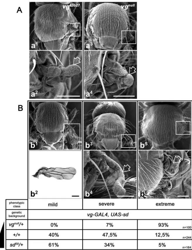
would significantly modify the expression of VG-SD targets. Previous results have shown that sd over-expression is deleterious for endogenous wing growth (Simmonds et al. 1998). Therefore, to understand the physiological relevance of the VG/SD ratio during development, it was important to evaluate the effect of VG/SD imbalance on wing growth and development, by using both mutants and over-expression of vg or sd. In order to increase SD over VG we over-expressed sd in the vg expression domain (vg-GAL4 driver) in three different genetic contexts: sd58/+, +/+ or vgnull/+ (fig 1B). We observed that sd over-expression led to a strong wing phenotype (fig 1B and (Simmonds et al. 1998). This wing phenotype was further enhanced upon VG reduction yielding up to 93% of extreme phenotypes (fig 1B) but, was reduced in an
sd hypomorphic context with only 5% of
extreme phenotypes. The data are in accordance with the hypothesis that an increased ratio of SD over VG is deleterious for normal expression of the dimer target genes involved in wing development. Conversely, vg over-expression did not induce any obvious developmental defects and wings remained completely normal (data not shown), demonstrating that an increased VG/SD ratio does not have a dominant negative effect on VG-SD function in the wing pouch. Thus, correct development and growth of the Drosophila wing disc implies a precise regulation of the respective expressions of SD and VG in vivo. These results suggest, as previously hypothesized by (Simmonds et al. 1998), that in physiological conditions, genes involved in wing growth (e.g. cell cycle genes) may fine-tune SD and VG expression levels to regulate wing growth and cell proliferation.
Cell cycles genes regulate VG-SD transcriptional activity
In the wing disc, wing pouch growth is known to be a highly homeostatic process (de la Cova et al. 2004; Reis & Edgar 2004). The ability of VG-SD to drive proliferation and cell cycle progression largely relies on the regulation of dE2F1 and its target genes by the dimer (Delanoue et al. 2004). So far,
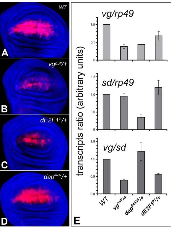
however, the effects of cell cycle regulators on VG-SD activity in feed back loops have not been investigated. To assess the potential action of cell cycle genes on the VG-SD dimer, we tested the effect of reducing the dosage of two antagonist regulators, dE2F1 and dap, on the dimer, using a VG-SD transcriptional activity reporter strain (fig 2). This strain contains a sensor transgene, ‘hsp70-GAL4db-sd’, in which the sd TEA DNA binding domain has been replaced by the GAL4 DNA binding domain. After a heat shock, a UAS-LacZ reporter gene is expressed only where SD::GAL4 dimerizes with a transcriptional co-activator (e.g. VG) (Vaudin et al. 1999). This strain thus provides a sensitive tool for observing VG-SD activity in the wing pouch. As a first positive test, we evaluated the effect of a VG decrease on VG-SD activity. Indeed, in
vgnull/+ wing discs, a significant
down-regulation of the VG-SD activity reporter construct was observed compared to control (fig 2A, B); vg is therefore not fully haplo-sufficient. In addition, we performed quantitative real time RT-PCR experiments and measured vg and sd transcript expression in vgnull heterozygous wing discs (fig 2E).
Compared to the w1118 control strain (vg and
sd arbitrary levels = 1), we observed a
significant decrease in the amount of vg (0.38 ± 0.05), whereas the level of sd remained comparable to that in the wild type strain (0.96 ± 0.07), leading to a decrease in the
vg/sd ratio. The diminution of the dimer
activity observed (fig 2B) is therefore likely due to competition between the SD::GAL4 chimeric construct and endogenous SD for a lower amount of the available VG transcriptional activator. Conversely, in a
sd1/+ heterozygote background, VG-SD
activity was greater than that in the control, probably due to a lower quantity of endogenous SD protein for the same amount of VG and SD::GAL4 protein (data not shown). This is in agreement with the
hypothesis that a precise VG/SD
stoichiometry is required during wing development.
To monitor the effects of cell cycle regulators on VG-SD, experiments with the reporter construct were carried out in heterozygous
dE2F1 or dap null mutants, which display no
wing phenotypes (Duronio et al. 1995). VG-SD activity was significantly reduced in heterozygotes for the dE2F191 amorphallele (fig 2C). RT-PCR experiments revealed a decrease in vg (0.68 ± 0.12) and an increase in sd (1.20 ± 0.21) transcripts compared to control (fig 2E). This led to a significantly lower vg/sd ratio, most likely responsible for the reduced VG-SD reporter activity observed (fig 2C). This result demonstrates that dE2F1 function is crucial for vg, sd normal expression and VG-SD activity. Since DAP is an inhibitor of cell cycle progression, the level of VG-SD sensor activity was also assessed in heterozygotes for the dap04454 amorphallele (de Nooij et al. 1996). Although no significant modulation of VG-SD activity could be detected (fig 2A,D), RT-PCR experiments revealed a strong decrease in both vg (0.44 ± 0.01) and sd (0.36 ± 0.07) expression levels compared to control...Thus, dap is required for normal vg and sd expression. Despite the reduced vg and sd levels, the vg/sd ratio remained similar to the control in this case (fig 2E), and VG-SD activity was thus unmodified. Therefore, to some extent, the vg/sd ratio, rather than absolute expression levels of vg and sd, directs reporter strain and VG-SD activities. Moreover, genetic interactions showed that both VG-SD activity and a normal vg/sd ratio were greatly restored, in the dE2F191/+; dap04454/+ double heterozygotes (data not shown). Our results demonstrate that cell cycle gene down-expression modulates down-expression of vg and sd and VG-SD activity. Moreover, dap and
dE2F1 loss of function display opposite
effects on VG-SD activity and, strikingly, on
sd expression. As a whole, this sheds light on
a process of feedback regulation between cell cycle regulators and VG-SD.
Ectopic dap induces sd and disrupts VG-SD activity
We next investigated whether the modulation of the respective expressions of vg and sd by cell cycle genes affects the regulation of wing disc growth and cell proliferation. We observed the strong requirement of dap for both vg and sd normal expression (see above
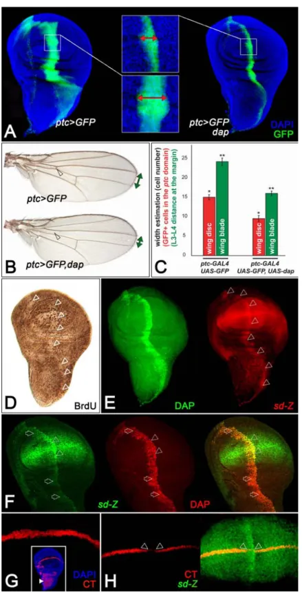
and fig 2). The next step was to test how dap intervenes in the process. The role of DAP protein in G1 arrest has been clearly demonstrated in Drosophila and dap ectopic expression induces cell cycle arrest without triggering apoptosis (Lane et al. 1996; Van de Bor et al. 1999). dap over-expression along the wing D/V boundary decreases proliferation and induces nicks at the wing margin (Delanoue et al. 2004). Accordingly, using the patched (ptc)-GAL4 driver, we found that ectopic dap restricts G1/S transition in BrdU pulse experiments (fig 3D), and decreases the number of ptc-expressing cells in the wing disc (fig 3A,C), and the adult wing blade (fig 3B,C).
To evaluate whether this cell cycle arrest was associated with VG-SD impairment, we tested the possible effect of over-expressing
dap on the expression of VG and SD. Since
no anti-SD antibodies are currently available, we used the sdETX4 strain, a P[lacZ] enhancer trap (see fig 6A). Ectopic dap, along the ptc domain, had no effect on VG (data not shown), but ectopically activated sd expression (fig 3E). This sd up-regulation was strong in the wing pouch, but was also triggered in hinge and notum regions (fig 3F).
Since sd induction in response to dap correlates with a G1-S transition delay and a decrease in cell proliferation (fig 3A-D), we tested the functional significance of this VG/SD imbalance.
We hypothesized that, if dap slows down proliferation by inducing sd, thus decreasing the VG/SD ratio, this might result in a wing phenotype that should be strengthened by further VG reduction. We tested this hypothesis by over-expressing dap at the margin (vg-GAL4 driver) of wild type and
vgnull/+ wings. As expected, nicks were
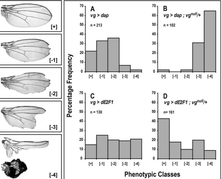
observed in a wild type context (fig 4A) and this phenotype was greatly enhanced in vg heterozygotes (fig 4B) suggesting that dap inhibits cell proliferation through impairment of VG/SD. Next, we tested the fate of cut (ct) at the D/V boundary of the disc as an assay of a VG-SD downstream target gene (fig 3G) (Guss et al.). CT expression was specifically lost in cells in which sd was induced in response to dap (fig 3H). Both ct and
wingless (wg) are also D/V-specific N
targets. Nevertheless, wg remained unaffected, suggesting that dap does not affect the N pathway. We concluded that dap over-expression leads to sd induction, VG-SD dimer imbalance and impairment of its function in wing development.
Ectopic dE2F1 rescues the vg83b27 mutant The results we obtained with dap led us to test whether dE2F1, which is required for VG-SD normal activity and antagonizes DAP, could possibly restore wing growth in a vg83b27mutant that displays no wing structures (fig1A). This mutant carries a complete deletion of the vg 2nd intron that spans the vgBE enhancer required for vg expression in the wing pouch, the remainder of the vg sequence being normal (Williams et
al. 1994).
Ectopic expression of both dE2F1-DP and
P35, a caspase inhibitor that prevents
E2F-induced apoptosis (Neufeld et al. 1998), was monitored in a vg83b27 mutant, using the
vg-GAL4 driver that allowed the recovery of
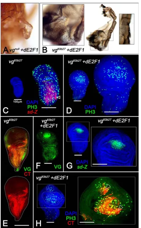
adults flies. dE2F1-DP expression restored growth of vg83b27 wing appendages, which often displayed distinct veins and margin (compare fig 5B insets and 1A). Neither the expression of P35 itself nor that of dIAP1, another caspase inhibitor, was sufficient to rescue the vg83b27 phenotype (Van de Bor et
al. 1999). Consistently, dE2F1-DP induced a
massive over-growth of the vg83b27 wing pouch (fig 5C, D) that was associated with significant proliferation. Nevertheless, in
vgnull mutants for which the entire vg
sequence is deleted, no such phenotypic rescue could be detected, and dE2F1-DP led to only a slight increase in hinge growth (compare fig 1A and 5A, B). This vgnull wing phenotype was rescued by vg-GAL4 driven expression of vg as expected (data not shown). Therefore, we concluded that the partial wing rescue of the vg83b27 mutant (but
not of the vgnull mutant) by dE2F1-DP, required the vg sequence but not the vgBE enhancer.
Moreover, in response to vg-GAL4 driven dE2F1-DP expression, VG was induced in the vg83b27 wing pouch (fig 5F), which does not normally display either VG or SD
endogenous expression (fig 5C,E and (Williams et al. 1991). VG induction was also obtained using the ptc-GAL4 driver, confirming that dE2F1 is able to induce vg expression, even outside the wing pouch (data not shown). These results demonstrate that vg expression is a prerequisite for rescue of the wing phenotype in response to dE2F1. In addition, using the sdETX4 enhancer trap, we found that ectopic dE2F1-DP also induced sd expression in the wing pouch (fig 5C, G). Given the role of VG-SD in cell proliferation (Delanoue et al. 2004), this up-regulation of expression of both vg and sd is consistent with the wing growth rescue induced by dE2F1. Therefore, we tested again the expression of CT, the dimer target, upon dE2F induction, as a read-out of the dimer activity. In the vg83b27 mutant, no CT expression was observed at the D/V boundary (fig 5E). When dE2F1-DP was over-expressed, CT was induced in the wing pouch, although in a different pattern than in a wild type disc (fig 3G). In addition, we often observed ectopic proliferating wing pouches that partly matched the wing marker CT, although these morphogenetic events might not give rise to viable rescued adults, (fig 5H).
We concluded that vg-GAL4 driven
dE2F1-DP-P35 ectopic expression, in a vg83b27
mutant, rescues vg/sd expression and VG-SD activity, which allow pouch cell proliferation and wing growth. All together, our results show that not only dE2F1 is required for normal VG-SD activity in a vg+ background (fig 2), but that ectopic dE2F1 rescues VG-SD activity in a vg hypomorphic context. Ectopic dE2F1 modulates sd expression in a homeostatic feedback loop
It has been shown that VG-SD induces
dE2F1 (Delanoue et al. 2004) and here, we
observed that dE2F1 can also induce VG-SD activity in a vg hypomorphic genetic context. Such a positive feedback loop might trigger uncontrolled wing pouch growth in a wild type context. So, since wing growth is known to proceed homeostatically, a compensatory mechanism must exist. To address this possibility, we assayed the effects of over-expressing dE2F1-DP+P35 driven by
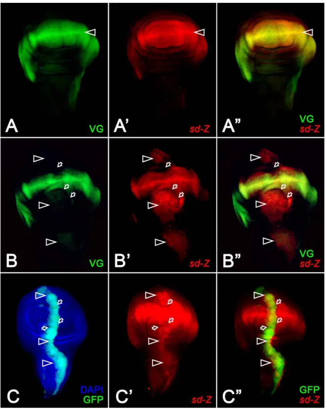
ptc-GAL4, in vg+ wing discs. dE2F1 induced folding of the epithelium layer and sd ectopic expression along the A/P boundary but mainly in the wing pouch (fig 6A’,B’,C’), without any obvious effect on VG expression in vg+ discs (fig 6A,B), unlike previous observations in the vg83b27 mutant. The P35 protein per se had no effect. Moreover,
dE2F1 over-expression with the vg-GAL4
driver, which allowed the recovery of adult flies, yielded nicks in the wings (fig 4C). These results demonstrate that dE2F1 over-expression induces sd and decreases the VG/SD ratio, which most probably impairs the dimer function in wing development. Thus, an excess of ectopic dE2F1 in vg+ discs is deleterious for wing development (fig 4C), whereas, in a vg83b27 mutant it rescues wing growth (fig 5B).
Next, to further understand how this cross-talk depends on the vg genetic context, we compared the response to dE2F1 expression in vg+ or vgnull/+. In this latter genotype,
which provided an intermediate vg
expression level (see fig 2), sd is less induced by dE2F1 than in vg+ discs (data not shown). This shows that a decrease in VG modifies the effect of ectopic dE2F1 on sd. Consistently, wing nicks induced by dE2F1 over-expression according to the vg-GAL4
driver (fig 4C) were partially rescued in a vgnull/+ heterozygote (fig 4D). Therefore, a
decrease in VG attenuates the deleterious effect of over-expressed dE2F1.
Interestingly, even if in a vg+ background expression of both dap and dE2F1 alters wing development (fig 4A,C), clear opposite behaviours are observed in vgnull/+ flies
where the dap induced phenotype is enhanced, while dE2F1 one is partially rescued (fig 4B,D). We concluded that sd induction and VG-SD impairment in response to dE2F1 expression are clearly dependent on the VG expression level. These results favour the hypothesis that sensor regulations coordinate VG-SD and cell proliferation effectors like dE2F1, and tend to ensure normal wing development. Our data suggest that in vivo, moderate excess in the proliferation regulator dE2F1 decreases VG-SD activity in a negative
feedback loop. This may reflect a homeostatic regulation of wing growth.
Discussion
Cell proliferation relies on the tight control of cell cycle genes, and, in the wing pouch, VG-SD is also critically required. Accordingly,
vg was shown to up-regulate dE2F1
expression and to antagonize the CKI dap (Delanoue et al. 2004). In this study, we investigated the effects of these two antagonistic proliferation regulators in the wing pouch of the disc, and tested the hypothesis that cell cycle genes fine-tune proliferation, through regulation of the respective expressions of vg and sd and VG-SD dimer activity, thereby providing a feedback control.
VG/SD ratio and wing growth
Modulation of the relative expression levels of vg and sd as an endogenous means of fine-tuning wing growth, had previously been suggested (Simmonds et al. 1998). Combined loss and gain of function experiments ascertained the requirement of a precise VG/SD ratio for normal wing development and showed that an excess in SD disrupts VG-SD function in wing growth (fig 1), and probably acts as a dominant-negative through titration of functional VG-SD dimers. Therefore, sd induction may efficiently restrain VG-SD function in vivo, and a similar effect may also be physiologically achieved down-regulating vg. Moreover, since SD DNA target selectivity is modified upon binding of VG to SD in vitro (Halder & Carroll 2001), we cannot discard the hypothesis that, in vivo too, VG-SD targets might be different from the targets of SD alone. This could explain to some extent the phenotypes observed when sd is induced. Dap induces sd and impairs VG-SD activity Our results show that the CKI member DAP, homogeneously expressed in the wing disc (Reis & Edgar 2004), regulates VG-SD function. dap heterozygotes display a wild type wing phenotype, reduced levels of both
vg and sd transcripts, but an almost normal vg/sd ratio, thus VG-SD activity is normal
phenotype could be detected. Therefore, the relative vg/sd stoichiometry, rather than absolute vg and sd expression levels, determines wing growth. Interestingly, it had been previously observed that dap
homozygous mutant adult escapers display duplication of the wing margin (Lane et al. 1996), indicating a role of DAP at the D/V boundary. This phenotype could be linked to an enhanced proliferation due to the absence of CKI function. Moreover, D/V-specific over-expression of dap alters wing margin structures (Van de Bor et al. 1999). This dap over-expression triggers both ectopic expression of sd and subsequent impairment of VG-SD activity associated with a proliferation decrease (fig 3).The associated wing phenotype is clearly enhanced in vg heterozygous flies (fig 4), providing evidence that dap over-expression affects VG/SD stoichiometry and represses VG-SD activity in wing development. This reveals a model in which, in the wing pouch, cell proliferation
down-regulation through cyclin/CDK
inhibition by DAP, may be enhanced by an additive reduction of VG-SD proliferation function. Such a mechanism probably participates in vivo in the control of balanced wing growth.
dE2F1 overexpression in different vg backgrounds
Our results also demonstrate that dE2F1-DP regulates VG-SD: the dE2F1 heterozygote displays a reduced vg/sd ratio due to a decrease in vg and an increase in sd transcripts, associated with reduced dimer activity, comparable to the vgnull/+ context
(fig 2). Thus, dE2F1 is required for vg normal expression. This supports the hypothesis that the slower proliferation observed in these contexts is linked to an imbalance in the dimer ratio.
Conversely, overexpressing dE2F1DP
-P35, in a vg83b27 hypomorphic mutant context, rescued expression of both vg and sd and normal VG-SD function, wing appendage specification and growth (fig 5). This was not observed in vgnull flies implying the necessity for vg sequences, but the second intron, missing in the vg83b27 mutant
(Williams et al. 1994) . In addition, we ascertained that not all the genes triggering cell cycle progression or cell proliferation can induce vg expression. Neither ectopic expression of CYC E, which promotes dE2F1-induced G1/S cell cycle transition, nor the growth regulator Insulin receptor (InR) was sufficient to elicit VG expression and wing growth in the vg83b27 mutant (data not shown). These results demonstrate that vg induction is a prerequisite for vg83b27 wing pouch growth in response to dE2F1 activity. In a vg+ genetic background, dE2F1 over-expression induced only sd, disrupting VG/SD stoichiometry (fig 6). Consistently, at the D/V boundary, wing notching was observed (fig 4). Therefore, although dE2F1 basically displays a positive role in proliferation, this sd induction in response to
dE2F1 over-expression was clearly associated with wing growth impairment. This effect was significantly weaker in a vg heterozygote background (fig 4), and a rescue of the wing phenotype was observed, supporting the hypothesis that VG/SD stoichiometry is restored. Therefore, sd induction by dE2F1 depends on the vg genetic context. This indicates that the effects of over-expressing dE2F1 differ depending on the growth-state of the wing pouch, which is tightly linked with the vg genotype.
Homeostatic regulations between dE2F1 and VG-SD
Clearly, feedback regulations rule growth of the wing disc. We have reported regulations in three different vg genetic contexts that can be analysed in the light of a homeostasis hypothesis. In the vg83b27under-proliferative wing pouch, ectopic dE2F1 expression coordinately increases vg and sd expressions in a positive feedback loop. This triggers VG-SD activity, and induces both cell proliferation and wing specification in the mutant. Conversely, no such crosstalk occurs in a correctly grown vg+ disc, where over-growth should be prevented. In this latter case, sd induction (VG/SD decrease) probably restrains the proliferation function of dE2F1. Consistently, wings were not overgrown, but notches were observed. This
phenotype was partially suppressed in a vg heterozygote background. As a whole, these results support the hypothesis that VG-SD/dE2F1 coordination tends to ensure normal wing growth and that the dimer does not trigger unrestricted cell proliferation in a
vg+ context, since an excess in dE2F1 attenuates VG-SD function in a negative feedback loop. Thus, molecular interactions between dE2F1, vg and sd, display a clear plasticity depending on the vg genetic context.
Fine-tuning wing cell proliferation.
Establishing and maintaining homeostasis is critical during development. This is achieved in part through a balance between cell proliferation and death. In mammals E2F1 and p21, the dacapo homolog, play a key role in this process. In the wing disc compensatory proliferation induced by cell death has been observed (Huh et al. 2004). However, the role of cell cycle genes in this process has not yet been established. How patterns of cell proliferation are generated during development is still unclear. It seems nevertheless likely that the gene responsible for regulating differentiation also regulates proliferation and growth (Skaer 1998). For instance, Hedgehog (HH) induces the expression of Cyclins D and E. This mediates the ability of HH to drive growth and proliferation (Duman-Scheel et al. 2002). In the same way, other data support a direct regulation of dE2F1 by the Caudal homeodomain protein required for anterio-posterior axis formation and gut development (Hwang et al. 2002). Wingless (WG) also displays both patterning and a cell cycle regulator function during Drosophila
development (Johnston & Sanders 2003). Here we show that growth control in the wing pouch seems to be achieved through both positive and negative feedback regulations linking dE2F1 and VG-SD, but also via additive impairment of VG-SD by DAP. In fact, in a vg+ background, over-expression of both dap and dE2F1 induces
sd, impairs VG-SD and alters wing
development. Nevertheless, clear opposite behaviours are observed in vgnull/+ flies
where dap-induced nicks are enhanced, while
those of dE2F1 are partially rescued (fig 4). This highlights the functional discrepancy between these two types of feedback regulation. We suggest that dap expression inhibits cell proliferation through a process involving both Cyclin-CDK inhibition and VG-SD impairment in the wing pouch. On the other hand, we propose that dE2F1 over-expression triggers an homeostatic response. It will either induce vg and sd to ensure proliferation (in a vg83b27 genotype), or decrease the VG/SD ratio in a vg+ context. In
this latter genotype, down-regulation
probably counteracts fundamental
proliferative properties of dE2F1 and governs homeostatic wing disc growth.
At late third instar, wing discs display a Zone of Non-proliferating Cells (ZNC) along the wing pouch D/V boundary (O'Brochta & Bryant 1985). It has been shown that, although dE2F1-DP is expressed in this area, its proliferative function is inactivated late, because of RBF1-induced G1 arrest (Duman-Scheel et al. 2004); (Johnston & Edgar 1998). Accordingly, although expression of
vg and sd presents a peak at the D/V
boundary, in late third instar, VG-SD activity is decreased in D/V cells, and it was suggested to result from an excess of SD (Vaudin et al. 1999). Therefore, the ZNC setting may also reflect a VG-SD/dE2F1 coordinated dialogue that triggers a decrease in proliferation signals in this area.
Previous studies of homeostatic control of cell proliferation in the wing reported that, to some extent, over-expression of positive or negative cell cycle regulators only weakly affects the overall division rate (Reis & Edgar 2004). For instance, although dap over-expression alters dE2F1 function in G1-S cell cycle transition, it also promotes
dE2F1 expression and function in G2-M
transition, preventing a decrease in the overall rate of cell division. Strikingly, cells seemed able to monitor each phase length and maintain cell cycle duration and normal proliferation in the wing pouch of the disc. Therefore, dE2F1 is a central component that enables cells to ensure normal proliferation in the wing disc and prevents imbalance in the process (Reis & Edgar 2004). The fact
that dE2F1 triggers quite different or opposite responses in vg+ or vg hypomorphic contexts suggests that the VG-SD/dE2F1 crosstalk plays a role in the same sort of homeostatic process that ensures entire wing growth.
We believe that such regulations are likely to reveal a precise physiological fine-tuning of
vg and sd by cell cycle effectors, promoting
an exquisite control of wing growth. Feedback loops between the developmental selector VG-SD and cell cycle effectors may stand for a control mechanism to guarantee that the tissue can sustain balanced growth and a reproducible size. Such a subtle mechanism, on a local scale, would correct the alterations in cell proliferation that may occur during development.
Experimental Procedures
Drosophila strains
The following strains were used : vgnull and
UAS-vg (Paumard-Rigal et al. 1998); (Zider et al. 1998), vg-GAL4 (gift from S. Carroll), vg83b27 (gift from J.Bell) vgBE-LacZ and vgQE -LacZ (Kim et al. 1996) (Williams et al.
1994), sd58 and the sdETX4 P[LacZ] enhancer trap mutant that do not display any wing phenotype in the heterozygous state (Campbell et al. 1992), UAS-sd (Varadarajan & VijayRaghavan 1999), dE2F191 (Duronio
et al. 1996), UAS-E2F1, UAS-DP, UAS-P35
(Neufeld et al. 1998), dap04454 (de Nooij et
al. 1996), UAS-dap (Lane et al. 1996), hsp-GAL4db-sd (Vaudin et al. 1999). Heat
shocks (38°C, 30 min) were performed 36 hours before puparium formation and wing discs were stained 24h after the heat shock. All other stocks come from the Bloomington
Drosophila Stock center (Indiana University)
Scanning Electron Microscopy (SEM) Adult flies were anesthetized with ethyl acetate (Sigma), mounted on stages and covered with a 40 nm layer of gold salts. Preparations were observed under a JEOL JSM 6100 Scanning Electron Microscope. Images were acquired using Genesis software.
Histology
Images were acquired with a DMR (Leica) microscope and processed with Adobe Photoshop. 5-Bromo-2’-deoxyuridine (BrdU) and 4'-6-Diamidino-2-phenylindole (DAPI) labeling of imaginal discs were performed as described (Delanoue et al. 2004). Antibody staining: rabbit anti-phospho-Histone H3 antibody (Upstate Biotechnology), anti-VG (gift from S. Carroll), anti-DAP (gift from C. Lehner) anti-βgalactosidase (Biodesign) antibodies, and mouse anti-βgalactosidase
(Jackson Immuno Labs), anti-CT
(Developmental Studies Hybridoma Bank (DSHB), University of Iowa) antibodies. We assessed the activity of the VG-SD transcriptional reporter strain 5 times
independently, using both
anti-βgalactosidase antibodies or the X-gal enzymatic reaction.
Quantitative Real time RT-PCR
Total RNA of 20 wing discs from third instar larvae was isolated by using tri reagent (Molecular Research Center Inc.) according to the manufacturer's instructions. One µg of RNA was used for reverse transcription into
cDNA according to the supplier’s
instructions (Roche). Real time PCR was conducted in a Light Cycler (Roche) with 45 cycles (15 sec 95°C, 15 sec 60°C for vg,
RP49 - 65°C for sd, 15 sec 72°C) 1 µl
cDNA, and PCR reaction mix from Roche. The following primers were used for amplification: sd: aatattgcaagtaatgagggccc / gacggtataatgtgatgggtggtg ; RP49 : ccgcttcaagggacagtatctg / cacgttgtgcaccaggaactt ; vg : cggcccactatggttcctatg / agcctgaggagactgccgtact. Differences in cDNA concentrations were adjusted by normalizing to RP49. For each gene, values were averaged over at least three independent measurements. The expression levels were calculated relative to the wild type genotype (w1118) (the expression in this genotype was set to 1). Two independent RNA isolation experiments were performed for all genotypes and means were calculated.
Acknowledgments
We are grateful to J. Bell, S. Carroll, R. Duronio, J. Nevins, C. Lehner, B. Edgar, P. Leopold, K. VijayRaghavan, C. Carré and J-R. Huynh for flies and reagents. We thank D. Montero for help with SEMs, A. Kropfinger for correcting the English, and B. Legois and M. Sanial who provided technical assistance. This work was supported by an “ Action Thématique concertée-vieillissement ” grant from the INSERM. K.L. was supported by the Fondation pour la Recherche Médicale..
References
Blair, S.S. (1995) Compartments and appendage development in
Drosophila. Bioessays 17, 299-309. Campbell, S., Inamdar, M., Rodrigues, V.,
Raghavan, V., Palazzolo, M. & Chovnick, A. (1992) The scalloped gene encodes a novel, evolutionarily conserved transcription factor required for sensory organ
differentiation in Drosophila. Genes
Dev 6, 367-379.
de la Cova, C., Abril, M., Bellosta, P., Gallant, P. & Johnston, L.A. (2004) Drosophila myc regulates organ size by inducing cell competition. Cell 117, 107-116.
de Nooij, J.C., Letendre, M.A. & Hariharan, I.K. (1996) A cyclin-dependent kinase inhibitor, Dacapo, is necessary for timely exit from the cell cycle during Drosophila embryogenesis.
Cell 87, 1237-1247.
Delanoue, R., Legent, K., Godefroy, N., Flagiello, D., Dutriaux, A., Vaudin, P., Becker, J.L. & Silber, J. (2004) The Drosophila wing differentiation factor Vestigial-Scalloped is required for cell proliferation and cell survival at the dorso-ventral boundary of the wing imaginal disc. Cell Death Differ 11, 110-122.
Dimova, D.K., Stevaux, O., Frolov, M.V. & Dyson, N.J. (2003) Cell
cycle-dependent and cell cycle-incycle-dependent control of transcription by the
Drosophila E2F/RB pathway. Genes
Dev 17, 2308-2320.
Du, W., Vidal, M., Xie, J.E. & Dyson, N. (1996) RBF, a novel RB-related gene that regulates E2F activity and interacts with cyclin E in Drosophila.
Genes Dev 10, 1206-1218.
Duman-Scheel, M., Johnston, L.A. & Du, W. (2004) Repression of dMyc
expression by Wingless promotes Rbf-induced G1 arrest in the
presumptive Drosophila wing margin.
Proc Natl Acad Sci U S A.
Duman-Scheel, M., Weng, L., Xin, S. & Du, W. (2002) Hedgehog regulates cell growth and proliferation by inducing Cyclin D and Cyclin E. Nature 417, 299-304.
Duronio, R.J., Brook, A., Dyson, N. & O'Farrell, P.H. (1996) E2F-induced S phase requires cyclin E. Genes Dev 10, 2505-2513.
Duronio, R.J., O'Farrell, P.H., Xie, J.E., Brook, A. & Dyson, N. (1995) The transcription factor E2F is required for S phase during Drosophila embryogenesis. Genes Dev 9, 1445-1455.
Goux, J.M. & Paillard, M. (1976)
[Incomplete penetrance at the vestigal locus of Drosophila]. C R Acad Sci
Hebd Seances Acad Sci D 283,
667-669.
Guss, K.A., Nelson, C.E., Hudson, A., Kraus, M.E. & Carroll, S.B. (2001) Control of a genetic regulatory network by a selector gene. Science 292, 1164-1167.
Halder, G. & Carroll, S.B. (2001) Binding of the Vestigial co-factor switches the DNA-target selectivity of the Scalloped selector protein.
Development 128, 3295-3305.
Halder, G., Polaczyk, P., Kraus, M.E., Hudson, A., Kim, J., Laughon, A. & Carroll, S. (1998) The Vestigial and Scalloped proteins act together to directly regulate wing-specific gene expression in Drosophila. Genes Dev 12, 3900-3909.
Harbour, J.W. & Dean, D.C. (2000) The Rb/E2F pathway: expanding roles and emerging paradigms. Genes Dev 14, 2393-2409.
Huh, J.R., Guo, M. & Hay, B.A. (2004) Compensatory proliferation induced by cell death in the Drosophila wing disc requires activity of the apical cell death caspase Dronc in a
nonapoptotic role. Curr Biol 14, 1262-1266.
Hwang, M.S., Kim, Y.S., Choi, N.H., Park, J.H., Oh, E.J., Kwon, E.J.,
Yamaguchi, M. & Yoo, M.A. (2002) The caudal homeodomain protein activates Drosophila E2F gene expression. Nucleic Acids Res 30, 5029-5035.
Johnston, L.A. & Edgar, B.A. (1998) Wingless and Notch regulate cell-cycle arrest in the developing
Drosophila wing. Nature 394, 82-84. Johnston, L.A. & Sanders, A.L. (2003)
Wingless promotes cell survival but constrains growth during Drosophila wing development. Nat Cell Biol 5, 827-833.
Kim, J., Sebring, A., Esch, J.J., Kraus, M.E., Vorwerk, K., Magee, J. & Carroll, S.B. (1996) Integration of positional signals and regulation of wing formation and identity by Drosophila vestigial gene. Nature 382, 133-138. Kolzer, S., Fuss, B., Hoch, M. & Klein, T.
(2003) defective proventriculus is required for pattern formation along the proximodistal axis, cell
proliferation and formation of veins in the Drosophila wing. Development 130, 4135-4147.
Lane, M.E., Sauer, K., Wallace, K., Jan, Y.N., Lehner, C.F. & Vaessin, H. (1996) Dacapo, a cyclin-dependent kinase inhibitor, stops cell
proliferation during Drosophila development. Cell 87, 1225-1235. Martin-Castellanos, C. & Edgar, B.A. (2002)
A characterization of the effects of Dpp signaling on cell growth and
proliferation in the Drosophila wing.
Development 129, 1003-1013.
Muller, H., Bracken, A.P., Vernell, R., Moroni, M.C., Christians, F.,
Grassilli, E., Prosperini, E., Vigo, E., Oliner, J.D. & Helin, K. (2001) E2Fs regulate the expression of genes involved in differentiation, development, proliferation, and apoptosis. Genes Dev 15, 267-285. Neufeld, T.P., de la Cruz, A.F., Johnston,
L.A. & Edgar, B.A. (1998) Coordination of growth and cell division in the Drosophila wing. Cell 93, 1183-1193.
O'Brochta, D.A. & Bryant, P.J. (1985) A zone of non-proliferating cells at a lineage restriction boundary in Drosophila. Nature 313, 138-141. Ohtani, K. & Nevins, J.R. (1994) Functional
properties of a Drosophila homolog of the E2F1 gene. Mol Cell Biol 14, 1603-1612.
Paumard-Rigal, S., Zider, A., Vaudin, P. & Silber, J. (1998) Specific interactions between vestigial and scalloped are required to promote wing tissue proliferation in Drosophila
melanogaster. Dev Genes Evol 208, 440-446.
Reis, T. & Edgar, B.A. (2004) Negative regulation of dE2F1 by cyclin-dependent kinases controls cell cycle timing. Cell 117, 253-264.
Simmonds, A.J., Liu, X., Soanes, K.H., Krause, H.M., Irvine, K.D. & Bell, J.B. (1998) Molecular interactions between Vestigial and Scalloped promote wing formation in Drosophila. Genes Dev 12, 3815-3820.
Skaer, H. (1998) Who pulls the string to pattern cell division in Drosophila?
Trends Genet 14, 337-339.
Van de Bor, V., Delanoue, R., Cossard, R. & Silber, J. (1999) Truncated products of the vestigial proliferation gene induce apoptosis. Cell Death Differ 6, 557-564.
Varadarajan, S. & VijayRaghavan, K. (1999) scalloped functions in a regulatory loop with vestigial and wingless to pattern the Drosophila wing. Dev
Genes Evol 209, 10-17.
Vaudin, P., Delanoue, R., Davidson, I., Silber, J. & Zider, A. (1999) TONDU (TDU), a novel human protein related to the product of vestigial (vg) gene of Drosophila melanogaster interacts with vertebrate TEF factors and substitutes for Vg function in wing formation. Development 126, 4807-4816.
Williams, J.A., Bell, J.B. & Carroll, S.B. (1991) Control of Drosophila wing and haltere development by the
nuclear vestigial gene product. Genes
Dev 5, 2481-2495.
Williams, J.A., Paddock, S.W., Vorwerk, K. & Carroll, S.B. (1994) Organization of wing formation and induction of a wing-patterning gene at the
dorsal/ventral compartment boundary.
Nature 368, 299-305.
Zider, A., Paumard-Rigal, S., Frouin, I. & Silber, J. (1998) The vestigial gene of Drosophila melanogaster is involved in the formation of the peripheral nervous system: genetic interactions with the scute gene. J Neurogenet 12, 87-99.
Figure 1 : VG-SD stoichiometric ratio is critical for wing growth.
(A) Thoraxes of vg83b27 (a1) and vgnull (a3) mutants display no growth of wing blade tissue, although some hinge structures are still present (arrows in a2 and a4). (B) vg-GAL4 driven over-expression of sd in sd58/+ ; +/+ or vgnull/+ genetic backgrounds providing a sequential decrease in VG/SD ratio. Wing phenotypes are classified into three distinct categories. Mild (b1) wings are significantly reduced, few veins and cross-veins are still distinguishable, (b2) Photonic magnification of a similar wing. Severe: (b3 and b4) Wing blade growth is severely impaired (arrow). Extreme: (b5 and b6) Thorax flight appendages are entirely missing. No hinge structure is recovered (arrow). All pictures except (b2) are Scanning Electron Micrographs (SEMs) and scale bars refer to 100mm. Phenotypic class percentages are presented for each genetic background. When the VG/SD ratio is decreased, a shift toward extreme phenotypes is observed.
Figure 2 : VG-SD transcriptional activity depends on cell cycle gene expression.
All third instar wing discs are orientated with the posterior compartment to the right and the ventral one to the top. These hsp70-GAL4db-sd, UAS-lacZ discs heat shocked and stained with anti-bgalactosidase antibodies (red) and the nuclear dye DAPI (blue), 24 hours later. Compared to the control (A), a significant decrease in VG-SD activity can be observed both in vgnull/+ (B) and dE2F191/+ (C), but not significantly in dap04454/+ discs (D). (E) Quantitative real time RT-PCR was performed in wing discs of these 4 genotypes. vg and sd transcripts were measured, adjusted by normalizing to RP49, and set to 1 in WT control discs. vg/sd transcripts ratio are shown. In vgnull/+ and dE2F191/+ wing discs, the vg/sd ratio is decreased to about a half of the value observed in control and other genotypes.
Figure 3: Over-expression of dap induces ectopic expression of sd.
(A) ptc-GAL4, UAS-GFP third instar wing discs over-expressing dap or not. The ptc domain width (red arrows) is estimated by the number of nuclei (DAPI) in the GFP+ domain at the margin. (B) Adult males wing blades of the same genotypes. Margin width (cells) between L3 and L4 veins (green arrows) is estimated by the number of bristles. The anterior cross-vein of dap over-expressing wings is often missing (arrowhead). (C) The widths of the wing disc ptc and adult L3-L4 at the margin measured in flies expressing UAS-dap or not, are presented on the graph. n=30 for each experiment. Student’s test : p<0.001 for both * and ** tests. (D-F, H) sdETX4/+ ; ptc-GAL4, UAS-dap wing discs. (D) BrdU incorporation reveals a decrease in S phase cells along the ptc domain (arrowheads). (E) Endogenous DAP is expressed homogeneously in the wing disc, while over-expressed DAP is detected along the ptc domain. sd-lacZ ectopic expression is induced, along the ptc domain, in response to dap (arrows). (F) Confocal sections. sd is strongly over-expressed at the posterior sharp edge of the ptc domain in the pouch (arrowheads), while ectopic expression outside the wing pouch is weaker (arrows). (G-H) are wing pouch magnification. (G) wild type D/V specific CT expression (open arrowhead) is magnified. (H) CT expression is lost
Figure 4: Effects of cell cycle gene expression in different vg backgrounds.
dap (A) or dE2F1+DP+P35 (C) are over-expressed along the D/V boundary of the disc (vg-GAL4 driver). Expression of P35 prevents cell death associated with dE2F1-DP over-expression. The defects in wings range from mild nicks at the tip of the wings to strong notching and wing size reduction and were quantified using an arbitrary scale of [-1] to [-4]. [+] indicates normal wings. In a vgnull/+ context, wing phenotypes associated with dap over-expression are enhanced (B), while dE2F1-induced nicks are partially rescued (D). Data in each plot are based on an analysis of at least 102 wings.
Figure 5: vg-GAL4 driven expression of dE2F1 rescues wing development in a vg83b27 context.
dE2F1 over-expression according to the vg-GAL4 driver is achieved with the use of a UAS-dE2F1, UAS-DP, UAS-P35 strain. (A) dE2F1 over-expression, in a vgnull mutant, triggers wing hinge growth, while no wing blade structure is observed (compare with fig 1A). (B) In a vg83b27 mutant, dE2F1 rescues wing appendage growth. Note the presence of wing blade features such as bristles, wing veins and margin (insets). (C-H) Third instar wing discs stained with the nuclear dye DAPI and the mitotic marker anti- phosphohistone H3 (PH3) antibodies while sd expression is monitored in sdETX4/+ females (C) In vg83b27 mutants,sd is weakly expressed in myoblasts (arrowhead in the notum) but not in the putative wing pouch, where proliferation is weaker (open arrowhead). (D) dE2F1 expression rescues vg83b27 wing disc growth. Proliferation is specifically triggered in the wing pouch that is recovered. (E) In vg83b27 discs, CT is expressed in all myoblasts, whereas VG is only weakly expressed in a subset of these adepithelial cells (dotted line). dE2F1 expression in a vg83b27 wing disc rescues expression of both VG (F) and sd (G) in the wing pouch. (H) dE2F1 also triggers CT expression (arrowheads) as visualized in the two proliferating wing pouches (surrounded). All scale bars refer to 100µm.
Figure 6 : Regulation of sd expression by dE2F1, in a vg+ background.
Third instar wing discs. sd expression was monitored in sdETX4/+ females. (A, A’, A”) Expression of VG and sd-lacZ colocalize mainly in the wing pouch, High levels are observed along the D/V boundary (arrowheads). (B-C) ptc-GAL4 ; UAS-GFP UAS-dE2F1, UAS-DP, UAS-P35 discs. (B, B’, B”) sd ectopic expression is induced along the ptc domain, in response to dE2F1 expression (arrowheads), whereas VG is not. Induction of sd in the notum is uncertain, since both vg and sd are weakly expressed in myoblasts in a wild type disc. dE2F1-induced proliferation promotes epithelium folding (arrows). (C, C’, C”) DAPI staining highlights the folds around the pouch (arrows) Note the ectopic expression of sd (arrowheads) along the entire ptc domain marked by GFP expression.
