Drug Eluting Prosthetic Joints through Drug Cluster
Morphology Control
by
Vincentius Jeremy Suhardi
S.B. Chemical Engineering California Institute of Technology, 2012S.M. Chemistry
California Institute of Technology, 2012
Submitted to the Harvard-MIT Division of Health Sciences and Technology in partial fulfillment of the requirements for the degree of
Doctor of Philosophy in Medical Engineering and Medical Physics at the
Massachusetts Institute of Technology June 2017
Massachusetts Institute of Technology 2017. All rights reserved.
Signature redacted
Signature of Author: _
Harvard-M vision of Health Sciences and Technology
March 29, 2017
Signature redacted
Certified by: _____
Ebru Oral, Ph.D. Professor of Orthopedic Surgery
Accepted by:
Signature redacted
Emery N. Brown, M.D., Ph.D.
Di or, Harvard-MIT Program in Health Sciences and Technology
Professor of Computational Neuroscience & Health Sciences and Technology
MASSACHUSETTS INSTITUTE
OF TECHNOLOGY 0
LRA E
LU
Dedication
To my parents Iwan Suhardi and Juliati Prajitno and to my wife, Anastasia Oktarina, for their relentless love and encouragement.
Drug Eluting Prosthetic Joints through Drug Cluster
Morphology Control
by
Vincentius Jeremy Suhardi
Submitted to the Harvard-MIT Division of Health Science and Technology on April 3rd 2017
in partial fulfillment of the requirements for the degree of Doctor of Philosophy in Medical Engineering and Medical Physics
More than one million joint replacements are performed in the USA annually. However, around 10 % of patients require revision surgery within 10 years with prosthetic joint infections (PJI) as a common reason. PJI has a recurrence rate of 16 %, a mortality rate of 2.5 %, and end-stage treatments involving arthrodesis and amputation. Most drug eluting polymers that were in development to address this problem failed due to toxic degradation products, insufficient drug release, and insufficient mechanical strength. The gold standard of treatment uses antibiotic eluting bone cement which has a mechanical failure rate of 26-60 % within 49-54 months if used under load bearing conditions. Therefore, despite advances in orthopedic materials, development of drug-eluting devices with effective, sustained delivery with the necessary mechanical strength for a fully load bearing joint implant has been elusive.
Here, we report the synthesis and application of a drug eluting, fully load bearing, and articulating joint
prosthesis that has superior mechanical strength and drug elution profile compared to the clinical gold standard, antibiotic eluting bone cement. We modified the eccentricity of drug clusters and percolation threshold in the polymeric matrix of Ultra-High Molecular Weight Polyethylene (UHMWPE), which resulted in maximized drug elution and mechanical strength retention. The optimized antibiotic eluting UHMWPE elutes antibiotic at a higher concentration for a longer period of time than antibiotic eluting bone cement while retaining the
mechanical and wear properties of clinically used UHMWPE joint prosthesis. After drug elution, the empty drug clusters in the polymer were filled with biological lubricants during articulation, which through a combination of weeping and elastohydrodynamic lubrication, reduced the overall wear rate of the UHMWPE. Treatment of Staphylococcus aureus infected lapine knee with the antibiotic eluting UHMWPE showed complete bacterial eradication without any detectable systemic side effect. Taken together, our study showed that the drug-eluting UHMWPE joint implants in this study are promising candidates for further clinical trial and as the next
generation prosthetic joints.
Thesis Supervisor: Ebru Oral, PhD
Acknowledgements
I have been very fortunate throughout my graduate studies. First, I am able to achieve the goal that I set myself when I enter the Ph.D. program: working on projects that can
someday truly change patients' lives. Second, I have been surrounded by amazing professors, scientists, and physicians that supported me in these projects to achieve a mutual goal of creating better care and treatment for patients. Third, I have interacted with extremely smart and imaginative peers at MGH, MIT, and HMS that are always willing to bounce ideas around and collaborate.
I would like to thank my principal investigator, Ebru Oral, Ph.D., who has always patiently guided me navigating through my projects with invaluable ideas, practical knowledge, and constructive criticism. Thank you for always being available to answer my questions and for always providing fresh perspective when I am at a dead end.
I would like to thank the director of the lab, Orhun Muratoglu, Ph.D., who always provided me with new insights, perspective, and always guided me to put on both my scientific and clinical cap as I am working on my thesis projects.
I would like to thank the team of orthopedic surgeons who has worked with me
throughout my thesis projects, Andrew Freiberg, MD, Henrik Malchau, MD, Harry Rubash, MD, and Hani Bedair, MD who always gave insight on what was needed in clinical practice and what would be the best way to use the new innovations.
I would like to thank my thesis committee chair and member, Richard Mitchell, MD, Ph.D., Edward K. Rodriguez, MD, Ph.D., and Jeffrey Karp, Ph.D., who have been very supportive and lend their invaluable time to provide very insightful feedbacks and ideas.
I would like to thank all of Harris Orthopedics Lab members who have helped me running experiments, animal studies, and be extra pairs of the ear to bounce ideas around.
I would like to thank my parents, lwan Suhardi, JD and Julia Prajitno, JD who gave me unconditional love and support, more than I could ever imagine. Thank you very much for being the one who hears my complaints and gave me encouragement to work smarter and harder. I'm so glad to have two amazing siblings, Angela and Matthew, that are always ready to help me whenever I need it.
I would like to thank my wife, Anastasia Oktarina, MD who provided an enormous amount of love, care, and support. Thank you for always listening to my stories, however boring those stories may be. Love you!
This acknowledgment is certainly incomplete, and to whomever, I accidentally did not
mention, please do not hold any grudge against me @. I am very grateful to everyone who has
Contents
Dedication
3
Abstract
5
Acknowledgement
7
Contents
9
List of Figures
14
List of Tables
20
List of Abbreviations
21
Prologue
23
Chapter 1: Background
26
Total Joint Arthroplasty...26
T otal hip arthroplasty
...
. . 26
T otal knee arthroplasty
...
27
Development of UHMWPE technologies to address total joint arthroplasty
co m p licatio ns
...
. . 2 7
Current complications related to TJA... 29
Incidence and economic burden of periprosthetic joint infection
...
31
Pathogenesis of prosthetic joint infection... 32
Risk factors for Prosthethic joint infection
...
33
Bacterial strains commonly found in PJI... 35
Involvem ent of biofilm in PJI... 37
D ia g n o sis of P J I
...
. 3 7
Prophylaxis approaches to reduce incidence of PJI... 40
Current treatment of patients with PJI... 41
Recommended antimicrobials for PJI
...
43
Antibiotic-eluting bone cement (ALBC) as gold standard for local delivery of
a ntib iotics in P JI
...
. 4 5
Problems with the current PJI treatments and proposed solutions... 48
Polyethylene as weight bearing material
...
50
Previous works on UHIMWPE with additives... 51
Chapter 2: Drug Percolation in Polymer Matrix and its Effect on Drug Elution
54
In tro d u ctio n
...
. 54
Thermodynamic of drug incorporation into polymer... 54
Kinetics of drug incorporation into polymer
...
55
Elution mechanism from drug-incorporated polymer
...
56
Higuchi equation for release of drug from non-swellable polymeric matrix . 58
P ercolation theory
...
. . 59
Percolation theory on eccentricity of clusters
...
61
Clinical implication of drug cluster morphology... 63 M e th o d s ... . . 6 4 R e s u lts ... . . 6 7
Heat resistance of vancomycin determined by thermogravimetric analysis (T G A )... . . 6 7 Spectral analysis of eluted vancomycin from vancomycin-eluting UHMWPE in comparison to pure antibiotics... 68 Ultraviolet-visible spectroscopy (UV-Vis) of vancomycin eluted from
U H M W P E ... . . 6 8 Fourier transform infrared (FTIR) spectroscopy of vancomycin eluted from U H M W P E ... . . 6 9 Nuclear Magnetic Resonance (NMR) of vancomycin eluted from
U H M W P E ... . . 7 0 Effect of Drug Cluster Eccentricity on drug elution of vancomycin eluting low density polyethylene (LDPE)... 71 Drug clusters interconnectivity and drug elution from vancomycin eluting U H M W P E ... . . 7 5 Effect of consolidation parameters (compression molding temperature,
Compression molding pressure, and vancomycin particle size) on drug elution from vancomycin eluting UHMWPE ... 78 Computational simulation on effect of drug cluster eccentricity on drug cluster inte rco n ne ctivity ... . 80 Incorporation of other antibiotics into UHMWPE ... 81 Heat resistance of antibiotics determined by thermos-gravimetric analysis
(T G A )... . . 8 1 Ultraviolet-visible spectroscopy (UV-Vis) of antibiotics eluted from
U H M W P E ... . . 8 5 FTIR analysis of antibiotics eluted from UHMW PE...86 NMR analysis of antibiotics eluted from UHMWPE ... 88
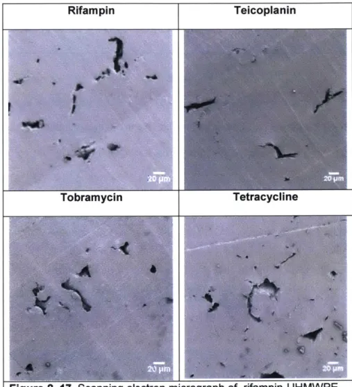
Scanning electron micrograph of antibiotic eluting UHMW PE ... 93
Effect of drug hydrophilicity/hydrophobicity on drug elution from drug eluting U H M W P E ... . . 9 5 D iscu ssio n ... . . 9 8 Most antibiotics can be incorporated into UHMWPE without any sign of thermal d e g ra d atio n ... . 9 8
Highly eccentric pore morhology in drug-eluting LDPE... 99 Highly eccentric pore morphology in drug-eluting UHMW PE. ... 102
Relation between drug type and elution rate from drug eluting UHMW PE. ... 103
C o n c lu s io n ... 1 0 5
Chapter 3: Tensile and Impact Properties of Drug Eluting UHMWPE 106
In tro d u ctio n ... 10 6 Contact mechanics of artificial joint in hip and knee...106 Theoretical basis of mechanical properties of polymers with fillers ... 108
Mechanical strength of filled polymer when fillers are weaker than the
p o ly m e r ... 1 0 9 Characterization of the mechanical strength of UHMWPE for joint
im p la n ts ... 1 1 1 M e th o d s ... 1 1 3
R e s u lts ... 1 1 5 The effect of drug cluster eccentricity on mechanical properties of low density
po lyethyle ne (LD P E ) ... 115
The effect of pore eccentricity on the mechanical properties of UHMWPE. 116 Mechanical properties of vancomycin eluting UHMWPE under various initial drug lo a d in g ... 1 1 8 Computational simulation of the effect of drug cluster eccentricity on mechanical s tre n g th ... 1 1 9 Effect of manufacturing parameters (compression molding temperature, compression molding pressure, and vancomycin particle size) on the mechanical Properties of vancomycin eluting UHMW PE ... 123
Effect of gamma irradiation dose on mechanical strength of vancomycin eluting U H M W P E ... 12 5 Effect of drug hydrophilicity/hydrophobicity on mechanical strength of drug eluting U H M W P E ... 12 7 D is c u s s io n ... 1 2 9 Effect of drug amounts and drug cluster eccentricity on mechanical strength of drug eluting polyethylene ... 129
Effect of drug type and compression molding parameters on mechanical strength o f U H M W P E ... 13 2 Effect of irradiation dose on mechanical strength of UHMWPE... 133
C o n c lu s io n ... 1 3 3 Chapter 4: Wear Resistance of Micro Textured Drug Eluting UHMWPE 135 In tro d u c tio n ... 1 3 5 W e a r of po lym e rs ... 135
Fluid lubrication at mechanical interfaces... 136
Wear mechanism of UHIMW PE ... 138
Relation between micro-texture and lubrication film thickness... 140
Relation between micro-texture and tribology properties ... 141
M e th o d s ... 14 2 R e s u lts ... 1 4 9 Wear resistance of vancomycin eluting UHMWPE ... 149
Formation of self-assembled micropatterns in drug eluting UHMW PE ... 151
Investigation of the relationship between self-assembled micropatterns in bupivacaine- eluting UHMW PE and wear resistance ... 153
Long-term wear testing of bupivacaine-eluting UHMW PE... 156
Topographical analysis of self-assembled micropatterns from bupivacaine-eluting U H M W P E (B upi-P E) ... 156
Evolution of the surface roughness of Bupi-PE during wear testing ... 158
Combination of micropatterns with previously developed method to reduce wear rate of U H M W P E ... 16 1 Effect of irradiation dose on the wear eate of MPC+Bupi-PE UHMWPE .... 165
Evolution of surface roughness of combined crosslinked and bupivacaine eluting UHMW PE (Bupi-PE) throughout wear testing ... 167
Mechanism of wear reduction in microtextured UHMW PE ... 170 Computational simulation of lubrication film thickness of microtextured and flat U H M W P E ... 17 3 D is c u s s io n ... 1 7 5 C o n c lu s io n ... 1 7 8
Chapter 5:
Chapter 6:
Chapter 7:
Pores
Drug Eluting UHMWPE with Highly Eccentric Drug Cluster Morphology for the
Treatment of Prosthetic Joint Infection
179
In tro d u ctio n
...
17 9
Gram-positive bacteria accounts for 80% of all PJI cases... 179
Antibiotic penetrates poorly to the infected joints and bone... 181
Previously reported antibacterial materials to address orthopaedic-associated
in fe ctio n s
...
1 8 2
Two-stage implant exchange as state-of-art treatment of PJI... 183
Complications of PMMA bone cement spacer as local antibiotic delivery
s y s te m
...
1 8 5
M e th o d s
...
1 8 6
R e s u lts
...
1 9 2
Optimization of mechanical strength and drug elution of vancomycin eluting
UHMW PE as a bearing surface in prosthetic joint implants... 192
Vancomycin elution and mechanical properties of optimized vancomycin eluting
UHMWPE and comparison against clinically used UHMWPE and antibiotic
e luting bo ne ce m e nt...
195
Antibacterial activity of VPE, BC1, and BC2
...
197
Irradiation grafting of vancomycin on surface of VPE prevents bacterial
a d h e re n ce
...
2 0 2
Optimization of combined rifampin-vancomycin eluting UHMWPE (RVPE)
A ctivity of R V P E
...
2 0 9
Kinetic of biofilm eradication by RVPE
...
215
D is c u s s io n
...
2 1 7
C o n c lu s io n
...
2 2 3
Pre-Clinical Safety and Efficacy Testing of Drug-Eluting UHMWPE for
Treatment of PJI
224
In tro d u c tio n
...
2 2 4
Both planktonic and biofilm bacteria are involved in prosthetic joint
In fe c tio n
...
2 2 4
Bacteria adhere to intra-articular components and bone-implant
In te rfa c e
...
2 2 6
Vancomycin is effective against planktonic bacteria and immature biofilm but not
against mature biofilm
...
227
Gold standard treatment in antibiotic-eluting bone cement spacer... 228
M e th o d s
...
2 2 9
R e s u lts
...
2 3 5
Murine planktonic bacteria infection model
...
235
Murine biofilm bacteria infection model...238
Lapine planktonic bacteria prosthetic joint infection model... 243
Lapine biofilm bacteria prosthetic joint infection model
...
250
D is c u s s io n
...
2 5 6
C o n c lu s io n
...
2 5 9
Sustained Delivery of Local Anesthetics from UHMWPE with Highly Eccentric
260
Intro d u ctio n
...
2 6 0
Current postoperative management for total joint arthroplasty
...
261
Antimicrobial activity of local anesthetics ... 265 Sustained delivery of local anesthetics using highly eccentric drug clusters-U H M W P E ... 2 6 6 M e th o d s ... 2 6 6 R e s u lt...2 7 2 Elution and mechanical properties of bupivacaine HCl (Bupi HCI) from Bupi HCI e luting U H M W P E ... 2 72 Elution of bupivacaine FB eluting UHMWPE ... 275 Elution and mechanical properties of Bupivacaine HCI (Bupi HCI), Bupivacaine Free Base (Bupi FB) and its combination from Bupi HCI eluting
U H M W P E ... 2 7 8 Measurement of minimum inhibitory concentration of bioluminescent S. aureus (Xen29) against Bupivacaine HCI, Bupivacaine FB, and FS ... 282 In vivo antibacterial activity of Bupi-PE in subcutaneous murine model... 283 In vivo analgesic activity of Bupi PE in murine model...285 D is c u s s io n ... 2 9 0 C o n c lu s io n ... 2 9 5
Epilogue 296
List of Figures
Figure 2.1. Vancomycin eluting PMMA bone cement with 15 wt % initial vancomycin loading...63
Figure 2.2. Thermogravimetric analysis (TGA) of vancomycin HCI powder
...
68
Figure 2.3. UV-Vis spectra of standard vancomycin and eluted vancomycin HCI...69
Figure 2. 4. FTIR spectra of eluted and standard vancomycin HCI.
...
70
Figure 2.5. NMR spectra of standard and eluted vancomycin HCI...71
Figure 2.6. SEM of Vancomycin eluting LDPE with 2- 10 wt % initial drug content and 2:1, 4:1,
16:1 LD PE granule : vanco particle ratio.
...
72
Figure 2.7. Vancomycin elution profile from vancomycin eluting LDPE with 2-10 wt % initial drug
content and 2:1, 4:1, 16:1 LDPE granule : vanco particle ratio... 73
Figure 2.8. Scanning electron micrograph and pCT Z-projection of vancomycin eluting
UHMW PE at
2 wt % ,
4
wt % ,
6
wt % ,
10
wt
% . ...
75
Figure 2. 9. Overall volumetric porosity and percent accessible pore of vancomycin eluting
UHMWPE at different initial vancomycin loading...
77
Figure 2.10. Elution profile of vancomycin eluting UHMWPE at different initial vancomycin
lo a d in g
...
7 7
Figure 2. 11. Elution profile of 4 wt % vancomycin eluting UHMWPE manufactured at different
in itia l va n co m yc in lo a d in g
. ...
7 9
Figure 2. 12. Computational simulation result of percent accessible cluster
...
81
Figure 2.13. Thermogravimetric analysis of various antibiotics incorporated into UHMWPE...83
Figure 2.14. UV-Vis Spectra of various antibiotics eluted from UHMWPE
...
85
Figure 2.15. FTIR Spectra of various antibiotics eluted from UHMWPE
...
88
Figure 2. 16. NMR Spectra of various antibiotics eluted from UHMWPE
...
88
Figure 2. 17. Scanning electron micrograph of various drug-eluting UHMWPE
...
93
Figure 2. 18. Elution rate of various drug-eluting UHMWPE
...
96
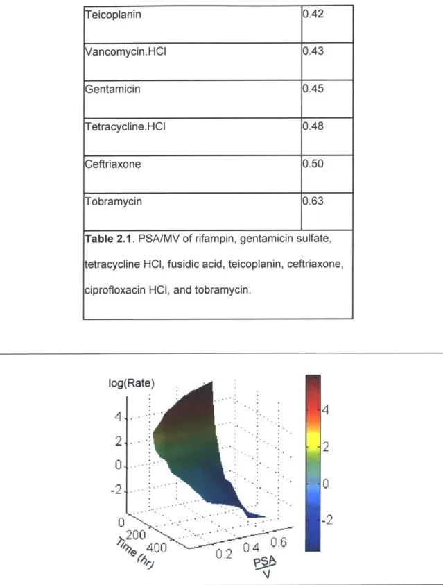
Figure 2.19. Elution rate throughout time vs PSA/MV of various drug-eluting UHMWPE...97
Figure 2.20. Schematic of solvent casting of Vancomycin eluting LDPE and compression
m olded vancom ycin eluting LD P E
...
101
Figure 2.21. Schematic of drug elution from antibiotic eluting UHMWPE...104
Figure 3.1. Mechanical Properties of Vancomycin eluting LDPE.
...
116
Figure 3.2.
Drug
cluster morphology and mechanical properties of vancomycin or NaCl eluting
Figure 3.3. Mechanical properties of vancomycin eluting UHMWPE. ... 118 Figure 3.4. Schematic of the finite element analysis on tensile dogbone samples with single
defe ct a nd d o u b le d efe ct...120 Figure 3.5. Percent change in mechanical properties with different eccentricity of the defect...122 Figure 3.6. Effect of compression molding temperature, compression molding pressure, and
vancomycin particle size on UTS, EAB, and IS ... 124 Figure 3.7. Mechanical properties of vancomycin eluting UHMWPE with and without drug
p a rtic le s iz e c o n tro ls ... 12 5 Figure 3.8. Mechanical properties of irradiated vancomycin eluting UHMWPE...126 Figure 3.9. Mechanical strength of other drug eluting UHMWPE...127 Figure 4.1 Scanning electron micrograph (SEM) of pre-eluted surface of vancomycin eluting
U H M W P E ... 1 5 0 Figure 4.2. Wear resistance of vancomycin-eluting irradiated UHMWPE containing 0-10 wt %
V a n c o m y c in ... 1 5 1 Figure 4.3 SEM micrograph of various drug eluting UHMWPE with initial 20% wt drug loading.... 152 Figure 4.4 Relation between drug types and wear rate of drug eluting UHMWPE. ... 153 Figure 4.5 Optical micrograph of surface of bupivacaine eluting UHMWPE post elution at 2-30 w t % initial bupivaca ine co nte nt ... 154
Figure 4.6 Relation between initial drug content and wear rate of bupivacaine eluting UHMWPE. ... 1 5 5 Figure 4.7 Wear rate of 20 % Bupivacaine-UHMWPE tested up to 3 million cycles...156 Figure 4.8 Surface topography of uneluted and eluted 20 % Bupivacaine-UHMWPE as obtained by sty lu s p rofilo m e try ... 15 7 Figure 4.9 Z-Projection of 20 consecutive micro-CT slices (25 pm slices) of the polymer phase a n d th e p o re p h a se ... 15 8 Figure 4.10 Surface texture of 20 wt % Bupivacacine before and after wear testing...158 Figure 4.11 Surface roughness contour obtained with AFM of 20 wt % Bupivacaine before and afte r w e a r te stin g ... 16 0 Figure 4.12 Progression of surface roughness (Ra) throughout wear testing of 20 wt %
B u p iv a c a in e . ... 1 6 1 Figure 4.13 Wear resistance of microtextured UHMWPE with various chemical treatments to reduce wear rate (75 kGy, MPC, and SLIPS) ... 162 Figure 4.14 Scanning electron micrograph of the surface of the MPC + microtextured UHMWPE and the corresponding EDAX carbon, phosphorous mapping ... 162
Figure 4.15 Scanning electron micrograph cutting through the thickness of the MPC +
microtextured UHMWPE and the corresponding EDAX carbon, phosphorous mapping...164
Figure 4.16 Wear rate of Bupi-PE and UHMWPE without bupivacaine irradiated at 0, 25, 50, 75 a n d 1 0 0 k G y . ... 1 6 6 Figure 4.17 Wear rate of 20 % MPC and normal UHMWPE (0% MPC) irradiated at 0, 25, 50, 75 a n d 1 0 0 k G y ... 1 6 6 Figure 4.18 Surface topography of radiation cross-linked Bupi-PE ( 75 kGy) before and after w e a r te s tin g ... 1 6 7 Figure 4.19 Surface roughness of radiation crosslinked Bupi-PE (75 kGy) before and after wear te s tin g ... 1 6 9 Figure 4.20 Progression of surface roughness (Ra) throughout wear testing of bupivacaine-e lu tin g U H M W P E . ... 17 0 Figure 4.21 Relation between compressive elastic modulus and initial bupivacaine loading co nce ntratio n in U H M W P E ... 17 1 Figure 4.22 Representative stress-strain curves of cyclic compression test (100 cycles total) w ith pe a k lo a d of 12 M P a ... 17 2 Figure 4.23 Fluorescence microscopy of surface of UHMWPE and Bupi-PE under no compression (0 MPa) and compression (8 MPa)...172
Figure 4.24 Computational simulation of lubrication film thickness due to elastohydrodynamic lubrication (EHD) and extruded lubricants due to weeping lubrication...175
Figure 5.1 Optimization field with respect to initial drug loading...194
Figure 5.2 Prototype of tibial insert (a) and acetabular liner (b) made from VPE...194
Figure 5.3 Elution profile of vancomycin eluting UHMWPE...195
Figure 5.4 Mechanical properties of vancomycin eluting UHMWPE and vancomycin eluting b o n e c e m e n t ... 1 9 6 Figure 5.5 Representative Kirby-Bauer agar diffusion test of VPE and BC1 after 24 hr in c u b a tio n ... 1 9 7 Figure 5.6 Area of inhibition throughout time of VPE, BC1, and BC2, against Staphylococcus aureus and Staphylococcus epidermidis. ... 198
Figure 5.7 Gross pictures of VPE or control UHMWPE-Titanium-Bone sandwich. ... 199
Figure 5.8 Bioluminescent imaging of live Xen29 that adhered to the titanium disc, cortical bone, and polyethylene component in the VPE and control groups...200 Figure 5.9 Quantification of the bioluminescent signal from the bacteria adherent to titanium
d isc a nd co rtica l bo ne d isc ... 2 0 1 Figure 5.10. Plate reculturing of bacteria post sonication of the titanium, bone, and polyethylene
Figure 5.11. Live/Dead stain of bacterial adherent to the surface of irradiated vancomycin
e luting U H M W P E ... 2 0 3 Figure 5.12. Sonicaton and reculturing of bacteria adherent to the surface of irradiated
vancom ycin eluting U H M W P E ... 204
Figure 5.13. Immunofluorescence staining of vancomycin grafted to the surface of irradiated vancom ycin eluting U H M W P E ... 205
Figure 5.14. Ratio of eluted rifampin and vancomycin throughout elution time at different initial d ru g s lo a d in g ra tio ... 2 0 7 Figure 5.15. Elution rate (a) and total drug eluted (b) of RVPE, VPE and BC2. ... 207
Figure 5.16. Mechanical properties of RVPE, VPE, and BC2...208
Figure 5.17 Prototype of tibial insert (a) and acetabular liner (b) made from combination of RVPE and non-antibiotic eluting UHMWPE...209
Figure 5.18. Schematic of 'sandwich; experiment to treat bacterial biofilm in between bone-titanium often found in patients with chronic PJI...210
Figure 5.19. Gross pictures of RVPE or control UHMWPE-Titanium-Bone sandwich ... 212
Figure 5.20. Bioluminescent imaging of live Xen29 that formed biofilm on the titanium disc, cortical bone, and polyethylene components...213
Figure 5.21 Quantification of the bioluminescent signal from the bioluminescent images shown a t F ig u re 5 .2 0 ... 2 1 4 Figure 5.22. Sonication and reculturing of bacteria adherent on the surface of the titanium, bone, and polyethylene components after 48 hr time points...215
Figure 5.23. Two-photon, live-dead imaging of the bacteria that adhered to the surface of titanium at 6 hr and 2 weeks after treatment. ... 216
Figure 5.24. Percent live dead as function of time of exposure to either control or RVPE...217
Figure 6.1. Gross picture of subcutaneously implanted control UHMWPE or VPE discs. ... 235
Figure 6.2. In vivo bioluminescence color map from Xen 29...236
Figure 6.3. Total bioluminescence quantification around the implants...237
Figure 6.4. Post-Mortem sonication of tissues and explants...237
Figure 6.5. Gross picture of subcutaneously implanted control RVPE discs and beaded titanium d is c s . ... 2 3 9 Figure 6.6. In vivo bioluminescence color map from Xen 29...239
Figure 6.7. Total bioluminescence quantification around the implants of mice receiving RVPE or co ntro l U H M W P E ... 2 4 1 Figure 6.8. Two-photon live-dead imaging of explanted UHMWPE and titanium discs. ... 241
Figure 6.9. Post-Mortem sonication of explanted titanium and polymer (UHMWPE) components.243 Figure 6.10. Gross picture of UHMWPE and Titanium Rod Implantation in the Lapine Joint
Infe ctio n M o d e l...2 4 5
Figure 6.11. Survival Curve of Rabbits Receiving Control UHMWPE , BC, or VPE ... 245
Figure 6.12. Kidney and Liver Function of Rabbits Receiving VPE...246
Figure 6.13. Necropsy bioluminescent map of rabbit knee infected with Xen 29...247
Figure 6.14. Total bioluminescence quantification of the rabbit knee receiving control UHMWPE o r V P E po st ne cro psy . ... 24 8 Figure 6.15. Two-photon live-dead imaging of explanted titanium rods. ... 248
Figure 6.16. Post-Mortem sonication of tissues and Explants...249
Figure 6.17. Gross picture of UHMWPE and Titanium Rod Implantation in the Lapine Joint In fe ctio n M o d e l...2 5 1 Figure 6.18. Bioluminescence of titanium rods prior to implantation ... 251
Figure 6.19. Survival Curve of Rabbits Receiving Control UHMWPE, BC, or RVPE. ... 252
Figure 6.20. Kidney and Liver Function of Rabbits Receiving RVPE. ... 253
Figure 6.21. Representative gross picture of the dissected knee from control, BC, and RVPE g ro u p s ... 2 5 3 Figure 6.22. Necropsy bioluminescent map of rabbit knee in control, BC, and RVPE infected with Xen 29 bioluminescent on titanium rod. ... 254
Figure 6.23. Two-photon live-dead imaging of explanted titanium rods from control, BC< and R V P E g ro u p s . ... 2 5 5 Figure 6.24. Post-Mortem sonication of tissues and Explants...256
Figure 7.1. Scanning electron micrograph of bupivacaine HCI eluting UHMWPE at different initial load ing of bupivacaine H C I...272
Figure 7.2. Elution Profile of Bupivacaine HCI eluting UHMWPE...274
Figure 7.3. Mechanical and Wear Properties of Bupivacaine HCI Eluting UHMWPE...275
Figure 7.4. Ionization state of bupivacaine determines its form in UHMWPE...277
Figure 7.5. Elution Profile of Bupivacaine FB eluting UHMWPE...278
Figure 7.6. Elution Profile of Combined Bupivacaine FB and Bupivacaine HCI eluting UHMWPE. ... 2 7 9 Figure 7.7. Mechanical and Wear Properties of Combined Bupivacaine FB and Bupivacaine HCI eluting UHMWPE. ... 281 Figure 7.8. Antibacterial activity of bupivacaine eluted from UHMWPE and standard
Figure 7.9. In vivo antibacterial activity of bupi-PE. ... 284 Figure 7.10. Schematic of in vivo testing of analgesic efficacy of bupi-PE ... 286 Figure 7.11. Stance and gait activity of the hind limbs post-implantation of bupi-PE or control
U H M W P E ... 2 8 7 Figure 7.12. Representative histology of the synovium capsule and the bone-implant interface of the rats receiving control UHMWPE or Bupi-PE ... 288 Figure 7.13. Equilibrium between Bupivacaine HCI and Bupivacaine FB in aqueous solution. ... 288 Figure 7.14. Comparison of bupivacaine release rate and total released bupivacaine from single injection, Expare TM, infusion pump, and bupivacaine eluting UHMWPE ... 291
List of Tables
Table 1.1 Cumulative incidence of PJI in THA based on number of year since implantation with conventional UHM W PE and HXLPE ... 30 Table 1.2 Cumulative incidence of PJI in TKA based on number of year since implantation with conventional UHMW PE and HXLPE ... 30
Table 1.3. Common bacteria causing PJI and the recommended antimicrobial treatment. ... 44
Table 2.1. PSA/MV of rifampin, gentamicin sulfate, tetracycline HCI, fusidic acid, teicoplanin, ceftriaxone, ciprofloxacin HCI, and tobram ycin. ... 97 Table 3.1. Defect eccentricity and average eccentricity tested for simulation with double defect. .121 Table 5.1. Type of Bacteria found in PJI patients reported from previous literature...180
Table 5.2. Calculation of RV-PE to Clinically Relevant Vancomycin to Rifampin Ratio. ... 206
Table 5.3. Calculation of RV-PE to Titanium-Bone interface Ratio...211 Table 5.4. Implant Design Based on VPE and RVPE and Comparison of Their Mechanical and Wear Rate to Conventional UHMWPE, Highly-Crosslinked UHMWPE (HXLPE), and ASTM
List of Abbreviations
AFM
Atomic force microscopy
ALBC
Antibiotic-eluting bone cement
BC
Vancomycin-eluting bone cement
Bupi FB
Bupivacaine freebase
Bupi HCI
Bupivacaine hydrochloride
Bupi-PE
Bupivacaine eluting UHMWPE (Layered 20 % Bupivacaine HCI in
UHMWPE on 20 % Bupivacaine FB in UHMWPE)
CoCr
Cobalt-chroium alloy
CRP
C-reactive protein
DAIR
Debridement, antibiotics, irrigation, and retention
EAB
Elongation to break
EDAX
Energy dispersive x-ray spectroscopy
EHD
Elastohydrodynamic lubrication
ESR
Erythrocyte sedimentation rate
FTIR
Fourier transform infrared spectroscopy
HXLPE
Highly-crosslinked UHMWPE
ICM-PJI
International consensus meeting on periprosthetic joint infection
IDSA
Infectious diseases society of America
IS
Impact strength
LDPE
Low-density polyethylene
MV
Molecular volume
NMR
Nuclear magnetic resonance
PJI
Prosthetic joint infection
PMMA
Poly(methyl methacrylate)
PROSTALAC Prosthesis with antibiotic-loaded acrylic cement
PSA
Polar surface area
Ra
Surface roughness
RVPE
6.7 % Vancomycin + 3.3 % Rifampin in UHMWPE
SEM
Scanning electron microscopy
SLIPS
Slippery liquid-infused porous surfaces
TGA
Thermogravimetric analysis
THA
Total hip arthroplasty
TJA
Total joint arthroplasty
TKA
Total knee arthroplasty
UHMWPE
Ultra-high molecular weight polyethylene
UTS
Ultiamte tensile strength
UV-Vis
Ultraviolet and visible spectroscopy
VPE
6.5 % Vancomycin in UHMWPE
VRE
Vancomycin-resistant Enterococcus
Prologue
More than one million joint replacement are performed in USA annually[1]. However, around 10 % of patients suffer complications and need to undergo revision surgery within 10 years [2]. One of the most common reasons for revision surgery is prosthetic joint infection (PJI) [2]. PJI is an increasing healthcare burden [3] with a recurrence rate of 16 % [4] and a mortality rate of 2.5 % [5]. End-stage treatments are severely morbid, including multiple revisions, resection arthroplasty, arthrodesis and amputation [6]. While attempts at developing drug eluting polymers have been made to address peri-prosthetic infection, they have been largely impeded due to toxic degradation products [7], insufficient drug release [8], insufficient mechanical strength [9], and difficulty in consistent large scale manufacturing [10]. To date, the only clinically available drug-eluting polymers for joint surgery are antibiotic-loaded bone cements (polymethyl methacrylate). Bone cements are used primarily for achieving early fixation of the bone-implant interface, providing stability and reducing the risk of loosening-related complications. They were not intended to be weight bearing and have a large incidence of in vivo fracture if used under bearing conditions [9]. Therefore, a drug-eluting polymeric surface designed specifically for weight bearing in joint arthroplasty revisions is urgently needed primarily for the prevention and treatment of peri-prosthetic joint infection but also for other conditions which may benefit from sustained delivery of therapeutic agents from the bearing surface such as pain or inflammation.
While bone cements were not developed primarily for weight-bearing and articulation, the incorporation of antibiotics in bone cements have further compromised their mechanical strength. Similarly, incorporation of drugs in polymers designed for weight bearing and articulation is expected to affect the mechanical strength and also the wear resistance, a crucial property of articulating surfaces. Wear particles from the articulating surface are associated with osteolysis and implant loosening, compromising the longevity of these functional devices. The
state-of-the art bearing surfaces in joint replacement comprise a cross-linked ultrahigh
molecular weight polyethylene (UHMWPE) 'soft' component articulating against a metal or
ceramic 'hard' surface to minimize wear. The total knee prosthesis consists of three
components: the femoral and tibial component are usually made of metal, while the plastic
spacer is usually made of UHMWPE [12]. The total hip prosthesis consists of four components:
The acetabular component, femoral head, and femoral stem are usually made of metal, while
the plastic liner is usually made of UHMWPE [12].Lubrication is disrupted by asperity contacts
and the wear of the surfaces are largely governed by the plasticity and large-strain deformation
of the polymeric component [11]. There is no literature on the wear behavior of
drug-incorporated UHMWPE.
The most important problems to be solved in the design of a drug-eluting bearing
surface are i) to minimize the effects of the incorporated drug on the mechanical properties of
the polymeric matrix, ii) to obtain sustained elution of the appropriate drugs specific to the
application; iii) to maximize lubrication and wear resistance. Understanding the effects of the
physical and chemical interactions of an incorporated drug with the polymer matrix can lay the
foundation by which processes can be developed to control the structure-property relationships
of the polymer based on these interactions. Understanding these interactions will also enable
the design of a controlled release profile of drugs most appropriate to the application. Finally, we
can also use our new understanding with other chemical and physical methods to manipulate
the morphology of the polymeric matrix to synergistic effect.
In this work, drug-incorporated polyethylene networks with modified morphologies were
developed. Appropriate therapeutic agents for the treatment of peri-prosthetic infection and
post-operative pain management were incorporated and the effects of drug chemistry on drug
elution are described. Medical device designs specific for joint replacement implants are
proposed for maximized mechanical strength of polyethylene matrices containing drugs and the
efficacy of these drug-eluting polyethylenes are demonstrated in vitro and in vivo. A new understanding of the effects of drug-incorporated UHMWPE on wear and lubrication is proposed. This work presents the first thorough description of a load-bearing surface with drugs designed to elute for therapeutic effect.
Chapter 1
Background
Total Joint Arthroplasty (TJA)
Arthritis (osteoarthritis and inflammatory arthritis) affects 52.5 million people in the United States annually [13] and it is expected to affect 78 million people by 2040 because of the aging population [14]. Joint arthroplasty, surgical procedure of replacing native joints with prosthetic joints, is the most effective treatment, especially for hip and knee arthritis, with more than one million arthroplasties performed in the USA annually[1]. To restore the joint function back to its normal function, prosthetic joints mimic the anatomical shape and function of the natural healthy joints, which will be detailed further in the sections below.
Total Hip Arthroplasty
The current design of total hip arthroplasty draws large similarity to the first metal on polymer prosthetic joints created by Professor Sir John Charnley in 1958 using Teflon for acetabular component and metal femoral component[15]. Because of its high wear rate, the material needed to be replaced after a couple of years[16]. As a result, in 1962, Charnley started using ultra high molecular weight polyethylene (UHMWPE) instead of Teflon for the acetabular component while continuing to use stainless steel as the femoral component[17]. Long-term follow-up (4-7 years) of 379 patients receiving Charnley prosthesis with UHMWPE acetabular component showed that 100 of patients were relieved of pain and were able to walk again[1 8].
There are four main components of total hip prosthesis (Figure P.1.): Acetabular cup, acetabular liner, femoral head, and femoral stem. The acetabular cup and femoral stem are
usually made of metal, while the acetabular liner and femoral head can be made of metal, UHMWPE, or ceramic. The acetabular cup and acetabular liner mimics the acetabulum of the pelvis, while the femoral head and femoral stem mimic the head and diaphysis of femur[19]. Metallic or ceramic femoral head articulating against UHMWPE acetabular liner are the most preferred choice of bearing surfaces, being used in more than 65 % of all primary joint replacement[20]. Because of the widespread use of UHMWPE in the hip joint prosthesis, in this thesis we focused on creating drug eluting UHMWPE that will be detailed later in this chapter.
Total Knee Arthroplasty
The current design of bicondylar knee implants were first designed in 1971 in Mayo Clinic, and named "Geometric" knee design[21]. The Geometric knee design was composed of two components: A metallic femoral bicondylar component and a UHMWPE tibial plateau, both of which were fixed in place using bone cement. The implants suffered from poor long-term fixation of the UHMWPE component[22], with overall 10-year survival rate of 69%[22]. Since then many modifications in the TKA implants design have been developed, including adding UHMWPE plugs as the patellar component[23], UHMWPE with metal backing[24], and mobile bearing TKA where the UHMWPE articulates against both the metal backing and the femoral component[25].
Modern TKA implants was usually comprised of three components: the femoral component, the tibial backing, and the tibial liner. The femoral component and tibial backing are made of metal, while the tibial liner is made of UHMWPE[12]. All the TKA implants currently used utilize metal femoral component articulating against UHMWPE tibial liner[1 2].
Development of UHMWPE Technologies to Address Total Joint Arthroplasty Complications
While the Charnley prosthesis in the hip and both Polycentric and Geometric prostheses in the knee significantly improved pain and walking function of patients receiving it, significant portions of the patients suffered complications related to the joint replacement. Among 106 UHMWPE components implanted by Charnley between November 1962 to December 1963 and followed up for 9-10 years, 4-6 % were infected, 1-2 % underwent aseptic loosening and 2 %
underwent dislocation[26]. In TJA, most revision surgeries are performed due to prosthetic joint infection and aseptic loosening (each contributes 25 % to the revision rates for TJA)[27, 28]. To address complications related to aseptic loosening, crosslinking technologies related to UHMWPE were developed to address complications related to TJA[29].
Ever since Charnley introduced low friction arthroplasty and used Teflon as part of the hip prosthesis, wear rate had been a concern. Wear particles promoted osteolysis and aseptic loosening, which ultimately required revision surgery[30]. Review of 11,543 revisions in hip arthroplasty in Sweden from 1979 to 1998 showed that 75.7 % of hip revisions occurred due to aseptic loosening[31]. To address the problem with insufficient wear resistance of UHMWPE,
highly crosslinked UHMWPE was developed[32].
Highly crosslinked UHMWPE (HXLPE) was created by exposing UHMWPE stock
material to ionizing radiation (gamma or electron beam) ranging from 50 to 105 kGy[32]. The irradiated material was then exposed to thermal treatment, by way of either annealing or remelting. After that, the material was machined to the final implant shape and then sterilized[32]. While radiation crosslinking of UHMWPE significantly decreased the wear rate, it also resulted in decreased the material ductility, fatigue resistance, and fracture resistance[33]. Because of the tradeoff between reducing wear rate and decreased fatigue and fracture resistance, manufacturers of the implants for total joint arthroplasty must decide on the optimum radiation dose to create an implant with low wear rate but also sufficient fatigue and fracture resistance[32]. Analysis of 1038 total hip replacements performed with highly crosslinked or
crosslinked UHMWPE significantly reduced in vivo wear rates and as a result, also reduced the risk for radiological osteolysis (mean risk ratio comparing highly crosslinked to conventional
UHMWPE was 0.40)[34]. Usage of highly crosslinked UHMWPE reduced the 10 year
cumulative percent revision in hip to 4-5 %, as compared to 6-13 % in conventional
UHMPWE[35].
While early clinical results of HXLPE have been excellent, long-term factors such as in vivo oxidation, which deteriorates mechanical strength and wear rate of HXLPE, may become the new limiting factor for the longevity of HXLPE in vivo[36]. In vivo oxidation of UHMWPE occurs through radiation induced free radicals, which may also be initiated in vivo by cyclic loading, and/or through reaction of lipids absorbed from the synovial fluid[37-39]. In order to minimize in vivo oxidation, highly crosslinked UHMWPEs stabilized with at least 500 ppm of the antioxidant vitamin E(VE-HXLPE) was added[40]. Accelerated in vitro oxidation of VE-HXLPE did not show any sign of oxidation nor decrease in mechanical strength compared to unoxidized VE-HXLPE[40, 41]. Unstabilized HXLPE, on the other hand, showed a 500% increase in total oxidation and a 70-80 % decrease in mechanical strength as compared to VE-HXLPE. [40, 41] A randomized clinical trial comparing VE-HXLPE against HXLPE for two years showed similar wear rates with no unexpected outcomes[40-42]. Because VE-HXLPE was only approved by FDA in 2007 for hip and in 2008 for knee, clinical data to compare longevity of VE-HXLPE vs HXLPE are not yet available[43].
Current Complications Related to TJA
Since the introduction of HXLPE in total hip arthroplasty (THA), incidence of aseptic loosening/osteolysis has significantly diminished [44]. Usage of HXLPE reduced the cumulative incidence of aseptic loosening by 60 % (2.3 % vs 0.92% with conventional vs HXLPE respectively) within ten years post implantation.[44] However, while HXLPE reduces the incidence of aseptic loosening, the cumulative incidence of PJI is similar to those of
conventional UHMWPE (Table 1.1). Other THA associated complications such as dislocation, fracture, and pain were also similar between conventional UHMWPE and HXLPE[44]. Overall, with HXLPE, PJI is the second most common reason for THA revision, second only to dislocation.
Number of years since Cumulative Incidence with Cumulative Incidence with
implantation conventional UHMWPE
(%)
HXLPE(%)
1 0.28 0.4
5 0.59 0.62
10 0.83 0.77
Table 1.1. Cumulative incidence of PJI in THA based on number of year since implantation with conventional UHMWPE and HXLPE[44].
In total knee arthroplasty (TKA), prostheses using HXLPE had a cumulative revision rate of 3.6 % at 10 years, much lower than the 5.7 % in prostheses using conventional UHMWPE[45]. The large decrease in cumulative revision rate was primarily due to reduced incidence of aseptic loosening in HXLPE vs conventional UHMWPE (0.77 % in HXLPE at 10 years vs 1.65 % in conventional UHMWPE at 10 years) [45]. Similar to the case with THA, while there is a large reduction in aseptic loosening with HXLPE in TKA, the cumulative incidence of revision due to other causes such as prosthetic joint infection, pain, and instability are similar to conventional UHMWPE (Table 1.2). [45] PJI was the top reason for TKA revision with HXLPE within one, five, and ten years of implantation. [45]
Number of years since Cumulative Incidence with Cumulative Incidence with
1 0.39 0.42
5 0.90 0.76
10 1.0 1.0
Table 1.2. Cumulative incidence of PJI in TKA based on number of year since implantation with conventional UHMWPE and HXLPE[45].
In summary, because HXLPE is currently used in 96 % of all THA[44] and 54 % of all TKA[45] with expected further increase in usage due to its wear resistance, incidence of aseptic loosening goes down as compared to conventional UHMWPE, while incidence of PJI becomes more prevalent. Therefore, it is important to develop better prophylaxis and treatment alternatives for PJI to reduce the overall incidence of revision arthroplasty.
Incidence and Economic Burden of Periprosthetic Joint Infection
PJI occurs in 2.05-2.18 % of patients who have undergone TKA and in 1.99-2.18 % of those with THA.1 PJI incidence in shoulder arthroplasty ranges form 0.8-1.1 %[46, 47], while PJI incidence in elbow arthroplasty is higher at 3.3 %[48]. The higher incidence of PJI in the elbow arthroplasty is attributed to the higher percentage of rheumatoid arthritis in patients undergoing elbow replacement and the limited amount of soft tissue surrounding the elbow, exposing it to infectious microorganisms.[49] The current annual incidence of PJI in the US is 7,100-15,000, and is predicted to be 55,000-75,500 by the year 2030. Adjusting for patient demographic factors such as age, sex, race, and census region, the risk of hip infection increased significantly from 2001 to 2012.[50] The risk for PJI was greatest in the first 2 years after implantation, during which time 60-70 % of PJI occurred.[51, 52]
PJI causes a significant morbidity and mortality to the patient, and also a significant burden to the economy. Compared to the average length of hospital stay for primary THA and TKA without PJI occurrence, an average of 10-12 days is added to the hospital stay for PJI.[50]
Patients who had PJI never return to the functionality of patients who did not have PJI.[53] In addition to significant morbidity, PJI leads to a high mortality rate: PJI treated with two-stage revision had up to 26 % all-cause mortality within two years[53]. Mortality rate as high as 45 % was reported for recurrent PJI.[54]
PJI treatment is estimated to cost as high as U$ 100,000 per case, which is five times higher than the cost of primary arthroplasty.[55] The cost is attributed to revision surgery being more resource-heavy, long rehabilitation time, and long-term usage of antibiotics and analgesics.[55] The cost of revision surgery due to infection is higher than the cost of revision for non-infectious causes[56]. In 2009, the total cost for the management of prosthetic joint
infections was ~ 800 million dollars; this number is predicted to be 1.6 billion dollars by
2020.[50] By all measures, PJI is a significant burden to the patient and the healthcare system.
Pathogenesis of Prosthetic Joint Infection
Bacterial seeding into the prosthetic joints is thought to occur through three main pathways: First, direct bacterial seeding during implantation; second, through hematogeneous spread from other body parts, and third, through recurrent infection[57]. Infection around medical implants is especially difficult to treat because of the dampened innate immune system[58] and bacterial biofilm formation on the surface of the implant which reduces the susceptibility to antibiotics of the bacterial population.[57] An animal infection study showed that 100,000 times less bacterial concentration is needed to induce infection in the presence of a foreign body as compared to the absence of a foreign body[59]. In fact, less than 100 colony-forming units of microorganisms were sufficient to cause an infection.[60] The increased susceptibility to infection in the presence of medical implant is partially due to local granulocyte defects in the environment surrounding the medical implant.[58, 60] In the presence of a foreign body such as a medical implant, neutrophils on the foreign surfaces release peptides that
Based on the time of onset, PJI can be acute or chronic. Seventy percent of PJI occurs acutely, and the remaining thirty percent occurs chronically[62]. Acute infections usually occur within three weeks of joint replacement, are confined to the joint space, do not invade the prosthetic-bone interface, and most do not involve the formation of bacterial biofilms. Patients with acute PJI usually present with fever, increased pain locally, around the surgical site,
erythema, and poor wound healing.10 On the other hand, chronic infection usually occurs at
least three weeks post-surgery, but can also occur months to years later, and patients present with a joint that is painful and warm, with an effusion. Since the bacteria have been occupying the prosthetic joint surface for weeks to months, most chronic PJls involve the formation of a biofilm, which allows the bacteria to adhere strongly to the prosthesis, shield themselves from host immune system, and make them 1000 to 1500 times more resistant to antibiotic treatment.
Acute infections usually occur through two main mechanisms: First, direct bacterial inoculation introduced during the surgery, and second, through contiguous spread of infection from superficial surgical site infections[63].
Chronic or late infections are predominantly due to hematogeneous spread.[64] The most common source of hematogenous spread is from a skin infection, respiratory tract infection, dental infection, and urinary tract infections.[65] Only 100-1000 CFU/ml bacteria in blood are needed to cause hematogenous infection of the prosthetic joints[60]. Overall, PJI resulting from hematogenous infection is rare: 1.2-6 % of reported PJI cases are due to hematogenous spread[66]. Of patients with confirmed bacteremia, only 6 % were found with hematogenous PJI[63]. However, some bacteria such as S. aureus causes a higher risk of hematogenous PJI: Several studies showed that S. aureus bacteremia is associated with 30-40 % risk of hematogenous PJI[63, 67, 68].In the majority of hematogenous PJI, bacteremia and onset of PJI occur almost simultaneously[63].
Various comorbidities and demographic factors such as diabetes[69, 70], preoperative anemia, cardiovascular disorders, chronic renal failure, nutritional status[57], body mass index[71], smoking and alcohol consumption status[72-74], rheumatoid arthritis[75], and usage of antibiotic eluting bone cement[76] have been shown to affect the risk for PJL.
Patient with diabetes had seven times higher risk for PJI than non-diabetic patients[69]. A separate study analyzing 1947 patients receiving TJA showed that diabetic patients occupied 22 % of the infected cohort, as opposed to 9 % in the uninfected cohort.[70] Blood glucose of > 200 mg/dL was found to double the risk of PJI as compared to patients with normal blood glucose[70]. The elevated risk for PJI in patients with diabetes is likely due to impaired wound healing[77] and elevated biofilm formation associated with elevated glucose levels[78].
Poor nutritional status has been associated with increased risk for PJI.[79] A patient is considered in the poor nutritional state if one of the following is satisfied: serum albumin<3.5 g/dL (healthy range 3.4-5.4 g/dL), total lymphocyte count <1500 cells/mm3, serum transferrin
levels < 200 mg/dL, or serum prealbumin <15 gm/dL.[80, 81] Patients with total lymphocyte count of <1500 cells/mm3 was found to have 5 times greater risk for PJI, and patients with
albumin levels of < 3.5 g/dL was found to have 7 times greater risk for PJI.[82] Separate study showed that patients who underwent PJI revision surgery and satisfied the poor nutritional status mentioned above were found to have 7 times higher infection rate than patients with good nutritional status.[79] The poor nutritional state has been shown to interfere with the synthesis of collagen and proteoglycan that is crucial for wound healing. Disruption of such processes may lead to persistent wound drainage and increased risk of infection.[81]
High body mass index (obesity) has been shown to have increased risk of PJI after THA and TKA (adjusted hazard ratio of 1.72 and 1.22 respectively).[71, 83] The risk of PJI elevated from 3.3 times higher in patients with BMI > 40 kg/M2 (as compared to patients with
7,181 primary TJA reported elevated PJI rate from 0.37 % in patients with normal BMI to 4.66 % in morbidly obese patients.[85]
Smoking has been shown to significantly increase the rate of surgical site infections[72], the rate of deep wound infections[74], and the rate of wound complications than nonsmokers.[73] The elevated risk of infection due to smoking is likely due to reduced blood flow to the healing tissue, impaired delivery of cellular and humoral immunity to the surgical site, disruption of immune cell function, and disruption of neutrophil defense mechanisms against
pathogens[86].
Alcohol consumption has been shown to increase the risk of infection in patients underwent major, non-cardiac surgery[87]. The elevated risk of infection is thought to originate from alcohol-induced disruption of the immune system that is crucial to eradicate microorganisms surrounding prosthetic joints.[88]
Patients with rheumatoid arthritis were reported to have elevated risk of PJI than patients without rheumatoid arthritis (hazard ratio = 1.71 for THA and 1.18 for TKA).[75] The PJI rate for patients with rheumatoid arthritis has been reported to be as high as 2.3 % in the first year[89]. While the reasons behind the elevated risk for infection due to rheumatoid arthritis are poorly understood, reports suggest that the disease itself and immunosuppressive therapies to treat it both contribute to increased risk for PJI[63].
Bacterial Strains Commonly Found in PJI
On average, a positive culture is obtained on 68-94 % of all patients with apparent PJI.[90]'[64, 91] Gram-positive bacteria account for 90-95 % of all culture-positive PJI,[64] with 54-80 % of PJls due to the gram-positive S. aureus and coagulase-negative Staphylococcus species such as S. epidermidis, or S. lugdunensis [64]. The percentage of PJI caused by S.aureus and coagulase negative Staphylococcus are relatively equal [63]. Gram-positive streptococci and enterococci account for -10 % of all PJI [63]. Some of the less common
![Table 1.1. Cumulative incidence of PJI in THA based on number of year since implantation with conventional UHMWPE and HXLPE[44].](https://thumb-eu.123doks.com/thumbv2/123doknet/14753539.581324/30.918.124.822.301.489/table-cumulative-incidence-number-implantation-conventional-uhmwpe-hxlpe.webp)
![Table 1.2. Cumulative incidence of PJI in TKA based on number of year since implantation with conventional UHMWPE and HXLPE[45].](https://thumb-eu.123doks.com/thumbv2/123doknet/14753539.581324/31.918.92.794.130.284/table-cumulative-incidence-number-implantation-conventional-uhmwpe-hxlpe.webp)