Cell‐Cell and Cell‐Medium Interactions in the Growth of Mouse Embryonic Stem Cells
by Nikhil V. Mittal B.Tech. Engineering Physics Indian Institute of Technology, Mumbai, 2000 S.M. Physics Massachusetts Institute of Technology, 2003 Submitted to the Department of Physics in partial fulfillment of the requirements for the degree of Doctor of Philosophy at the Massachusetts Institute of Technology June 2010 © 2010 Nikhil Mittal. All rights reserved. The author hereby grants to MIT permission to reproduce and to distribute publicly paper and electronic copies of this thesis document in whole or in part in any medium now known or hereafter created. Signature of author:_____________________________________________________________________ Department of Physics May 14, 2010 Certified by:___________________________________________________________________________ Professor Joel Voldman Department of Electrical Engineering and Computer Science Thesis Supervisor Certified by:___________________________________________________________________________ Professor Alexander van Oudenaarden Department of Physics Thesis Co‐supervisor Accepted by:__________________________________________________________________________ Professor Krishna Rajagopal Associate Head for Education, Department of PhysicsContents
Abstract ... 6 Chapter 1: Introduction ... 8 1.1 Growth of cells in culture ... 8 1.2 Communication between cells ... 9 1.3 Embryonic stem cells ... 11 1.4 Motivation ... 13 1.5 Thesis Aims and Organization ... 16 1.6 Summary ... 17 Chapter 2: An nDEP Microwell Array for studying intercellular signaling ... 18 2.1 Technologies for studying homotypic cell‐cell communication ... 18 2.2 Device Principle ... 20 2.3 Technologies for making clusters of cells ... 20 2.4 Materials and Methods ... 23 2.5 Results and Discussion ... 26 2.5.1 Biocompatibility of the nDEP Microwell Array ... 26 2.5.2 An Alternate Loading Protocol for the nDEP Microwell Array ... 28 2.6 Summary ... 32 Chapter 3: Studying mESC Self‐renewal using the Bio Flip Chip ... 34 3.1 Introduction ... 34 3.2 Materials and Methods ... 35 3.3 Results ... 38 3.3.1 Patterning clusters of cells using the BFC ... 38 3.3.2 Variation of colony‐forming efficiency with the number of cells in a cluster ... 40 3.3.3 Self‐renewal potential and its variation with the number of cells in a cluster ... 42 3.3.4 Variation of colony‐forming efficiency and colony area with plating density ... 44 3.4 Discussion ... 45 3.5 Summary ... 48 2Chapter 4: mESC cultures contain an autocrine survival factor ... 50 4.1 Materials and methods ... 50 4.2 Results ... 52 4.2.1 Variation of growth rate with plating density ... 52 4.2.2 Post‐attachment post‐lag growth of mESCs ... 52 4.2.3 Conditioned Medium improves mESC growth ... 54 4.2.4 Interpreting and improving upon growth data ... 55 4.2.5 mESC Cultures contain an Autocrine Growth‐supportive Large Molecule ... 56 4.2.6 Length of attachment period and lag phase ... 58 4.2.7 The autocrine growth factor(s) in mESC cultures is (are) not mitogenic, but improve(s) survival ... 59 4.3 Discussion ... 61 4.4 Summary ... 62 Chapter 5: Cyclophilin A is an autocrine survival factor in mESC cultures ... 66 5.1 Materials and Methods ... 66 5.2 Results ... 67 5.2.1 Determining protein concentrations in cell cultures using numerical modeling ... 68 5.2.2 Previously known mESC secreted proteins are not growth‐supportive for mESC ... 70 5.2.3. Proteomic analysis of mESC conditioned medium ... 72 5.2.4 Mass spectrometric analysis of gel slices ... 74 5.2.5 Assaying proteins detected via mass spectrometry ... 75 5.3 Discussion ... 76 5.4 Summary ... 77 Chapter 6: Nutritional Aspects and Numerical Modeling of mESC growth ... 80 6.1 Introduction: Modeling the growth of cells and communication between cells ... 80 6.2 Materials and Methods ... 80 6.3 Results ... 82 6.3.1.1 Net effect of nutrient depletion and metabolite production on mESC growth ... 82 6.3.1.2 Amino Acids ... 83 6.3.1.3 Glucose ... 86 3
6.3.1.4 Vitamins and Salts ... 87 6.3.1.5 Metabolites ... 88 6.3.2 Numerical modeling of mESC growth ... 88 6.4 Discussion ... 90 6.5 Summary ... 91 Chapter 7: Conclusions ... 92 7.1 Thesis contributions ... 92 7.2 Future directions ... 93 References ... 100 Appendix 1: Dielectrophoresis (DEP) ... 110 Appendix 2: Supplementary Data for Chapter 3 ... 114 Appendix 3: Supplementary Data for Chapter 5 ... 120 Appendix 4: Supplementary Information for Chapter 6 ... 124 Appendix 5: Our Cell Lines ... 128 Acknowledgements ... 130 4
5
Cell‐Cell and Cell‐Medium Interactions in the Growth of Mouse Embryonic Stem Cells by Nikhil V. Mittal Submitted to the Department of Physics on May 14th 2010 in partial fulfillment of the requirements for the degree of Doctor of Philosophy at the Massachusetts Institute of Technology
Abstract
Embryonic stem cells serve as powerful models for the study of development and disease and hold enormous potential for future therapeutics. Due to the potential for embryonic stem cells (ESCs) to provide a variety of tissues for use in regenerative medicine, there has been great interest in the identification of factors that govern the differentiation of ESCs into specific lineages. Much of this research builds on previous studies of the role of intercellular signaling in the specification of various cell types in the developing embryo. However, relatively little work has been done on understanding the role of cell‐cell communication in the self‐renewal of ESCs.
In the first part of this thesis I describe the development and testing of new devices for studying intercellular signaling ‐ the nDEP microwell array and the Bio Flip Chip (BFC). We used the BFC to show that cell‐cell interaction improves the colony‐forming efficiency and the self‐renewal of mouse ESCs. Further, we demonstrate that the interaction is at least partly diffusible.
In the next part of the thesis I describe our use of more traditional assays to validate the results obtained using the BFC and to further explore the role of diffusible signaling in the survival of mouse ESCs. We demonstrate the existence of an optimal density for 2‐day culture of mouse ESCs. Further, we demonstrate that the increase in growth with plating density (103‐104 cells/cm2) is at least partly due to the existence of one or more survival‐enhancing autocrine factor(s) in mouse ESC cultures, and that one of these factors is Cyclophilin A.
Finally, we demonstrate that changes in the low molecular weight composition of the medium are likely responsible for the decrease in growth at high plating densities (>104 cells/cm2). We use a numerical model to show that competition between the positive effect (on growth) of autocrine survival factors and the negative effect of nutrient depletion can account for the observed optimal growth density. Our study provides new insight into the processes underlying, and optimization of, growth in cell types that lack contact inhibition such as cancer cells and stem cells. Thesis Supervisor: Joel Voldman Title: Associate Professor of Electrical Engineering and Computer Science 6
7
Chapter 1: Introduction
This thesis describes the influence of secreted factors, nutrient depletion, and metabolite accumulation on the growth of mouse embryonic stem cells in culture. In this first introductory chapter I will first describe some of the factors that motivated this study. I will begin by providing some background on cell growth and embryonic stem cells.
1.1 Growth of cells in culture
The growth of cells in culture has been studied quantitatively for almost 80 years. Oscar Richards1 appears to be the first person to attempt to fit microbial (yeast) growth data with numerical models (1928). However, Jacques Monod2 was the first person to develop mechanistic models of microbial (bacterial) growth that accounted for the effects of nutrient concentrations. In particular, he explored the existence and effects of a “limiting nutrient” in the medium, and showed that the growth of bacteria is well described by the equation: , where n is the concentration of cells, µm is the
maximum growth rate, s is the concentration of the limiting nutrient, and Ks is the nutrient
concentration which supports half the maximum specific growth rate.
Starting around 1990, with the advent of packaged frozen foods, there was much greater interest in understanding the growth of cells, especially bacteria such as Listeria monocytogenes (the bacterium responsible for listeriosis, a potentially lethal condition) on various kinds of (food) substrates3. Additionally, around the same time, there was growing interest within the biotechnology industry, in the growth of cells for the production of proteins for therapeutic and diagnostic purposes4, 5. This production of proteins required the use of large bioreactors and led to increased interest in the use of models to optimize the yield of proteins in these reactors6. Most of these models were still Monod‐type models except that the availability of computers made it much easier to simulate the effects of depletion of more than one nutrient. Typically, glucose and glutamine were found to be the limiting nutrients. Additionally, models incorporated the effects of the accumulation of metabolites such as lactate and ammonia in the medium, and the (usually) inhibitory effects of these metabolites on cell growth6. Such models appear to sufficient for describing the growth of certain types of cells such as bacteria, yeast, Chinese hamster ovary (CHO) cells and hybridomas (the two types of cells often used for producing proteins) etc. However, for most mammalian cells, in addition to nutrient depletion, cell‐cell communication/signaling is often an important modulator of cell growth. In the next section I will discuss the various types of cell‐cell communication. 8
1.2 Communication between cells
Cells interact with one another both in vivo and in vitro. According to the Gene Ontology (GO) database, as of this writing, 454 of 11570 (4%) currently annotated human proteins are directly involved in cell‐cell signalinga. Cell‐cell interactions can be classified into two types:
(i) Diffusible signaling. In this type of signaling, one cell produces a molecule (ligand) which is subsequently released from its surface. The ligand then diffuses in the medium or through the extracellular matrix (hence the name diffusible signaling), until it is bound by a receptor molecule on the surface of the same cell, or another cell. The capture of the ligand by the same cell as it is released from is referred to as autocrine capture, while capture by a receptor on a different cell is called paracrine capture. The ligand is sometimes also referred to as a soluble factor, and this type of signaling is therefore also known as soluble signaling.
Autocrine signaling was originally studied in the context of cancer. In the 1980s an autocrine model for cancer growth was widely investigated – i.e. a model that postulated that cancers were caused by cells responding to an excess of growth factors (GFs) that they secrete themselves. This hypothesis is usually ascribed to Sporn and Todaro7. One known oncogene, the sis oncogene, encodes a protein similar to a growth factor, the platelet‐derived growth factor (PDGF) and produces cancers via this mechanism8. Some cancers are due to overproduction of GF receptors (Her2 in some breast cancers) which make these cells proliferate rapidly in response to normal levels of growth factors. However, many oncogenic mutations are in proteins downstream from growth factor receptors.
Autocrine signaling via growth factors can modulate the growth rate in two ways8‐10. The total growth rate is the difference between the rate at which cells divide (proliferation rate), and the rate at which cells die (death rate). Growth factors typically increase the proliferation rate and/or decrease the death rate. Most mammalian cells require the presence of growth factors in the early G1 phase of the cell cycle in order to proceed to the S phase. This mechanism links growth factor concentration and proliferation rate, i.e., growth factors are required for mitosis and therefore such required growth factors are referred to as mitogens. However, in mammalian cells, there appears to be cross‐wiring between pathways regulating the cell‐cycle and pathways that trigger cell death9, 10. As a result, growth factors can also affect the death rate of cell9, 10. Paracrine signaling plays a very important role in development where gradients of secreted factors are used to create various regions in an initially homogenous embryo8. Paracrine signaling may also play a vital role in the regulation of adult stem cells by niche cells11 – it is thought that adult stem cells receive cues from surrounding cells that tell them when they need to produce more cells to replace dead cells in the tissue. Endocrine signaling refers to a subset of paracrine signaling where cells from one organ produce a factor that cells in another organ respond to. a The GO database uses the following definition for cell‐cell signaling: Any process that mediates the transfer of information from one cell to another. In the mouse genome, 295 of 11165 annotated genes are involved in cell‐cell signaling. 9
(ii) Contact signaling. Contact signaling can be further sub‐divided into two types –
a. Signaling through gap junctions. Gap junctions provide channels through which two cells in contact can exchange small molecules and ions (generally < 1.5 kDa)12. The channels consist of two connexons, which are each made up of six connexin proteins (Figure 1.1). In mammals, more than 20 kinds of connexins have been identified. Gap junctional communication is particularly important in the heart, where it serves to transmit ions so as to induce the coordinated contraction of cells13.
Figure 1.1 Schematic of gap junctions between two cells. Each junction consists of two connexons which are comprised of six
connexin molecules each. Image courtesy of Mariana Ruiz (Wikicommons).
b. Signaling via cadherin molecules. Cadherins are the molecules that hold cells together. It is these molecules that are believed to be responsible for the well‐known phenomenon of contact inhibition seen in cultures of endothelial and epithelial cells14. A recent study has shown that some cadherins do this by inhibiting the activity of epidermal growth factor receptors, which are found to be closely associated with cadherins15. The name “cadherin” derives from the fact that these molecules need the presence of calcium ions to perform their adhering function.
To summarize, as regards growth in particular, two types of intercellular cell signaling are important modulators: autocrine signaling via growth factors often provides a positive feedback effect on growth, and cadherin interactions often lead to growth inhibition.
In particular, we have investigated the role of cell‐cell signaling in the growth of embryonic stem cells (though some results from our studies likely apply to growth in other cell types as well). In the next section I will describe some of the basic properties of embryonic stem cells, and previous work on their growth.
1.3 Embryonic stem cells
Embryonic stem cells (ESCs) are cells that have the potential of being able to produce any cell type in the adult animal. This property is referred to as pluripotency. These cells are derived from the inner cell mass of an early stage embryo, called the blastocyst (Figure 1.2). Human embryos reach the blastocyst stage 4‐5 days after fertilization, and consist of 50‐150 cells. ESCs have been isolated in a number of different organisms – while mouse ESCs (mESCs) were first isolated in 198116, 17, human ESCs (hESCs) were first isolated in 199818.
Figure 1.2 ESC derivation and properties. The fertilization of an oocyte by a sperm cell leads to the formation of a zygote. The
zygote divides rapidly to form a morula, and then a blastocyst by day 4‐5 in humans. The blastocyst consists of two types of cells – inner cells (on the inside) and trophoblast cells (on the outside). Embryonic stem cells are derived from the inner cell mass of a blastocyst. Embryonic cells are pluripotent i.e. they can form any cell type in the adult animal via a process called differentiation. However, ESCs cannot generate the whole organism without the aid of trophoblast cells. Only cells in the morula can form the whole organism and are referred to as totipotent cells. Figure courtesy of Mike Jones (Wikicommons).
ESCs generate organ‐specific cells such as neurons, cardiomyocytes etc. via a process called differentiation or commitment. Cells that are partially committed (relative to ESCs) will be able to form some subset of cell types found in the adult animal and are referred to as multipotent cells. Cells that are completely committed will either not divide, or divide to form daughter cells with the same phenotype as the parent (unipotent cells). While ESCs can differentiate to form various cell types, they can also divide to form two daughter ESCs, a process referred to as self‐renewal. Self‐renewal is obviously crucial for the expansion of ESCs while maintaining their pluripotency.
Given that there are ~36000 known (mouse) genes as of this writing, a complete molecular‐level description associated with any state of a cell is challenging to define. Pluripotency is no exception. Several studies have tried to obtain a “molecular signature” associated with pluripotency (for example, 11
Ivanova et al.19 and Ramalha Santos et al.20) by comparing the transcriptional profile of ESCs with committed cells such as bone marrow cells using microarrays. Several hundred genes were found to be differentially expressed by ESCs. However, the candidate genes obtained from such experiments will have to be validated. Additionally, the overlap between candidate genes determined by different studies is far from complete21. Finally, a recent study has indicated that several proteins are expressed differentially by ESCs as compared to committed cells, but there are no significant differences in RNA concentrations indicating that post‐translational regulation may be important for maintenance of a pluripotent state22.
Nevertheless, smaller‐scale studies have implicated a large number of genes as being required for the maintenance of pluripotency. Perhaps not surprisingly, almost all major signal transduction pathways seem to play a role (reviewed in Liu et al.23). For historical reasons, one pathway that has received considerable attention is the Stat3 pathway. mESCs were initially propagated using a feeder layer consisting of embryonic fibroblast cells16, 17 (in a medium supplemented with bovine calf serum). Subsequently, it was found that the requirement for the feeder layer could be replaced by adding leukemia inhibitory factor (LIF) to the medium24, 25. LIF binds to a heterodimer complex consisting of the LIF receptor (LIF‐R) and another receptor subunit called Membrane glycoprotein 130 (gp130). This binding event leads to the activation of several pathways, of which, the activation of Stat3 has been shown to be essential for the maintenance of a pluripotent state in mouse ES cells26, 27, though not in human ES cells28. Besides Stat3, Oct‐429, 152, Sox‐230, 31, and Nanog32‐34 are three other transcription factors shown to be essential for developing and maintaining the pluripotent state. These factors appear to be essential in mouse as well as human ES cells (see above references).
1.3.1 Growth of embryonic stem cells
Circling back to self‐renewal, note that it actually consists of two processes – maintenance of pluripotency, as well as cell division. While most research on self‐renewal has focused on understanding the signaling and transcriptional networks involved in the maintenance of the ESC pluripotent state, comparatively few studies have investigated pathways involved in ESC growth and death under conditions shown to maintain pluripotency. Below I review some of the latter studies. Cell growth is intimately tied to progression through the cell cycle, and several studies have focused on understanding cell cycle control in mESCs. mESC growth in culture is characterized by a short cell cycle (11–16 hours), primarily owing to a reduction in the duration of G1 phase (as compared to somatic cells, reviewed in Orford and Scadden35). This short G1 phase is due to a lack of the mitogen‐dependent early G1 phase of the cell cycle, owing to constitutive Cyclin E – CDK2 activity36. This constitutive activity of Cyclin E ‐ CDK2 in turn appears to be driven at least partially by a constitutively active Ras protein expressed specifically in ES cells, called Eras153, which stimulates PI3K, which is upstream of Cyclin E37.
Very little work has been done thus far on understanding the role of cell‐cell signaling in the growth of mESCs. Since these cells appear to lack the mitogen‐dependent early G1 phase of the cell cycle, it seems unlikely that cell interactions could modulate the length of the cell cycle. However, it is plausible that cell interactions could modulate the survival of these cells. mESCs typically have survival/colony‐forming efficiencies of ~30%38, while hESCs have survival/colony‐forming efficiencies in the range of 1‐10%39. 12
Studies have shown that the addition of EGF, ANG‐2, Activin, or Nodal proteins to mouse ESC (mESC) cultures results in improved growth40, 41, 154. However, in these studies they did not test for secretion of these proteins by ESCs, i.e. they did not (attempt to) fully demonstrate that these pathways are constitutively active in mouse ES cultures and that the factors are acting in an autocrine fashion. For example, Ogawa et al.40 demonstrated that addition of Activin or Nodal proteins to a culture improves
their growth. Additionally they show that inhibition of Activin‐Nodal signaling downstream of the receptor binding event (by Smad7 expression or the specific inhibitor SB‐431542), led to decreased growth. However, in this latter assay (especially in the control) it is possible that cells were responding to Activin/Nodal present in the supplied medium and not cell‐secreted proteins. Because ESCs grow as colonies, juxtacrine signaling could also presumably play a growth‐supportive role. Todorova et al. used pharmacological blockers of gap junctional communication and Connexin‐43 small‐ interfering RNA to inhibit signaling via gap junctions in mESCs42. They demonstrated that gap junctional communication enhances either the growth rate or the attachment efficiency (or both) of mESCs. They did not attempt to separate the potential contributions of attachment efficiency and growth rate to the observed decrease in fold growth over 72 hours. It is clear that an investigation into the role of intercellular signaling in the growth of ESCs is relevant to basic biology. Below I will describe some of the other areas of research that motivated our work.
1.4 Motivation
For my thesis I have investigated the growth of mESCs under conditions supporting self‐renewal. Below I highlight some of the issues that motivated this work. 1.4.1 Biophysics of cell growth In sections 1.1 and 1.2 I described two major factors that influence the growth rate of cells in culture, namely, (i) depletion of nutrients from the medium, and (ii) cell‐cell signaling. The depletion of nutrients can also be thought of as a means of indirect communication in that cells are consuming materials out of a common pool. In general, such communication provides a number of positive and negative feedback loops that control cell phenotype. One mechanism for controlling the strength of the feedback is to vary the amount of signal that cells can exchange.
Now let us consider a more concrete example. Most mammalian cell types seem to produce and respond to one or more growth factors in an autocrine fashion8. This provides a positive feedback to the rate of growth. Such systems have been studied in considerable detail by the Lauffenburger lab (MIT) and the Shvartsman lab (Princeton). In particular, one can modulate the strength of the feedback by altering the density at which cells are cultured ‐ higher cell densities result in higher densities of the growth factor in the medium, which will result in a higher growth rate (until one reaches a saturation regime). Thus, in general, growth rate increases with cell density43.
However, eventually one will enter a saturating regime, where additional increases in growth factor concentration do not result in improved growth127. Additionally, at high plating densities we expect several kinds of negative feedback to become important – the depletion of nutrients, the consequent 13
production of metabolites, and contact inhibition of growth6, 14. In the simplest case, the combination of a single positive feedback mechanism along with one or more negative feedback mechanisms will result in the existence of a single density at which the growth will be optimal. While it is possible and even likely that biologists have had experimental evidence for this hypothesis for a long time, published observations demonstrating an optimal density for cell cultures have been available only recently44. Additionally, while the above mechanisms qualitatively account for such an optimal density, a quantitative assessment is lacking. Our work seeks to fill‐in some of the holes in the understanding of factors that determine the optimal density for cell culture.
1.4.2 Potential for the use of ESCs in regenerative medicine
Regenerative medicine includes the study and development of artificial organs, specially‐grown tissues and cells (including stem cells), laboratory‐made compounds, and combinations of these approaches for the treatment of injuries and disease45. According to the 2007 report of the Scientific Registry of Transplant Recipients, every year this decade ~2000 people have died while waiting for a liver transplant. The waiting list for kidney transplants at the end of 2007 consisted of ~80,000 patients. Thus there is clearly a large unmet need for new sources of cells and tissues. However, perhaps the most exciting applications of regenerative medicine will be in developing therapies for conditions for which there are no widely successful, existing therapies. Examples of such conditions include degenerative diseases of the central nervous system (Alzheimer’s disease, Parkinson’s disease) and spinal cord injury. Another possibility is that tissue‐engineered organs may function better than transplanted organs – for e.g., according to the above report, the 5‐year survival rate for liver transplant recipients is currently only around 50%.
In most research to‐date, researchers have used more committed cells for developing new tissue46. For example, in a landmark study in 2006, Atala et al. expanded patient bladder cells on bladder‐shaped collagen scaffolds and implanted the resulting constructs back into patients, resulting in successful enhancement of bladder volume47. Similarly, neonatal cardiac myocytes have been used for developing cardiac tissue constructs48. Finally, while embryonic stem cells are derived from an embryo and are pluripotent, adult organisms also contain stem cells (adult stem cells, ASCs) that are multipotent, and several recent studies have used mesenchymal stem cells from adult bone marrow for therapeutic purposes49. Mesenchymal stem cells have been shown to have the ability to form bone, cartilage, and fat cells, and, more controversially, skin cells, neurons, and hepatocytes (reviewed by Kuhn and Tuan50) .
The advantage of using more committed cell types such as bladder cells or myocytes is that this avoids the process of differentiating less committed cells to the required celltype, processes that are not ready for use in the clinic. An important step forward in this regard is worth noting; Geron Corporation received FDA clearance in early 2009 to begin the world’s first human clinical trial of ESC‐based therapy for spinal cord injury. The therapy involves the injection of hESC‐derived oligodendrocyte progenitor cells directly into the lesion site of the patient's injured spinal cord. However, the trial was placed on hold in mid‐August 2009 because “in one preclinical study a higher frequency of animals developed cysts in the injury site than had been seen in numerous foregoing preclinical studies” with their progenitor cells (www.geron.com). Results of this trial are eagerly awaited. Indeed, once differentiation protocols
such as those for oligodendrocyte progenitors have been developed, ESCs may offer some advantages over more committed cells such as adult stem cells (ASCs), for developing tissues51: (i) Most importantly, we do not currently have protocols for obtaining ASCs for several tissue types. Secondly, while tissues can often be developed from “seed” samples of whole tissue, obtaining such a sample, at least from a live individual, is currently extremely impractical for certain kinds of tissue such as CNS tissue. (ii) More committed cells typically divide slower than less committed ones, if they divide at all. For e.g., the doubling time for mESCs is 11‐16 hours35, while the doubling time for mouse hematopoietic stem cells is approximately 3 days52, and for mouse mesenchymal stem cells is approximately 2.5 days164. Mature hepatocytes do not appear to divide at all (unless injured)46. For (more) committed that do divide in culture, this still means that developing tissues from them may take longer, depending on the time required for differentiation of ESCs.
(iii) While conditions for maintaining ESC self‐renewal are well established, conditions for the self‐renewal of adult stem cells are not. For e.g., ESCs can be propagated practically indefinitely53, but current protocols allow HSCs to be expanded only ~20‐fold in vitro155.
Certainly, there are many challenges to be addressed before hESCs can be used for developing therapeutic constructs. The most important is the development of reliable differentiation protocols for obtaining functional committed cells. Another is the reliable expansion of hESCs. hESCs are propagated in most labs using a combination of mouse fibroblast feeder layers, and bovine serum‐supplemented medium54. However, when expanding cell populations for clinical use, it is preferable to do so in feeder‐ free, serum‐free medium, to avoid the risk of transmitting pathogens and immunoreactive substances55. Note that the FDA actually does allow the use of bovine serum for culturing cells that may be used therapeuticallyb, as long as the following criterion is met: “The residual amount of serum or additives in the final product should be determined, and shall not exceed 1:1,000,000”. However, a bigger concern with the use of sera is lot‐to‐lot variability56, which may make reproducible differentiation of ESCs very challenging. A third issue is that ESCs seem to be prone to karyotypic instability57. Finally, it is very important to exclude undifferentiated cells from the transplanted population, because these cells may form teratomas16, 17.
The most direct application of this thesis relates to trying to find solutions for the second point mentioned above, i.e. development of media for the expansion of ESCs. Another issue to consider in this regard is the time required for expansion of stem cells to the population size required for therapy. The recent discovery that somatic cells can be “reprogrammed” to ES‐like cells58 (called induced pluripotent stem (iPS) cells) has suggested a route by which autologous grafts could be developed. This approach would circumvent the potential immune rejection of engineered grafts and/or the need for immunosuppressant therapy. Typical organs contain 109‐1011 cells. Given a doubling time of 30‐60 hours for hESCs39, and assuming that iPS cells should have similar doubling times, to grow a clonal population bhttp://www.fda.gov/biologicsbloodvaccines/guidancecomplianceregulatoryinformation/guidances/cellularandgen etherapy/ucm072987.htm 15
of 1010 cells (i.e. starting for a single cell), and then differentiate them, would take several months. Importantly, this time is a function of the colony‐forming efficiency (CFE). If the CFE is 100%, then for a doubling time of 50 hours, it would take approximately 2 months to grow a clonal population 1010 cells. However, if the CFE is only 10%, this time would be about 5 months – additional time, which in the real world, a patient may or may not have. Thus, methods for improving the expansion rate of ESCs, while still maintaining pluripotentiality, could be clinically relevant. One way to find ideal media would be to try different growth factors. However, our approach is a more analytical one, where we have tried to find mechanisms already used by mESCs to self‐regulate their growth, and see whether these mechanisms can be exploited to obtain faster growth. While we have focused on mESC growth, our approach is easily extendable to the study of hESC and iPS cell growth. 1.4.3 Understanding the development of cancers In recent years, the notion that cancers may be generated and supported by relatively small populations of cells has gained ground (for a recent review, see Visvader et al.59). This small population is posited to have stem cell‐like properties, and hence the cells comprising this population are referred to as cancer stem cells. One possible origin of these cells is from normal stem cells that have become deregulated, and thus studies of the growth of stem cells may provide clues as to how this deregulation comes about. 1.4.4 Understanding differences between mESCs and hESCs mESCs have relatively high colony‐forming efficiencies (CFE, 30‐50% in our studies), and relatively short doubling times (11‐16 hours35) as compared to hESCs (CFE of 0.1‐10%, doubling times 30‐60 hours39). A deeper understanding of the factors underlying these differences will further our understanding of stem cell biology and mammalian development. 1.4.5 Developing new cell lines in the laboratory Finally, methods for increasing the growth rate of ESCs especially when growing at low densities will be useful for developing new cell lines in the lab via clonal selection.
1.5 Thesis Aims and Organization
As described above, there are several reasons why the study of the growth of ESCs in culture is interesting, and methods that improve the growth could be potentially useful in the clinic and the laboratory. The main, broad aim of my thesis was to elucidate factors that affect the growth of mESCs in culture. It is known that one of the important factors that affects the growth rate of cells is homotypic cell interactions. The other factors that affect the growth rate of cells are the availability of nutrients and the accumulation of metabolites in the medium, and contact inhibition. However, ESCs are not believed to possess contact inhibition of growth60. Thus, my specific thesis aims were as follows: Aim 1: In support of Aim 2, to develop and test devices for screening for interactions between cells and their effects on particular processes and/or phenotypes. In chapters 2 and 3 I will describe my contributions to the development and of testing of novel devices that can be used to screen for cell
interactions. We used these devices to obtain initial evidence that mESCs communicate with one other via a diffusible signal that improves their survival. Aim 2: Elucidate and understand factors affecting mESC growth by a) Measuring culture parameters for mESCs, in particular i. Elucidating mechanisms of interaction and quantifying their effects on mESC growth ii. Quantifying nutrient depletion, metabolite production, and their effects of mESC growth
Aim 2a)i. is one of the primary aims of the thesis and experiments related to this aim are described in chapters 2‐5. Experiments related to nutrient depletion and related effects are described in chapter 6.
b) Using a numerical model to test the sufficiency of the above measurements. The numerical model and results are described in chapter 6.
And,
Aim 3: Also, in support of Aim 2ai, to determine the identity of signaling proteins in mouse ESC cultures. We performed gel analysis and mass spectrometry of mESC conditioned medium. These experiments are described in detail in chapter 5.
1.6 Summary
In this chapter I provided some basic information about the growth of cells in culture and embryonic stem cells – their origin, properties, and the role they may play in regenerative medicine in the future. I also discussed some of the motivation for studying the growth of ESCs. In addition to making contributions to basic biology we use this as a model system to better understand mechanisms that determine the optimal growth density for cells in culture, and we seek to improve defined media for ESC expansion. Cell‐cell communication is one of the key modulators of cell growth, and while technologies for studying intercellular communication exist, they have limitations, especially as screening tools. I will discuss this in more detail in the next chapter. Additionally, in the following two chapters I will describe the development and testing of two novel devices that can be used to screen for the effects of both diffusible and juxtacrine cell interactions in a particular process/phenotype.
17
Chapter 2: An nDEP Microwell Array for studying intercellular signaling
As mentioned in section 1.5, one of the aims of my thesis research was to develop new technologies to study cell‐cell interaction. Below I describe some of the existing technologies for studying this aspect of cell physiology, and unmet needs in this area.2.1 Technologies for studying homotypic cellcell communication
Many of the methods used for studying this aspect of cell biology are the same as the methods used for studying other cellular (intracellular) processes. Two examples are: (a) Genetic manipulations: A gene may be deleted (“knocked out”), or additional copies of a gene may be added into a cell (“knocked in”), so as to modulate the levels of the corresponding protein61. Finally, the transcription of a gene can be controlled by inserting DNA into the cell, in which the gene is downstream of an inducible promoter.(b) RNA interference: A relatively new technique which is gaining popularity is the disruption of RNA corresponding to a particular gene using either short hairpin RNA (shRNA) or small interfering RNA (siRNA)62. This technique is called RNA interference, or RNAi.
The above techniques typically modulate the activity of only one gene at a time. Thus, they are useful for studying the role of a particular gene. On the other hand, these approaches are not useful if one is interested in screening for the existence of an interaction. Alternately, large‐scale RNAi screens are performed, which, however, are currently very expensive to perform (see also 7.1.1). Finally, certain signaling proteins have both intracellular and extracellular roles. For example, Cyclophilin A which we will discuss later in this thesis is found in the cytoplasm as well as extracellularly. Intracellularly it aids in the folding of proteins63 while extracellularly it is a diffusible factor64. Similarly, some recent studies suggest that certain connexins such as Connexin43 have intracellular as well as extracellular functions65. Use of the above techniques will modulate both ‐ the internal and external concentration of such proteins. Thus it will not be possible to distinguish between the effects of the signaling role and other roles of such proteins using the above methods.
Additional molecules/techniques used by biologists specifically to study cell interactions include:
(i) Antibodies: Antibodies against particular receptors can be added to the culture medium, which reduce the ability of the natural ligand to then bind those receptors8. Note that for such a “blocking antibody” to be useful however, it is important that it does not itself activate the receptor. Similarly, antibodies to cadherin molecules can be used to reduce cadherin‐mediated (contact‐based) signaling. This approach is again specific and typically used to block one protein per antibody.
(ii) Density variation: A common method for varying diffusible signaling is to vary the plating density of cells43. However, one has to be careful when using this method because a denser culture will also metabolize the contents of the medium more quickly. This approach is useful
when screening for the presence of a diffusible interaction because changing the density modulates the amount of signal exchanged across all diffusible species.
(iii) Conditioned medium studies: Another popular method for studying diffusible signaling involves collecting medium from one culture and then growing a fresh set of cells in this “conditioned medium”. This conditioned medium will contain cell‐secreted factors. As a control, another set of cells is grown in fresh medium, and thus the effects of the cell‐secreted factors can be assayed. Conditioned medium collected from one cell type (say cell type A) could be applied to either a different cell type (cell type B), or a fresh culture of the same cell type (cell type A). In the latter case the idea is that even though the conditioned medium contains the same secreted proteins that the cells in the culture are producing, it takes some time for the concentration of secreted factors to reach physiologically relevant values, so the culture with the conditioned medium has a head start (relative to the control culture). Then by comparing the conditioned medium culture with the fresh medium culture, inferences can be made about the effects of the factors produced by the cells. Here again, as above, one must be careful to distinguish effects of molecules depleted from the original medium versus molecules added into the medium. (iv) Microtechnology‐based approaches: Finally, a few microtechnology‐based approaches exist for studying cell‐cell signaling. Some of these approaches provide means to construct cell pairs170, 171. By comparing the phenotype of cell pairs versus single cells, the effect of interactions can be
determined. Additionally, our group is investigating the use of fluid flow for modulating diffusible signaling by using the flow to transport secreted molecules away from regions of the device that contain cells172. The following observations are worth making: a) Other than (ii)‐(iv) above, all the other methods listed typically require knowledge of proteins – the ligands/receptors/cadherins/connexins produced by the cell. Then the level of that protein can be altered. Additionally, these other methods are primarily designed to modulate one gene at a time. b) While (ii) and (iii) above can be used to modulate the total diffusible signal, they cannot be used for modulating contact‐based signals.
c) Approaches (i)‐(iii) above are widely used. The microtechnology‐based approaches are recent and have not been widely adopted as yet. We are unaware of any widely used, specialized techniques for studying the effects of contact‐based cell interactions. Of course, the more general methods described above can certainly be used to modulate the levels of expression of a particular cadherin or connexin. One technique that is used to determine the existence of gap junctions is to inject a small dye such as luciferase into one cell which is in contact with other cells and then observing whether the dye spreads to the other cells66. In the next section I will describe the principle behind a device that builds upon the microtechnology‐ based cell‐pairing approach described above. 19
2.2 Device principle
My initial research was done in collaboration with Adam Rosenthal, an alumnus of our lab. Adam had been working on this project before I joined the lab, and had started to develop devices that could modulate the amount of interaction between cells67. As shown in the schematic below (Figure 2.1a), one way to increase the amount of contact between cells is to create clusters of cells with an increasing number of cells per cluster. Of course, as one does this, one also increases the amount of diffusible signaling between the cells. As a result, the system can be used as a screening tool, to look for the role of both types of signaling in a particular phenotype. A very similar approach has been developed independently by Chris Chen’s group (see section 3.4 for a detailed comparison).
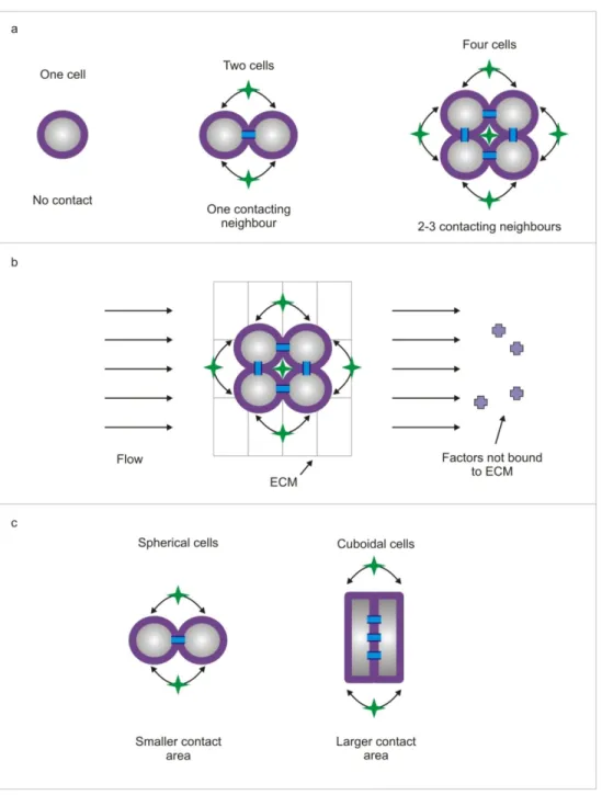
If one specifically wants to modulate only the contact signal, one could then imagine placing these clusters in a perfusion bioreactor, such that factors produced by the cells are swept away downstream (Figure 2.1b). However, some fraction of factors will diffuse through the extracellular matrix (ECM), and the flow may not penetrate into the ECM, so it is plausible that some diffusible signaling will persist (unpublished data from our lab).
A second way to modulate contact signaling would be to change the shape of cells (Figure 2.1c). Cuboidal cells could be made to have a larger surface area in contact with another cell as compared to spherical cells, which could result in increased contact interaction. However, changing the shape of cells also leads to changes in cytoskeletal tension68, and it would be difficult to segregate the effects of altered cytoskeletal tension and contact interaction. As a result, we have taken the cluster‐based approach, and used it to study and modulate both juxtacrine and diffusible cell interactions.
2.3 Technologies for making clusters of cells
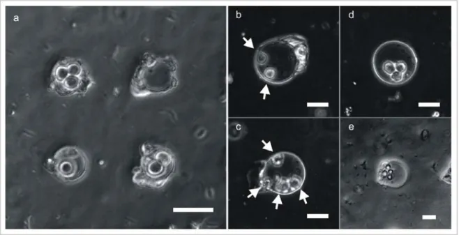
Next, we must find a technology for making clusters of small number of cells. Several technologies are available, in principle. Perhaps the simplest option would be to make wells with a diameter of ~30‐50 µm in a substrate, and trap cells in these wells. If the wells are seeded randomly, we would expect to obtain clusters containing different numbers of cells. Such an approach does, in fact, work (Figure 2.2a). However, when working with adherent cells, the cells in the resulting clusters have no room to attach (spread) and grow. A potential way to fix this would be to make larger wells, but now one cannot guarantee that cells will end up in contact with one other (Figure 2.2b‐c), which is what we desire (above section).
Figure 2.1 a. By making clusters of cells, one can modulate the amount of contact interaction experienced by a cell. A single cell
does not experience any contact interaction. In a cluster of two cells, each cell has one neighbor; in a cluster of 4 cells, each cells is in contact with two neighboring cells. But the making of clusters also results in an increase in the local cell density, and a corresponding increase in the local concentration of diffusible factors. b. By placing the clusters under flow, it is possible to eliminate/reduce some types of diffusible interaction; the fluid flow will carry away factors unbound to the extracellular matrix (ECM) downstream. c. Another way to modulate the contact interaction between cells is to change the shape of cells such that they have varying amounts of their surface in contact with another cell. 21
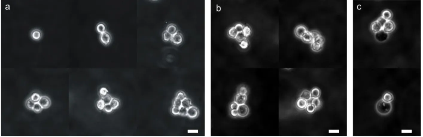
Figure 2.2 a. Clusters of small numbers of cells can be made by seeding cells onto a substrate containing wells with a diameter
of 30 µm. b, c. When cells are seeded into 60 µm (diameter) wells, they sometimes do not form clusters. The white arrows indicate the location of cells. d, e. When mESCs are seeded into 60 µm wells however, most often they do form clusters. Scale bars represent 30 µm in all images.
In practice, for mESCs, which is the cell type we have investigated in detail, we later found that in >90% of cases clusters did form even in the larger wells (Figure 2.2d‐e). We hypothesize that this is due to the random motion of cells within the well, combined with the fact that when these cells are brought into contact with one other, they stick to each other. Some evidence for the “stickiness” of mESCs is provided in chapter 3. However, we do not know whether such spontaneous clustering would occur on other cell types. Similar arguments apply for clusters that could be generated using surface patterning69 instead of microwells.
To avoid the issues mentioned above we decided to use electrical traps to create clusters of cells. If the traps are ~30‐50 µm wide, they would allow cluster creation as shown in Figure 2.2a. However, importantly, one can subsequently turn the fields (and traps) off, allowing cells to proliferate, as well as move around. Additionally, it is plausible that electric traps may be strong enough to “trap” clusters of cells, yet weak enough that they don’t prevent the subsequent growth and motility of cells. Still, since one cannot claim that the mechanisms by which electric fields affect cells are completely understood, it is usually advisable to turn off the electric fields after the clusters have been created.
There are primarily three kinds of electrical traps used to pattern cells, they are traps based on electrophoresis, positive dielectrophoresis (pDEP), and negative dielectrophoresis (nDEP). Electrophoresis has been used to pattern cells inside physical structures70, 71, but is generally considered to be unsuitable because it leads to the generation of radicals at the electrodes (due to the electrolysis of water), which can damage cells. While pDEP has been used by several groups to pattern cells72, as well as create cell clusters73, 74, it requires cells to be in a medium with low conductivity to guarantee that the cells are more polarizable than the media. In contrast, nDEP can be performed with cells
suspended in commonly used cell‐culture media. This presents a significant advantage because it allows one to use the traditional media formulations optimized for cell culture. This avoids any gross viability issues associated with placing sensitive cells in artificial media75. The physics of DEP is described in greater detail in Appendix 1.
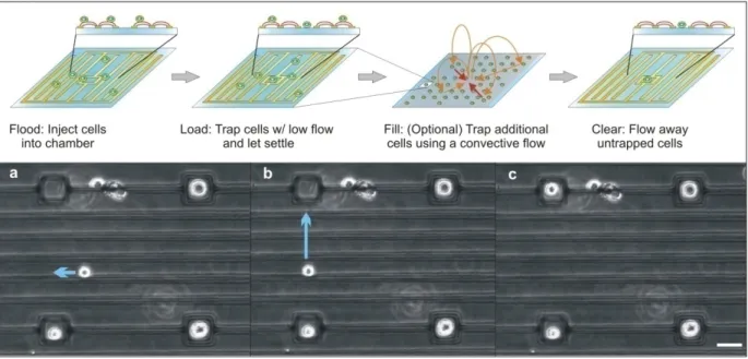
Adam Rosenthal from our lab had previously developed an nDEP trap that could be used to hold micron‐ sized beads at chosen locations on a substrate67, 76. Subsequently, he modified the design, so as to be able to pattern single cells67, 75. This required adding interdigitated electrodes to minimize non‐specific cell adhesion (Figure 2.3) and determining operating parameters that minimized heating and electric field exposure. The resulting structures are termed nDEP microwells to reflect the fact that they present an electrical microwell to incoming cells, only allowing cell‐substrate attachment inside the DEP trap. While the initial device was designed to trap only single cells, we believe that it would be straightforward to increase the well size, so as to be able to create and trap clusters of cells.
Figure 2.3 Image of an nDEP microwell array with HL60 cells trapped at most trap locations (white circles within the black
rectangles). The inset shows an HL60 cell trapped inside a single nDEP microwell. Scale bar represents 100 µm. I made two main contributions to the development of the nDEP microwell array: i. I performed bio‐compatibility testing of the system ii. I developed an alternate loading protocol for loading the system with high efficiency Both the above are described in greater detail below. The results of this work were published in Lab on a Chip75.
2.4 Materials and Methods
ModelingWe extracted electric‐field data from simulations using the commercial field solver Comsol Multiphysics (Comsol, Inc., Burlington, MA). Temperature modeling was also done using Comsol Multiphysics. After using the Electromagnetics module to determine the resistive heating in the flow chamber, we used the Heat Conduction module to determine the temperature at various locations on the device. We determined the transmembrane potential using the maximum value of
23
the electric field experienced by a trapped cell. For cell parameters, we used a membrane capacitance of 1.6 µF/cm2, membrane conductivity of 0.22 S/cm2, cytoplasmic conductivity of 0.75 S/m and cytoplasmic dielectric constant of 75. Medium conductivity is 1.5 S/m and dielectric constant is 80. These values are all taken from Huang et al.
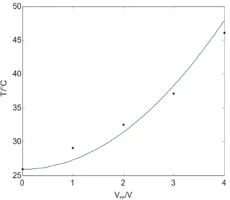
Modeling temperature rises in microsystems is often inaccurate because the exterior boundary conditions (between the device and ambient) are not known or well controlled. In our case, we set the exterior of the device to a fixed temperature boundary condition of room temperature. To determine whether this would result in realistic temperature modeling, we experimentally verified the calculated temperatures. For this we used a slightly different device where we fabricated an on‐ chip resistor specifically for measuring temperature (using the temperature coefficient of resistivity of Au). These experiments were performed by Adam Rosenthal. As seen in Figure 2.4, the simulated and measured temperatures are comparable, validating our modeling approach. Thus, we believe that our thermal simulations are adequate design tools for avoiding thermal stresses.
Figure 2.4 Validation of temperature modeling: Modeled temperatures (solid line) and measured temperatures (dots) versus
voltage.
Electrode traps
We designed the DEP microwells as square electrodes with an inner square side length of 25 µm, and two other line electrodes spaced 10 µm away. All electrode widths were 10 µm. The microwells were in a 10 x 10 square array, with a trap‐to‐trap distance of 200 µm. In‐between the DEP microwells we placed 3 other electrodes spaced by 30 µm each. These electrodes set up an interdigitated electrode array‐like configuration (Figure 2.3).
We fabricated the electrodes by patterning gold onto Pyrex wafers. We cleaned wafers for 10 min in a Piranha solution (3:1 H2SO4:H2O2), blew them dry with N2, and then dehydrated them for 30 min at 225oC. We then used the image‐reversal photoresist Hoechst AZ‐5214 (Celanese, Somerville, NJ)
and photolithography to define the electrode patterns. Finally, we evaporated 10 nm of titanium and 200 nm of gold onto the slides followed by resist dissolution and metal liftoff in acetone.
Flow chamber and packaging
We created the flow chambers using a silicone gasket (19 x 6 x 0.5 mm; Grace Bio‐Labs, Inc., Bend, OR). A 4‐5 mm thick sheet of baked PDMS (Sylgard 184, Dow Corning, Midland, MI) was used to form the roof of the chamber. We cored two holes in the PDMS sheet using syringe needles (0.065’’ outer diameter; Hamilton, Reno, Nevada). We then inserted PEEK tubing (Upchurch Scientific, Oak Harbor, WA) through these holes to provide fluidic access to the chamber, and applied epoxy to hold it in place. Finally, we clamped the chamber top and gasket to the bottom electrode slide using two binder clips for easy assembling and disassembling. The chamber was clamped at an angle to the electrode array to allow for loading with a lower density of cells (Figure 2.7). Wires were electrically connected to the electrodes using conductive epoxy (Circuit Specialists, Mesa, AZ).
Fluidics
We connected the two inputs of a four‐way valve (V‐101D, Upchurch Scientific, Oak Harbor, WA) to a 3‐mL syringe filled with cell suspension and a 5‐mL syringe filled with media. The 5‐mL syringe was controlled using a syringe pump (KD Scientific 210C, Holliston, MA). One output on the four‐way valve was connected to the flow chamber using PET tubing (.03’ I.D., 0.048’ O.D.; Becton Dickinson and Co., Sparks, MD), and the other was connected to waste.
Cell culture
We cultured cells in 20 cm2 dishes (Nunc, Rochester, NY). GFP‐expressing HeLa cells (a generous gift from Dr. Sangeeta Bhatia) were maintained in Dulbecco's Modified Eagle Medium (DMEM; Gibco, Grand Island, NY) supplemented with 10% fetal calf serum, 100 µg mL–1 penicillin, and 100 µg mL–1 streptomycin and incubated in 7.5% CO2 at 37 °C. We passaged them at preconfluency no more
than 25 times. HL60 cells (ATCC, Manassas, VA) were cultured in RPMI medium with additions as above. Prior to patterning experiments, we released cells into suspension and concentrated them to 106 cells/ml. HL60 cells were fluorescently labeled with chloromethylfluorescein diacetate (CMFDA; Molecular Probes, Eugene, OR; 20 µM, 30 minutes) for identification at (ex/em) 492/517 nm wavelengths.
Imaging
Images were taken on a Zeiss Axiovert 200 microscope (Carl Zeiss MicroImaging, Inc., Thornwood, NY) using a Spot RT Color camera (Diagnostic Instruments, Inc., Sterling Heights, MI). For fluorescence imaging we used a X‐Cite 120 illumination system (Exfo Life Sciences, Ontario).
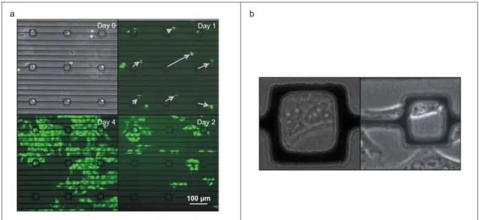
Proliferation experiments
To demonstrate that cells patterned in our device are able to proliferate, we first patterned GFP‐ expressing HeLa cells in our device. After allowing the cells to attach for 30 minutes, we disconnected the patterning fluidics described above and attached a media containing syringe to
25
the fluidic input port of the device. Then we gently placed the device in a dish containing DMEM. The DMEM served to increase the local humidity in the dish and greatly reduce the rate of evaporation of media from the device. We subsequently fed the cells once a day using the attached media‐containing syringe. Electrical excitation Sine wave excitation at 10 MHz was generated by an Agilent 33250A signal generator (Agilent, Palo Alto, CA). The signal was measured using a digital oscilloscope (Tektronix TDS 2024, Beaverton, OR).
2.5 Results and Discussion
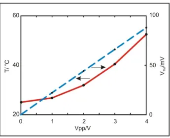
2.5.1 Biocompatibility of the nDEP Microwell ArrayBiocompatibility is one of the basic requirements of any device meant for use with cells. Three mechanisms have been described by which alternating electric fields can affect normal cell physiology77, namely, (i) Joule heating of the cell suspension, (ii) induction of large transmembrane potentials ultimately leading to electro‐poration of the plasma membrane78 or the membranes of intracellular organelles79, and finally, (iii) generation of reactive oxygen species80, 81.
While detailed investigations of the stress response induced by DEP forces and electric fields have been performed by others in our lab81, I have focused on the nDEP microwell architecture in particular, and on modeling, and aspects of gross physiology such as attachment and proliferation. We designed the nDEP microwell array to operate well beneath the damage thresholds associated with cell heating and membrane loading. We excluded any investigation of damage to intracellular structures since the fields responsible for these effects have been reported to be two orders of magnitude higher than the fields in our device79. To avoid any harmful effects due to Joule heating, we wanted to avoid temperature excursions above physiological levels (37oC for mammalian cells). We simulated the temperatures experienced by cells at various voltages (Figure 2.5). Simulations included conductive heat transfer and neglected convection, which is typically minimal in these systems (Pe<1). We see that the temperature scales with the square of the applied voltage, as expected. Using a typical room temperature of 25 oC, we can avoid thermally stressing the cells by keeping the applied voltage to ≤ 2.5 V. 26