For permissions, please e-mail: journals.permissions@oup.com
Novel NLRP3/cryopyrin mutations and
pro-inflammatory cytokine profiles in Behçet’s
syndrome patients
Şahru Yüksel
1, Elif Eren
1, Gülen Hatemi
2, Ali Can Sahillioğlu
1, Yetiş Gültekin
1, Duygu Demiröz
1,
Cezmi Akdiş
3, İzzet Fresko
2and
Nesrin Özören
11Molecular Biology and Genetics Department, Apoptosis and Cancer Immunology Laboratory (AKiL), Bogazici University,
Bebek-Istanbul 34342, Turkey
2Department of Internal Medicine, Division of Rheumatology, Cerrahpasa Faculty of Medicine, Istanbul University,
Aksaray-Istanbul 34300, Turkey
3Swiss Institute of Allergy and Asthma Research, University of Zurich, Davos 7270, Switzerland Correspondence to: N. Özören; E-mail: nesrin.ozoren@boun.edu.tr
Received 31 August 2012, accepted 15 August 2013
Abstract
Behçet’s syndrome (BS) is a systemic inflammatory disorder with unknown etiology. Features of both innate and adaptive immunity have been claimed in the pathogenesis of BS. To test the possible dysregulation of the NLRP3/cryopyrin (Nod-like receptor with a pyrin domain 3) inflammasome, as a result of mutation(s), we performed single-strand conformation polymorphism analyses and/ or sequencing of all the coding regions and intron–exon boundaries of NLRP3/cryopyrin and ASC (apoptosis-associated speck-like protein containing CARD) genes from Turkish BS patients and healthy controls. At the same time, we determined pro-inflammatory cytokine secretion profiles of peripheral blood cells in response to LPS treatment using ELISA. BS patients with vascular involvement showed significantly increased levels of TNF-α release at 2-, 4- and 8-h post-treatment and significantly increased IL-1β levels were detected at 2 h (P = 0.005) and 4 h (P = 0.025) (n = 10). We identified four mutations in the NLRP3/cryopyrin gene, V200M (n = 3/104) and T195M (n = 1/104), in BS patients but none in control samples. No mutations were detected in the ASC gene. The effect of these NLRP3/cryopyrin mutants on ASC speck assembly and IL-1β secretion was tested and the V200M mutant was shown to induce IL-1β secretion. Thus, it is likely that certain mutations in NLRP3/cryopyrin in combination with yet unknown other factors may contribute to the pro-inflammatory cytokine profiles in BS patients.
Keywords: ASC, autoinflammation, IL-1β, NLR, TNF-α Introduction
Behçet’s syndrome (BS) is a systemic inflammatory disease with manifestations including recurrent oral and genital ulcera-tions, and vasculitis involving the skin, mucosa, joints, eyes, veins, arteries, nervous and gastrointestinal systems. While some groups included BS among autoimmune disorders, the absence of specific antibodies and the episodic nature of the clinical findings led some authors to include BS among auto-inflammatory diseases (1). A recent review claimed that both innate and adaptive immune responses play substantial parts in the pathogenesis of the disease (2). Elevated levels of acute phase proteins and recurrent inflammatory attacks are asso-ciated with increased secretion of pro-inflammatory cytokines including IL-1β, TNF-α and IL-6. Both in the active and inactive BS patients, increased pro-inflammatory cytokine secretion and superoxide generation have been reported (3, 4).
Inflammasomes are multiprotein complexes controlling IL-1β secretion. The NLRP3/cryopyrin (Nod-like receptor with a pyrin domain 3) inflammasome includes ASC (apop-tosis-associated speck-like protein containing CARD) (TMS1/ PyCard) and procaspase-1. Interestingly, it has been shown that oligomers of ASC can form specks, presumably pyropto-some complexes that can activate caspase-1 in response to pro-inflammatory stimuli (5).
IL-1β is a significant pro-inflammatory cytokine contrib-uting to the establishment of the symptoms in autoinflam-matory diseases and the NLRP3/cryopyrin inflammasome is the major regulator of caspase-1 activation leading to the cleavage of pro-IL-1β and the secretion of the mature cytokine (6). We hypothesized that IL-1β may be the earli-est cytokine to be secreted; initiating the pro-inflammatory Head1=Head2=Head1=Head1_AfterHead2
Head2=Head3=Head2=Head2_AfterHead3
doi:10.1093/intimm/dxt046
state in BS attacks and that mutations in inflammasome com-ponents might contribute to de-regulated IL-1β secretion.
In order to establish the general cytokine secretion profiles of newly diagnosed and untreated BS patients, we performed a time-dependent release study of IL-1β, TNF-α and IL-6 for the first time. Furthermore, we analysed for the first time the cytokine release profiles of BS patients falling into the mucocu-taneous, vascular and uveitis/ocular involvement subtypes. Simultaneously, all of the coding regions and intron–exon boundaries of the inflammasome components, NLRP3/cryopy-rin and ASC, were screened for the presence of mutations.
Methods
Patients and controls for peripheral blood stimulation and ELISA study
Newly diagnosed BS patients, who fulfilled International Study Group (ISG) criteria published in The Lancet (7) and had active disease, were included in the peripheral blood stimu-lation and ELISA study. These patients were not using any medications at the time of blood collection and had not used immunosuppressives in the past. This group was defined as the functional study cohort. There were 51 patients (34 men, 17 women, mean age 31.7 ± 6.5) in this group. Among these, 29 had mucocutaneous, 12 had eye and 10 had vas-cular involvement. One of the patients with eye disease also had neurological disease. All of the patients with eye involve-ment had active uveitis with a drop of at least two lines on the Snellen chart in visual acuity. Patients in the vascular group had deep vein thrombosis and/or pulmonary artery aneu-rysms. Mucocutaneous patients had only skin and mucosa lesions.
Thirty-seven healthy controls (22 men, 15 women, mean age 29.1 ± 9.2) were also included. Both the controls and patients in this group were also included in the genetic screen.
Patients and controls for genetic study
A total of 50 patients who fulfilled ISG criteria and 50 controls (30 men, 20 women, mean age 27.4 ± 11) were screened for mutations in all nine coding exons of NLRP3/cryopyrin and three exons of ASC genes. Only for the exon 3 of the NLRP3/ cryopyrin, which is a hot spot for mutations in several autoin-flammatory disorders, we increased the number of patients to 104 (72 men, 32 women, mean age 34.3 ± 7.1).
Among these, 43 had mucocutaneous, 39 had eye, 6 had neurological and 19 had vascular involvement. Two of these patients had both eye and vascular involvement and one had both neurological and eye involvement.
This study was approved by the Bogazici University Institutional Review Board for Research with Human Subjects. Informed consent was obtained from all patients and control subjects.
Cell culture and ELISA
Blood samples were collected into EDTA-Vacutainer tubes (BD, Plymouth, UK) and within 2 h diluted 1:3 with prewarmed RPMI supplemented with 10% fetal bovine serum (FBS), 2 mM glutamine and antibiotics (Gibco, Grand Island, NY, USA).
The diluted blood was incubated in 48-well tissue culture plates (0.5 ml per well) (TPP, Trasadinge, Switzerland) with 100-ng ml−1 ultra-pure LPS (Escherichia coli 0111:B4; Alexis
Biochemicals, San Diego, CA, USA), at 37°C in a CO2 incu-bator (5%) for the indicated time points. Untreated cells were collected at the same time and tested at the 24-h time point. At the end of incubation, cells were transferred to microfuge tubes and centrifuged (Spectrafuge 16M Microcentrifuge; Labnet, Woodbridge, NJ, USA) at 3000 r.p.m. for 3 min and supernatants were aliquoted and frozen for determination of cytokine secretion. IL-1β, TNF-α and IL-6 levels in the super-natants were measured by a commercially available ELISA kit (R&D Biosystems, Minneapolis, MN, USA) according to the manufacturer’s instructions. Standard curves of known concentrations of recombinant human cytokines were used to calculate the cytokine concentration (pg ml−1) in each
indi-vidual assay.
Genomic DNA isolation and screening of mutations/ polymorphisms
A modified version of salting-out method was used for the extraction of genomic DNA from whole blood samples. All coding regions including exon–intron boundaries of NLRP3/ cryopyrin (11 fragments), pyrin (two PCR reactions for exon 2 and exon 10) and ASC (three fragments) were amplified by PCR using specific primers. Primer pairs are available upon request. Sequencing was done in MGH DNA Core Facility, Boston, MA, USA; Macrogen Inc., Seoul, South Korea and Iontek Inc., Istanbul, Turkey.
Statistical analysis
SPSS version 18 (IBM Corporation, Armonk, NY, USA) was used for all statistical analyses. Data were given as mean and standard deviation. Statistical analysis for IL-1β, TNF-α and IL-6 in untreated cells or those at, 2 h, 4 h, 8 h and 24 h was ana-lysed by independent samples t-test (two tailed) and analysis of variance for comparison of the means in patient and con-trols. Each treatment was performed in triplicates. The results were considered significant when the P value was <0.05.
Site-directed mutagenesis
NLRP3/cryopyrin V200M mutation was generated by PCR ampli-fication of pcDNA3-NLRP3/cryopyrin wild-type (WT)-FLAG (a gift from Gabriel Nunez, University of Michigan, Ann Arbor, MI, USA) with two sets of primers: F1 (5′-AAGAGATGAGCCGAAGT GGG-3′)/R1 (5′-CTTAATGGGACTCATGGGGCTCTCACACG-3′) and F2 (5′-CGTGTGAGAGCCCCATGAGTCCCATTAAG-3′)/R2 (5′-CAAACTGGAAAGGAAGAAGACG-3′). R1 and F2 primers were complementary to each other and contained the under-lined mutant nucleotides. Resulting PCR products were mixed in the same amounts after gel purification and amplified with F1/R2 primers to obtain a final product containing the mutations. This fragment and pcDNA3-WT NLRP3/cryopyrin-FLAG vector were digested by using PmlI and SacII restriction enzymes, ligated using T4 DNA ligase and transformed into competent E. coli TOP 10. Positive colonies for the mutation were selected by col-ony PCR and confirmed by sequencing. All restriction enzymes and T4 DNA ligase were purchased from New England Biolabs (Ipswich, MA, USA).
A similar protocol was applied for the generation of T195M mutant. In this case, F1 (5′-AAGAGATGAGCCGAAGTGGG-3′)/ R1 (5′-GGGGCTCTCACACATCTTGGTCTTGC-3′) and F2 ( 5 ′- G C A A G A C C A A G AT G T G T G A G A G C C C C - 3 ′) / R 2 (5′-CAAACTGGAAAGGAAGAAGACG-3′) primer sets were used. The final product was digested with PmlI and SacII enzymes and re-ligated into pcDNA3-NLRP3/cryopyrin-FLAG vector. All primers were purchased from MGH Harvard DNA Core Facility.
Generation of pLenti-Ef1a-EGFP-ASC
The ASC gene cDNA was cloned from the pcDNA3-ASC plas-mid (a gift from Prof. Gabriel Nunez, University of Michigan) into the pEGFP-C3 (Clontech, Mountain View, CA, USA) vec-tor by restriction digestion with HindIII-EcoRI enzymes. The resulting enhanced green fluorescent protein (EGFP)-ASC fragment was cloned into the NheI-EcoR I double-digested pLenti-Ef1a-AKIL vector, which has a polylinker suitable for cloning from pcDNA3 and Clontech vector series (including
NheI, HindIII, XbaI, EcoRI, NotI, EcoRV, NdeI restriction sites). Lentivirus production
HEK293FT cells in 10-cm plates (TPP) were transfected with 4-μg pLenti-Ef1a-EGFP-ASC, packaging vectors pCMVdel-taR8.74 and pMD2.G (contains vesicular stomatitis virus-gly-coprotein) by the calcium phosphate method. Two days after transfection, supernatants containing virus were filtered with 0.45-μm filters, aliquoted and frozen at −80°C.
Transduction and stable line production
HEK293FT cells were plated 1 day before transduction. Virus-containing vials were thawed at room temperature and mixed with polybrene (Sigma-Aldrich, Taufkirchen, Germany) at a final concentration of 4 μg ml−1. Media on cells were removed
and virus-containing medium was added dropwise; 5 h later, the cells’ media were changed. One week later, the trans-duced HEK293FT cells were serially diluted and transferred into 96-well plates (TPP) so that one cell or less could fall into each well. One of the subclones, EGFP-ASC_C8 was chosen for its high of GFP+/GFP− cell ratio (90%) and low frequency
of spontaneous speck formation (<1%).
Transfection of EGFP-ASC stable lines with FLAG-NLRP3/ cryopyrin variants, quantification of ASC specks and IL-1β secretion
EGFP-ASC HEK293FT stable lines were plated on 6-well plates (TPP). On the next day, cells were transfected with either 20 ng of pcDNA3-FLAG-NLRP3/cryopyrin _WT, _ V200M, _T195M or _R260W or empty pcDNA3, with 50-ng caspase-1 and 500-ng IL-1β vectors. Transfection was per-formed in triplicate wells for each construct and the experi-ment was replicated three times.
Five images were captured from each well at random locations with an inverted fluorescence microscope (n = 15) (Zeiss Observer Z1; Carl Zeiss Microscopy, Thornwood, NY, USA). All images used for speck counting were taken under identical conditions. Cells were imaged with a ×10 objective. EGFP-ASC specks in the entire visual field were counted.
The two-tailed unpaired Student’s t-test was employed to compare speck counts.
Additionally, IL-1β release by these cultured cells was measured in triplicates by ELISA using a commercially avail-able IL-1β ELISA kit (R&D Biosystems).
EGFP-ASC HEK293FT cells were cultured for 24 h after transfection in DMEM without FBS. Then the supernatants were collected and the proteins were precipitated with trichlo-roacetic acid by incubation on ice for 10 min, followed by a wash with acetone. Then the protein precipitates were dis-solved in 50 μl 0.5% NP40 lysis buffer and loaded on SDS-PAGE. The proteins were transferred to a polyvinylidene difluoride membrane in wet transfer for 2 h at 100 V in 4°C. Following 1 h blocking with 5% BSA, the membrane was incubated overnight at 4°C with 1:1000 anti-IL-1β antibody (Cell Signaling; 2022) in 5% BSA. Then the membrane was incubated for 2 h at room temperature in 1:2000 anti-rabbit secondary antibody (Cell Signaling; 7074) in 5% BSA and visualized using Lumi-Light Western Blotting Substrate (Roche Applied Sciences; 12015200001).
Results
Elevated levels of TNF-α secretion detected in LPS-stimulated BS patients’ cells
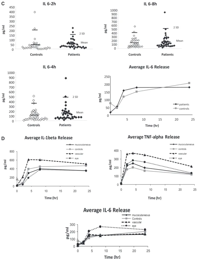
Peripheral blood cells collected from BS patients, with active lesions who had not received any treatment at the time of blood collection (functional study group), and healthy con-trols were stimulated with LPS to establish time-dependent IL-1β, TNF-α and IL-6 secretion profiles (Fig. 1A–C). Mean IL-1β, TNF-α and IL-6 levels secreted at 2, 4, 8 and 24 h after stimulation with LPS, among vascular, mucocutaneous and ocular BS patients, and healthy controls are given in Table 1
and shown in Fig. 1D.
BS patients with vascular involvement had significantly higher IL-1β levels at 2 and 4 h after LPS stimulation (P < 0.001, F3df = 6.59 and P = 0.039, F3df = 2.92, respectively) and higher TNF-α levels at 2 and 8 h after LPS stimulation (P = 0.033, F3df = 3.05 and P = 0.022, F3df = 3.38, respectively), compared with mucocutaneous BS, ocular BS and healthy control groups. The levels among the mucocutaneous BS, ocular BS and healthy control groups were similar for these time points (P = 0.58, F2df = 0.58; P = 0.166, F2df = 1.84; P = 0.383, F2df = 0.956; P = 0.214, F2df = 1.28, respectively).
When the total group of BS patients was compared with healthy controls, IL-1β and IL-6 levels were not different at any of the time points after LPS stimulation. The BS group had a trend for higher TNF-α secretion at 2 h (251 ± 173 SD versus 178 ± 147 SD, P = 0.048), 4 h (280 ± 181 SD versus 200 ± 164, P = 0.046) and 8 h (240 ± 178 versus 169 ± 146,
P = 0.056) after stimulation. The values for mean IL-1β,
TNF-α and IL-6 levels secreted at 2, 4, 8 and 24 h after stimulation with LPS for the total BS group versus healthy controls are given in Table 2.
The IL-6 levels in the ocular subgroup were higher than the other groups, starting from 4 h post-treatment. However, the dif-ference did not reach statistical significance, probably due to the limited number of patients in this subgroup (n = 7) (Fig. 1D).
The IL-1β and IL-6 levels in a few untreated samples were higher in some healthy controls, but the cytokine levels in
untreated samples were not compared since these samples were studied 24 h after blood collection, which might have resulted in spontaneous cytokine release.
Novel mutations identified in the NLRP3/cryopyrin gene
Screening of all nine exons including intron–exon boundaries of the NLRP3/cryopyrin gene resulted in the identification of a
A
C
D
Fig. 1. IL-1β, TNF-α and IL-6 secretion from LPS-stimulated blood cells in BS patients and healthy controls. (A) TNF-α (patients n = 51, controls
n = 34), (B) IL-1β (patients n = 51, controls n = 36) and (C) IL-6 (patients n = 37, controls n = 30) release from BS patients’ and healthy con-trols’ whole blood samples after LPS treatment (100 ng) analysed at 2-, 4-, 8- and 24-h post-stimulation. Untreated control at 0 h was collected together with 24-h treatment samples. First bar shows mean and the second mean ± 2 SD. (D) Average TNF-α (vascular n = 10, eye n = 12 and mucocutaneous n = 29), IL-1β (vascular n = 10, eye n = 12 and mucocutaneous n = 29) and IL-6 (vascular n = 6, eye n = 7 and mucocutaneous n = 21) release in BS patient subgroups compared with healthy controls.
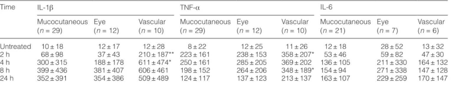
novel T195M mutation in one and V200M (V198M) mutation in three patients among 104 BS patients, whereas no mutations were found in 49 healthy controls (Fig. 2A). Patients bearing the V200M mutation are from either the eye, mucocutaneous or neurological subgroups, whereas the patient with T195M belongs to the mucocutaneous subgroup.
In order to check whether the V200M mutation exists in healthy family members, we sequenced the available genetic material from two brothers and a grandmother in one BS patient carrying the mutation and who had eye involvement. The V200M mutation was not detected in the tested healthy individuals (Fig. 2B).
Only one of the V200M carrier patients in the mucocuta-neaous subgroup was suitable for the functional cytokine study, since the other two were already being treated for the disease. In this patient, IL-1β secretion levels were higher than the average values for the whole functional study group at 2 and 4 h and only at 2 h compared with the mucocutane-ous subgroup (Table 3).
Interestingly, the Q705K (rs35829419) (HapMap-CEU %11, Hapmap-JPT %0) polymorphism in the NLRP3/cryopyrin gene appears with an increased frequency in both Turkish controls (n = 16/116) and BS patients (n = 13/109). Other polymor-phisms especially in the NOD/NACHT/NAD domain of NLRP3/ cryopyrin were found as a result of sequencing (Table 4). When analysed statistically, the frequency differences in patients and controls do not appear to be significant. However, the H260H (1526 C→ T) polymorphism that we find in 1/49 controls has not
been reported to date. In our study, we did not find changes in any of the three exons and intron–exon boundaries of the ASC in 50 BS patients and 50 unrelated controls tested.
Besides NLRP3/cryopyrin exons, we wanted to see whether mutations in the inflammasome regulatory protein PYRIN may be involved in BS, so we sequenced a subset of patients with high IL-1β secretion levels and/or carrying a mutation in NLRP3/cryopyrin for MEFV exon 10 (n = 26). Five patients have the M694V mutation (19.2%), previously linked with familial Mediterranean fever, one has M680I and another one has A744S. The V200M patient with neurological disease also has a MEFV M694V mutation.
Mutant NLRP3/cryopyrin’s effect on ASC speck formation
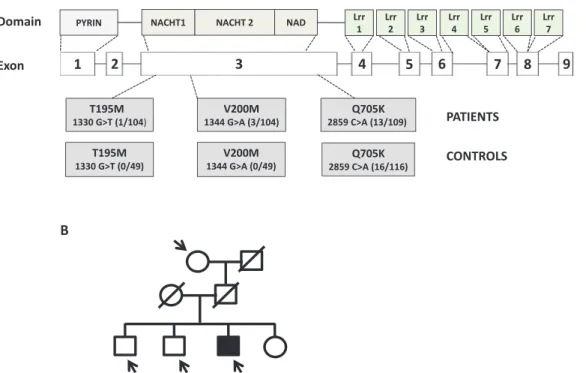
We studied whether V200M and T195M NLRP3/cryopyrin over-expressing cells are more prone to ASC speck forma-tion in comparison with WT NLRP3/cryopyrin-expressing cells. EGFP-ASC stable HEK293FT cells were transfected with equal amount of FLAG-NLRP3/cryopyrin_WT, _V200M, _T195M or _R260W plasmids and the number of speck for-mation in five representative visual fields from three different wells were counted under identical settings of the inverted fluorescent microscope (Fig. 3A and B).
As expected, we observed that transfection of 20 ng of WT NLRP3/cryopyrin induced speck formation (average number of specks per visual field= 17.6 ± 5.6) significantly, compared with control empty pcDNA3-transfected cells (9.2 ± 2.4,
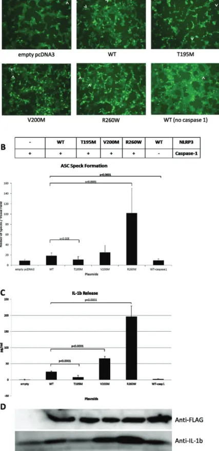
Table 1. Cytokine secretion profiles of BS patient subgroups
Time IL-1β TNF-α IL-6
Mucocutaneous
(n = 29) Eye (n = 12) Vascular (n = 10) Mucocutaneous (n = 29) (n = 12)Eye Vascular (n = 10) Mucocutaneous (n = 21) Eye (n = 7) Vascular (n = 6) Untreated 10 ± 18 12 ± 17 12 ± 28 8 ± 22 12 ± 25 11 ± 26 12 ± 18 28 ± 52 13 ± 32 2 h 68 ± 98 37 ± 43 210 ± 187** 223 ± 161 238 ± 153 358 ± 207* 53 ± 46 59 ± 82 47 ± 30 4 h 300 ± 315 188 ± 178 611 ± 474* 250 ± 161 285 ± 205 369 ± 202 136 ± 105 211 ± 330 164 ± 132 8 h 399 ± 436 381 ± 407 606 ± 461 198 ± 152 264 ± 206 348 ± 189* 154 ± 94 271 ± 338 147 ± 128 24 h 352 ± 391 354 ± 386 509 ± 489 124 ± 117 137 ± 123 213 ± 137 163 ± 107 229 ± 259 170 ± 147 IL-1β, TNF-α and IL-6 secretion from LPS-stimulated blood cells in subgroups of BS patients. Results are given as means in pg ml−1 ± standard
deviations.
*P value <0.05, **P value <0.001.
Table 2. Cytokine secretion profiles of BS patients and healthy controls
Time IL-1β TNF-α IL-6
Patients (n = 51) Controls (n = 36) Patients (n = 51) Controls (n = 34) Patients (n = 37) Controls (n = 30)
Untreated 11 ± 20 154 ± 226 10 ± 23 18 ± 22 16 ± 31 74 ± 104
2 h 88 ± 125 60 ± 72 252 ± 173* 178 ± 148 53 ± 49 45 ± 78
4 h 333 ± 350 393 ± 363 303 ± 25* 200 ± 164 156 ± 166 119 ± 123
8 h 432 ± 433 449 ± 449 241 ± 179* 162 ± 142 175 ± 174 149 ± 133
24 h 384 ± 407 470 ± 469 145 ± 125 117 ± 118 177 ± 152 204 ± 206
IL-1β, TNF-α and IL-6 secretion from LPS-stimulated blood cells in BS patients and healthy controls. Results are given as means in pg ml−1 ±
standard deviations. *P value <0.05.
P < 0.0001). On the other hand, transfection of R260W mutant
NLRP3/cryopyrin, previously reported to cause spontaneous IL-1β release in over-expression studies, increased speck numbers significantly (109.4 ± 48.3 specks) compared with WT NLRP3/cryopyrin-transfected cells. Besides, T195M mutant NLRP3/cryopyrin-transfected cells have a slight but significant decrease in the number of specks (12 ± 6) in com-parison with WT NLRP3/cryopyrin-transfected cells, whereas V200M mutant NLRP3/cryopyrin-transfected cells did not form a significantly higher number of specks (25.3 ± 13.4) (Fig. 3B). NLRP3/cryopyrin expression was verified for each condition (Fig. 3D). These data suggest that, unlike R260W, the T195M and V200M mutant NLRP3/cryopyrin proteins do not have a pronounced positive effect on ASC oligomerization.
Mutant NLRP3/cryopyrin’s effect on IL-1β secretion
Any effect of NLRP3/cryopyrin mutants on IL-1β secretion was tested in the EGFP-ASC HEK293FT stable line (Fig. 3C). As
expected, transfection of the R260W mutant increased IL-1β secretion significantly (196 ± 33.6 pg ml−1, P < 0.0001)
com-pared with WT (24.77 ± 2.57 pg ml−1; 0.81 ± 1.2 pg ml−1 for empty
plasmid-transfected cells, Fig. 3C). V200M also increased IL-1β release (65.93 ± 6.9 pg ml−1, P < 0.0001), whereas the
T195M mutation decreased IL-1β release (8.01 ± 6.21 pg ml−1, P < 0.0001) in comparison with WT NLRP3/cryopyrin.
Secreted mature IL-1β was also tested by western blot-ting (Fig. 3D) and results correlate with ELISA results. Furthermore, the IL-1β secretion pattern is in line with the ASC speck formation.
Discussion
Our study shows that BS patients with vascular involvement have higher IL-1β and TNF-α levels after stimulation with LPS, compared with BS patients with eye involvement, BS patients with only mucocutaneous involvement and healthy controls (Fig. 1D). Cytokine levels of IL-1β and TNF-α in BS patients with eye and mucocutaneous involvement were similar to healthy controls (Fig. 1D). These findings reflect the clinical observation that BS patients with vascular involvement are the ones who have fever and other constitutional symptoms, especially during active disease. Moreover, elevation of acute phase reactants is also a feature of vascular BS, which alarms the clinician to this type of involvement.
There are several findings indicating the presence of dif-ferent pathogenetic mechanisms underlying difdif-ferent types of organ involvement in BS. The presence of various symp-tom clusters in BS, such as the association of deep vein thrombosis and superficial thrombophlebitis or the associa-tion of acne, arthritis and enthesopathy, raises the possibility
Fig. 2. Novel T195M mutation and other genetic changes were identified in the NLRP3/cryopyrin gene. (A) Genetic mutations and
polymor-phism found in BS patients and healthy controls. (B) Genetic analysis of a patient family with V200M mutation.
Table 3. Cytokine secretion profile of BS patient with V200M
mutation IL-1β TNF-α IL-6 Untreated 27.2 ± 3.9 6.9 ± 0.5 18.1 ± 0.6 2 h 289.2 ± 21.8 342.7 ± 20.9 30.8 ± 2.2 4 h 343.6 ± 24.8 274.8 ± 39.9 50.9 ± 5.9 8 h 303.0 ± 48.8 194.4 ± 3.4 55.3 ± 10.4 24 h 341.4 ± 39.8 74.0 ± 27.1 48.7 ± 5.6 IL-1β, TNF-α and IL-6 secretion from LPS-stimulated blood cells in a patient with V200M mutation. Results are given as means of tripli-cates readings in pg ml−1 ± standard deviations.
Tab
le 4.
Single nucleotide polymorphisms in NLRP3/cr
yopyrin exon 3 for BS patients and healthy contr
ols Nucleotide changes Patients Contr ols Total population Homozygote WT Heter ozygote Homozygote variant Total variant Homozygote WT Heter ozygote Homozygote variant Total variant Homozygote WT Heter ozygote Homozygote variant Total variant p.T221T 82.7% (86/104) 16.35% (17/104) 0.96% (1/104) 17.3% (18/104) 83.67% (41/49) 16.32% (8/49) 0% (0/49) 16.32% (8/49) 83% (127/153) 11.11% (17/153) 0.65% (1/153) 16.99% (26/153) 1409 ACC>ACT rs7525979 p.A244A 18.27% (19/104) 52.89% (55/104) 28.85% (30/104) 81.73% (85/104) 12.25% (6/49) 69.39% (34/49) 18.37% (9/49) 91.84% (45/49) 16.34% (25/153) 58.17% (89/153) 25.49% (39/153) 83.66% (128/153) 1478 GCG>GCA rs3806268 p.H260H 100% (104/104) 0% (0/104) 0% (0/104) 0% (0/104) 97.96% (48/49) 2.04% (1/49) 0% (0/49) 2.04% (1/49) 99.35% (152/153) 0.65% (1/153) 0% (0/153) 0.65% (1/153) 1526 CAC>CA T New p.R262R 0.96% (1/104) 3.85% (4/104) 95.19% (99/104) 99.04% (103/104) 0% (0/49) 6.12% (3/49) 93.88% (46/49) 100% (49/49) 0.96% (1/153) 4.58% (7/153) 94.77% (145/153) 95.42% (146/153) 1532 CGA>CGG rs4925543 p.D312D 97.22% (105/108) 2.78% (3/108) 0% (0/108) 2.78% (3/108) 98% (49/50) 2% (1/50) 0% (0/50) 2% (1/50) 97.47% (154/158) 2.53% (4/158) 0% (0/158) 2.53% (4/158) 1682 GAC>GA T rs143840033 p.P342P 98.15% (106/108) 1.85% (2/108) 0% (0/108) 1.85% (2/108) 98% (49/50) 2% (1/50) 0% (0/50) 2% (1/50) 98.1% (155/158) 0% (0/158) 1.9% (3/158) 1.90% (3/158) 1772 CCC>CCT rs41311573 p.L346L 98.15% (106/108) 1.85% (2/108) 0% (0/108) 1.85% (2/108) 96% (48/50) 4% (2/50) 0% (0/50) 4% (2/50) 97.47% (154/158) 2.53% (4/158) 0% (07158) 2.53% (4/158) 1784 CTG>CT A rs144616577 p.L413L 98.15% (106/108) 1.85% (2/108) 0% (0/108) 1.85% (2/108) 100% (50/50) 0% (0/50) 0% (0/50) 0% (0/50) 98.73% (156/158) 1.27% (2/158) 0% (0/158) 1.27% (2/158) 1983 CTG>TTG rs148478875 p.S436S 64.82% (70/108) 26.85% (29/108) 8.33% (9/108) 35.19% (38/108) 58% (29/50) 40% (20/50) 2% (1/50) 42% (21/50) 62.66% (99/158) 31% (49/158) 6.33% (10/158) 37.34% (59/158) 2054 TCC>TCT rs34298354 p.H465H 99.07% (107/108) 0.93% (1/108) 0% (0/108) 0.93% (1/108) 100% (50/50) 0% (0/50) 0% (0/50) 0% (0/50) 99.37% (157/158) 0.63% (1/158) 0% (0/158) 0.63% (1/158) 2141 CAC>CA T
rs111400208 Single-nucleotide polymorphisms in BS patients and healthy contr
Fig. 3. The effects of NLRP3/cryopyrin mutations on inflammasome activation. (A) ASC speck formations. Inverted fluorescent microscopy images of stable ASC-EGFP-expressing HEK293FT cells transfected with empty vector, FLAG-NLRP3/cryopyrin_WT, _V200M, _T195M, _R260W and _WT in the absence of caspase-1. ASC specks are shown with a white arrow. The images represent cells transfected with 20 ng of the corresponding NLRP3/cryopyrin, 50-ng caspase-1 and 500-ng IL-1β plasmids. (B) Quantification of ASC speck numbers. (C) Measurement of IL-1β release by ELISA. (D) Western blot of secreted mature IL-1β protein.
that the pathogenesis of BS involves more than one biologic pathway (8). This is also supported by evidence from drug trials. Colchicine, which is an effective drug in skin lesions and arthritis of BS patients, has no efficacy in eye or vascu-lar involvement. The difference in cytokine secretion profiles among different types of BS patients in this study is more evi-dence for the presence of different mechanisms in the patho-genesis of BS.
There are controversial studies regarding the serum IL-1β levels (9–11) in BS patients some of which report normal and some elevated levels. Mege et al. (3) found increased TNF-α, IL-1β, IL-6 and IL-8 levels in culture conditions after stimu-lation with LPS 16 h post-treatment, whereas Slobodin et al. (12) reported that TNF-α production after LPS stimulation was similar in BS patients and healthy controls. The advantage of our study is that we included only patients with active lesions at the time of blood collection, who had not received any medication including colchicine. Moreover, analyzing clinical subgroups separately gave us the opportunity to evaluate the role of each cytokine in each type of involvement.
Another novelty brought by this paper is that we studied the kinetics of cytokine secretion in BS patients and com-pared this to healthy controls. This is important because not only the level of inflammation, but also the timing is thought to be important in BS. A typical example for this is the pathergy phenomenon, where the reaction is clinically indistinguishable from healthy controls in the beginning. However, after 24 h, the reaction begins to subside in healthy controls, in contrast to BS patients where the inflammation is prolonged up to 72 h. This was also shown in the tissue level by skin biopsies of the needle prick sites at 0, 8 and 48 h after the prick. Cellular infil-tration patterns, cytokine and chemokine levels were similar in BS patients and healthy controls for the first 8 h. However, after 8 h, increased influx of mature dendritic cells, monocytes and lymphocytes as well as increased cytokine and chemokine levels continued only among BS patients (13). Our study shows that whole blood cytokine levels after LPS stimulation are higher, especially for the vascular BS patients, but the cytokine release kinetics of IL-1β, TNF-α and IL-6 are parallel in BS patients and healthy controls (Fig. 1D).
A randomized controlled trial and several open studies showed that TNF-α antagonists are effective in the man-agement of BS. Especially, patients with eye, neurologic and gastrointestinal involvement, resistant to other drugs have benefitted from TNF-α antagonists. Case series show-ing efficacy of IL-1 antagonists have also been published. The elevated IL-6 levels that we have observed in ocular BS patients may be important with this respect. Elevated IL-6 lev-els were previously shown in cerebrospinal fluid and serum of BS patients with neurologic involvement, another subgroup of patients that may sometimes be associated with ocular involvement. These findings point out to the possibility of using IL-6 antagonists in such patients (Fig. 1D).
IL-1β is a key cytokine in autoinflammatory disorders. Dominant mutations in the inflammasome component NLRP3/ cryopyrin have been documented to cause higher levels of IL-1β secretion. For instance, the R260W mutant NLRP3/ cryopyrin has been reported to cause spontaneous IL-1β release by monocytes isolated from Muckle–Wells syndrome (MWS) patients (14).
We have found three patients carrying the V200M NLRP3/ cryopyrin mutation and a novel T195M NLRP3/cryopyrin mutation all in the heterozygous state (Fig. 2A). V200 and T195 are conserved among primates and higher mam-mals such as cows, but not in mice and rats. For one of the V200M mutation carriers, genetic material from healthy family members was tested negative for this mutation. V200M has been reported in one study to have an incidence of 1/97 for BS patients (15). None of these previous reports examines the functional importance of the V200M for inflammasome assembly. Interestingly, a clinical case report about a British family with three MWS patients carrying the heterozygous V200M mutation gave a complete response to Anakinra treat-ment (16). This finding supports the fact that V200M might be important in the disease pathogenesis. However, the low incidence in our study and presence of the mutation in the healthy Caucasian populations with an allele frequency of 0.0074 (17) make it impossible to draw strong conclusions about its significance in BS pathogenesis.
We investigated the functional consequence of the V200M and T195M mutant NLRP3/cryopyrin on inflammasome assembly and activation via the speck formation and IL-1β secretion assays. The V200M mutant tends to show a posi-tive effect on the formation of specks, albeit not significantly (Fig. 3A and B), but the significant effect of this mutant on IL-1β secretion was confirmed by ELISA and western blot analyses in a more solid and quantitative manner (Fig. 3C
and D). Although this increase in IL-1β secretion was not as pronounced as R260W’s effect, still V200M appears to be a moderately activating mutation.
Recently, the MEFV M694V mutation has been shown to be associated with BS in the Turkish population (18). PYRIN has been previously shown to activate NLRP3/cryopyrin inflam-masome and combination of PYRIN and NLRP3/cryopyrin mutant proteins did not interfere with inflammasome activa-tion (19). The presence of both NLRP3/cryopyrin V200M and MEFV M694V mutations in one of the four patients carrying NLRP3/cryopyrin mutations suggests that mutations of inflam-masome components and/or regulators, such as NLRP3/cry-opyrin and PYRIN, may have a combinatorial effect on the cytokine release profiles of BS patients. It was shown very recently that treatment of BS patients suffering from acute intraocular inflammation with XOMA 052, a humanized anti-IL-1β antibody, resulted in reduction of inflammation in all seven patients, spotlighting the importance of the IL-1β path-way in BS pathogenesis (20).
Overall, our data show that NLRP3/cryopyrin and/or ASC by themselves are unlikely to be responsible for BS patho-genesis or to account for the observed a cytokine profiles. It is clear that BS is a complex disorder with possible involve-ment of multiple genes and pathways (such as MEFV, HLA-B51, MICA, etc.) along with a contribution from environmental factors. Mutations in proteins, such as TLR4 and TLR2, known to regulate the inflammation process via the NF-κB pathway, controlling TNF-α and pro-IL-1β levels, were recently shown to be associated with BS (18, 21). Besides Toll-like recep-tors, other complexes controlling caspase-1 and caspase-5 activation such as AIM-2, NLRC4/Ipaf and other NLRP inflam-masomes are still plausible mutation targets that may predis-pose individuals to develop BS.
Funding
Bogazici University Research Fund (BAP-06HB103 to Ş.Y.); European Molecular Biology Organization Strategic Development and Integration Grant (SDIG-1468 to N.Ö.); Turkish Education Foundation (TEV) Graduate Degree Scholarship to Y.G. and D.D.
Acknowledgements
The authors thank Assoc. Prof. Andrzej Furman (Bogazici University, Institute of Environmental Sciences, Istanbul, Turkey) for help with the statistical analysis of cytokine profiles. We would also like to express our gratitude to Prof. Gabriel Nunez (University of Michigan, Ann Arbor, MI, USA) for the various plasmids, as mentioned in the text. HEK293FT cell was a kind gift from Dr Maria Soengas (CNIO, Madrid, Spain). Lentiviral packaging vectors were provided by Prof. Karl Deisseroth (Stanford University, Stanford, CA, USA). We would like to thank Prof. Hasan Yazıcı (Istanbul University Cerrahpaşa Medical School, Istanbul-Turkey) for a critical review of the manuscript. Conflict of interest: Authors declare no conflicts of interest.
References
1 Gül, A. 2005. Behçet’s disease as an autoinflammatory disorder. Curr. Drug Targets Inflamm. Allergy 4:81.
2 Direskeneli, H. 2006. Autoimmunity vs autoinflammation in Behcet’s disease: do we oversimplify a complex disorder? Rheumatology (Oxford) 45:1461.
3 Mege, J. L., Dilsen, N., Sanguedolce, V. et al. 1993. Overproduction of monocyte derived tumor necrosis factor alpha, interleukin (IL) 6, IL-8 and increased neutrophil superoxide generation in Behçet’s disease. A comparative study with familial Mediterranean fever and healthy subjects. J. Rheumatol. 20:1544.
4 Düzgün, N., Ayaşlioğlu, E., Tutkak, H. and Aydintuğ, O. T. 2005. Cytokine inhibitors: soluble tumor necrosis factor receptor 1 and interleukin-1 receptor antagonist in Behçet’s disease. Rheumatol. Int. 25:1.
5 Fernandes-Alnemri, T. and Alnemri, E. S. 2008. Assembly, purifi-cation, and assay of the activity of the ASC pyroptosome. Methods Enzymol. 442:251.
6 Martinon, F., Mayor, A. and Tschopp, J. 2009. The inflammas-omes: guardians of the body. Annu. Rev. Immunol. 27:229. 7 International Study Group for Behçet’s Disease. 1990. Criteria for
diagnosis of Behçet’s disease Lancet 5:1078.
8 Hatemi, G., Fresko, I., Tascilar, K. and Yazici, H. 2008. Increased enthesopathy among Behçet’s syndrome patients with acne
and arthritis: an ultrasonography study. Arthritis Rheum. 58:1539.
9 Sayinalp, N., Ozcebe, O. I., Ozdemir, O., Haznedaroğlu, I. C., Dündar, S. and Kirazli, S. 1996. Cytokines in Behçet’s disease. J. Rheumatol. 23:321.
10 Memisoglu, H., Aksu, H. S. Z., Erken, E. and Göcük, M. 1991. Serum interleukin-1 level in Behçet’s disease. In Oduffy, J. D. and Kökmen, E., eds, Behçet’s Disease, p. 387–391. Dekker, New York, NY.
11 Treudler, R., Orfanos, C. E. and Zouboulis, C. C. 1997. Increased serum levels of interleukin I-beta and basic fibroblast growth fac-tor in severe Adamandiades-Behçet’s disease. In Hamzaou, M., ed., Behçet’s Disease, p. 33–37. Adhoua, Tunis.
12 Slobodin, G., Toukan, Y., Rosner, I. et al. 2007. LPS-stimulated pro-duction of TNF-alpha by peripheral blood monocytes in patients with Behcet’s disease. Clin. Rheumatol. 26:764.
13 Tunc, R., Keyman, E., Melikoglu, M., Fresko, I. and Yazici, H. 2002. Target organ associations in Turkish patients with Behçet’s disease: a cross sectional study by exploratory factor analysis. J. Rheumatol. 29:2393.
14 Agostini, L., Martinon, F., Burns, K., McDermott, M. F., Hawkins, P. N. and Tschopp, J. 2004. NALP3 forms an IL-1beta-processing inflammasome with increased activity in Muckle-Wells autoinflam-matory disorder. Immunity 20:319.
15 Koné-Paut, I., Sanchez, E., Le Quellec, A., Manna, R. and Touitou, I. 2007. Autoinflammatory gene mutations in Behçet’s disease. Ann. Rheum. Dis. 66:832.
16 Hawkins, P. N., Lachmann, H. J., Aganna, E. and McDermott, M. F. 2004. Spectrum of clinical features in Muckle-Wells syndrome and response to anakinra. Arthritis Rheum. 50:607.
17 Aksentijevich, I., Putnam, C. D., Remmers, E. F. et al. 2007. The clinical continuum of cryopyrinopathies: novel CIAS1 mutations in North American patients and a new cryopyrin model. Arthritis Rheum. 56:1273.
18 Kirino, Y., Zhou, Q., Ishigatsubo, Y. et al. 2013. Targeted rese-quencing implicates the familial Mediterranean fever gene MEFV and the toll-like receptor 4 gene TLR4 in Behçet disease. Proc. Natl Acad. Sci. USA 110:8134.
19 Yu, J. W., Wu, J., Zhang, Z. et al. 2006. Cryopyrin and pyrin acti-vate caspase-1, but not NF-kappaB, via ASC oligomerization. Cell Death Differ. 13:236.
20 Gül, A., Tugal-Tutkun, I., Dinarello, C. A. et al. 2012. Interleukin-1β-regulating antibody XOMA 052 (gevokizumab) in the treatment of acute exacerbations of resistant uveitis of Behcet’s disease: an open-label pilot study. Ann. Rheum. Dis. 71:563.
21 Liang, L., Tan, X., Zhou, Q. et al. 2013. IL-1β triggered by pep-tidoglycan and lipopolysaccharide through TLR2/4 and ROS-NLRP3 inflammasome-dependent pathways is involved in ocular Behçet’s disease. Invest. Ophthalmol. Vis. Sci. 54:402.