Extensibility and rheology of collenchyma I. Creep
relaxation and viscoelasticity of young and senescent cells
Michel Jaccard and Paul-Emile Pilet
Institute of Plant Biology and Physiology of the University, 1005 Lausanne, PI. de la Riponne, Switzerland
(Received July 28, 1975)
The extensibility of collenchyma cells was methodologically analyzed; extensibility (elasticity and plasticity) and rheology of this tissue are discussed. Experimental conditions are described. Rheological data for young and senescent cells were ob-tained; they were found to be not Boltzmannian. The permanent strain is not viscous, and is larger for young cells than older ones.
Previous observations (17, 18) have shown that collenchyma cells are useful for extensibility experiments. The problem of the extensibility of these cells will be considered here methodologically. In order to describe the analyzing technique, the results of creep relaxation will be compared with those related to physical analysis of polymers. First, several general questions have to be discussed.
Clearly, the elongation rate is related both to turgor pressure and deform-ability of the wall (8, 20). O n the other hand, wall extensibility is essential for growing cells (5, 14). I t depends on the metabolism and has been very often associated with protein formation (4, 7). The deformability of the wall can be analyzed also by comparing it with the physical properties of some polymers. The aim of such assays is to discover the physical nature of the wall by analyzing stress relaxation by rheological measurements. This can be easily done by simple tension with a pulled bar.
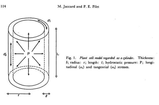
Measuring extensibility and deformability is not an easy task; the stress developed in the cell wall is not a simple tension, but a hydrostatic one. A cellular model was proposed (11) and is presented in Fig. 1. The cell can be thought of as a cylinder (thickness: 8; radius: r ; length: 1) bearing hydrostatic pressure (P). The stress of the inner side is composed of tangential (at) and longitudinal stress (ai). Simple calculation shows that:
CTt=2<TI = P-r/8 ( I )
If the walls were isotropic, cell growth would occur radially. This does not in fact occur because of the transversal orientation of the microfibrils in growing cells. Such microfibrils should act as rings incompassing a rubber tube and preventing radial extension, but not longitudinal extension. Such a phenomenon, called multinet growth (21, 23), is much discussed to day. This leads to neglect of at
with regard to the problem of wall extension. However, some studies have shown 1113
Fig. 1. Plant cell model regarded as a cylinder. Thickness:
S; radius: r; length: /; hydrostatic pressure: P; longi-tudinal {oi) and tangential (at) stresses.
that one effect of at is to cause a longitudinal stress a{ which would be subtracted from <?i (10). In the case of the cell, it is therefore possible, on a first approach, to reduce the effect of turgor pressure (P) to a simple tension, the force (F) value of which would be:
and to regard the extensibility analysis as a rheological measure of simple tension. The situation of cells from epicotyl, hypocotyl and root is of greater complexity (11, 13, 20). For instance, the edge cells partly bear the turgor pressure of the central cells, which are under compression, whereas the peripheral cells are under tension (epidermis, collocytes). The present model can be used for calculating the force to be applied to such organs, but is too simple for several reasons: in-homogeneity of the organs and tissues, misappreciation of the distribution of ten-sions and the radial evolution of the physical characteristics of the walls. Con-sequently, it is quite difficult to totally accept this model. For young tissues, applying longitudinal tension causes the appearance of tensions which are not longitudinal for not cylindrical cells. Further, the joint effect of the turgor pre-ssures of two cells under compression produces both longitudinal and transversal tensions due to the mechanical characteristics of the wall. Those tensions might induce migration of matrix polymers inside the wall (21). For short time analysis, such a reaction may be disregarded.
Collenchyma cells, with primary walls (18, 19, 22), seem to be a choice material for such an experiment. Like the epidermis, it is subject to a force which is chiefly longitudinal (13, 22). This clearly indicates that the collenchyma has to be characterized by a typical passive growth (11). The shape of the cells is close to a cylinder and the wall thickness considerably reduces the effect of undesirable tensions.
limit of any organ (Apium petioles for instance), make it easy to prepare. Reduc-tion of the hydrostatic pressure to a sole longitudinal force is thus more valuable. Therefore, rheological determinations will be preferred to extensibility tests, which however will not be disregarded.
Materials and methods
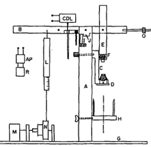
Apium graveolens plants were cultivated in a greenhouse. Senescent petioles from the exterior of the plant as well as young petioles from the heart were used. From each of them, bundles of collocytes were taken at random {1,2). As soon as the collocytes were obtained, the epidermis was partly dissociated by fine scrap-ing. This method enabled the bundles of collocytes to be isolated by 3 cm without notably perturbing the tissue; the parenchyma cells, still attached to the bundle, only participate negligibly in the extensibility analysis. The fiber were then placed for 10 min in a 0.2 M citrate buffer (pH 7.0). Such incubation allows formation of an osmotic equilibrium and also stops cell contraction owing to its state of tension which has often been observed. The bundle was then fixed between the pincers of a creep apparatus (Fig. 2) and at once incubated in the same buffer solution.
• Properly set, the apparatus enables creep determinations and stress strain curves. A beam (B) mounted on ball-bearings lies on its bearer (A), with equalized pincers fixed to it. The inferior pincers (D) are mobile (C) and are allowed to vary the initial length of the fiber. A tube (H) vertically moving enables the fiber to be plunged during the time of the test. A counterbalance insures the equilibrium of the whole. The applying force is produced by rolling up a thread around a pillow block (N) and fixing it to a dynamometer (L). A reversible synchronous motor (M) sets the pillow block in motion, producing stress strain curves at variable speed. The displacement of the beam is recorded by a trans-ducer Tesa (CDL) held by (I). The arm of the transtrans-ducer lies on one of the two adjustable trays (K). The signal is amplified (AD) and transmitted to a Metrohm recorder (R). A blocking system (J) can stop the beam when desired. This apparatus permits varying the force applied from 5 to 500 g and analysis
Fig. 2. Creep apparatus employed for measuring extension of isolated fibers of collenckyma cells. (For
of displacements from 6// to 2.2 mm with 5% precision. In the present study, the sudden appearance of a stress is attained by hanging a weight to the beam (B); this method can only be used for relaxation periods of over one second.
Traction analysis requires a material in the shape of an L length bar, cylindri-cal and prismatic. The collocytes bundles approximately correspond to this model. To be stretched lengthwise, they must be held by two pincers which locally produce an inhomogeneous field of stress. The L length must be important enough so that the effect of that field will be neglected.
In the case of collenchyma cells and the forces used, the usual length (L: 25 mm) is suitable. Assuming that the surface (S) of the fiber is constant, the applied value of the force F is usually transformed to stress:
a=F/S (III) For extensibility measurements, the value of the force F used to stretch collen-chyma cells can be estimated by the method first proposed by Lockhardt (9).
Assuming that S=area of the fiber == 0.16 mm2 and P=turgor pressure=7 atm,
using formula II, F =: 11.2 g. However, this estimation does not seem satisfying: collenchyma cells, as edge cells, partly bear the turgor pressure of the central cells of the petiole and are subject to passive growth (11). Probably the value of the real force is greater that the calculated force above. For rheological measure-ments, the surface S should be the wall surface, but such a notion is not devoid of ambiguity, considering the possible variation of degrees in hydrophility. Dividing F by the weight of dried walk also raises some difficulties (5). It seemed simpler to use F in g in the results. The samples had to be submitted to a variable series of force values. Pulling on the ends of the fiber with a force F causes the length L to increase by J L which is usually transformed into the strain:
(IV) Each result refers to the analysis of at least 10 fibers and was repeated twice.
Results and discussion
With simple tension tests, there are two possibilities: analyzing a=o(t) in regard to the variation of e, and analyzing £=s(t) in regard to the variation of cr. The latter was used. It corresponds better to the simulation of hydrostatic pressure. The creep function g(t) is:
g(t)=e(t)/a ( V )
when cr is applied at zero time and held constant. Usually, when measuring e(t), an instantaneous strain e\ and a time-dependant strain Et, can be obtained (Fig. 3). After time T, the stress can be quickly removed. The strain decreases;
there is recovery. The recovery also contains an instantaneous strain eri and
a time-dependent strain £d(t—T). Other deformations are:
total strain: £tot=ei+£t(T) ( V I )
Fig. 3. Creep curves for collenchyma cells. T h e
strain (%) is expressed in relation to time (sec). A: senescent cells; B: young cells; C: applied force. JEO5-tn OJO 10 O8 l o . 4 0.2 0.0 _——' c 1 • r ElID E, (BJ E. And 20 10 60 TIME (SEC) 80
permanent strain: limEr(t)=£P
elastic strain: £ei=£tot—er>
(VIII) ( I X ) Using (VI), (VII), (VIII), we find ep=EP(T). In our determinations T = 3 6
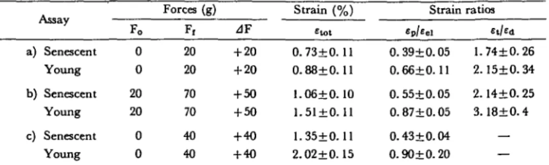
sec was used; it causes stabilization of the creep curves. e(t) for F=20 g is given in Fig. 3, with the detailed values presented in Table la.
When a force of 40 g is used, etot is greater for young fibers than for older ones (Table lc). The thickening of the cell walls caff partly explain these results which seem to be generalized for any force (17). Note that the relation £p/eei
is different, even if <=tot, with a force applied on senescent and young tissues, does not differ significantly (Table la). Conditions of linear viscoelasticity being satisfied, the response can be reduced to the finite or infinite sum of two ideal elements placed in series or parallel: the Hooke solid (a=E-e) which is symbolized by a spring, and the viscous liquid (cr=ij-de/dt) represented by a dashpot. The
Table 1 Stress-strain relations for collenchyma cells (senescent or young)
a) b) c) Assay Senescent Young Senescent Young Senescent Young Fo 0 0 20 20 0 0 Forces (g) F, 20 20 70 70 40 40 zlF + 20 + 20 + 50 + 50 + 4 0 + 40 Strain (%) Etot 0.73+0. 11 0.88±0.11 1.06+0.10 1.51±0.11 1.35±0.11 2.02±0.15 Strain Ep/Eel 0.39+0.05 0.66+0.11 0.55+0.05 0.87+0.05 0.43+0.04 0.90+0.20 ratios E,/Ed 1.74+0.26 2.15±0.34 2.14+0.25 3.18±0.4 — — Each result±standard error is the average of 10 values.
Table 2 Data showing collenchyma cells (senescent or young) as non-linear material
Collenchyma te° (%) sel (20 g)/eei (50 g)
Senescent 0. 6±0. 11 0. 75+0. 15 Young 0. 7±0. 28 0.64±0.09 Each result±standard error is the average of 10 values.
" ^e=£tot (50g, T)|c lio.-stot (50g, T ) | «p.
For further explanations, see Table 1 and page 1118.
general body is the generalized Voigt model {15). Its relaxation analogy is the generalized Maxwell-Viechert model (25). The interest of such analogy is to distinguish sp, eei, et, £r> £d, graphically, which is much better than using an
equation, and to link the various parameters more efficiently to the verifications of the physical theories.
Linear viscoelasticity condition is next. If g(t) has die same value, whatever tension a we use, the body is called viscoelastic linear or Boltzmannian. This is the generalization of the linearity of stress and strain (26). For a given tem-perature, a Boltzmannian body is generally permanent from die molecular point of view (75). The stress or strain does not interfere to modify the structure (crys-talline, amorphous). But the walk of the collenchyma cells are not of the Boltzmannian type. Three facts easily prove this:
1) A force F=20g is used. With this result and in regard to the Boltzmannian hypothesis, the theoretical value of etot(T) for 50 g is calculated and compared with experimental one. Table 2 shows that:
J« = «tot(T, 50 g)|ealo.-«tot(T, 50 g)|exp.>0
This clearly indicates the linear viscoelasticity condition is not satisfied. However: £ei(20g)/£ei(50g)>0.4
2) As pretreatment, F = 7 0 g is applied then removed and er(t) is allowed to
be stabilized. A force of 20 g is re-applied and compared with a bulk of fiber
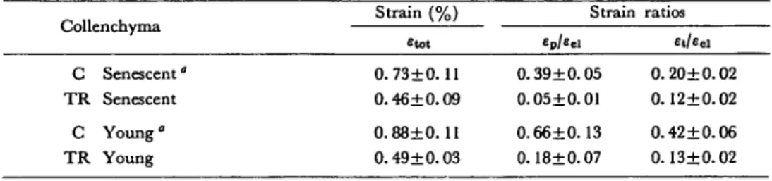
Table 3 Effect of a preliminary extension on the stress strain relations for collenchyma cells (senescent or young)
Strain (%) Strain ratios Collenchyma — etot C Senescent" O.73±O. 11 0.39±0.05 0.20±0.02 TR Senescent 0.46±0.09 0.05±0.01 0.12±0.02 C Young" 0.88±0. 11 0.66±0.13 0.42±0.06 TR Young 0.49±0.03 0.18±0.07 0.13+0.02 Each result±standard error is the average of 10 values.
C: control fibers. Force applied=20 g.
TR: treated fibers. A force of 70 g was applied as pretreatment; after equilibration of deformation, the force was removed and substituted with one of 20 g.
not pretreated: ep decreased with the pretreatment as well as the ratio £t/£ei (Table 3). A permanent strain, which is not of viscous nature (in the Boltzmannian sense) but which depends on the stress applied formerly, exists in et and ei. De-formation of senescent or young tissues contains no linear elements of the plastic type with strain hardening.
3) However, when the strain is small, almost all material may behave with linear viscoelasticity. The limit of linear viscoelasticity should be interesting to observe. When F=20 g, the permanent strain is also not of viscous nature (in the Boltzman-nian sense), then the stabilization of the creep curve could not be defined. If the body is viscoelastically linear et(T)=ea(T). This proposition is not verified: £t(T)/ea(T)>l (see Table 1). With forces F around 10 g, the last proposition is also not verified (unpublished results). This could indicate that the limit of linear viscoelasticity is smaller than the force which stretches collenchyma cells in the petiole. However, this proposition may be taken with circumspection, the experimental conditions in the creep system being not the same as in the petiole.
Conclusion
The results presented here indicate the importance of ep for distinguishing
the senescent from young tissues. The permanent strain is not viscous. Col-lenchyma cell walls have no linear components. A similar result was obtained and reported by Cleland with Avena coleoptiles (6). Therefore, the elongation of the collenchyma cells is not only due to exogenous stresses, but also requires a wall loosening as in the case of the epidermis (21). Similar deformation obtained for young and senescent cells suggests some identical physical properties of their walls.
To attempt simplifying the analysis of living material, we assumed that hydro-static pressure could be regarded as turgor pressure, which could be (11) identified with longitudinal tension. But clearly the cell walls cannot be considered a physical body, the evolution of which was only related to the variation of some external parameters such as stress and temperature. The complexity of the wall structure, its hydrophility and the ionic interactions inside the wall necessitate insertion of some parameters related to the thermodynamics of diluted solutions and ir-reversible processes. The rheology of the walls, therefore, depends on the phy-siological situation of the tissue (5, 12, 16). Consequently, it is absolutely necessary to inhibit all metabolic processes when measuring; and the tissues were immersed in boiling ethanol then submitted to pronase or congealed (2). But such treat-ments destroy the gel of the walls and the studies undertaken later might not give a real picture of the rheological characteristics dependent on weak links and van der Waal forces (3) of the walls. In the case of collenchyma cells, because the walls are strongly hydrophilous, such treatments are not acceptable. If the analysis is rapid, it seems to be clear that the characteristic changes would be extremely small in comparison with the destruction caused by the above treatments.
References
( 1) Burstrom, H. G., I. Uhrstrom and R. Wurscher: Growth, turgor, water potential and Young's modulus in pea intemodes. Physiol. Plant. 20: 213-231 (1967).
(2) Cleland, R.: Extensibility of isolated cell walls: measurement and changes during cell elon-gation. Planta 74: 197-209 (1967).
( 3) Cleland, R.: A dual role of turgor pressure in auxin induced cell elongation in Aetna coleoptile. ibid. 77: 182-191 (1967).
( 4) Cleland, R.: Protein synthesis and wall extensibility in the Avena coleoptile. ibid. 95: 218— 226 (1970).
( 5 ) Cleland, R.: Cell wall extension. Ann. Rev. Plant Physiol. 22: 197-220 (1971).
( 6) Cleland, R.: The mechanical behavior of isolated Avena coleoptile walls subjected to constant stress. Plant Physiol. 47: 805-811 (1971).
( 7 ) Davies, P . J . : Current theories on the mode of action of auxin. Bot. Rev. 39: 139-171(1973). ( 8) Green, P. B., R. O. Erickson and J. Buggy: Metabolic and physical control of cell elongation
rate. Plant Physiol. 47: 423-430 (1971). (9) Jaccard, M. and P. E. Pilet: In preparation.
(10) Kamiya, N., M. Tazawa and T. Takata: The relation of turgor pressure to cell volume in Nitella with special reference to mechanical properties of the cell wall. Protoplasma 57: 501— 521 (1963).
(//) Lockhart, J. A.: Cell extension. In Plant Biochemistry, p. 826-849, Edited by J. Bonner and J. E. Varner. Acad. Press, New York, 1965.
(12) Masuda, Y.: Auxin induced cell expansion in relation to cell wall extensibility. Plant & Cell Physiol. 10: 1-9 (1969).
(13) Morr6, D. J. and J. Bonner: A mechanical analysis of root growth. Physiol. Plant. 18: 635-649 (1965).
(14) Olson, C. A., J . Bonner and D. J. Morre: Force extension analysis of Avena coleoptile cell walls. Planta 66: 127-133 (1965).
(15) Persoz, B.: In Introduction a V'etude de la Rhiologie, p. 34-35, Dunod 1960. (16) Pilet, P. E . : In Les parois cellulaires, p . 101-114, Doin 1971.
(17) Pilet, P. E., M. Jaccard and R. Magliocco: Mesure de l'extensibilite des cellules de Col-lenchyme. C. R. Acad. Sc. 277: 1637-1640 (1973).
(18) Pilet, P. E. and J. Cl. Roland: Growth and extensibility of collenchyma cells. Plant Sc. Utters 2: 203-207 (1974).
(19) Preston, R. D.: In Physical Biology of Plant Cell Walls, p. 312-315, Chapman and Hall Ltd., 1974.
(20) Ray, P. M. and A. W. Ruesink: Osmotic behaviour of oat coleoptile tissue in relation to growth. J. Gen. Physiol. 47: 83-101 (1963).
(21) Roelofsen, P. A.: Ultrastructure of the wall in growing cells and its relation to the direction of growth. Adv. in Bot. Research 2: 69-149 (1965).
(22) Roland, J . Cl.: Organisation de la membrane paraplasmique du Collenchyme. J. de Micro-scopic 5: 323-348 (1966).
(23) Roland, J. Cl. and P. E. Pilet: Implications du plasmalemme et de la paroi dans la croissance des cellules v6getales. Experientia 30: 441-451 (1974).
(24) Yamagata, Y., R. Yamamoto and Y. Masuda: Auxin and hydrogen ion action on light grown pea epicotyl segments II. Effect of hydrogen ions on extension of isolated epidermis. Plant & Cell Physiol. 15: 833-841 (1974).
(25) Yamamoto, R., K. Shinozaki and Y. Masuda: Stress relaxation properties of plant cell walls with special reference to auxin action. Plant & Cell Physiol. 11: 947-956 (1970).