The kidney during hibernation and arousal from hibernation. A natural model of organ preservation during cold ischaemia and reperfusion
9
0
0
Texte intégral
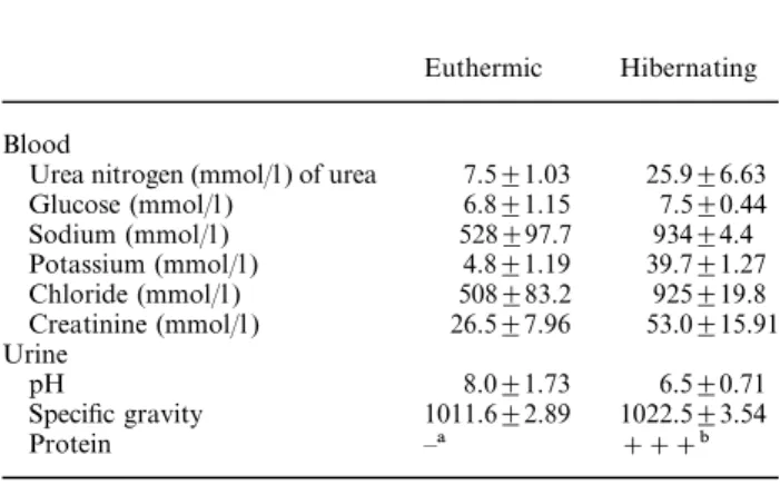
(2) Kidney cortex during hibernation. 1983. Urine samples (500–1000 ml ) were obtained by puncture from the bladder in five dormice (two hibernating, three euthermic). In the case of euthermic dormice, urine spontaneously emitted by animals before sacrifice was pooled with the bladder urine where needed. Mixed blood samples were obtained immediately after decapitation by collecting blood dripping from the trunk. Electrolyte concentrations were measured by an ion-selective electrode technique. Other blood and urine analyses were performed utilizing commercially available kits with an automated multiparametric analyser (BM/Hitachi 917, Boehringer Mannheim, Milan, Italy). Kidneys were rapidly removed from all dormice after sacrifice, cut in small fragments with a razor blade and immersion-fixed for 1.5 h in 2% glutaraldehyde in 0.1 M cacodylate buffer at 4°C, pH 7.4. Post-fixation was done in 1% osmium tetroxide and 1% potassium ferricyanide (final concentrations) in distilled water for 1 h at 4°C. Tissue fragments were then dehydrated through ascending concentrations of acetone and embedded in a mixture of epon and Araldite. Toluidine-blue-stained semithin sections were used for selection of cortical areas of interest and ultrathin sections were obtained on a Reichert Ultracut E ultramicrotome. Ultrathin sections were stained with lead citrate and viewed and photographed in a Zeiss EM 10 electron microscope operated at 60–80 kV.. Results In all the hibernating and arousing dormice the urinary bladder was extremely dilated with urine and both kidneys appeared of normal size in comparison with euthermic individuals. Urine and blood analyses The results of these are shown in Table 1. The plasma concentrations of electrolytes were obviously higher in hibernating than in euthermic dormice. Similarly, the concentrations of creatinine and urea nitrogen were twofold and fourfold respectively, in hibernating individuals. In the urine of hibernating dormice, pH values were lower and specific gravity was higher than in Table 1. Blood and urine analyses in euthermic (n=3) and hibernating (n=2) dormice. Euthermic Blood Urea nitrogen (mmol/l ) of urea Glucose (mmol/l ) Sodium (mmol/l ) Potassium (mmol/l ) Chloride (mmol/l ) Creatinine (mmol/l ) Urine pH Specific gravity Protein Means±SEM. a not detectable. b>300 mg/dl.. 7.5±1.03 6.8±1.15 528±97.7 4.8±1.19 508±83.2 26.5±7.96 8.0±1.73 1011.6±2.89 –a. Hibernating. 25.9±6.63 7.5±0.44 934±4.4 39.7±1.27 925±19.8 53.0±15.91 6.5±0.71 1022.5±3.54 +++b. euthermic. The urine of hibernating dormice was characterized by the appearance of proteinuria, which was not detectable in euthermic individuals. The renal corpuscle (Figures 1–6) In active, euthermic dormice (Figure 1), the renal corpuscle showed the usual structural components of the mammalian kidney. Glomerular vessels contained variable amount of blood and showed a flat, fenestrated endothelium. Pedicels of podocytes contacted the glomerular basal lamina (BL) and were separated by filtration slits. The BL comprised an internal and external lamina rara and a lamina densa in between. The average thickness of BL was 1500 nm. In dormice hibernating at low temperature, the ultrastructure of most renal corpuscles was quite similar to that of euthermic animals (Figure 2). Focally, endothelial cells of glomerular capillaries were enlarged with abundant, electron-lucent perinuclear cytoplasm ( Figure 3). In these corpuscles, podocytes were prominent, due to different degrees of cell swelling. In these cells (Figure 5) the Golgi apparatus was conspicuous, sometimes multiple, and it was surrounded by many small vesicles. Vesicles were also found scattered in the cell. Pedicels of podocytes were regularly spaced along the BL. The BL was continuous and appeared of regular texture and thickness (Figure 6). However, areas of BL wrinkling could be found. Mitochondria were of regular shape and size. The cytoskeleton was obvious and consisted of bundles of filaments and tubules. In renal corpuscles of hibernating dormice the capsular space was usually closed. In arousing dormice (body temperature about 26°C ), capillary loops in a large number of glomeruli were dilated and engorged with blood. Here, endothelial cells were often swollen, with a clear ‘empty’ cytoplasm. The urinary and capsular spaces were well appreciable. Podocytes were often frankly oedematous with a clear cytoplasm and the usual complement of organelles. The ultrastructure of the renal ultrafilter was regular. In many other instances, the ultrastructure of podocytes resembled the euthermic (Figure 4). The renal tubules (Figures 7–10) In active dormice (not shown), proximal tubule cells showed prominent microvilli and an obvious apical endocytic apparatus; the basal labyrinth was well developed and mitochondria had a few electron-dense granules. In the distal convoluted tubule, epithelial cells were provided with short microvilli and prominent endoplasmic reticulum. Dilated cisternae of the endoplasmic reticulum containing floccular material were a regular finding in many of these cells ( Figure 10, inset). In hibernating dormice ( Figure 7), cells of the proximal convoluted tubules were regularly provided with packed, long microvilli occupying the tubular lumen. At the base of microvilli an impressive endocytic apparatus (Figure 7, inset) consisting of coated pits, coated vesicles, apical dense tubules and endosomes was found together with large clear vesicles and dense.
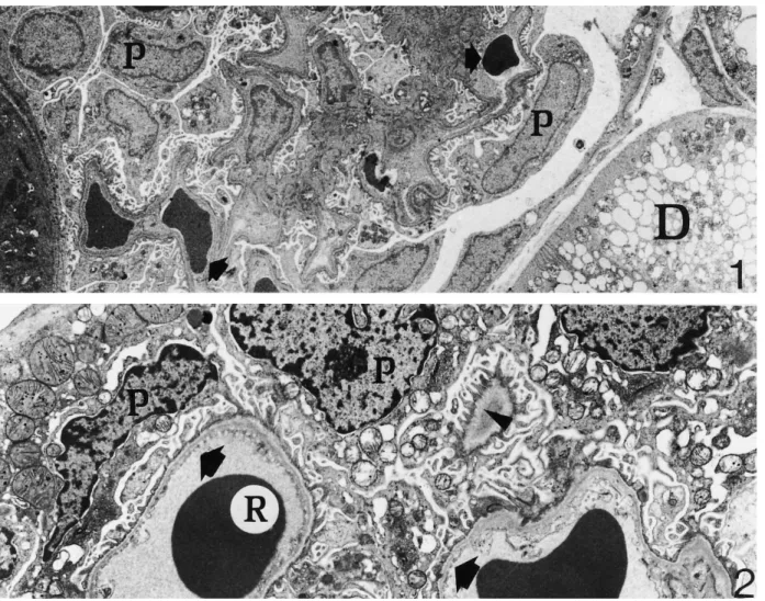
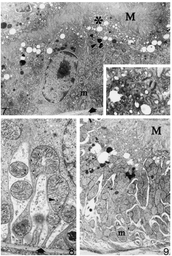
(3) 1984. C. Zancanaro et al.. Fig. 1. Renal corpuscle of an euthermic, active dormouse. Note podocytes (P) and glomerular capillary vessels (arrows). Part of a distal convoluted tubule (D) and a proximal convoluted tubule ( left bottom corner) are also shown ×3000. Fig. 2. Renal corpuscle of a dormouse hibernating at a body temperature of about 10°C. The ultrastructure of podocytes (P) and fenestrated endothelial vessels (arrows) is very well preserved. Arrowhead: regularly spaced pedicels of podocytes reach the basal lamina on a tangentially sectioned capillary vessel. (R) red blood cell×6300.. bodies. A tall membranous labyrinth ( Figure 8) containing many oval to elongated mitochondria was found in the basal portion of the tubular epithelial cell. Mitochondria had homogeneously dense internal matrix with electron-dense granules. Distal convoluted tubule cells ( Figure 10) showed very short microvilli and a modest basal labyrinth. In many cells, the cytoplasm was mainly occupied by cisternae of the endoplasmic reticulum filled with flocculent material. In arousing dormice, epithelial cells of the proximal convoluted tubules (Figure 9) showed a well-developed apical endocytic apparatus albeit less prominent than in hibernating dormice. Mitochondria had shorter cristae, a clearer internal matrix and fewer electron-dense granules. In the distal convoluted tubules the endoplasmic reticulum showed fewer dilated cisternae (not shown).. Discussion Hibernation is a complex condition involving dramatic changes in the function of organ systems and indi-. vidual tissues. During hibernation, body temperature decreases close to ambient temperature, body functions are reduced to a minimum, metabolism is depressed, and animals enter a lethargic state lasting days or weeks (for a recent review of metabolic and biochemical adaptation during dormant state, see Ref. [3]). The mechanisms responsible for mammalian hibernation have not been fully elucidated. Hibernation is a seasonal torpor involving neurophysiological and neuro-endocrine adaptation leading to ‘the coordinated reduction of the activity state of a specific subset of key regulatory enzymes or proteins in the cell and the activities of the processes that they control’ [3]. In general, entry in (and maintenance of ) the depressed metabolic state does not require extensive gene expression, albeit structural changes in nuclear constituent are found in hibernation [4–6 ]. As far as the renal function during hibernation is concerned, it has been shown in some of the smaller hibernators that urine production is much reduced, in association with striking reduction of renal blood flow, reduction or cessation of the glomerular filtration rate [7] and reduction.
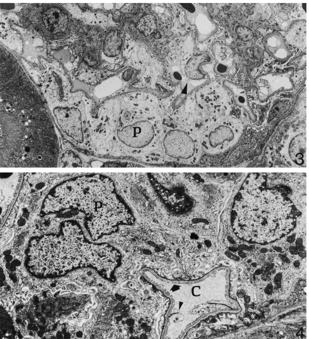
(4) Kidney cortex during hibernation. 1985. Fig. 3. Renal corpuscle of a hibernating dormouse. Some podocytes (P) are oedematous as well as capillary endothelial cells (arrowhead). However, the general ultrastructure of renal corpuscle cells is well-maintained. Bottom left corner: portion of a proximal convoluted tubule ×3000. Fig. 4. Renal corpuscle of an arousing dormouse. Both podocytes (P) and a capillary vessel (C ) show very good ultrastructural preservation. Arrow: fenestrations of endothelial cell. Arrowhead: portion of endothelial cell blebbing in the capillary lumen×6300.. of the free water reabsorption rate (TCH O) [1]. These 2 changes are similar to those induced by hypothermia in non-hibernating mammals [1]. However, renal blood flow can continue during hibernation, albeit greatly reduced [8]. During periodical arousal, hibernating animals show transient increase of body temperature and restore normal blood flow to the kidney, glomerular filtration rate, and urine production [9]. In the present study we investigated the kidney cortex of the dormouse Muscardinus avellanarius during hibernation and arousal from hibernation by means of transmission electron microscopy. Results were compared with. findings in fully active, euthermic individuals. The following points were demonstrated: (i) The ultrastructure of the kidney cortex is well preserved during both hibernation (i.e. a condition where renal blood flow and glomerular filtration rate are permanently reduced ) and arousal from hibernation (i.e. a period of rapidly resuming body temperature and renal blood flow). (ii) The most apparent ultrastructural changes during hibernation take place in glomerular endothelial cells and podocytes. Changes extend to a larger number of such cells during arousal..
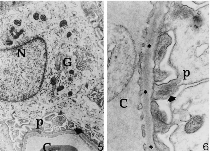
(5) 1986. C. Zancanaro et al.. Fig. 5. Renal corpuscle of a hibernating dormouse. The fine structure of an oedematous podocyte is shown. Note the well-organized Golgi apparatus (G) and pedicels (p). N, nucleus of podocyte; C, capillary vessel; arrow, fenestration of endothelial cell ×11 700. Fig. 6. Renal corpuscle of a hibernating dormouse. At high magnification the renal ultrafilter appears to be morphologically intact. C, glomerular capillary vessel. p, pedicel of podocyte containing packed filaments (arrow). Asterisks: basal lamina ×41 400.. (iii) In the proximal tubule, the apical reabsorbing zone of epithelial cells is activated during hibernation, in association with proteinuria. Measurement of GFR or urine output were not performed in this study; however, the regular finding of urine-filled bladder in hibernating and arousing dormice [see also Ref. 10] strongly suggests persistent, albeit greatly reduced, urine production in the kidney during hibernation. Accordingly, blood analyses ( Table 1) showed marked increase of the plasma concentrations of electrolytes, urea nitrogen and creatinine in hibernating animals. Despite such functional changes, electron microscopy showed that the general organization and fine structure of the renal ultrafilter is preserved during hibernation. Specifically, both the BL and the pedicels/filtration slits complex appear to be essentially unaffected during hibernation. Earlier studies in different species showed, in osmium or formaldehyde/osmium fixed material, that hibernation was associated with BL thickening [11] or irreversible structural and permeability changes [12] similar to those found in proteinuria or protamine sulphate administration [13]. Probably, improved fixation pro-. cedures explain the above discrepancy. A reduction of the endothelial pore number and epithelial filtration slits during hibernation have been consistently reported both qualitatively [11] and quantitatively [14] in different species. This would imply a reduced permeability of the ultrafilter. Due to the limited number of available animals, we did not perform morphometric analyses of our material but, from a qualitative point of view, the density of endothelial pores was found to be similar in hibernating and euthermic dormice. However, any quantitative evaluation of endothelial pores should be considered with caution because of their uneven distribution along the capillary endothelial cell [15]. Qualitatively, a tendency of pedicel number to increase was appreciable in hibernating dormice; this could be an adaptive response to reduced glomerular filtration rate. Podocytes often showed a certain degree of oedema during hibernation, which was more frequent in arousing dormice. This could be related to reduced metabolic activity of these cell, associated with entry of sodium and leakage of potassium; chloride would then enter the cell and an isotonic gain of water occurs. During arousal, podocyte oedema is even more.
(6) Kidney cortex during hibernation. 1987. Fig. 7. Hibernating dormouse. Portion of a proximal convoluted tubule showing a fully preserved ultrastructure as well as an impressive apical endocytic apparatus (asterisk). M, long, packed microvilli in the tubular lumen; m, mitochondria; arrow, basal labyrinth; arrowhead, lysosome-like structures. Inset, detail of the apical endocytic apparatus ×7500, ×75 000. Fig. 8. Hibernating dormouse. Basal labyrinth in a proximal convoluted tubule cell. Arrow, infolding of plasma membrane; arrowhead, electron-dense granule in the inner mitochondrial matrix ×20 000. Fig. 9. Arousing dormouse. Epithelial cells of the proximal convoluted tubule are morphologically well preserved. Note the slightly dilated intercellular space among folds of the basal labyrinth (asterisk). M, long, packed microvilli in the tubular lumen; m, mitochondria containing many electron-dense granules ×6300..
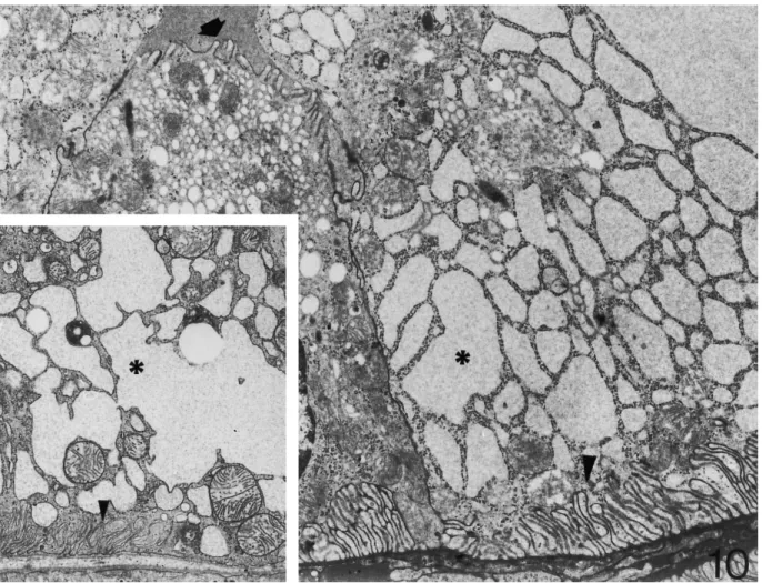
(7) 1988. C. Zancanaro et al.. Fig. 10. Distal convoluted tubule of hibernating dormouse. Many epithelial cells show dilated cisternae of the endoplasmic reticulum (asterisk) and a small basal labyrinth (arrow head). This pattern is quite similar to the euthermic condition (inset). Arrow, short microvilli projecting in the tubular lumen ×8000, ×8000.. appreciable, possibly in association with the increasing blood flow in the presence of still metabolically depressed podocytes. On the basis of reduced arterial pressure and glomerular filtration rate found in hibernation, it could be inferred that morphological changes in proximal tubules of hibernating dormice would be striking. In, for example, experimental ischaemic acute kidney failure early ultrastructural alterations of tubules are loss of microvilli and intense vacuolization [16 ]. Moreover, the kidney tubule is a major site of damage during ischaemia associated with organ storage for transplantation as well as following reperfusion in the host [17] and continuing efforts are made to improve the structural preservation of organs procured for transplantation since functional dependence is placed on the preserved kidney after transplant. Renal tubules in the cortex of hibernating dormice were exposed for days to very low blood flux in deep hypothermia and showed excellent preservation in comparison with euthermic individuals. Interestingly, disruption of microfilaments as well as fragmentation of microtubules occurring in epithelial cells of the proximal tubule. during ischaemia [18,19] did not apparently take place in hibernating dormice. These changes contribute to loss of cell polarity and altered function in experimental renal ischaemia. Instead, tubular cells of hibernating dormice show a fully preserved brush border and apical endocytic apparatus, thereby indicating that they possess a preserved cytoskeleton. Further, the more evident ultrastructural change in proximal tubule cells of hibernating dormice was a well organized, expanded endocytic apparatus. This finding suggests that epithelial cells are able to increase reabsorption during hibernation, possibly in response to increased protein content of the tubular fluid. In accordance with this hypothesis, marked proteinuria was found in hibernating dormice ( Table 1), which was not detectable in euthermic individuals. Increase of protein in the tubular fluid could be a consequence of the slow blood flow in the glomerulus, which would allow in turn a longer filtration time. The reported changes in proximal tubule morphology during hibernation are generally consistent with findings in bats [20], Eliomys quercinus [21], and Spermophilus lateralis [14]. Therefore, a mechanism of increased reabsorption at the.
(8) Kidney cortex during hibernation. proximal convoluted tubule could be active in many different species of hibernators. From the above discussion, it appears that re-setting of kidney function at low systemic arterial pressure, GFR, and temperature only involve slight structural changes in the renal corpuscle and cortical tubules. The finding of only minor ultrastructural changes in the kidney of arousing dormice undergoing ‘reperfusion’ further suggest that the whole process is finely regulated to prevent lesion. Although renal ischaemia, kidney storage for transplantation, and hibernation are not strictly comparable, due for example to the presence of very slow, constant kidney blood flow in the latter [8], these conditions share features such as intracellular acidosis, hypothermia, and hypoxia. Therefore, understanding of the mechanisms involved in kidney preservation during hibernation could be relevant to human kidney pathology and transplantation. For example, kidney transplantation would be improved by both extending the time that kidneys may be stored and the quality of kidney structural preservation. Unfortunately, little is known about the specific biochemical and cellular mechanisms which allow maintenance and preservation of cell and organ structure in hibernation. It has been shown that erythrocytes have a greatly extended life span during the periods of low body temperature of hibernation [22]. In general, tissues and cells of hibernators are better able than those of non-hibernators to withstand long-term storage at low temperature, possibly because of their better regulation of cations [23,24]. At the organ level, the renin–angiotensin–aldosterone system seems to be functioning during hibernation in the presence of greatly reduced mean arterial pressure [25,14], together with increased urinary excretion of ADH [26 ]. Indirect support to this view came from the finding of a morphologically active glomerular zone (producing aldosterone upon stimulus of renin through angiotensin) in the adrenal gland of hibernating dormice [27]. In the last years there has been a dramatic explosion of knowledge on local and systemic factors influencing blood flow and renal tubule control mechanisms (e.g. atrial natriuretic factors, nitric oxide, prostaglandins, endothelins etc.). The relevance of such factors in the kidney of hibernating animals has not been assessed. Only a short report appeared in the literature showing decreased plasma concentrations of atrial natriuretic factor in hibernating marmots [28]. Therefore, there is clearly a need for new, integrated structural and biochemical investigation of the hibernating kidney. Acknowledgements. CZ and MM were the recipients of a fellowship in the frame of the exchange programme between the Consiglio Nazionale delle Ricerche (Italy) and the Swiss National Science Foundation.. 1989. 3. 4. 5.. 6. 7.. 8. 9. 10. 11. 12.. 13. 14.. 15.. 16. 17.. 18. 19.. 20.. 21. 22.. References 1. Zatzman ML. Renal and cardiovascular effects of hibernation and hypothermia. Cryobiology 1984; 21: 593–614 2. Zancanaro C, Malatesta M, Vogel P et al. Ultrastructural and. 23. 24.. morphometrical analyses of the brown adipocyte nucleus in a hibernating dormouse. Biol Cell 1993; 79: 55–61 Storey KB, Storey JM. Metabolic rate depression and biochemical adaptation in anaerobiosis, hibernation and estivation. Q Rev Biol 1990; 65: 145–174 Malatesta M, Zancanaro C, Martn EKL et al. Cytochemical and immunocytochemical characterization of nuclear bodies during hibernation. Eur J Cell Biol 1994; 65: 82–93 Malatesta M, Zancanaro C, Tamburini M et al. Novel nuclear ribonucleoprotein structural components in the dormouse adrenal cortex during hibernation. Chromosoma 1995; 104: 121–128 Tamburini M, Malatesta M, Zancanaro C et al. Dense granular bodies: a novel nucleoplasmic structure in hibernating dormice. Histochem Cell Biol 1996; 106: 581–586 Volkert WA, Tempel GE, Musacchia XJ. Renal function in hypothermic hamsters. In: Jansky L, Musacchia XJ, eds. Regulation of Depressed Metabolism and Thermogenesis. Thomas, Springfieid Ill.: 1976: 258–273 Bullard RW, Funkhouser GE. Estimated regional blood flow by Rubidium 86 distribution during arousal from hibernation. Am J Physiol 1962; 302: 266–270 Lesser RW, Moy R, Passmore JC, Pfeiffer EW. Renal regulation of urea excretion in arousing and homeothermic ground squirrels (Citellus colombianus). Comp Biochem Physiol 1970; 36: 291–296 Zancanaro C, Vogel P, Fakan S. The bladder wall under extreme stress condition: ultrastructural observations in a hibernating mammal. J Submicrosc Cytol Pathol 1993; 25: 617–621 Zimny ML, Rigamer E. Glomerular ultrastructure in the kidney of a hibernator animal. Anat Rec 1966; 154: 87–94 Amon H. Untersuchungen an der Niere des Siebenschla¨fers (Glis glis L.) im Winterschlaf und im sommerlichen Wachzustand— IV. Electronenmikroscopische Befunde an Glomerula von Jungtieren. Z Zellforsch 1967; 76: 394–354 Seiler MW, Venkatachalam MA, Cottran RS. Glomerular epithelium: structural alterations induced by polycations. Science 1975; 189: 390–393 Anderson DG, Lopez GA, Bewernick S et al. Changes in renal morphology and renin secretion in the golden-mantled squirrel (Spermophilus lateralis) during activity and hibernation. Cell Tissue Res 1990; 262: 99–104 Bulger RE, Eknoyan G, Pucell DJ II et al. Endothelial characteristics of glomerular capillaries in normal, mercuric chlorideinduced and gentamicin-induced acute renal failure in the rat. J Clin Invest 1983; 72: 128–141 Racussen LC. Structural correlates of renal electrolyte alterations in acute renal failure. Min Electrolyte Metab 1991; 17: 72–88 Rohr MS. Renal allograft acute tubular necrosis. II. A light and electron microscopic study of biopsies taken at procurement and after revascularization. Ann Surg 1983; 197: 663–671 Kellerman PS, Bogusky RT. Microfilament disruption occurs very early in ischemic proximal tubule cell injury. Kidney Int 1992; 42: 896–902 Abbate M, Bonventre JV, Brown D. The microtubule network of renal epithelial cells is disrupted by ischemia and reperfusion. Am J Physiol 1994; 267 (Renal Fluid Electrolyte Physiol 36): F971–978 Rosenbaum RM, Melmem A, Sobel H. Normal seasonal and experimentally induced changes in kidney of active summer and hibernating winter bats: histochemical and electron microscopic observations. In: Fisher KO, Dawe AR, Lyman CP, Schonbaum E, South FE, eds. Mammalian Hibernation III. American Elsevier, New York: 1967, 295–304 Soria Milla MA, Coca-Garcia MC. Ultrastructure of the proximal convoluted tubule in the hibernating garden dormouse (Eliomys quercinus L.). Cryobiology 1986; 23: 537–542 Brock MA. Production and life span of erythrocytes during hibernation in the golden hamster. Am J Physiol 1960; 198: 1181–1186 Willis JS. The possible roles of cellular K for survival of cells at low temperature. Cryobiology 1972; 9: 351–366 Willis JS, Baudys˘ova´ M. Retention of K+ in relation to cold.
(9) 1990 resistance of cultured cells from hamster and human embryos. Cryobiology 1977; 14: 511–516 25. Kastner PR, Zatzman ML, South FE et al. Renin–angiotensin–aldosterone system of the hibernating marmot. Am J Physiol 1978; 234: R178–182 26. Bloch R, Canguilhem B. Cycle saisonnier d’e´limination urinaire de l’aldoste´rone chez un hibernant Cricetus cricetus. Influence de la tempe´rature. C R Soc Biol 1966; 160: 1500–1502. C. Zancanaro et al. 27. Zancanaro C, Malatesta M, Vogel P et al. Ultrastructure of the adrenal cortex of hibernating, arousing, and euthermic dormice, Muscardinus avellanarius. Anat Rec 1997; 249: 359–364 28. Zatzman ML, Thornhill GV. Plasma levels of atrial natriuretic factor in non hibernating and hibernating marmots. Cryobiology 1989; 26: 196–198. Received for publication: 31.8.98 Accepted in revised form: 15.2.99.
(10)
Figure
+3
Documents relatifs
Similarity aggregation method (Marcotorchino, 1981) Input: n individuals described by p qualitative variables,.. or: a symmetric square array of
Les chercheurs plébiscitent une approche de type coopérative, mais ils estiment qu’un soutien insti- tutionnel non-financier leur est indispensable pour légitimer
better diuresis recovery, lower blood IL-18 (first week post-transplantation) and reduced fibrosis and creatinine levels at day 90, in the W-M101 group, suggests that the early
In a subsequent quantitative proteome analysis of differentially treated cells, we then wanted to address the question of whether synergistic effects on the cell biological level
LA DÉPENSE NATIONALE POUR LA FORMATION PROFESSIONNELLE CONTINUE ET L’APPRENTISSAGE EN 2004 L’État transfère aux Régions les aides aux employeurs d’apprentis.. 24
[r]
In this paper, we adapt the results of [19] to the setting of [8], that is to perforated domains that are defined as a local perturbation of the periodically perforated
To reveal a potential competition between plants and microorganisms, we also assessed plant P uptake, fluctuations in rapidly avail- able inorganic P (measured as resin-extractable
