Prolonged decrease in heart rate variability after elective hip
arthroplasty
S. C. U. MARSCH, K. SKARVAN, H . - G . SCHAEFER, B. NAEGELI, R. PAGANONI, I. CASTELLI AND D . SCHEIDEGGER
SUMMARY
The pattern of postoperative heart rate variability may provide insight into the response of the autonomic nervous system to anaesthesia and surgery. We have obtained spectra/ (fast Fourier transform) and non-spectral indices of heart rate variability from e/ectrocardiographic recordings, sampled during continuous perioperative Hotter monitoring in 15 otherwise healthy patients with an uncomplicated postoperative course, undergoing elective hip arthroplasty with either spinal or general anaesthesia. In both groups, total spectral energy 1 Hz), low-frequency spectral energy (0.01-0.15 Hz) and high-frequency spectral energy (0.15-0.40 Hz) decreased after surgery to 32% (95% confidence interval (Cl) 10.5; P < 0.01), 29% (95% Cl 12.5; P < 0.07; and 33% (95% Cl
12.5; P < 0.01) of their preoperative values, re-spectively, and these indices remained suppressed for up to 5 days. Non-spectral indices decreased to a similar extent. These findings indicate a sub-stantial and prolonged postoperative decrease in both parasympathetic and sympathetic influence on the sinus node. (Br. J. Anaesth. 1994; 72: 643-649)
KEY WORDS
Anaesthetic techniques: subarachnoid. Anaesthesia: general. Heart: heart rate Monitoring: electrocardiograph^. Surgery orthopaedic.
Although sympathetic nervous activity may be assessed using plasma or urine measurements of noradrenaline and its metabolites, these concentra-tions depend not only on the release of noradrenaline into plasma but also on reuptake from plasma, which might be altered in the postoperative period. Because of its ubiquitous and short-lived transmitter, para-sympathetic nervous activity cannot be determined by plasma or urine measurements of acetylcholine. However, over the past decade, analysis of heart rate variability has emerged as a powerful tool to investigate the autonomic nervous system and es-pecially parasympathetic nervous activity [1-4]. When analysis of heart rate variability is combined with 24-h ambulatory electrocardiography (Holter monitoring), cardiac autonomic activity can be assessed continuously in a non-invasive manner.
Heart rate variability denotes fluctuations in beat-to-beat intervals (e.g. respiratory sinus arrhythmia). Although there is a complex interrelation between beat-to-beat fluctuations and prevailing average heart rate, heart rate variability does not simply reflect overall heart rate or vice versa [5]. Heart rate variability can be quantified mathematically by simple statistical measures (e.g. SD) or by more sophisticated techniques, such as approximate en-tropy or power spectral analysis. The power spec-trum of electrocardiographic RR intervals shows two distinct frequency bands: a high-frequency band (0.15—0.4 Hz) indicating parasympathetic tone and a low-frequency band (< 0.15 Hz) which is mediated jointly by the sympathetic and parasympathetic nervous systems [1—4].
Induction of general anaesthesia is associated with a decrease in heart rate variability [6—8]. Donchin, Feld and Porges found that the amplitude of respiratory sinus arrhythmia recovered to pre-operative baseline values within 30 min after arrival in the recovery room, after having been depressed after induction and during maintenance of general anaesthesia [8]. The long-term effects of anaesthesia and surgery on the pattern of heart rate variability have not been investigated systematically.
Heart rate variability is affected by a variety of pathological states and is influenced by changes in posture [2, 3, 9]. Thus studies on perioperative heart rate variability have to take into account preoperative and postoperative morbidity. In addition, electro-cardiographic recordings for subsequent power spectral analysis require a standardized posture, which is difficult to achieve during the perioperative period. However, at our institution, management of patients undergoing elective hip arthroplasty is highly standardized and, apart from short periods of mobilization, patients remain in bed in a supine position for several days. Moreover, surgery can be
STEPHAN C. U. MARSCH, M.D., KARL SKARVAN, M.D., HANS-GERHARD SCHAEFER, M.D., F.R.C.A., BARBARA NAEGELit, M.D., RETO PAGANONI, M.D., ITALO CASTELLI^, M.D., DANIEL SCHEIDEGGER, M.D., Department of Anaesthesia, Kantonsspital, 4301 Basel, Switzerland. Accepted for Publication: December 6, 1993.
Present addresses:
tDepartment of Cardiology, Kantonsspital, 4031 Basel, Switzerland.
^Kantonsspital Olten, 4600 Olten, Switzerland. Correspondence to S.C.U.M.
performed under spinal anaesthesia, which at the sensory level required for adequate analgesia and surgical conditions does not affect the pattern of heart rate variability [10]. The aim of the present study was to determine the pattern of perioperative heart rate variability in apparently healthy patients after elective hip arthroplasty under spinal anaes-thesia.
PATIENTS AND METHODS
The study was approved by the Ethics Committee of the University of Basle and written, informed consent was obtained from every patient. We studied 12 consecutive patients of both sexes with osteo-arthritis undergoing elective unilateral hip arthro-plasty under spinal anaesthesia. Preoperative ex-clusion criteria included neuropathy, diabetes mell-itus, chronic alcohol abuse, presence of an artificial pacemaker, angina, myocardial infarction, coronary bypass grafting, congestive heart failure, claudi-cation, stroke, transient ischaemic attack, hyper-tension, smoking, cardiac medication (digitalis, nit-rates, beta adrenergic blockers, calcium channel blockers, angiotensin converting enzyme inhibitors), atrioventricular conduction block, left or right bundle branch block, left ventricular hypertrophy with "strain" pattern or severe deviation of the ST segment (^1.5 mm in one lead) on ECG, measured before operation. Exclusion criteria during the period of Holter monitoring were any rhythm other than sinus rhythm > 2 h day"1, fever > 38 °C > 1
day, clinical evidence of pulmonary embolism, post-operative complications (e.g. bleeding) requiring surgical intervention, angina, myocardial infarction, electrocardiographic evidence of myocardial is-chaemia or ventricular arrhythmias > Lown class Illb. Perioperative myocardial ischaemia was defined as descending or horizontal depression of the ST segment > 0.1 mV > 60 s measured 60 ms after the J point. Myocardial infarction was defined as elevation in the ST segment combined with ^ 2 samples of serum creatine kinase MB isoenzyme concentrations ^ 50 i.u. litre"1, a new Q-wave on
postoperative 12-lead ECG or evidence of acute myocardial infarction at autopsy.
To examine if the marked and prolonged decrease in heart rate variability during the postoperative period found in this study was unique to spinal anaesthesia, the perioperative pattern of heart rate variability was determined additionally in five con-secutive patients who requested general anaesthesia for elective hip arthroplasty. Preoperative exclusion criteria for this group were rhythm other than sinus rhythm, presence of an artificial pacemaker, atrio-ventricular conduction block, left or right bundle branch block, neuropathy, diabetes mellitus or chronic alcohol abuse.
Perioperative care
The skin overlying the operation site was prepared with a disinfectant and kept covered with an occlusive dressing within 2 h after beginning Holter monitoring in the afternoon of the day before operation in all patients, who were thereafter kept in
bed. After premedication with bromazepam 1.5-4.5 mg orally, 90 min before surgery, spinal anaes-thesia was performed using 0.5 % isobaric bupiv-acaine. General anaesthesia was induced with thiopentone 3 mg kg"1, followed by orotracheal
in-tubation, facilitated with atracurium 0.5 mg kg"1,
and maintained with enflurane and 70% nitrous oxide in oxygen and fentanyl 0.2-0.4 mg. Surgery was performed with patients supine and in all patients bone cement (methylmethacrylate) was used. Duration of surgery and amount of blood loss were recorded. After surgery, patients were trans-ferred to the recovery room where they stayed for at least 2 h and were then admitted to the ward. Patients were mobilized by physiotherapists twice daily for 15 min according to a standardized protocol starting on the morning of the first day after operation. Apart from mobilization, patients remained supine in bed. Pain was treated with paracetamol 1000 mg orally and methadone 0.1 mg kg"1 s.c. on demand. Patients
received heparin 5000 i.u. s.c. twice daily, beginning after administration of spinal anaesthesia. Oral anti-coagulation with coumarin was started on the third day after operation. Body temperature was measured at least twice daily during the period of Holter monitoring. All patients were interviewed and examined daily by one of the investigators.
Electrocardiographic data
A 12-lead ECG was obtained before operation and after completion of Holter monitoring. Electro-cardiographic data were monitored continuously and stored on tapes for a period of 6 days, starting on the afternoon of the day before operation. An am-bulatory three-channel electrocardiographic monitor (Holter monitor Marquette 8500, Marquette, Milwaukee, WI, U.S.A.) with three modified bipolar leads (CM3, modified CM5, modified aVF) was used. Tapes were processed and analysed on a computerized analyser (Marquette Laser SXP). The algorithm identifies the QRS complex and stores it in different classes, each class containing QRS com-plexes of identical shape. For every class, one of the following diagnoses is provided: normal beat, ectopic beat (ventricular or supraventricular) or artefact. This classification of beats was controlled visually on a screen (Marquette Laser SXP) and corrected manually when necessary. Special care was taken to identify ectopic beats and artefacts which had been classified incorrectly as normal beats by the com-puter algorithm and exclude them from further analysis. Spectral and non-spectral indices of heart rate variability were obtained subsequently using commercially available software (software version 5.8, Marquette).
Non-spectral measures included: (1) mean heart rate averaged over periods of complete hours; (2) SD about the mean RR interval (SD RR), which is computed from all consecutive RR intervals over a specified period (24-h periods in the present study). This index is a measure of global variability; (3) SD of all RR intervals during a 5-min period (SD ANN). The mean RR intervals of consecutive 5-min periods are averaged over 24 h and the SD about this mean is defined as SD ANN. This measure shows how much
heart rate differs during each 5-min period (288 measurements during a 24-h sampling period) from the overall day-long mean heart rate, SD ANN is thus sensitive to circadian changes in heart rate varia-bility; (4) mean of all 5-min SD: the SD about the RR interval computed from consecutive 5-min periods are summed and averaged. SD reflects variability within 5-min periods and therefore is sensitive to short-term variations, whereas variations that de-velop over longer periods (e.g. circadian changes) are ignored; (5) percentage proportion of adjacent RR intervals differing by more than 50 ms (pNN50); and (6) root mean square of the difference in successive RR intervals (rMSSD). Both pNN50 and rMSSD are computed from consecutive triplets of normal beats. Each triplet defines two adjacent coupling intervals. For calculation of pNN50, the difference between the two coupling intervals is compared with 50 ms and the number of differences over 50 ms is expressed as percentage of all differ-ences tested. For calculation of rMSSD, the dif-ference between the two adjacent coupling intervals is squared and summed. At the end of the test, the sum is divided by the number of contributions. As pNN50 and rMSSD reflect changes from one QRS cycle to the next, both variables are most sensitive to the highest frequency components of heart rate variability.
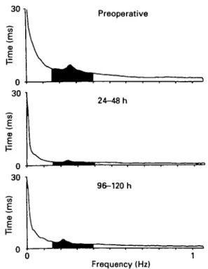
The spectral algorithm sampled heart rate at 469-ms intervals; 256 samples (120 s of data) were multiplied by a Harming window to minimize spectral leakage and then converted to the frequency domain using fast Fourier transform; 30 consecutive spectra (1-h data) were averaged and the resulting spectral plots printed. Thus the algorithm provided spectral plots on an hour-by-hour basis. In addition, composite spectral plots over 24-h periods were computed (fig. 1). For each of these spectral plots, the algorithm calculated the amplitude (i.e. the square root of the area under the power spectrum over the frequency studied) for total spectral energy (0.01-1 Hz), low-frequency spectral energy (0.04-0.15 Hz) and high-frequency spectral energy ((0.04-0.15- (0.15-0.4 Hz). The values for amplitude were subsequently squared so that spectral measures represent the areas under the spectral power-frequency curve over the frequencies indicated in units of ms2. Low frequency
to high frequency ratios were calculated by dividing low frequency spectral energy by high frequency spectral energy.
High-frequency spectral energy is related in-versely to ventilatory frequency, which in turn is reflected by the centre frequency of the high-frequency peak [3]. As the algorithm used for spectral analysis in the present investigation does not provide centre frequencies, we estimated the centre frequency of the high-frequency component visually from the spectral plots. Using this rough approach, we found no difference in the centre frequency of the high-frequency peak between preoperative and post-operative recordings.
The postoperative period was denned as the first complete 1 h of electrocardiographic data collected after spinal anaesthesia had waned (sensorimotor level below SI), or the first complete 1 h of
electro-Preoperative
Frequency (Hz)
FIG. 1. Composite power spectral plots (0.01-1.0 Hz) of the preoperative period (top panel), the second day after operation (middle panel) and the fifth day after operation (bottom panel) in a typical patient undergoing elective hip arthroplasty under spinal anaesthesia. The areas representing high-frequency spectra]
energy are shaded for clarity.
cardiographic data collected after tracheal extuba-tion. The first day after operation was denned as the first 24 h of the postoperative period.
Statistical analysis
Results are expressed as mean (SEM), unless other-wise stated. Differences in patient characteristics were compared using the unpaired Student's t test and Fisher's exact test, as appropriate. Two-way analysis of variance for repeated measurements followed by Duncan's multiple range test was used to assess the pattern of heart rate variability within groups. Two-way analysis of variance followed by an unpaired Student's t test at each stage was used to assess differences in the indices of heart rate variability between the groups. Least squares linear regression analysis was used to assess the dependence of heart rate variability on heart rate. P < 0.05 was considered statistically significant.
RESULTS
Two of the 12 patients in the spinal group were excluded (one because of preoperative myocardial ischaemia and one because of postoperative ven-tricular arrhythmia, Lown class IVa). In the re-maining 10 patients, 0.5 % isobaric bupivacaine 19.2 (1.9) mg was injected into the subarachnoid space resulting in a median sensory level of T8. Spinal anaesthesia had waned (sensorimotor level below SI) after 245 (38) min. There was no significant dif-ference between patients undergoing spinal anaes-thesia and the five patients undergoing general anaesthesia, with respect to duration of preoperative
TABLE I. Patient characteristics (mean (SEM or range) or number)
Age(yr) Sex(M:F)
Duration of surgery (min) Intraoperative blood loss
(ml) Spinal anaesthesia (n = 10) 69 (53-76) 4:6 122 (14.8) 675(71) General anaesthesia (n = 5) 64 (58-79) 3:2 120(12.7) 840(116) 120 100- 802 0 0 - 150- 100-Preop. 2 3 4
Days after op.
Preop. 2 3
Days after op.
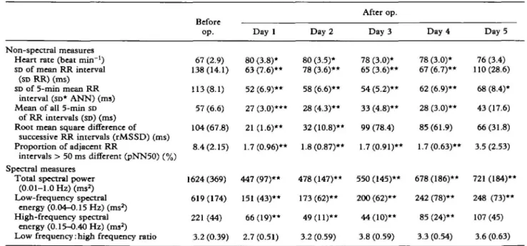
FIG. 2. SD about the mean RR intervals (SD RR) during the perioperative period in 10 patients undergoing spinal ( • ) and five patients undergoing genera] (H) anaesthesia (mean, SEM). Preop.
= Preoperative period. * P < 0 . 0 5 ; **P<0.01 vs preoperative values.
Holter monitoring (17.3 (1.7) and 16.8 (2.1) h, respectively), duration of surgery and intraoperative blood loss (table I). There was no significant difference between preoperative and postoperative body temperature which did not exceed 37.5 °C in any patient at any time.
A significant decrease in spectral and non-spectral indices of heart rate variability occurred during the early postoperative period (table II, figs 1-3). In only
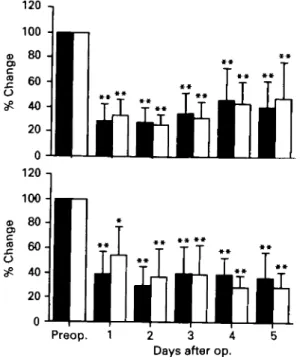
FIG. 3. Postoperative pattern of heart rate variability expressed as percentage change in total spectral energy (TP) (0.01-1.0 Hz) in 10 patients undergoing elective hip arthroplasty under spinal ( • ) and five patients undergoing elective hip arthroplasty under genera] ( • ) anesthesia (mean, 95% confidence intervals). Preop. = Preoperative period. *P<0.05; **P < 0.01 vs preoperative
values.
two patients had heart rate variability reached preoperative baseline values by the end of the fifth day after operation. Compared with baseline values, no significant change in the low frequency to high frequency ratio occurred in any patient during the postoperative period (fig. 4). There were no signifi-cant differences in indices of heart rate variability between patients who had spinal anaesthesia and those who had general anesthesia (figs 2-4).
The algorithm of power spectral analysis computes variables of heart rate variability for intervals of complete hours only, and when Holter monitoring had been commenced, sampling periods could not be co-ordinated with the beginning of anaesthesia or surgery. This resulted in a small number of intra-operative values and, therefore, a reliable interpret-ation of intraoperative heart rate variability was not possible. However, there were at least three values for every patient between the onset of spinal
TABLE II. Perioperative heart rate variability
Non-spectral measures Heart rate (beat min"1)
SD of mean RR interval (SD RR) (ms) SD of 5-min mean RR
interval (SD* ANN) (ms) Mean of all 5-min SD
of RR intervals (SD) (ms) Root mean square difference of
successive RR intervals (rMSSD) (ms) Proportion of adjacent RR
intervals > 50 ms different (pNN50) (%) Spectral measures
Total spectral power (0.01-1.0 Hz) (ms2)
Low-frequency spectra] energy (0.04-0.15 Hz) (ms2)
High-frequency spectra] energy (0.15-0.40 Hz) (ms2)
Low frequency:high frequency ratio
in 10 patients *P < 0.05, **P Before op. 67 (2.9) 138(14.1) 113(8.1) 57 (6.6) 104 (67.8) 8.4(2.15) 1624(369) 619(174) 221 (44) 3.2 (0.39)
undergoing elective hip arthroplasty under < 0.01 vs preoperative value. Day 1 80 (3.8)* 63 (7.6)** 52 (6.9)** 27 (3.0)*** 21 (1.6)** 1.7(0.96)** 447 (97)** 151 (43)** 66(19)** 2.7 (0.51) Day 2 80 (3.5)* 78 (3.6)** 58 (6.6)** 28 (4.3)** 32 (10.8)** 1.8(0.87)** 478(147)** 173(62)** 49(11)** 3.2 (0.59) i After op. Day 3 78 (3.0)* 65 (3.6)** 54 (5.2)** 33 (4.8)** 99 (78.4) 1.7(0.91)** 550 (145)** 200 (62)** 44(10)** 3.8 (0.59) spinal anaesthesia Day 4 78 (3.0)* 67 (6.7)** 62 (6.9)** 28 (3.0)** 85(61.9) • 1.7(0.63)** 678(186)** 242 (78)** 85 (24)** 3.3 (0.54) (mean (SEM)). Day 5 76 (3.4) 110(28.6) 68 (8.4)* 43 (17.6) 66(31.8) 3.5 (2.53) 721 (184)** 248 (73)** 107 (45) 3.6 (0.63)
120 100
-Preop. 2 3 4
Days after op.
FIG. 4. Postoperative percentage changes in low- ( • ) and high-CD) frequency spectral energy in 10 patients undergoing elective hip arthroplasty under spinal (top panel) and five patients undergoing elective hip arthroplasty under general (bottom panel) anaesthesia (mean, 95 % confidence intervals). Preop. = Pre-operative period. Note that the postPre-operative decrease in low-frequency (LF) and high-low-frequency spectral energy (HF) is similar which results in an unchanged LF-to-HF ratio. *P <
0.05; **P < 0.01 vs preoperative values.
anaesthesia and the time when spinal anaesthesia had waned. In none of the patients did these values differ from preoperative values, while in all patients heart rate variability during spinal anaesthesia significantly exceeded postoperative values (Wilcoxon's rank sum test).
A significant correlation was found between the perioperative percentage change in heart rate varia-bility (HRV) and heart rate (HR): HRV (% of preoperative value) = 250 % - 2.5 HR (beat min"1), r
= 0.56, P < 0.001.
DISCUSSION
Beat-to-beat variation in haemodynamic variables is thought to reflect the dynamic response of several feedback loops to perturbations in cardiovascular homeostasis [11]. Thus any interaction with the neural reflex arc, target organ responsiveness or both, might alter the pattern of heart rate variability. As neither spectral nor non-spectral indices enable us to identify the site or the mechanisms of these interactions, the exact explanation of the post-operative decrease in heart rate variability has to await further study. However, to exclude the possi-bility that the prolonged decrease in heart rate variability was unique to spinal anaesthesia, peri-operative heart rate variability was assessed ad-ditionally in patients requiring general anaesthesia for hip arthroplasty and a similar pattern was found. Thus a substantial decrease in heart rate variability occurred after a standard operation performed under
two different anaesthetic techniques while peri-operative care was standardized. Immobility per se does not influence heart rate variability [12]. As none of the additional drugs given during the peri-operative period (benzodiazepines, methadone, paracetamol, heparin, coumarin) is known to exert significant depressant and prolonged effects on the heart, autonomic nervous system, or both, and the doses of these drugs were relatively small, it is unlikely that perioperative medication was the main cause of the decrease in postoperative heart rate variability. We conclude therefore that the post-operative decrease in heart rate variability reflects changes caused by tissue trauma. Surgery is associ-ated with neuroendocrine, metabolic and acute phase responses characterized, among others, by increase in plasma concentrations of catecholamines and release of cytokines [13-16]. Any of these changes and as yet undefined biochemical mediators may be involved in the genesis and maintenance of the decrease in postoperative heart rate variability.
After surgery, high-frequency spectral energy decreased to approximately one-third of its pre-operative value. Since high-frequency spectral en-ergy appears to be mediated solely by the para-sympathetic nervous system [1-3], this indicates that during the postoperative period, the influence of the parasympathetic nervous system on the sinus node was depressed significantly.
Low-frequency spectral energy is considered to be mediated jointly by the sympathetic and para-sympathetic nervous systems. [1-4, 17]. However, regardless of the underlying mechanisms, any in-crease in sympathetic nervous activity results in an increase in low-frequency spectral energy in relative terms and a shift in the low frequency to high frequency ratio towards a predominant low fre-quency segment [1-4, 18]. In the present study, postoperative low-frequency spectral energy in ab-solute terms decreased, while the low-frequency segment in relative terms and the low frequency to high frequency ratio remained unchanged. There is no evidence so far of unchanged or decreased postoperative sympathetic nervous activity. In con-trast, postoperative increase in plasma concentra-tions of noradrenaline have been reported and increased noradrenaline concentrations were found up to 2 days after surgery [13,14], which indicates an increase rather than a decrease in postoperative sympathetic nervous activity. Even under exper-imental conditions of total vagal block, sympathetic stimulation has been reported to lead to a marked increase in low-frequency segment in both absolute and relative units [1]. Thus despite depression of the parasympathetic nervous system, indicated by the decrease in high-frequency spectral energy, sym-pathetic stimulation during the postoperative period should have resulted in a significant shift in the low frequency ratio to high frequency ratio towards a predominant low-frequency segment. We conclude therefore that the sympathetic influence on the sinus node decreased after surgery.
Previous studies have shown that neuropathic, pharmacological, traumatic or surgical interruption of autonomic efferent pathways reduced heart rate
variability [1, 10, 11, 19, 20]. Thus a reversible func-tional uncoupling between sympathetic centres and the sinus node could explain the decrease in low-frequency spectral energy and the unchanged low frequency to high frequency ratio, despite an increase in postoperative sympathetic nervous ac-tivity. A possible mechanism for this uncoupling could be desensitization of beta adrenergic receptors, which has been reported to occur in the presence of elevated plasma concentrations of catecholamines [21, 22]. Alternatively, neural transmission in auto-nomic cardiac efferent nerves might be affected.
We found a significant correlation between the postoperative increase in heart rate and the decrease in heart rate variability. There is, however, no evidence so far for a simple and inverse relationship between heart rate and heart rate variability [4, 5, 17, 18]. In contrast, Hirsch and Bishop reported that increased resting heart rate is associated with an increase in the corner frequency (i.e. the breathing frequency above which respiratory arrhythmia am-plitude declines with any further increase in breath-ing frequency) [23]. Thus an increase in heart rate should be associated with an increase rather than a decrease in high-frequency spectral energy. We suggest therefore that both increased heart rate and decreased heart rate variability result from a common cause, that is the postoperative decrease in para-sympathetic influence on the sinus node, rather than one being the result of the other.
The clinical implications of our findings are not yet clear. However, Fleisher, Hawes and Rosenbaum found that a decrease in postoperative heart rate variability was associated with an increase in cardiac complications in high-risk patients undergoing vas-cular surgery [24]. The present study demonstrates that after surgery a substantial decrease in heart rate variability can occur in the absence of cardiac or other complications. The decrease in heart rate variability occurred regardless of the anaesthetic technique used and, therefore, most likely reflects changes caused by surgical trauma. Further studies are thus necessary to determine if and how the postoperative pattern of heart rate variability, and consequently its usefulness as a non-invasive pre-dictor of cardiac complications, depends on the site and the extent of the operation performed.
Ewing and colleagues reported normal heart rate variability in young men several days after leg and pelvic fractures [12]. To the best of our knowledge there are no other data on the effects of trauma on heart rate variability. However, the pattern of heart rate variability might emerge as a measure of the extent of the (operative) trauma or a non-invasive index of recovery, or both.
REFERENCES
1. Rimoldi O, Pierini S, Ferrari A, Cerutti S, Pagani M, Malliani A. AnaJysis of short-term oscillations of R-R and arterial pressure in conscious dogs. American Journal of Physiology 1990; 258: H967-H976.
2. Pomeranz B, Macaulay RJB, Caudill MA, Kurz I, Adam D, Gordon D, Kilborn KM, Barger AC, Shannon DC, Cohen
RJ, Benson H. Assessment of autonomic function in humans by heart rate spectral analysis. American Journal of Physiology 1985; 248: H151-H153.
3. Pagani M, Lombardi F, Guzzetti S, Rimoldi O, Furlan R, Pizzinelli P, Sandrone G, Malfatto G, Dell'Orto S, Piccaluga E, Turiel M, Baselli G, Cerutti S, Malliani A. Power spectral analysis of heart rate and arterial pressure variabilities as a marker of sympatho-vagal interaction in man and conscious dog. Circulation Research 1986; 59: 178-193.
4. Pagani M, Mazzuero G, Ferrari A, Liberari D, Cerutti S, Vaitl D, Tavazzi L, Malliani A. Sympathovagal interaction during mental stress. A study using spectral analysis of heart rate variability in healthy control subjects and patients with a prior myocardial infarction. Circulation 1991; 83 (Suppl. II): II43-II51.
5. Ewing DJ, Hume L, Campbell IW, Murray A, Neilson JMN, Clarke BF. Autonomic mechanisms in the initial heart rate response to standing. Journal of Applied Physiology:
Res-piratory, Environmental and Exercise Physiology 1980; 49:
809-814.
6. Galletly DC, Corfiatis T, Westenberg AM, Robinson B. Heart rate periodicities during induction of propofol-nitrous oxide-isoflurane anaesthesia. British Journal of Anaesthesia 1992; 68: 360-364.
7. Kato M, Komatsu T, Kimura T, Sugiyama F, Nakashima K, Shimada Y. Spectral analysis of heart rate variability during isoflurane anesthesia. Anesthesiology 1992; 77: 669-674. 8. Donchin Y, Feld JM, Porges SW. Respiratory sinus
ar-rhythmia during recovery from isoflurane-nitrous oxide anaesthesia. Anesthesia and Analgesia 1985; 64: 811-815. 9. Malliani A, Pagani M, Lombardi F, Cerutti S. Cardiovascular
neural regulation explored in the frequency domain. Cir-culation 1991; 84: 482^92.
10. Pruett JK, Yodlowski EH, Introna RPS, Buggay DS, Crumrine RS. The influence of spinal anesthetics on heart rate variations. Pharmacology 1991; 10: 51-55.
11. Akselrod S, Gordon D, Ubel FA, Shannon DC, Barger AC, Cohen RJ. Power spectrum analysis of heart rate fluctuation: a quantitative probe to beat-to-beat cardiovascular control. Science 1981; 213: 220-222.
12. Ewing DJ, Neilson JMM, Shapiro CM, Stewart JA, Reid W. Twenty four hour rate variability: effects of posture, sleep, and time of day in healthy controls and comparison with bedside tests of autonomic function in diabetic patients. British Heart Journal 1991; 65: 239-244.
13. Helbo-Hansen S, Fletcher R, Lundberg D, Nordstrom L, Werner O, Stahl E, Norden N. Clonidine and the sympathico— adrenal response to coronary artery by-pass surgery. Ada Anaethesiologica Scandinavica 1986; 30: 235-242. 14. Fell D, Chmielewski A, Smith G. Postoperative analgesia
with controllcd-relcase morphine sulphate: comparison with intramuscular morphine. British Medical Journal 1982; 285: 92-94.
15. Douglas RG, Shaw JHF. Metabolic response to sepsis and trauma. British Journal of Surgery 1989; 76: 115-122. 16. Stahl W. Acute phase protein response to tissue injury.
Critical Care Medicine 1987; 15: 545-550.
17. Inoue K, Miyake S, Kumashiro M, Ogata H, Yoshimura O. Power spectral analysis of heart rate variability in traumatic quadriplegic humans. American Journal of Physiology 1990; 258: H1722-H1726.
18. Lombard F, Sandrone G, Pempruner S, Sala R, Garimoldi M, Cerutti S, Baselli G, Pagani M, Malliani A. Heart rate variability as an index of sympathovagal interaction after acute myocardial infarction. American Journal of Cardiology 1987; 60: 1239-1245.
19. Sands KE, Appell ML, Lilly LS, Schoen FJ, Mudge GH, Cohen RJ. Power spectrum analysis of heart rate variability in human cardiac transplant recipients. Circulation 1989; 79: 76-82.
20. Kitney RI, Byrne S, Edmonds ME, Watkins PJ, Roberts VC. Heart rate variability in the assessment of the autonomic diabetic neuropathy. Automedica 1982; 4: 155-167. 21. Strasser RH, Stiles GL, Lefkowitz RJ. Translocation and
uncoupling of the beta-adrenergic receptor in rat lung after catecholamine promoted desensitization in vivo. Endocrin-ology 1984; 115: 1392-1400.
Bristow MR. Myocardial catccholamine and neuropeptide Y humans: how breathing pattern modulates heart rate, depletion in failing ventricles of patients with idiopathic American Journal of Physiology 1981; 241: H620-H629. dilated cardiomyopathy. Correlation with beta-adrencrgic 24. Fleisher LA, Hawes AD, Rosenbaum SH. Approximate receptor downregulation. Circulation 1992; 85: 46-53. entropy of heart rate as a correlate of postoperative ventricular 23. Hirsch JA, Bishop B. Respiratory sinus arrhythmia in dysfunction. Anesthesiology 1993; 78: 683-692.