2005/130
Dual concentration-dependent activity of thyroglobulin type-1
domain of testican: specific inhibitor and substrate of
cathepsin L
Primozˇ Meh1, Miha Pavsˇicˇ1, Vito Turk1, Antonio Baici2and Brigita Lenarcˇicˇ1,3,*
1Department of Biochemistry and Molecular Biology,
Jozef Stefan Institute, Jamova 39, SI-1000 Ljubljana, Slovenia
2Department of Biochemistry, University of Zurich,
Winterthurerstrasse 190, CH-8057 Zurich, Switzerland
3Department of Chemistry and Biochemistry, Faculty of
Chemistry and Chemical Technology, University of Ljubljana, Jamova 39, SI-1000 Ljubljana, Slovenia * Corresponding author
e-mail: brigita.lenarcic@ijs.si
Abstract
The thyroglobulin type-1 (Tg-1) domain is a protein mod-ule that occurs in a variety of secreted and membrane proteins and is recognised as a potent inhibitor of cys-teine peptidases. We present here some properties of the Tg-1 domain of human testican, a modularly organised proteoglycan secreted mainly by brain cells, the exact in
vivo function of which is not yet clear. The domain was
prepared as a recombinant protein in a Pichia pastoris expression system and its activity was demonstrated by specific and selective inhibition of cathepsin L (Kis0.14 nM). Interaction at high enzyme and inhibitor
concentrations resulted in degradation of the domain by cathepsin L, which was not observed under conditions used for the determination of kinetic parameters. No inhibitory activity could be detected for cathepsin K, but it exhibited a very similar degradation pattern. Homology modelling provided a good explanation for the different behaviour observed with the two enzymes. Firstly, the steric fit between the interfaces of testican domain and cathepsin L is stabilised by numerous favourable forces, while no such interactions are evident in the complex with cathepsin K, and repulsive interactions even prevent access of the domain to the active site of papain. Sec-ondly, the prolonged first loop of the domain occupies a position near the catalytic cysteine residue in a more substrate-like manner, enabling cleavage of the Gly22
-Ala23bond.
Keywords: cysteine peptidases; extracellular
proteolysis; inhibition kinetics; proteoglycans; testican; thyropin.
Introduction
Thyroglobulin type-1 (Tg-1) domains are recognised as unique sequence motifs of 65–80 residues. They occur
in a variety of modularly organised proteins that are either secreted or located in membranes (Molina et al., 1996; Lenarcˇicˇ and Bevec, 1998). A few years ago, it was established that some proteins containing a Tg-1 module function as inhibitors of the papain-related cysteine pep-tidases. So far, five proteins have been characterised as true inhibitors, the p41 invariant chain fragment (Bevec et al., 1996), chum salmon egg cysteine peptidase inhib-itor (Yamashita and Konagaya, 1996), equistatin (Lenarcˇicˇ et al., 1997), saxiphilin (Lenarcˇicˇ et al., 2000) and testican (Bocock et al., 2003). All these inhibitors differ signifi-cantly in their selectivity for target enzymes. For example, the p41 fragment can discriminate between two closely related enzymes, cathepsin L and cathepsin S, inhibiting only the former (Bevec et al., 1996). Only one of the two Tg-1 domains present in saxiphilin inhibits cathepsins L or B (Lenarcˇicˇ et al., 2000) and, in the case of the three equistatin domains, the first one exhibits broad specific-ity against papain-like cysteine peptidases, while the subsequent two are non-inhibitory (Lenarcˇicˇ and Turk, 1999; Galesˇa et al., 2003). Furthermore, the second Tg-1 domain of equistatin is unique in inhibiting the aspartic peptidase cathepsin D (Galesˇa et al., 2003). The general inhibitory mechanism of a Tg-1 domain was revealed by the crystal structure of the p41 fragment in complex with cathepsin L (Guncˇar et al., 1999). The wedge shape and the three-loop arrangement of the p41 fragment interacts with the active site cleft in a non-specific manner, where-as the additional contacts between regions of the inhib-itor with the right domain of the enzyme are supposed to be crucial for the specificity. The structure of the p41 fragment is stabilised by three disulfide bridges and is also well defined in the absence of enzyme (Chiva et al., 2003).
Testican is a proteoglycan expressed predominantly in brain (Bonnet et al., 1996). At the genetic level, two addi-tional forms of testican have been identified, testican-2 (Nagase et al., 1996; Vannahme et al., 1999) and testi-can-3 (Nakada et al., 2001). The characteristic feature of all variants is their modular organisation. The signal sequence on the N-terminal end is followed by a short part unique to testican, the follistatin domain, which also includes a Kazal domain, the calcium-binding domain, by the thyroglobulin type-1 domain (Tg-1), and finally by a short C-terminal sequence where two glycosaminogly-can attachment sites are located. The latter modification is suggestive of its involvement in cell adhesion process-es and extracellular matrix organisation (Hartmann and Maurer, 2001). Lysosomal peptidases are known to be secreted into the peri/extracellular space, where they take part in remodelling or pathological degradation of extracellular matrix proteins in processes such as osteo-arthritis, angiogenesis and inflammatory response (Baici
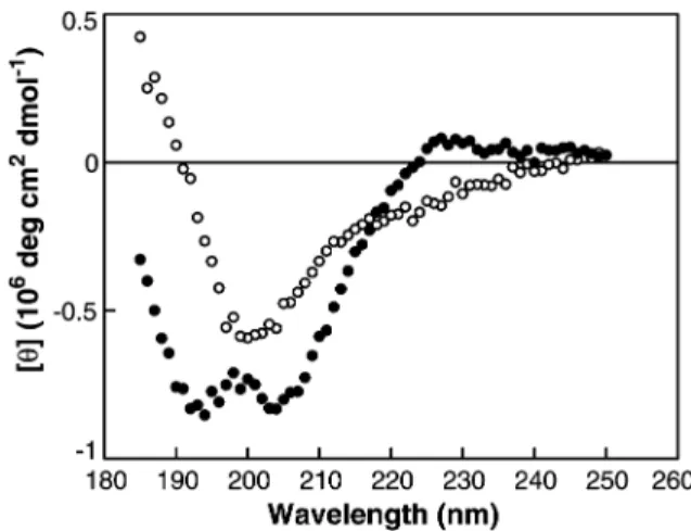
Figure 1 CD spectra of the testican Tg-1 domain under normal and denaturing conditions.
CD spectra were recorded using a protein concentration of 0.1 mg/ml in 10 mMsodium phosphate buffer, pH 5.60. (d)
Tes-tican Tg-1 domain measured at 48C; and (s) tesTes-tican Tg-1 domain in the presence of 12.5mMDTT, measured at 888C. et al., 1995; Chapman et al., 1997; McGrath, 1999).
Recently, a model of the involvement of the p41 invariant chain Tg-1 domain in the regulation of extracellular cathepsin L activity was presented (Fiebiger et al., 2002). It was shown that a stable complex of cathepsin L/p41 fragment is formed within antigen presenting cells and is afterwards secreted into the extracellular milieu, where the Tg-1 domain of p41 most probably acts to protect the enzyme and is displaced by a physiological substrate when necessary.
In the present study we characterised the Tg-1 domain, a 7-kDa fragment of the 49-kDa full-length human testican, which shows significant sequence sim-ilarity to the Tg-1 domains of the p41 invariant chain and equistatin, for which the specificities against different peptidases have already been determined in detail (Bevec et al., 1996; Lenarcˇicˇ and Turk, 1999; Lenarcˇicˇ et al., 2000). Using the expression system of Pichia
pasto-ris, we produced the Tg-1 domain in an apparently native
form. Molecular modelling and functional analyses revealed new features about the interaction of testican Tg-1 domain with its target papain-like enzymes.
Results and discussion
Preparation of recombinant Tg-1 domain of testican
P. pastoris proved to be a highly effective heterologous
system for generating functional Tg-1 domains of equi-statin in high yield (Galesˇa et al., 2003). In the present study, we utilised the same system to produce the Tg-1 domain of human testican. The cDNA construct for Tg-1 domain coding for 70 amino acids (Gly310–Glu379, testican
unprocessed precursor counting) was introduced into the
P. pastoris GS115 strain and the level of the expressed
protein in media was followed by sodium dodecyl sul-fate-polyacrylamide gel electrophoresis (SDS-PAGE) (data not shown). The domain was purified by three chro-matographic steps and at the end a single band on SDS-PAGE corresponding to a molecular mass of approximately 8 kDa was obtained (data not shown). In the final purity assessment by N-terminal sequencing, we detected three slightly different populations of domains. In approximately 60% of the molecules two Glu-Ala repeats (out of three) were missing at the N-terminus, in 25% of the molecules one Glu-Ala repeat had been removed, and 15% of the molecules had the sequence of the intact Tg-1 domain. Processing of a-MF propep-tide from the recombinant protein by the Kex2 endopep-tidase appeared to be efficient, while the efficiency of Ste13 in removing the Glu-Ala repeats was not absolute. Similarly, it was observed that the two Glu-Ala repeats begin the sequences of previously reported Tg-1 domains of equistatin expressed in P. pastoris (Galesˇa et al., 2003), confirming the fact that the precise action of Ste13 depends on the surrounding amino acid sequence and on the tertiary structure of the protein of interest (Brake et al., 1984; Cereghino and Cregg, 2000). A yield of 10 mg of pure, active testican Tg-1 domain was typ-ically obtained from 1 l of culture medium.
CD spectrum of testican Tg-1 domain
The CD spectrum of the Tg-1 domain of testican is cha-racterised by two peaks, a positive one at 230 nm and an intense double trough at 204 nm (Figure 1). The spec-trum differs slightly from the spectra of the individual Tg-1 domains of equistatin (Galesˇa et al., 2003) in two respects, by having a pronounced double trough and by the fact that both peaks are shifted to slightly higher wavelengths. As a comparison, the first domain of equis-tatin has negative peaks at 200, 194 and 198 nm, while the positive peak, which reflects the aromatic contribu-tion (Woody, 1994) is present only in the second domain at 220 nm. A similar strongly positive band at 226 nm has also been observed with synthetic p41icf Tg-1 domain (Chiva et al., 2003). The small differences observed in the CD spectra are probably a consequence of slightly different backbone conformations of individual domains, which might in turn correspond to different specificities. On the other hand, it appears that the gen-eral features of all known Tg-1 domain spectra are char-acteristic of the thyropin fold. It very much resembles spectra of a group of b-proteins in which the a-helix con-tent is low (Koepf et al., 1999; Kidricˇ et al., 2002). When the testican domain was thermally unfolded, the spec-trum exhibited the characteristics of a random coil (Fig-ure 1).
Inhibition of cathepsin L by the Tg-1 domain of testican
An important characteristic of thyroglobulin type-1 domains is their selectivity in inhibiting different papain-like enzymes (Bevec et al., 1996; Lenarcˇicˇ et al., 1997, 2000). To investigate the inhibitory capacity of the testi-can Tg-1 domain we considered the human lysosomal cysteine peptidases cathepsins B, K, L, S, and X, and the plant cysteine peptidase papain, as well as the aspar-tic peptidase cathepsin D and the serine peptidase tryp-sin. Only cathepsin L was inhibited, while all of the other enzymes were not affected, even at high inhibitor con-centrations. Cathepsin K rapidly cleaved the Tg-1
Scheme 1 Reaction mechanism proposed for cleavage of the Tg-1 domain by cathepsin L.
domain (see next section), while cathepsins B, S, X, and D, as well as papain and trypsin, did not. For cathepsin L, Eq. (1) was used to calculate ys, yzand l (see
Mate-rials and methods). yswas a hyperbolic function of
wTg-1x and dropped to zero at saturating inhibitor concentrations, yz was independent of wTg-1x, and l
depended linearly upon wTg-1x over the whole concen-tration range explored. The inhibitor affected Km, but not
the limiting rate V. This behaviour is characteristic of a fully competitive, slow-binding inhibition mechanism, in which the enzyme-inhibitor complex is formed in a single step (Morrison, 1982). Cleavage of the Tg-1 domain by cathepsin L was observed under second-order reaction conditions at high enzyme and Tg-1 concentrations (next section). The proposed complete reaction mechanism, including the substrate used to monitor reaction, is shown in Scheme 1.
I* collectively represents all possible fragments gen-erated by proteolysis. Following the first attack on the most susceptible bond, two new peptides are generated and then smaller peptides are formed from these. If these are substrates for the enzyme, the system will consist of a mixture of substrates with different affinities. Thus, the path leading to I* contains multiple steps that remain kinetically inaccessible to experimental verification and is represented by the global constant k*. Best-fit par-ameters and their standard errors, calculated by non-linear regression, were: k3s(8.94"0.35)=106 M-1 s-1;
k-3s(1.28"0.23)=10-3 s-1; and Kisk-3/k3s(1.4"0.3)=10 -10M. There was no evidence that the Tg-1 domain was
cleaved by proteolysis, thus behaving as a substrate, during assays with catalytic enzyme concentrations. In this case the steady-state slope had turned to a con-cave-upward profile for long reaction times. For calcu-lation we used the concentration of the Tg-1 domain as determined spectrophotometrically. A titrated concentra-tion of the Tg-1 domain was not available, since the only enzyme with which this would be possible is cathepsin L. Titration inevitably involves the use of both enzyme and inhibitor at relatively high concentrations, and under these conditions the inhibitor is degraded (see below). The high quality of the CD spectrum (Figure 1), which shows a remarkable difference between the native and the unfolded polypeptide, suggests that our testican Tg-1 domain preparation was properly folded and active. The inhibitory behaviour of the isolated Tg-1 domain
towards cathepsin L is thus very similar to that of intact testican, with Kivalues of 0.14 nM(this study) and 0.7 nM
(Bocock et al., 2003). These results are in good agree-ment with our previous studies on equistatin (Lenarcˇicˇ et al., 1997; Galesˇa et al., 2003) and the p41 fragment invariant chain (Bevec et al., 1996; Fiebiger et al., 2002), in which we demonstrated that the Tg-1 domains fully retain their functionality after excision from the rest of the parent molecule. Therefore, the testican Tg-1 domain may serve as a good model for further cha-racterisation of the action of this type of inhibitor.
Degradation of Tg-1 domain of testican by cathepsins L and K
The kinetic measurements for inhibition of cathepsin L by testican Tg-1 domain were carried out with Tg-1 domain/ cathepsin L molar ratios in the range 130:1–550:1 and picomolar enzyme concentrations. When molar ratios of 1:1–6:1 and concentrations of enzyme and inhibitor in the micromolar range were used, extensive degradation of the inhibitor molecule was observed. Reaction pro-gress was followed by HPLC separation of the frag-ments, and degradation products were analysed by matrix-assisted laser desorption/ionisation time-of-flight mass spectrometry (MALDI-TOF). A large number of frag-ments observed (Tables 1 and 2) were due to the pres-ence of three differently expressed forms of testican Tg-1 domains and to partial reduction of the disulfide bridges during the process of cleavage. For the purposes of anal-ysis we have omitted fragments that evidently arise from the minor E-4and Gly1 forms. The time-dependent
deg-radation of the domain enabled us to reveal the sequence of cleavage events. Within the tight cathepsin L/Tg-1 tes-tican domain complex, the most susceptible bond appeared to be Gly22–Ala23. These amino acids are
locat-ed at the bottom of the first loop in the close vicinity of the cathepsin L active site. After this primary cleavage, the first loop was attacked at two further sites: (i) between Gln12-Lys13 and/or Asn9-Arg10, resulting in the
excision of corresponding short peptides that occupy the active site (Lys13-Gly22 and Arg10-Gly22, respectively); (ii)
the bonds between Tyr34-Lys35 and Lys35-Ala36 became
exposed at the same time, and their cleavage provoked the complete separation of the first loop from the rest of the molecule. Prolongation of the incubation time brought about only the release of disulfide-bonded frag-ments (Table 1).
The interaction of cathepsin K with the testican domain resulted in its rapid cleavage (Table 2). No inhibition of the enzyme could be detected, although it cannot be excluded that, during the interaction, the domain may enter the active site of cathepsin K as thyropin-like inhib-itors normally do, i.e., with all three loops (Guncˇar et al., 1999). The most exposed region of the Tg-1domain to cathepsin K was again the extended first loop, where the main fragmentation took place (Ser15-Lys16, Leu21-Gly22
and Gly22-Ala23). Prolongation of the incubation time
resulted in additional cleavages between Gln12-Lys13and
on the C-terminal part of the Tg-1 molecule (Ala57-Gly58
and Ala64-Val65).
Cysteine peptidases of the papain family have been shown to have primary substrate specificity at the P2
position and to have a preference for hydrophobic amino acids (McGrath, 1999; Turk et al., 1998). Despite struc-tural similarities, cathepsins L and K display differences in their binding preferences for P2residues. Cathepsin L
favours both aromatic and aliphatic amino acid residues (Phe, Tyr, Trp, Val, Leu, Ile), while cathepsin K favours aliphatic amino acids (Ile, Leu, Val) and is unique in posi-tioning Pro in the S2subsite, a property responsible for
its potent intrahelical collagenolytic activity (Garnero et al., 1998; Lecaille et al., 2002). Our present results show that the amino acid residues at P2 subsites of the
iden-tified cleavage points of the testican Tg-1 domain reflect the specificity of the cathepsin considered (Tables 1 and
2). Cathepsin L shows broader selection of amino acids at the P2 position by utilising Tyr, Leu, Ile and Val, and
can also employ Met and Ala, while cathepsin K is limited to Leu and Ile, with the one exception for Gly.
Evidence from experiments conducted under different conditions suggests that the dual nature of the Tg-1 tes-tican domain as substrate and inhibitor of cathepsin L is due to the very low rate of degradation of the EI complex according to Scheme 1. An estimate for the rate of deg-radation of the Tg-1 domain was obtained from the time-dependent degradation of the inhibitor after incubating 0.39mM cathepsin L with 2.3mM Tg-1 domain at 248C and pH 5.50 (data not shown). The final degradation
products of the Tg-1 domain were no longer inhibitory, as ascertained by incubating them with cathepsin L and measuring the residual activity with Z-Phe-Arg-MCA (MCA, 4-methyl-7-coumarylamide) as substrate. Since evaluation of all the individual constants contributing to
k* in Scheme 1 was impossible, quantification was
car-ried out by measuring the decreasing area of the HPLC peak corresponding to the Tg-1 domain and reaction profiles were analysed by the integrated Michaelis-Men-ten equation. Calculations afforded a first-order rate con-stant of 1.33=10-4 s-1 (half-lifes5200 s), equivalent to
V/Kmfor the most susceptible bond of the Tg-1 domain,
i.e., the first one cleaved by cathepsin L. Considering this result and the enzyme concentration used (0.39mM), a first-order rate constant of 7.5=10-9 s-1
(half-lifes9.2=107 s) can be calculated for an enzyme
con-centration of 22 pM, i.e., the concentration used for
measuring the inhibition constant (see the preceding section). Hence, the rate of Tg-1 domain degradation at catalytic enzyme concentration is approximately 18 000-fold lower than at high enzyme concentrations. Degra-dation is virtually invisible, and the Tg-1 domain behaves as an inhibitor. Cathepsin L used for the experiments shown in this paper was prepared in E. coli from inclusion bodies and was obtained by renaturation, thus making the presence of contaminating bacterial peptidases very unlikely. In order to rule out the possibility that Tg-1 domain degradation was not due to cathepsin L but to a contaminant enzyme, we repeated the cleavage experi-ment and fragexperi-ment analysis using the same Tg-1 domain preparation and cathepsin L from two additional sources, namely the natural enzyme purified from human kidney and a recombinant form from Pichia pastoris. With all of the three different enzyme preparations we obtained the same cleavage products (data not shown).
Structural analysis of models
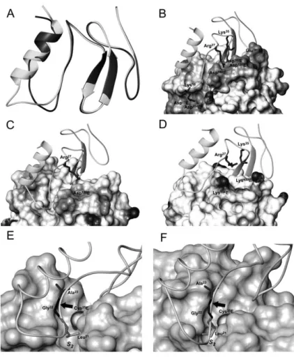
To explain the experimentally observed specific interac-tion of the Tg-1 domain of testican with cathepsin L, sev-eral models of their complex were built and compared to the models of complexes of the domain with cathepsin K and papain. The models were derived by a homology modelling procedure with amino acid sequence identity between the Tg-1 domain of testican and the template structure (p41 fragment) being above 30%. The major differences between the two molecules are present in the first subdomain and are a consequence of the insertion of five amino acids in the testican Tg-1 domain com-pared to the p41 fragment (Figure 2A). In the modelled domain, the extended a-helical region contributes to hydrophobic packing, and the region following the a-helix forms an enlarged loop that may have a more flex-ible conformation, similar to the corresponding loop in the p41 fragment, as observed from its NMR structure (Chiva et al., 2003). The interactions between the testican Tg-1 domain and cathepsin L in the modelled complex are electrostatic, polar and hydrophobic. Lys18interacts
electrostatically with Asp71E and Asp114E, and Arg27and
Lys35 with Asp137E, Asp160E and Asp162E, the latter three
forming the S19 binding site (the letter E denotes enzyme
residues). Polar interactions occur between Ser42 and
both Cys25E and Gln21E, the latter representing the
oxy-anion hole. Hydrophobic interactions predominantly involve Leu21, which fits into the deep and hydrophobic
S2 binding pocket of cathepsin L, and are mainly
con-fined to the first and second loops of the domain (Figure 2B). These interactions are similar to interactions observed in the crystal structure of the p41 fragment complex with cathepsin L. Models of complexes of the domain with cathepsin K and papain (Figure 2C,D, respectively) show the same general binding mode, although the majority of favourable electrostatic interac-tions observed in the cathepsin L complex are not pres-ent. These observations are consistent with the finding that neither cathepsin K nor papain was inhibited by the Tg-1 domain. Furthermore, the surface of papain near the domain in the modelled complexes is positively charged, which may prevent binding of the domain to the enzyme (Figure 2D). These results lead to the conclusion that the domain binds to both cathepsin L and K, but not to papain.
It is still not possible to establish whether proteolytic cleavage of the domain by cathepsins L and K is a con-sequence of the geometry of the prolonged first loop or of the amino acid composition at the P4-P1/P19 site, or
maybe a combination of both. For example, testican and the first domain of equistatin exhibit a very similar arrangement of amino acid residues at P4-P1/P19
posi-tions within the first loop, i.e., Ser-Leu-Leu-Gly/Ala and Gly-Leu-Ile-Gly/Ala, respectively (Lenarcˇicˇ et al., 1997). Only testican is cleaved by cathepsin L, which points to the position of the extended loop as a decisive factor. This could be explained by the more substrate-like nature of the extended first loop of the testican Tg-1 domain compared to the corresponding loop of the first domain of equistatin, which presumably is tighter and similar to the corresponding loop in the p41 fragment. However, another prerequisite for proteolytic cleavage is the acces-sibility of the scissile bond for nucleophilic attack of the sulfur atom of the active Cys residue (Figure 2E,F). This accessibility is in turn governed by the residues sur-rounding the putative cleavage site and their conforma-tion. Finally, it should be noted that the interacting region of the testican Tg-1 domain is clearly different from a classical substrate, since it is not linear but rather com-posed of two parts, one of them interacting with S bind-ing subsites and the other with S9 bindbind-ing subsites. Nevertheless, the modelling results suggest that binding of the domain to the enzyme in an orientation similar to that found in the cathepsin L-p41 fragment complex is a prerequisite for proteolytic cleavage of the domain, since this is the only orientation that permits close contact between the domain and the enzyme.
Physiological aspects
There are several reports about the proteolytic process-ing of molecules that contain Tg-1 domains. Two C-ter-minal testican fragments (cleavage within the Tg-1 domain at position Tyr343-Lys344 and Arg369-Lys370) with
attached chondroitin sulfate chains were found as the major proteoglycans in human seminal plasma (Bonnet et al., 1992). The Tyr343-Lys344 bond (testican
unproces-sed precursor counting) corresponds to the Tyr34-Lys35
Figure 2 Three-dimensional models of testican Tg-domain in complex with target enzymes.
(A) Superimposition of the testican Tg-1 domain model (grey) and the p41 fragment (black). The insertion of five amino acids in the testican domain, as compared to the p41 fragment, results in an extended a-helix and altered geometry of the first loop. (B–D) Models of complexes of testican Tg d-1 domain and cathepsin L (B), cathepsin K (C) and papain (D). The surface of enzymes is shaded to represent the electrostatic potential: negatively charged residues are shown in grey and positively charged residues in black. Positively charged amino acid residues of the testican domain involved in favourable (cathepsins L and K) or unfavourable (papain) electrostatic interactions are shown as black sticks. (E,F) Testican Tg-domain displayed over the surfaces of the active sites of cathepsins L (E) and K (F). The position of the scissile bond (marked with an arrow) in the testican Tg d-1 domain relative to the active site cysteine residue (Cys25E) is similar for both cathepsin L and K. In this substrate-like orientation, the P
2residue (Leu21) fits into the hydrophobic S2binding pocket of the enzyme. Images were generated using the program MOLMOL (Koradi et al., 1996).
detected after the interaction of the domain with cathep-sin L. Interestingly, the Tg-1 domain was also proposed as a target cleavage region during the conversion of large plasma testican into smaller stable forms, although it was shown that processing can be blocked by certain serine peptidase inhibitors (BaSalamah et al., 2001). Neverthe-less, in the present work we demonstrated that the Tg-1 domain of testican is very susceptible to cleavage by the two cysteine peptidases cathepsin L and cathepsin K. Therefore, the role of the testican Tg-1 domain must be different from that of the Tg-1 domain of the major his-tocompatibility complex class II associated invariant chain (p41 Ii), the processing of which has been studied in more detail (Fiebiger et al., 2002). Two key enzymes are crucial for processing p41 Ii, cathepsins S and L (Len-non-Dumenil et al., 2002) and, from the point of view of the Tg-1 domain, it remains intact after proteolysis and can serve either as an inhibitor of cathepsin L activity or as a chaperone in maintaining the pool of mature enzyme outside the cell (Fiebiger et al., 2002). The major differ-ence between the two domains is the insertion of five amino acids on the N-terminal part of the testican
domain, which probably has a great impact on the geom-etry and position of the first loop and consequently on the susceptibility to proteolytic attack.
In the light of the results reported here, testican may behave as an inhibitor or as a substrate of cathepsin L, depending on the relative concentrations available. In a close pericellular environment, where cathepsin L is likely to reach considerable local concentrations, proteolytic fragmentation of testican can be expected. Conversely, at a certain distance from a cathepsin L-releasing cell, the enzyme concentration is likely to be sufficiently low with respect to the proteoglycan to make proteolysis insignificant relative to inhibition. Further studies will be needed to focus on the possible physiological role of Tg-1 domains in the peri/extracellular space, where several cysteine peptidases are known to play an important role in remodelling of extracellular matrix proteins (McGrath, 1999). A number of modularly organised proteins with diverse functions, such as testican, nidogen, thyroglo-bulin, Trop and SMOC that contain one or more copies of Tg-1 domains, are considered to be extracellular proteins.
Materials and methods Materials
Materials for cloning, expression and purification were obtained from the following sources: polymerase chain reaction (PCR) primers from MWG Biotech (Ebersberg, Germany); restriction enzymes, DNA ligase and deoxynucleotide from Promega (Mad-ison, USA); Taq polymerase from Fermentas (Vilnius, Lithuania); salts and media for P. pastoris from Sigma-Aldrich (St. Louis, MO, USA); P. pastoris expression kit, pPIC9 plasmid and P.
pas-toris strain GS115 from Invitrogen (Carlsbad, CA, USA); Phenyl
Sepharose, Sephadex G-50 and SP Sepharose FF from Amer-sham Pharmacia Biotech (Uppsala, Sweden); and Z-Phe-Arg-4-AMC, Z-Phe-Val-Arg-Z-Phe-Arg-4-AMC, Z-Arg-Arg-AMC and Z-Phe-Arg-pNA from Bachem AG (Bubendorf, Switzerland). 2,4-Dinitrophenyl-Gly-Phe-Phe-Trp-OH was synthesised according to Klemencˇicˇ et al (2000) and the cDNA coding for the testican protein was kindly provided by C.J.S. Edgell (University of North Carolina, USA). Papain was prepared as described in Blumberg et al. (1970), human cathepsins B, D, L, K, S as in Kuhelj et al. (1995), Kregar et al. (1977), Dolinar et al. (1995), Hwang and Chung (2002) and Bro¨mme et al., (1993), respectively, and cathepsin X according to Klemencˇicˇ et al. (2000). Bovine trypsin was obtained from Sigma-Aldrich.
Cloning and expression of testican Tg-1 domain The DNA segment coding for the Tg-1 domain of testican (res-idues Gly310–Glu379) was amplified with the primers 59-GGA TCC CTC GAG AAA AGA GAG GCT GAA GCT GGT CTC CCT TGC CAG AAT GAA ATG AAC AGA ATT-39 (sense), and 59-AAT TCG CGG CCG CTT ACT CCT CTT CAC AGC T-39 (antisense) using a plasmid encoding full-length testican as a template. The ampli-fied DNA was digested with XhoI and NotI restriction endonu-cleases and ligated into pPIC9 digested with the same enzymes. The recombinant construct was amplified in Escherichia coli strain DH5a and the correct construction of the plasmid was confirmed by restriction analysis and DNA sequencing using an ABI PRISM 310 DNA sequencer (Perkin Elmer, Wellesley, MA, USA). After linearisation of the pPIC9 construct with SalI, the
Pichia pastoris strain GS115 was transformed by electroporation
with Gene Pulser (Bio-Rad, Hercules, CA, USA) according to the manufacturer’s protocol. The transformed cells were plated on MD agar plates w0.34% yeast nitrogen base (YNB), 1% ammo-nium sulfate, 400mg/l biotin, 2%D-glucose, 1.5% agarx. Result-ing colonies were used to inoculate 10 ml of BMGY medium (1% yeast extract, 2% peptone, 100 mM potassium phosphate, pH 6.0, 0.34% YNB, 1% ammonium sulfate, 400mg/l biotin, 1% glycerol) and grown overnight at 308C in a shaking incubator at 250 rpm. Cells were collected by centrifugation (2500 g for 5 min at 238C) and gently resuspended in 10 ml of BMMY medium (same as BMGY medium but containing 0.5% methanol instead of 1% glycerol). Cultures were incubated for an additional 96 h with daily supplements of 50ml of methanol. Clone production was monitored by electrophoretic analysis of the supernatants on 12% polyacrylamide SDS gels (Laemmli, 1970). The colony with the highest expression level of testican Tg-1 domain was used to prepare protein in larger quantities, using 2-l shake-flasks under the conditions specified above. The cells were pel-leted and the supernatant was stored at -208C.
Purification of the Tg-1 domain
Ammonium sulfate was added to culture supernatants to a final concentration of 1.0M and the pH was adjusted to 6.0. The protein solution was loaded onto a phenyl-Sepharose column equilibrated with 0.1 M phosphate buffer containing 1.0 M
ammonium sulfate. Proteins were eluted with a linear gradient of ammonium sulfate (from 1 to 0M) in 0.1Mphosphate buffer, pH 6.0 and analysed by SDS-PAGE, in which the Tg-1 domain of testican migrated as a band with a molecular mass of approx-imately 8 kDa. Fractions containing testican domain were further purified on a Sephadex G-50 column equilibrated with 0.01M
Tris/HCl buffer, pH 7.3 in 0.1M NaCl. Final purification of the testican domain was achieved by ion-exchange chromatography on SP Sepharose FF pre-equilibrated with 0.01Macetate buffer, pH 4.5. Proteins were eluted with a linear gradient of NaCl (0–0.5M) in the same buffer. The concentration of the Tg-1 domain was determined spectrophotometrically using an absorption coefficient of 9890M-1cm-1at 280 nm.
N-Terminal sequence determination
N-Terminal sequencing was carried out by automated Edman degradation using an Applied Biosystems liquid pulse sequencer 475A (Foster City, CA, USA) connected on line to a phenylthio-hydantoin analyser 120A from the same manufacturer.
Circular dichroism spectroscopy
The far-UV CD spectrum of recombinant Tg-1 domain of testican was measured at 48C on an Aviv 60DS spectrometer (Piscata-way, NJ, USA) using a quartz cell with 1-mm path length. In the wavelength region 250–185 nm, data points were recorded at 1-nm intervals with a dwell time of 10 s. Protein was dissolved at 0.1 mg/ml in 10 mMsodium phosphate buffer, pH 5.6. For thermal denaturation, the sample was exposed to 888C for 1 h in the presence of 12.5mMdithiothreitol (DTT) and the spectra were recorded at 888C. To confirm the absence of turbidity, the absorbance of protein solutions was scanned from 350 to 240 nm on a Lambda 18 spectrophotometer (Perkin Elmer) prior to CD measurement.
Kinetic measurements
Enzymes were assayed following standard procedures using the substrate Z-Phe-Arg-MCA for papain, cathepsins L and cathep-sin K, Z-Arg-Arg-MCA for cathepcathep-sin B, Z-Phe-Val-Arg-MCA for cathepsin S, and 2,4-dinitrophenyl-Gly-Phe-Phe-Trp-OH for cathepsin X. Inhibitory effects of the testican Tg-1 domain were studied by incubating it with an enzyme for 30 min at 258C before adding the substrate and measuring the residual activity. The kinetic inhibition mechanism of cathepsin L by the Tg-1 domain was investigated under pseudo-first-order conditions essentially as described for the equistatin Tg-1 type inhibitor (Lenarcˇicˇ et al., 1997). Testican Tg-1 domain (2.9–12 nM final concentration) and the fluorogenic substrate Z-Phe-Arg-MCA (10mMfinal concentration) were dissolved in 1.97 ml of 0.1M
sodium acetate buffer, pH 5.50 containing 1 mMEDTA, at 248C. The reaction was started by the addition of 30ml of cathepsin L, which was pre-activated with DTT, giving a final enzyme con-centration of 22 pM. Progress curves were recorded for 30–60 min on a C-61 fluorimeter (Photon Technology Interna-tional, Lawrenceville, GA, USA) and the conditions chosen assured less than 8–10% substrate consumption during each experiment. Primary data consisted of an exponential rise in product concentration followed by a linear steady state, a typical characteristic of slow-binding inhibition (Morrison, 1982). The general, integrated rate equation wEq. (1)x was fitted to data to obtain best fits of the parameters ys(velocity at steady state), yz(velocity at time zero), and l (apparent pseudo-first-order rate constant for exponential approach to a steady state).
Figure 3 Amino acid sequence alignment of the p41 fragment and testican Tg-1 domain.
Alignment was carried out with the algorithm of the Modeler program (Sˇali and Blundell, 1993). Identical amino acids are indicated by shaded boxes. The position of inserted amino acids is underlined. Numbers above the sequences denote the positions of loops.
ylt
w xPsy tq y yy 1yeŽ .Ž .yl (1)
s z s
ys, yzand l were then plotted against the Tg-1 concentration to determine the inhibition mechanism.
The Selwyn (1965) method, which consists of plotting pro-gress curves as product concentration vs. enzyme concentration multiplied by time, was used to check any loss of enzyme activ-ity by denaturation, adherence to the cuvette walls or other causes during the relatively long incubation required by the experiments. There was no enzyme activity loss for measure-ment times of up to at least 20 min, so that data collected in this time range were selected for calculations.
Proteolytic cleavage of Tg-1 domain of testican The proteolytic degradation of testican Tg-1 domain (2.1–3.5mM
final concentration) by activated cathepsin L (0.39mMfinal con-centration) or cathepsin K (40 nMfinal concentration) was carried out in 0.1Msodium acetate buffer, pH 5.50, containing 1 mM
EDTA. The mixtures were incubated for different periods of time at 258C. The progress of degradation was followed on an reverse-phase HPLC system (HP 1100, Hewlett-Packard, Palo Alto, CA, USA) using a CromSpher C18 (Varian, Palo Alto, CA, USA) column equilibrated with 0.1% (v/v) trifluoroacetic acid in water. Elution of peptides was carried out with 90% (v/v) ace-tonitrile containing 0.1% (v/v) trifluoroacetic acid. In control experiments, testican Tg-1 and cathepsins L and K were sepa-rately incubated under the same conditions for 60 min at 378C. Progress curves for Tg-1 domain degradation by cathepsin L were analysed using the integrated Michaelis-Menten equation as described by Duggleby (2001).
Identification of cleavage sites by MALDI-TOF Samples for identification of cleaved bonds were prepared as described under Proteolytic cleavage of Tg-1 domain of testican. Mixtures of peptides were desalted on a reverse-phase HPLC system with a short run (2 min) in an acetonitrile gradient and analysed by MALDI-TOF. Desalted peptides were dried and dis-solved in a saturated solution of a-cyano-4-hydroxycinnamic acid in 0.1% trifluoroacetic acid (v/v), 60% (v/v) acetonitrile and placed on the MALDI target. MALDI data were recorded on a Bruker Biflex III (Billerica, MA, USA) instrument equipped with a scout ion source. Spectra were acquired with pulsed ion extrac-tion in reflectron mode using a 337-nm nitrogen laser.
Homology modelling
The models of complexes of testican Tg-1 domain with papain and cathepsins K and L were calculated with the homology modelling program Modeler (Sˇali and Blundell, 1993) using the crystal structures of papain (Kamphuis et al., 1984), cathepsin K (Zhao et al., 1997) and p41 fragment complexed with cathepsin L (Guncˇar et al., 1999) as templates (PDB codes 9PAP, 1ATK and 1ICF, respectively). Prior to modelling, the template struc-tures were cleaned of water molecules and non-native atoms (oxygen atoms on oxidised Cys25 in papain and E-64 in the cathepsin K structure). In the initial modelling stage, the align-ments of the Tg domains (Figure 3) and of the peptidases were used, along with template structures to build 20 models of each complex. During this stage the segment of the Tg-1 domain
including residues Cys4–Leu14 was restrained to an a-helical conformation based on predictions by the PsiPred program (McGuffin et al., 2000). For each complex, one model was cho-sen from the pool of the initial 20 and the segment Ser15–Ala23 of the domain was subjected to a long simulated annealing refinement protocol implemented in Modeler. Of the 20 refined models, five models with the lowest value of the Modeler objec-tive function were checked with PROCHECK (Laskowski et al., 1996) and analysed.
Acknowledgments
The financial support of the Ministry of Education, Science, and Sport is acknowledged. We also thank Mojca Trstenjak Preban-da, Ivica Klemencˇicˇ and Marko Mihelicˇ for providing cathepsins K, X and S, respectively, and Roger Pain for helpful discussion and critical reading of the manuscript.
References
Baici, A., Lang, A., Ho¨rler, D., Kissling, R., and Merlin, C. (1995). Cathepsin B in osteoarthritis: zonal variation of enzyme activ-ity in human femoral head cartilage. Ann. Rheum. Dis. 54, 289–297.
BaSalamah, M.A., Marr, H.S., Duncan, A.W., and Edgell, C.J. (2001). Testican in human blood. Biochem. Biophys. Res. Commun. 283, 1083–1090.
Bevec, T., Stoka, V., Pungercˇicˇ, G., Dolenc, I., and Turk, V. (1996). Major histocompatibility complex class II-associated p41 invariant chain fragment is a strong inhibitor of lysosomal cathepsin L. J. Exp. Med. 183, 1331–1338.
Blumberg, S., Schechter, I., and Berger, A. (1970). The purifica-tion of papain by affinity chromatography. Eur. J. Biochem.
15, 97–102.
Bocock, J.P., Edgell, C.J., Marr, H.S., and Erickson, A.H. (2003). Human proteoglycan testican-1 inhibits the lysosomal cysteine protease cathepsin L. Eur. J. Biochem. 270, 4008–4015.
Bonnet, F., Perin, J.P., Maillet, P., Jolles, P., and Alliel, P.M. (1992). Characterization of a human seminal plasma glycosamino-glycan-bearing polypeptide. Biochem. J. 288, 565–569. Bonnet, F., Perin, J.P., Charbonnier, F., Camuzat, A., Roussel, G.,
Nussbaum, J.L., and Alliel, P.M. (1996). Structure and cellular distribution of mouse brain testican. Association with the postsynaptic area of hippocampus pyramidal cells. J. Biol. Chem. 271, 4373–4380.
Brake, A.J., Merryweather, J.P., Coit, D.G., Heberlein, U.A., Masiarz, F.R., Mullenbach, G.T., Urdea, M.S., Valenzuela, P., and Barr, P.J. (1984). a-Factor-directed synthesis and secre-tion of mature foreign proteins in Saccharomyces cerevisiae. Proc. Natl. Acad. Sci. USA 81, 4642–4646.
Bro¨mme, D., Bonneau, P.R., Lachance, P., Wiederanders, B., Kirschke, H., Peters, C., Thomas, D.Y., Storer, A.C., and Ver-net, T. (1993). Functional expression of human cathepsin S in Saccharomyces cerevisiae. Purification and characteriza-tion of the recombinant enzyme. J. Biol. Chem. 268, 4832–4838.
Cereghino, J.L. and Cregg, J.M. (2000). Heterologous protein expression in the methylotrophic yeast Pichia pastoris. FEMS Microbiol. Rev. 24, 45–66.
Chapman, H.A., Riese, R.J., and Shi, G.P. (1997). Emerging roles for cysteine proteases in human biology. Annu. Rev. Physiol.
59, 63–88.
Chiva, C., Barthe, P., Codina, A., Gairi, M., Molina, F., Granier, C., Pugniere, M., Inui, T., Nishio, H., Nishiuchi, Y., et al. (2003). Synthesis and NMR structure of p41icf, a potent inhibitor of human cathepsin L. J. Am. Chem. Soc. 125, 1508–1517.
Dolinar, M., Maganja, D.B., and Turk, V. (1995). Expression of full-length human procathepsin L cDNA in Escherichia coli and refolding of the expression product. Biol. Chem. Hoppe-Seyler 376, 385–388.
Duggleby, R.G. (2001). Quantitative analysis of the time courses of enzyme-catalyzed reactions. Methods 24, 168–174. Fiebiger, E., Maehr, R., Villadangos, J., Weber, E., Erickson, A.,
Bikoff, E., Ploegh, H.L., and Lennon-Dumenil, A.M. (2002). Invariant chain controls the activity of extracellular cathepsin L. J. Exp. Med. 196, 1263–1269.
Galesˇa, K., Pain, R., Jongsma, M.A., Turk, V., and Lenarcˇicˇ, B. (2003). Structural characterization of thyroglobulin type-1 domains of equistatin. FEBS Lett. 539, 120–124.
Garnero, P., Borel, O., Byrjalsen, I., Ferreras, M., Drake, F.H., McQueney, M.S., Foged, N.T., Delmas, P.D., and Delaisse, J.M. (1998). The collagenolytic activity of cathepsin K is unique among mammalian proteinases. J. Biol. Chem. 273, 32347–32352.
Guncˇar, G., Pungercˇicˇ, G., Klemencˇicˇ, I., Turk, V., and Turk, D. (1999). Crystal structure of MHC class II-associated p41 Ii fragment bound to cathepsin L reveals the structural basis for differentiation between cathepsins L and S. EMBO J. 18, 793–803.
Hartmann, U. and Maurer, P. (2001). Proteoglycans in the nerv-ous system: the quest for functional roles in vivo. Matrix Biol.
20, 23–35.
Hwang, H.S. and Chung, H.S. (2002). Preparation of active recombinant cathepsin K expressed in bacteria as inclusion body. Protein Express. Purif. 25, 541–546.
Kamphuis, I.G., Kalk, K.H., Swarte, M.B., and Drenth, J. (1984). Structure of papain refined at 1.65 A˚ resolution. J. Mol. Biol.
179, 233–256.
Kidricˇ, M., Fabian, H., Brzin, J., Popovicˇ, T., and Pain, R.H. (2002). Folding, stability, and secondary structure of a new dimeric cysteine proteinase inhibitor. Biochem. Biophys. Res. Commun. 297, 962–967.
Klemencˇicˇ, I., Carmona, A.K., Cezari, M.H., Juliano, M.A., Julia-no, L., Guncˇar, G., Turk, D., Krizˇaj, I., Turk, V., and Turk, B. (2000). Biochemical characterization of human cathepsin X revealed that the enzyme is an exopeptidase, acting as car-boxymonopeptidase or carboxydipeptidase. Eur. J. Biochem.
267, 5404–5412.
Koepf, E.K., Petrassi, H.M., Sudol, M., and Kelly, J.W. (1999). An isolated three-stranded antiparallel b-sheet domain that unfolds and refolds reversibly; evidence for a structured hydrophobic cluster in urea and GdnHCl and a disordered thermal unfolded state. Protein Sci. 8, 841–853.
Koradi, R., Billeter, M., and Wu¨thrich, K. (1996). MOLMOL: a program for display and analysis of macromolecular struc-tures. J. Mol. Graph. 14, 29–32, 51–55.
Kregar, I., Urh, I., Umezawa, H., and Turk, V. (1977). Purification of cathepsin D by affinity chromatography on pepstatin Sepharose column. Croat. Chem. Acta 49, 587–592. Kuhelj, R., Dolinar, M., Pungercˇar, J., and Turk, V. (1995). The
preparation of catalytically active human cathepsin B from its precursor expressed in Escherichia coli in the form of inclu-sion bodies. Eur. J. Biochem. 229, 533–539.
Laemmli, U.K. (1970). Cleavage of structural proteins during the assembly of the head of bacteriophage T4. Nature 227, 680–685.
Laskowski, R.A., Rullmannn, J.A., MacArthur, M.W., Kaptein, R., and Thornton, J.M. (1996). AQUA and PROCHECK-NMR: programs for checking the quality of protein structures solved by NMR. J. Biomol. NMR 8, 477–486.
Lecaille, F., Choe, Y., Brandt, W., Li, Z., Craik, C.S., and Bro¨mme, D. (2002). Selective inhibition of the collagenolytic activity of human cathepsin K by altering its S2 subsite specificity. Bio-chemistry 41, 8447–8454.
Lenarcˇicˇ, B. and Bevec, T. (1998). Thyropins: new structurally related proteinase inhibitors. Biol. Chem. 379, 105–111. Lenarcˇicˇ, B. and Turk, V. (1999). Thyroglobulin type-1 domains
in equistatin inhibit both papain-like cysteine proteinases and cathepsin D. J. Biol. Chem. 274, 563–566.
Lenarcˇicˇ, B., Ritonja, A., Sˇtrukelj, B., Turk, B., and Turk, V. (1997). Equistatin, a new inhibitor of cysteine proteinases from
Actin-ia equina, is structurally related to thyroglobulin type-1
domain. J. Biol. Chem. 272, 13899–13903.
Lenarcˇicˇ, B., Krishnan, G., Borukhovich, R., Ruck, B., Turk, V., and Moczydlowski, E. (2000). Saxiphilin, a saxitoxin-binding protein with two thyroglobulin type 1 domains, is an inhibitor of papain-like cysteine proteinases. J. Biol. Chem. 275, 15572–15577.
Lennon-Dumenil, A.M., Bakker, A.H., Wolf-Bryant, P., Ploegh, H.L., and Lagaudriere-Gesbert, C. (2002). A closer look at proteolysis and MHC-class-II-restricted antigen presentation. Curr. Opin. Immunol. 14, 15–21.
McGrath, M.E. (1999). The lysosomal cysteine proteases. Annu. Rev. Biophys. Biomol. Struct. 28, 181–204.
McGuffin, L.J., Bryson, K., and Jones, D.T. (2000). The PSIPRED protein structure prediction server. Bioinformatics 16, 404–405.
Molina, F., Bouanani, M., Pau, B., and Granier, C. (1996). Char-acterization of the type-1 repeat from thyroglobulin, a cys-teine-rich module found in proteins from different families. Eur. J. Biochem. 240, 125–133.
Morrison, J.F. (1982). The slow-binding and slow, tight-binding inhibition of enzyme-catalysed reactions. Trends Biochem. Sci. 7, 102–105.
Nagase, T., Seki, N., Ishikawa, K., Ohira, M., Kawarabayasi, Y., Ohara, O., Tanaka, A., Kotani, H., Miyajima, N., and Nomura, N. (1996). Prediction of the coding sequences of unidentified human genes. VI. The coding sequences of 80 new genes (KIAA0201–KIAA0280) deduced by analysis of cDNA clones from cell line KG-1 and brain. DNA Res. 3, 321–329, 341–354.
Nakada, M., Yamada, A., Takino, T., Miyamori, H., Takahashi, T., Yamashita, J., and Sato, H. (2001). Testican 2 abrogates inhi-bition of membrane-type matrix metalloproteinases by other testican family proteins. Cancer Res. 61, 8896–8902. Selwyn, M.J. (1965). A simple test for inactivation of an enzyme
during assay. Biochim. Biophys. Acta 105, 193–195. Sˇali, A. and Blundell, T.L. (1993). Comparative protein modeling
by satisfaction of spatial restraints. J. Mol. Biol. 234, 779–815.
Turk, D., Guncˇar, G., Podobnik, M., and Turk, B. (1998). Revised definition of substrate binding sites of papain-like cysteine proteases. Biol. Chem. 379, 137–147.
Vannahme, C., Schubel, S., Herud, M., Gosling, S., Hulsmann, H., Paulsson, M., Hartmann, U., and Maurer, P. (1999). Molecular cloning of testican-2: defining a novel calcium-binding proteoglycan family expressed in brain. J. Neuro-chem. 73, 12–20.
Woody, R.W. (1994). Contributions of tryptophan side chains to the far-ultraviolet circular dichroism of proteins. Eur. Biophys. J. 23, 253–262.
Yamashita, M. and Konagaya, S. (1996). A novel cysteine pro-tease inhibitor of the egg of chum salmon, containing a cysteine-rich thyroglobulin-like motif. J. Biol. Chem. 271, 1282–1284.
Zhao, B., Janson, C.A., Amegadzie, B.Y., D’Alessio, K., Griffin, C., Hanning, C.R., Jones, C., Kurdyla, J., McQueney, M., et al. (1997). Crystal structure of human osteoclast cathepsin K complex with E-64. Nat. Struct. Biol. 4, 109–111.