162 Brief Reports CID 1999; 28 (January)
Two weeks later, he was seen in our lipid clinic. He was thin the dose. It is not yet known if use of ritonavir is safe for patients with abnormal lipid values at baseline; however, further investiga-(weight, 63 kg) and muscle wasting was noted, but otherwise
the examination findings were unremarkable. The CD4/ lym- tion is warranted. phocyte count was 517/mm3, and the viral load, as determined
by HIV-RNA PCR, was 8,815 copies/mL. Blood glucose, hemo- R. Clark Perry, Herbert E. Cushing, Mark A. Deeg, and
globin Alc, thyroid-stimulating hormone, and serum amino- Melvin J. Prince*
transferase levels were normal. The triglyceride level, however, Divisions of Endocrinology and Metabolism and of Infectious Diseases, had again risen to 25.24 mmol/L (2,234 mg/dL). The total cho- Department of Medicine, Indiana University School of Medicine; and Endocrinology Section, Richard Roudebush Veterans Affairs Medical lesterol value was 7.26 mmol/L (281 mg/dL), and that of HDL
Center, Indianapolis, Indiana cholesterol was 0.41 mmol/L (16 mg/dL). Gemfibrozil therapy
was initiated. Antiretroviral therapy was reinstituted with
zido-vudine, didanosine, delaviridine, nelfinavir, and saquinavir, and References no complications were noted. Currently his cholesterol level is
1. Carpenter CC, Fischl MA, Hammer SM, et al. Antiretroviral therapy for 4.91 mmol/L (190 mg/dL), and the triglyceride level is 4.79 HIV infection in 1997: updated recommendations of the International
mmol/L (424 mg/dL). AIDS Society – USA Panel. JAMA 1997; 277:1962 – 9.
Markowitz et al. [3] demonstrated triglyceride level elevations 2. Danner SA, Carr A, Leonard JM, et al. A short-term study of the safety, pharmacokinetics, and efficacy of ritonavir, an inhibitor of HIV-1 prote-in excess of 200% over baselprote-ine values prote-in 65% of patients receivprote-ing
ase. N Engl J Med 1995; 333:1528 – 33. ritonavir. These effects on serum lipids appear to be dose-related
3. Markowitz M, Saag M, Powderly WG, et al. A preliminary study of rito-and can be seen as early as 1 week after initiation of therapy with
navir, an inhibitor of HIV-1 protease, to treat HIV-1 infection. N Engl ritonavir [2].
J Med 1995; 333:1534 – 9. The mechanisms leading to hyperlipidemia with ritonavir
4. Eagling VA, Back DJ, Barry MG. Differential inhibition of cytochrome are not known. Although ritonavir inhibits the CYP3A4
iso-P450 isoforms by the protease inhibitors ritonavir, saquinavir, and indi-form of cytochrome P450 [4], there is no definitive evidence navir. Br J Clin Pharmacol 1997; 44:190 – 4.
that inhibition of this enzyme leads to hyperlipidemia [5]. Re- 5. Gylling H, Vanhanen H, Miettinen TA. Hypolipidemic effect and mecha-cently, it has been suggested that protease inhibitors may alter nism of ketoconazole without and with cholestyramine in familial hyper-the structure or function of hyper-the peroxisome proliferator – acti- cholesterolemia. Metabolism 1991; 40:35 – 41.
6. Carr A, Samaras K, Chisholm DJ, Copper DA. Pathogenesis of HIV-1 vated receptor type gamma (PPAR-gamma), but this has yet
protease inhibitor – associated peripheral lipodystrophy, hyperlipidaemia, to be studied fully [6].
and insulin resistance. Lancet 1998; 351:1881 – 3. HIV infection alone is associated with hypertriglyceridemia,
7. Constans J, Pellegrin JL, Peuchant E, et al. Plasma lipids in HIV-infected patients: and the triglyceride level correlates inversely with the CD4/
a prospective study in 95 patients. Eur J Clin Invest 1994;24:416–20. lymphocyte count [7]. The degree of hypertriglyceridemia
attrib-8. Feingold KR, Krauss RM, Pang M, Doerrler W, Jensen P, Grunfeld C. The uted to HIV infection, however, is generally less than that
de-hypertriglyceridemia of acquired immunodeficiency syndrome is associ-scribed in this case [8]. Furthermore, triglyceride levels tend to
ated with an increased prevalence of low density lipoprotein subclass be normal in HIV-infected individuals who have no manifesta- pattern B. J Clin Endocrinol Metab 1993; 76:1423 – 7.
tions of AIDS [9]. 9. Grunfeld C, Pang M, Doerrler W, Shigenaga JK, Jensen P, Feingold KR.
Clinicians need to be familiar with the association between rito- Lipids, lipoproteins, triglyceride clearance, and cytokines in human im-navir and hyperlipidemia, and serum lipids should be monitored munodeficiency virus infection and the acquired immunodeficiency
syn-drome. J Clin Endocrinol Metab 1992; 74:1045 – 52. during the early stages of ritonavir treatment or with escalation of
aortic-valve regurgitation, moderate mitral-valve regurgitation, and
Myocardial Infarction, Culture-Negative Endocarditis,
a vegetation (5 1 14 mm) on the right coronary leaflet of the
and Chlamydia pneumoniae Infection: A Dilemma?
aortic valve. There was no history of fever, weight loss, or shaking chills in the preceding weeks.
The role of Chlamydia pneumoniae infection in cardiovascular
The axillary temperature was 36.27C, pulse was 110/min, and diseases remains unclear. Herein we describe a case in which the
blood pressure was 120/70 mm Hg. A 3/6 holosystolic murmur many aspects of possible involvement are illustrated.
and a 2/6 diastolic decrescendo murmur were noted. Skin and A 67-year-old woman previously in good health was admitted
mucous membranes as well as funduscopic findings were normal. for pulmonary edema. An electrocardiogram showed typical signs
The C-reactive protein level was 114 mg/L. The leukocyte count of acute myocardial infarction. Echocardiography revealed severe
was 13,400/mm3
, with a normal differential, and the serum chemis-try was compatible with recent myocardial infarction. Three pairs of aerobic and anaerobic cultures of blood drawn before adminis-Reprints or correspondence: Dr. H. J. Schaad, Department of Medicine, tration of antibiotics remained negative. Antibiotic therapy with University Hospital, 3010 Bern-Inselspital, Switzerland (heinz.schaad@
high-dose intravenous flucloxacillin, later replaced by penicillin insel.ch).
and gentamicin, was initiated. Body temperature wasõ377C, ex-Clinical Infectious Diseases 1999; 28:162 – 3
cept for two spikes to 38.47C on days 14 and 16. Doxycycline and q 1999 by the Infectious Diseases Society of America. All rights reserved.
1058–4838/99/2801 – 0040$03.00 rifampicin were added on day 14.
163
CID 1999; 28 (January) Brief Reports
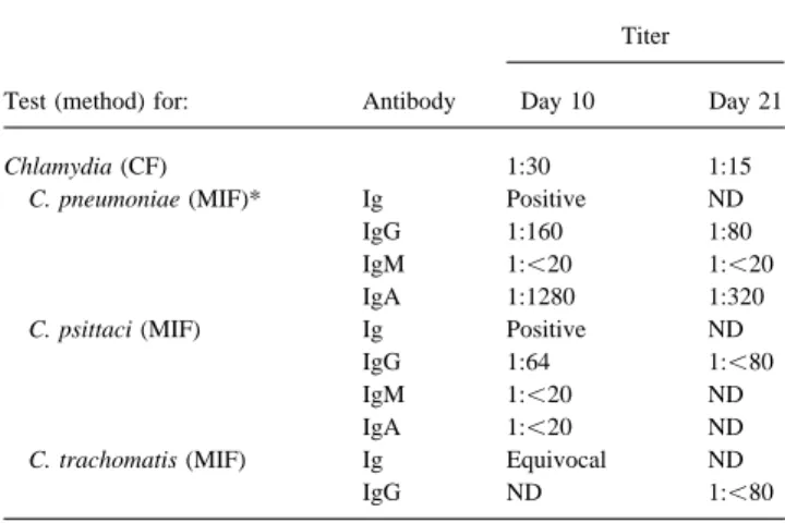
Table 1. Serological evaluation of a 67-year-old woman presenting and IgA after surgery and adequate antibiotic treatment. Evaluation
with acute pulmonary edema. for IgM was always negative, suggesting either reinfection or
reac-tivation of C. pneumoniae infection. In contrast to the patient Titer described by Marrie et al. [2], our patient had no fevers, sweats, chills, or productive cough. That patient had declining IgM titers Test (method) for: Antibody Day 10 Day 21
during the observation period, suggesting primary infection. In another report, an increase in IgG titers from 1:64 to 1:512,
fol-Chlamydia (CF) 1:30 1:15
lowed by a decrease to 1:32 without IgM response, suggested C. pneumoniae (MIF)* Ig Positive ND
reinfection [3]. The third case also involved increasing IgG against
IgG 1:160 1:80
C. pneumoniae, without IgM [4].
IgM 1:õ20 1:õ20
IgA 1:1280 1:320 In none of the previously published cases was PCR examination C. psittaci (MIF) Ig Positive ND of the valve performed. Immunohistochemistry proved to be nega-IgG 1:64 1:õ80 tive when performed, as it was in our case. In a recent report, IgM 1:õ20 ND nonrheumatic stenotic aortic valves without evidence of endocardi-IgA 1:õ20 ND tis were frequently positive for C. pneumoniae, either by immuno-C. trachomatis (MIF) Ig Equivocal ND
staining or PCR [5]. These findings question the relevance of the
IgG ND 1:õ80
positive PCR in our case. Although it is conceivable that our patient did have C. pneumoniae endocarditis with concurrent myo-NOTE. CF Å complement fixation; MIF Å microimmunofluorescence;
NDÅ not determined. Slides from MRL Diagnostics, Cypress, CA. cardial infarction, an etiologic role for this pathogen in any of * Results confirmed by S.-P. Wang (Department of Pathobiology, University these events is not proven.
of Washington, Seattle).
This case report suggests that C. pneumoniae infection, with its tropism for the human vascular system, may be simultaneously activated in various parts of the system, such as coronary arteries and heart valves, producing complex diseases [6]. As evidence of On day 15, pain and swelling of the right knee were noted.
Cultures of joint fluid remained sterile. On day 18, the results of the widespread presence of C. pneumoniae in the cardiovascular system increases, clinicians are likely to face more diagnostic di-various serological examinations were reported and revealed a
positive reaction for C. pneumoniae (see table 1), while antibodies lemmas, as in our case. to Brucella, Coxiella burnetii, Legionella, Bartonella, and
Trepo-nema were undetectable. On day 19, the aortic valve was replaced. Heinz J. Schaad, Raffaele Malinverni, Lee Ann Campbell,
The right coronary leaflet was partially destroyed and contained a and Lukas Matter
grayish vegetation with a diameter ofÇ4 mm. Aerobic and anaero- University Hospital (Department of Medicine and Medizinische Poliklinik) and the Institute for Medical Microbiology, University of bic cultures of valve tissue remained negative. PCR for C.
pneu-Berne, pneu-Berne, Switzerland; and Department of Pathobiology, University moniae (performed at the Department of Pathobiology, University
of Washington, Seattle, Washington, USA of Washington, Seattle [1]) was positive in a specimen from the
left aortic leaflet but negative in material from the right leaflet.
References The postoperative course was complicated by severe bleeding
in the operative area, necessitating repeated surgery with mass 1. Campbell LA, Perez-Melgosa M, Hamilton DJ, Kuo CC, Grayston JT. transfusions. The patient died 15 days later, following progressive Detection of Chlamydia pneumoniae by polymerase chain reaction. J multiorgan failure due to invasive candidiasis. Necropsy was not Clin Microbiol 1992; 30:434 – 9.
2. Marrie TJ, Harczy M, Mann OE, et al. Culture-negative endocarditis proba-permitted by the patient’s relatives.
bly due to Chlamydia pneumoniae. Clin Infect Dis 1990; 161:127 – 9. A MEDLINE search revealed only three reports of possible and
3. Dumont D, Mathieu D, Alemanni M, Eb F, Manigand G. Endocardite probable endocarditis due to C. pneumoniae [2 – 4]. There was
d’Osler probablement due a` Chlamydia pneumoniae (souche TWAR). evidence of myocardial infarction in our patient, possibly due to
Presse Med 1990; 19:1054. a thromboembolic event. In fact, coronary angiography showed
4. Norton R, Schepetiuk S, Tuck WK. Chlamydia pneumoniae pneumonia normal coronary arteries except for a complete obstruction of the
with endocarditis. Lancet 1995; 345:1376 – 7.
first marginal vessel. The recently diagnosed aortic insufficiency 5. Juvonen J, Laurila A, Juvonen T, et al. Detection of Chlamydia pneumoniae with a large vegetation and observations during surgery were com- in human nonrheumatic stenotic aortic valves. J Am Coll Cardiol 1997;
patible with infective endocarditis. 29:1054 – 9.
Microbiological evaluation revealed an evolving antibody re- 6. Danesh J, Collins R, Peto R. Chronic infections and coronary heart disease: is there a link? Lancet 1997; 350:430 – 6.
sponse to C. pneumoniae, with rapidly decreasing titers of IgG