Repetitive proteins from the flagellar cytoskeleton of
African trypanosomes are diagnostically useful antigens
M. IMBODEN1, N. MULLER2, A. HEMPHILL2, R. MATTIOLI3 andT. SEEBECK1 1Institute of General Microbiology, University of Bern, Baltzerstrasse 4, 3012 Bern, Switzerland 2
Institut fiir Parasitologie, Universitdt Bern, La'nggasstrasse 122, 3012 Bern, Switzerland 3
International Trypanotolerance Centre (ITC), PMB 14, Banjul, The Gambia (Received 25 July 1994; revised 8 September 1994; accepted 15 September 1994)
SUMMARY
Trypanosome infection of mammalian hosts leads, within days, to a strong early response against a small, distinct number of parasite proteins. One of these proteins is the variable surface glycoprotein (VSG). Most of the others are apparently non-variable, intracellular trypanosome proteins. Two of these antigens I2 and I17 are now characterized at the molecular
level. Both exhibit a highly repetitive amino acid sequence organization, but they show no sequence similarity either to each other or to any other proteins known to date. Preliminary serological analyses indicate that both allow the early, sensitive and specific detection of infections with different species of trypanosomatids, making them interesting candidates for the development of diagnostic tools for trypanosomiasis detection.
Key words: Trypanosoma brucei brucei, Trypanosoma congolense, Trypanosoma vivax, cytoskeleton, repetitive protein, antigen, diagnosis.
INTRODUCTION
The strong immune response of a mammalian host infected with African trypanosomes is mainly di-rected against the variant surface glycoprotein, VSG (Cross, 1990) and is not protective (Seed & Sechelski, 1987; DeGee, Levine & Mansfield, 1988). This lack of protectivity is because of the high variability of the parasite's surface coat during the course of the infection. Much effort has been directed towards the search for invariant antigens both as tools for diagnosis and as potential targets for vaccine de-velopment. Such antigens are often high molecular weight proteins and associated with the flagellum or the cytoskeleton of the parasite (Lafaille et al. 1989; Ruiz et al. 1990; dos Santos et al. 1992). Muller et al. (1992) have identified 2 invariant, repetitive high molecular weight proteins associated with the cyto-skeleton of the parasite which exhibited a high immunodiagnostic sensitivity. Time-course studies of the infection demonstrated that these 2 antigens were representatives of a whole group of high molecular weight (HMW) antigens which are all recognized very early in infection (Muller et al. 1993). Based on these findings, we have designed a screening strategy specifically aimed at identifying additional members of this group. A cDNA library from bloodstream T. gambiense was screened with affinity-purified antibodies against high molecular weight cytoskeleton proteins. T h e present study reports the identification of two new antigens, I2 and
I17, by this strategy. Both exhibit a high repetitive
sequence organization, and both are structural elements of the cytoskeleton.
MATERIALS AND METHODS
Trypanosomes
The trypanosomes used for cytoskeletal prepara-tions, Western blot analysis, immunofluorescense and immunogold electron microscopy were all pro-cyclic forms of Trypanosoma brucei brucei clone EATRO 427. They were grown in SDM-79 medium (Brun & Schonenberger, 1979) supplemented with haemin and 5 % foetal calf serum. T h e cells were harvested during exponential growth at a cell density of about 5 x l O6/ m l .
Sera
Trypanosome cytoskeletons were prepared as de-scribed by Hemphill et al. (1991). Briefly, trypano-somes from 10 ml of culture were washed 3 times in M M E (10 mM Mops/1 mM MgCl2/0-2 mM E G T A ,
pH 6-9) and incubated in M M E containing 0 5 % Triton X-100 for lOmin on ice. After a centrifug-ation at 3000 £ for lOmin the Triton X-100-insol-uble cytoskeleton fraction was washed once in PBS (137 mM NaCl/2-7 mM KC1/8-1 mM N a2H P O4/
1-5 mM KH2PO4, pH 72) and resuspended in 100 ml
of PBS. Rats were inoculated intraperitoneally with this cytoskeleton material either in complete (1st inoculation) or incomplete (2nd and 3rd inoculation) Freund's adjuvants. Blood was finally collected by cardiac puncture. Sera from experimentally infected mice were taken 14 days after an experimental infec-tion with the LouTat 1 strain of T. b. brucei (DeGee
et al. 1988). The bovine sera used for the evaluation Parasitology (1995), 110, 249-258 Copyright © 1995 Cambridge University Press
of the diagnostic sensitivity originated from cyclical or experimental infections of N'Dama and Zebu cattle with either T. b. brucei, T. vivax or T.
congo-lense. The control sera originated from young (10
months) Zebu cattle. All bovine sera were kindly provided by Dr R. Mattioli, International Trypano-tolerance Centre (ITC) in Banjul, The Gambia.
Lambda gtll cDNA expression of T. b. gambiense
The cDNA expression library from bloodstream form T. b. gambiense (stock TREU 1285) was constructed in the cloning vector Agtl 1 as described (Barnes et al. 1989) and was kindly provided by Dr M. Selkirk (Department of Biochemistry, Imperial College of Science, Technology and Medicine, London).
Immunoscreening of a cDNA library and construction of recombinant A lysogen
The Agtl 1 expression library of T. b. gambiense was screened for clones corresponding to high molecular weight proteins. The antibody used for screening was prepared as follows. Whole procyclic trypano-some lysates (see below) were fractionated by SDS-PAGE and blotted onto nitrocellulose filters. After staining with 0-04% Ponceau S in 10% acetic acid, the high molecular weight region of the filter ( > 180 kDa) was excised and used for affinity-purification of the antibodies from hyperimmune rat serum raised against trypanosome cytoskeletons (Muller et al. 1992).
Affinity purification of antibodies on fusion protein
Affinity purification of antibodies from rat anti-cytoskeleton hyperimmune serum on /?-galactosidase fusion proteins was done as described by Muller et al. (1992). The affinity-purified antibodies were used at a dilution of 1:10 for Western blot analysis, immunofluorescence and immunogold electron mi-croscopy.
(Laemmli, 1970). The cytoskeletal fraction was obtained by resuspending 4-5 x 10⁸ cells in MME containing 0-5 % Triton X-100 and incubating them for lOmin on ice (Hemphill et al. 1991). For the preparation of the flagellar fraction, cytoskeletons were resuspended in 2 ml of 0-1 x MME containing 1 M NaCl (Schneider, Hemphill & Seebeck, 1988). After sonication for 15s the suspension was kept on ice for 20 min. The cytoskeletons and the flagellar suspension as well as the supernatant of the cyto-skeletons were all precipitated with 4:1 methanol/ chloroform (Wessel & Fliigge, 1984) to remove salts and detergent and the final protein pellets were solubilized in 1 ml of sample buffer. The Western blot analysis for the determination of the diagnostic sensitivity of I2 and I17//?-galactosidase fusion
pro-teins was performed as described by Muller et al. (1992). Serum antibodies to /?-galactosidase and other components of E. coli were previously removed by pre-adsorption of the bovine sera to a lysate of Agtl 1 lysogens from strain Y 1089 (Huynh, Young & Davis, 1985). The peroxidase-dependent colour reaction was performed using either 3,3'-diamino-benzidine tetrahydrochloride as described by Vogel
et al. (1988) or Enhanced Chemoluminescence
(ECL, Amersham) according to the manufacturer's specifications.
DNA sequencing and hybridization
Nested deletions of DNA fragments were prepared with exonuclease III double-stranded nested de-letion kit (Pharmacia, Sweden); subclones were double-strand sequenced using the Sequenase DNA sequencing kit (United States Biochemical Cor-poration). For hybridization analysis, DNA was blotted onto nylon filters and was hybridized in 0-lxSSC (15 mM NaCl, 1-5 mM Na3citrate), 4x
Denharts, 008% SDS, 80 mM sodium phosphate pH 6-5, 80 mg/ml herring sperm DNA at 65 °C for 14 h. The filters were washed 3 times for 10 min in 0-lxSSC, 0 1 % SDS for 20 min at 65 °C. Both hybridizations were done using purified inserts of AI2 and AI17 respectively.
Subcloning of Agtl 1 inserts
The cloning of recombinant AI2 and AI17 cDNA
inserts into Bluescript plasmid KS-plus (Stratagene) was performed as described by Rindisbacher et al. (1992).
Western blot analysis
The trypanosomal fractions used for the Western blot analysis were prepared as follows. For whole cell lysates 4-5 x 10⁸ trypanosomes were washed once in MME and then lysed in 1 ml of sample buffer
Immunofluorescence
Trypanosomes were cultivated as described above. After 3 washes in PBS, cells (approximately 107/ml) were applied to cover-slips previously coated with
100/^g/ml polylysine and were allowed to settle for 1 h in a moist chamber at room temperature. Cover-slips were then rinsed twice in MME and treated with 2% formaldehyde in MME for 15 min. The fixed trypanosomes were permeabilized in methanol at — 20 °C for 10 min and subsequently rehydrated in PBS for 30 min. For the preparation of cyto-skeletons, cells were extracted on the cover-slips with MME containing 0-5 % Triton X-100 for 5 min
at room temperature prior to fixation. After rehy-dration, cover-slips were incubated for 1 h in blocking buffer (PBS containing 100 mM L-lysine and 1 % BSA). For immunostaining, cover-slips were incubated for 45 min in a moist chamber at room temperature with affinity-purified antibody diluted 1:10 in blocking solution. After washing the cover-slips 6 times in PBS, FITC-conjugated rabbit anti-rat Ig (Dakopatts, Denmark; diluted 1:100 in blocking solution) was applied as described for the affinity-purified antibodies. After removing excess second antibody by washing 6 times in PBS, cover-slips were mounted on a slide using a mixture of gelvatol/glycerol in PBS as embedding medium (Lawson, 1983).
Immunogold electron microscopy
Washed trypanosomes were allowed to settle onto a Formvar carbon-coated grid for 1 h prior to ex-traction in MME containing 0-5% Triton X-100 as described above. After rinsing the cover-slips in MME, the cytoskeletons were fixed in 3-7 % para-formaldehyde in MME, incubated in blocking buffer and exposed to the antibodies as described for immunofluorescence, with the modification that the second antibody was an affinity-purified goat anti-rat IgG coupled to 10 nm colloidal gold (Janssen, Beerse, Belgium), diluted 1:5 in 20 mM Tris-HCl, pH 82, containing 1 % BSA. After washing the grids 6 times for 5 min in PBS, they were fixed with 2% glutaraldehyde in PBS for 20 min. Grids were then washed 6 times in H2O, and negative staining was
carried out in 3 changes of 1 % uranyl acetate, 20 s each. Finally, grids were air-dried and electron micrographs were taken with a Philips EM 300 microscope operating at 60 kV.
RESULTS
High molecular weight proteins associated with the cytoskeleton are antigens
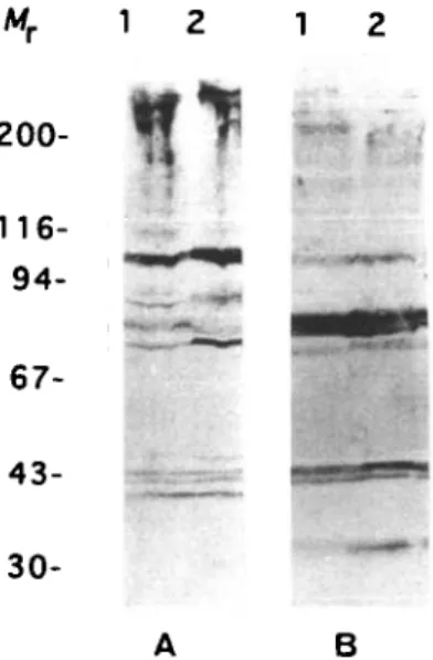
When whole trypanosome extracts were anlaysed by immunostaining with infected bovine serum, a discrete banding pattern was observed (Fig. 1 A). Several distinct bands could be discerned in the 30—100 kDa region, and heavy staining was observed in the high molecular weight range, in agreement with earlier observations (Miiller et al. 1992). Unexpectedly, the staining patterns obtained with whole cell lysates (Lane 1) and with purified cytoskeletons (Lane 2) were very similar, indicating that antibodies in the infected serum are mainly directed against cytoskeletal components. This no-tion is further supported by the observano-tion that a qualitatively similar staining pattern was also ob-tained with hyperimmune rat serum raised against trypanosomal cytoskeletons (Fig. 1 B).
200-% :<• 67- 43- 30-B
Fig. 1. Western blot analysis of whole trypanosomal cell extracts (Lane 1) and cytoskeletons (Lane 2) probed with (A) a bovine serum taken 14 days after infection and (B) with an anti-cytoskeleton rat hyperimmune serum. Molecular weight markers are given in kDa.
B Fig. 2. Immunofluorescent staining of fixed trypanosomal cytoskeletons probed with (A) anti-cytoskeleton rat hyperimmune serum and (B) affinity-purified anti-HMW antibody.
In order to select antibodies with specificity against high molecular weight (HMW) proteins, the > 180 kDa region of gel-fractionated whole trypano-some extracts was used for affinity purification of antibody from hyperimmune anti-cytoskeleton serum. Immunofluorescence microscopy shows that hyperimmune serum against cytoskeletons reacts with almost the entire cell (Fig. 2 A) whereas affinity-purified HMW-specific antibodies predominantly stain the flagellum (Fig. 2B).
Results presented in Figs 1 and 2 indicate that most of the host antibodies formed in the first 2 weeks of infection react with components of the cytoskeleton. Furthermore, many of the antibodies which are specific for HMW proteins react with components of the flagellum, suggesting that the flagellum is a source of strongly immunogenic antigens of high molecular weight.
A
1 PHSVSRHNGQ TQXAVGYAPS FSBLBABASI VVLSLGPFAR 41 BPVCSAVNID SBSEKHDNLL OVLLTAOSH7 SSBCIPIBIV 81 RIPLCNCMR 90 BDLTKAEELDBPVADTEVABKBPTDSBVIPBKBIPDTBAASBQPA (1) 135 T (2) 180 T (3) 225 A (4) 270 T (5) 315 A <6) 360 A (7) 405 T
429 YIBRDSLRSL THABAKVSAB KKAAHPHRBI VVKKSALT7A 469 WANLVIVSPV HRRCSSHSAN RLPPCPRAIN GPYVWYPA'
kb = > -cc -cc o o = > -Hind •£ cc o cco UJ
£
B 1 30 59 B7 116 145 174 202 231 259 287 315 344 372 (1) (2) (3) (4) (5) (6) (7) (8) (9) .A Q (10) .A Q (11) .A VA (12) .A Q (13) .A AA. . . (14) LAVBALBELEEPQQ1/PAEAQPBAQ PEGDX k Q 399 GBALVOLDVB EPD*Fig. 3. Sequence analysis of the inserts in AI2 and AI17.
(A) Deduced amino acid sequence from the AI2 cDNA
sequence. Between amino acids 90 and 428 the sequence consists of seven and a half highly conserved repeat units of 45 amino acids. One amino acid can be either alanine or threonine. (B) Amino acid sequence deduced from the AI17 cDNA sequence. The sequence consists
of 14 highly conserved repeat units of either 29 or 28 amino acids. The first amino acid (leucine) of the given sequence does not belong to the repeat. At 2 positions within the repeat amino acid exchanges occur: from valine to alanine, and from glutamine to leucine or to valine plus alanine or to alanine plus alanine. The C-terminus of I17 consists of 13 unrepeated amino acids.
In both panels, identical amino acids are indicated with a period and in-frame stops are marked by asterisks. The sequences have been deposited in the
GenBank/EMBL data library under the accession numbers Z36280 (I2) and Z36281 (I17).
Screening of a T. b. gambiense expression library with an affinity-purified antibody
In order to identify genes for such HMW flagellar antigens, a Agtl 1 library of bloodstream T. b.
gambiense (Barnes et al. 1989) was screened with the
anti-HMW protein antibody. The phages recovered from this screening represented a number of dif-ferent genes, two of them, I2 and I17, were further
analysed in this study.
0-5 —
0-3 —
0-15 —
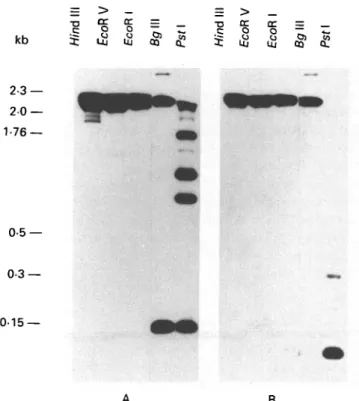
A B Fig. 4. Southern blot analysis of digested genomic
Trypanosoma brucei brucei DNA. The DNA was
hybridized with an I2-specific probe (A) and with an I17
-specific probe (B). Size markers are Hin&\\\ fragments of phage A DNA.
Afr
200-
116-B
Fig. 5. Western blot analysis of different trypanosomal fractions. Lane 1 contains whole trypanosomal cell extracts, Lane 2 contains the cytoskeletal fraction of trypanosomes, Lane 3 corresponds to the supernatant of the cytoskeletal fraction and Lane 4 contains a flagellar fraction of cytoskeletons. In (A) the filter was probed with affinity-purified anti-I2//?-galactosidase fusion
protein antibody and in (B) with affinity-purified anti-I17//?-galactosidase fusion protein antibody. Molecular
weight makers are given in kDa.
I2 and /1 7 both show repetitive sequence motifs
The inserts of recombinant AI2 and AI17 were
subcloned into Bluescript plasmid KS-plus and were sequenced as described in the Materials and Methods section. The analysed fragment of I2 has a length of
2004 bp. Its sequence has an in-frame stop codon at position 1518, followed by an untranslated stretch and a poly-A tail (not shown). This suggests that the
cloned cDNA fragment of I2 represents the
C-terminus of the I, gene. A 1017 bp stretch of the translated part (Fig. 3 A) consists of a highly conserved 135 bp repeat. Only a single amino acid position of the repeat is not fully conserved and contains either alanine or threonine. The 3' end of the coding sequence (300 bp) is not repetitive. The analysed fragment of I17 has a length of 1539 bp. The
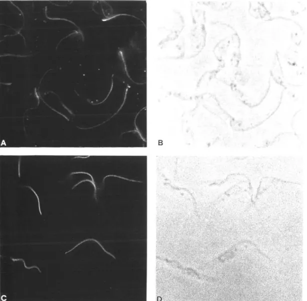
Fig. 6. Intracellular localization of I2 and I17 by immunofluorescence microscopy (A and C). The cytoskeletal
preparation of procyclic Trypanosoma brucei brucei was probed with affinity-purified anti-I2/I17//?-galactosidase fusion
protein antibody. The corresponding phase-contrast micrographs are shown in B and D.
1233, followed by about 300 bp of untranslated region and a poly-A tail, which again indicates that the sequenced fragment represents the C-terminal part of the I17 gene. T h e derived amino acid sequence
of I17 is shown in Fig. 3B. Most of the I17 sequence
(1179 pb) consists of a highly conserved repeat of 87 bp with only 3 positions not fully conserved. The C-terminus of I17 is formed by a short stretch (14
amino acids) of non-repetitive sequence. Database searching with both the I2 and I17 amino acid
sequences revealed no similarity with any known sequences.
Genomic organization of the I2 and /1 7 gene
The cDNA sequence of I2 and I17 showed that both
genes are internally repetitive. The genomic organi-zation of the genes of I2 and I17 was subsequently
determined by restriction mapping. Genomic DNA
of T. b. brucei was digested with restriction enzymes which either do or do not cut within the repeat sequence and was analysed by Southern blotting. Hybridization of genomic digests with an I2-specific
probe (Fig. 4A) revealed that Pst I and Bglll, which both cut once within the I2 repeat, generate a
strongly hybridizing fragment, with the size of the repeat unit (135 bp). Hindi 11, EcoRV and EcoRl which do not cut within the repeat all generate large fragments ( > 15 kDa). This observation indicates that I2 consists of the conserved 135 bp repeat
throughout most of its length. The additional fragments (1-2 kb) generated by Pst I may be indicative of occasional point mutations within the repeat.
Similarly, hybridization with the I17-specific probe
(Fig. 4B) gives a strong signal in the upper molecular weight range ( > 15 kb) for enzymes which do not cut in the repeat (Hindlll, EcoRV, EcoRl and Bgl
B
Fig. 7. Immunogold labelling of I2 (A) and I17 (B) with negatively stained whole-mount cytoskeleton preparation
probed with the corresponding affinity-purified antibodies (see Materials and Methods section). (A) Gold particles are present mainly along the paraflagellar rod (pfr) of the flagellum. (B) Gold particles cover the area between the paraflagellar rod and the axoneme (ax) of the flagellum.
1 2 3 4 5 6 7 8
9 10 11 12 13 14 15 16 17 18• p -
• S
19 20
•*••#
4
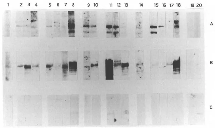
BFig. 8. Western blot analysis demonstrating the diagnostic sensitivity of I2 and I17. Protein extracts from E. coli
lysogens of AI2 (A), AI17 (B) and Agtll (C) were blotted and cut into strips. Each strip was then incubated with a
bovine serum. The sera originated from cyclical infections by Trypanosoma vivax (Lanes 2-4), T. congolense (Lanes 5-8), T. b. brucei (Lanes 1, 9, 10), or from mixed infections by T. b. brucei/T. vivax (Lanes 11-13) or T. b. brucei/T.
congolense (Lane 14) or from experimental infections with T. congolense (Lanes 15-18). Strips 19 and 20 were
incubated with two different uninfected sera.
II) whereas Pst I, which cuts once within the repeat, generates a very strong band at about 90 bp. This correlates well with the size of the repeat defined by sequence analysis (87 bp). The additional two faint bands correspond to dimers and trimers of the repeat unit. This is in good agreement with the available sequence data which show that the Pst I restriction site is missing in three of the 14 repeats sequenced. In contrast to I2, the I17 hybridization probe does not
reveal additional bands, indicating that the whole I17
gene has a repeated structure.
/2 and lxl are both high molecular weight proteins
The screening strategy used for gene isolation, as well as sequence and hybridization data predict that I2 and I17 both are high molecular weight proteins.
In order to confirm this prediction at the protein level, the following Western blot analysis was performed. Whole cells, cytoskeletons and flagella of trypanosomes were prepared (see Materials and Methods section) and fractionated by S D S - P A G E . After transfer to nitrocellulose, they were probed with affinity-purified antibodies against I2 and I17
//?-galactosidase fusion proteins. Both antibodies re-vealed several distinct bands in the high molecular weight ( > 1 8 0 k D a ) region. Both proteins are detected by immunostaining of whole cell extracts,
in Triton-insoluble cytoskeletons and in salt-extracted flagella (Fig. 5) indicating that both proteins are tightly associated to the flagellar cyto-skeleton.
Localization of I2 and I17
Having established the tight association of I2 and I17
with the flagellar cytoskeleton, an immunofluores-cent staining of cytoskeletons and whole cells with affinity-purified anti-I2 and anti-I17 antibodies was
performed. The results of this staining are shown in Fig. 6. Both I2 and I17 antibodies induce a bright
fluorescence along the flagellum. As both antibodies stain the flagellum with a very similar pattern, a more detailed localization was required. This was achieved by immunogold electron microscopy (Fig. 7). Antibody against I2 reacts with the paraflagellar
rod (Schlaeppi, Deflorin & Seebeck, 1989) sug-gesting that I2 is a component of this structure. In
contrast, antibody against I17 specifically stains the
interface between the paraflagellar rod and the axoneme. While immunogold electron microscopy thus confirms the biochemical and immunofluores-cence data indicating a close association of I2 and I17
with the flagellum, it further demonstrates that the detailed localization of the two proteins within the flagellum is quite distinct.
Immunodiagnostic potential of I2 and /17
In order to evaluate the potential of I2 and I17 for
diagnostic purposes, the respective /9-galactosidase fusion proteins were used to analyse, by Western blotting, 18 sera from N'Dama and Zebu cattle with parasitologically confirmed infections with T. b.
brucei, T. congolense, T. vivax, as single and as mixed
infections. Also, two uninfected sera were included as controls. Fig. 8 demonstrates that 15/18 infected sera reacted with I2 and 17/18 with I17 but none with
the /9-galactosidase control protein. All sera reacted with at least one of the two recombinant antigens. The serum which was taken 5 days after infection reacted with both recombinant antigens. No re-activity was seen with the 2 control sera.
DISCUSSION
In an earlier study we identified two trypanosomal antigens to which antibodies are produced early in an infection, MARP1 and GM-6 (Miiller et al. 1992; Hemphill et al. 1992), as high molecular weight proteins both associated with the cytoskeleton. As both of them exhibited a high immunodiagnostic sensitivity (90%), we wanted to know if African trypanosomes contain such additional cytoskeletal antigens. Therefore we have designed a screening protocol for the identification of these antigens. The present study described two antigens isolated by this strategy, I2 and I17. Both are high molecular weight
proteins (MW > 180 kDa), both are highly intern-ally repetitive and consist predominantly of con-served repeat units of 45 and 29 amino acids, respectively. Thus I2 and I17 share several important
features with GM6 and MARP1, but all 4 proteins show neither amino acid sequence similarity nor immunological cross-reactivity (data not shown). I2
and I17 are both located in the flagellum, though at
different locations within this structure. The data presented in Fig. 8 indicate that I2 and I17 might be
of diagnostic interest in that they represent proteins which (i) are strongly immunogenic already in the early phase of infection, (ii) elicit an appreciable antibody response in all infected hosts, and (iii) are highly conserved between the African trypano-somatids (T. brucei, T. congolense or T. vivax).
The finding that these trypanosome antigens are highly repetitive is in line with what we already have described for two other early antigens, MARP-1 and GM6 (Muller et al. 1992, 1993). These observations contribute to the emerging overall picture that the host antibody response both in the African trypano-somiases as well as in South American Chagas disease is strongly directed against parasite proteins which have the common denominators of being (i) very large and (ii) highly internally repetitive with a very high degree of sequence conservation between the individual repeat units (Ibanez et al. 1988; Hoft et al.
1989; Lafaille et al. 1989; Duncan, Gay & Donelson, 1991; Pollevick et al. 1991; Burns et al. 1993). In addition, all these antigenic proteins are intracellular proteins, with the exception of SAPA, which is a GPI-anchored surface protein (Pollevick et al. 1991). Several aspects of the molecular characteristics of these antigens are noteworthy. Already the mere fact that proteins consist of so many so well-conserved repeat units is peculiar. Highly repetitive proteins are not common in biology, and where they exist, e.g. spectrin in the erythrocyte, they exhibit a large extent of sequence variation between individual repeat units (Alcina et al. 1988). Conceptually, an internally repetitive organization of a protein might have evolved because the protein in question inter-acts with a repetitive substrate and is under evol-utionary pressure to form many identical interactions with the substrate. This line of argument would be particularly cogent for MARP-1, which was orig-inally identified as a microtubule-associated protein of trypanosomes (Schneider et al. 1988; Hemphill et
al. 1992) and thus interacts with a lattice of regularly
spaced tubulin dimers. However, although similar pressures should exist in other organisms, no comparable degree of repetitiveness is found else-where. Many microtubule-associated proteins from different organisms have been characterized and, where repetitive domains have been found, they consist of a small number of not very highly conserved repeat units (Lewis, Wang & Cowan, 1988).
These findings argue that it is not the biological function of the various internal trypanosome anti-gens which exert the pressure to build up highly repetitive proteins and to maintain the observed near-perfect sequence conservation. So what does ? Is it conceivable that the high molecular weight repetitive proteins have been generated by the trypanosome as devices for liberating, upon cell destruction by host defence mechanisms, large amounts of identical, immunologically active pep-tides which stimulate the immune system? Since many of the infecting trypanosomes are invariably destroyed at the site of an infection, this concept would guarantee a rapid and effective stimulation of the host's immune system, some parts of which might be beneficial for the parasite. The concomitant stimulation of antibodies against the peptides would be of no consequence to the trypansome because the target proteins are safely sheltered inside the cell. A similar situation is the strong stimulation of the host's immune response by the variant surface coat (VSG) which is the most prominent external antigen of the trypanosome (Cross, 1990). Here a different VSG-type population of parasites is already growing when the response against the former one is fully raised. Thus the parasite, after having stimulated a response against itself, can survive without any disadvantage.
While we clearly need to learn much more about the possible roles of these repetitive proteins in the course of an infection, the available data demonstrate that they could play a most useful role, in a more practical sense, as diagnostic reagents.
We would like to thank Toni Wyler for his hospitality at the electron microscope and Yvonne Schlatter for her technical support. This work was supported by grant No. 31-30870.91 of the Swiss National Science Foundation and a grant of the Stanley Thomas Johnson Foundation.
R E F E R E N C E S
ALCINA, A., HARGREAVES, A. J., AVILA, J. & FRESNO, M. (1988). The detection of a spectrin-like protein in
Trypanosoma cruzi with a polyclonal antibody. Cell Biology International Reports 12, 979-85.
BARNES, D. A., MOTTRAM, J., SELKIRK, M. & AGABIAN, N. (1989). Two variant surface glycoprotein genes distinguish between different substrains of
Trypanosoma brucei gambiense. Molecular and Biochemical Parasitology 34, 135—46.
BRUN, R. & SCHONENBERGER, M. (1979). Cultivation and in vitro cloning of procyclic culture forms of
Trypanosoma brucei in semi-defined medium. Ada Tropica 36, 289-92.
BURNS, J. M., SHREFFLER, W. G., BENSON, D. R., GHALIB,
H. w., BADARO, R. & REED, s. G. (1993). Molecular
characterization of a kinesin-related antigen of
Leishmania chagasi that detects specific antibodies in
African and American visceral leishmaniasis.
Proceedings of the National Academy of Sciences, USA
90, 775-9.
CROSS, G. A. M. (1990). Cellular and genetic aspects of antigenic variation in trypanosomes. Annual Reviews
of Immunology 8, 83-110.
DE GEE, A. L., LEVINE, R. F. & MANSFIELD, J. M. ( 1 9 8 8 ) . Genetics of resistance to the African trypanosomes. VI. Heredity of resistance and variable surface glycoprotein specific immune response. Journal of
Immunology 140, 283-8.
DOS SANTOS, C. N. D., KRIEGER, M. A., ALMEIDA, E., LAFAILLE, J. J., GOLDENBERG, S. & GALLER, R. ( 1 9 9 2 ) .
Trypanosoma cruzi flagellar repetitive antigen
expression by recombinant Baculovirus — towards an improved diagnostic reagent for Chagas disease.
Biotechnology 10, 1474-7.
DUNCAN, L. R., GAY, L. S. & DONELSON, J. E. ( 1 9 9 1 ) . African trypanosomes express an immunogenic protein with a repeating epitope of 24 amino acids.
Molecular and Biochemical Parasitology 48, 11-16.
HEMPHILL, A., AFFOLTER, M. & SEEBECK, T. ( 1 9 9 2 ) . A novel microtubule-binding motif identified in a high molecular weight microtubule-associated protein from
Trypanosoma brucei. Journal of Cell Biology 117,
95-103.
HEMPHILL, A., SEEBECK, T. & LAWSON, D. ( 1 9 9 1 ) . T h e
Trypanosoma brucei cytoskeleton: ultrastructure and
localization of microtubule-associated and spectrin-like proteins using quick-freeze, deep-etch, immunogold electron microscopy. Journal of
Structural Biology 107, 222-20.
HOFT, D. F., KIM, K. S., OTSU, K., MOSER, D. R., YOST, W. J.,
BLUMIN, J. H., DONELSON, J. E. & KIRCHHOFF, L. V. (1989). Trypanosoma cruzi expresses diverse repetitive protein antigens. Infection and Immunity 57, 1959-67. HYUNH, T. V., YOUNG, R. A. & DAVIS, R. W. ( 1 9 8 5 ) .
Construction and screening cDNA libraries AgtlO and Agtll. In DNA Cloning 1 (ed. Glover, D. M.), pp. 49-78. Oxford: IRL Press.
IBANEZ, C. F., AFFRANCHINO, J. L., MACINA, R. A., REYES, M. B., LEGUIZAMON, S., CAMARGO, M. E., ASLUND, L.,
PETTERSON, u. & FRASCH, A. c. c. (1988). Multiple Trypanosoma cruzi antigens containing tandemly
repeated amino acid sequence motifs. Molecular and
Biochemical Parasitology 30, 27—34.
LAFAILLE, J. J., LINSS, J., KRIEGER, M. A., SOUTO-PADRON,
T., DE SOUZA, w. & GOLDBERG, s. (1989). Structure and expression of two T. cruzi genes encoding antigenic proteins bearing repetitive epitopes. Molecular and
Biochemical Parasitology 35, 127-36.
LAEMMLI, u. K. (1970). Cleavage of structural proteins during the assembly of the head of the bacteriophage T4. Nature, London 227, 680-5.
LAWSON, D. (1983). Epinemin: a new protein associated with vimentin filaments in non-neural cells. Journal of
Cell Biology 97, 1891-905.
LEWIS, S. A., WANG, D. & COWAN, N. J. ( 1 9 8 8 ) . Microtubule-associated protein MAP2 shares a microtubule-binding motif with tau protein. Science,
242, 936-9.
MtJLLER, N., HEMPHILL, A., IMBODEN, M., DUVALLET, G.,
DWINGER, R. H. & SEEBECK, T. (1992). Identification and characterization of two repetitive non-variable
antigens from African trypanosomes which are recognized early during infection. Parasitology 104, 111-20.
MULLER, N., IMBODEN, M., DETMER, E., MANSFIELD, ] . M. &
SEEBECK, T. (1993). Cytoskeleton-associated antigens from African trypanosomes are recognized by self-reactive antibodies of uninfected mice. Parasitology
107,411-17.
POLLEVICK, G. D., AFFRANCHINO, J. L., FRASCH, A. C. C. &
SANCHES, D. o. (1991). The complete sequence of a
shed acute-phase antigen of Trypanosoma cruzi.
Molecular and Biochemical Parasitology 47, 247-50.
RINDISBACHER, L., HEMPHILL, A. & SEEBECK, T. ( 1 9 9 3 ) . A repetitive protein from Trypanosoma brucei which caps the microtubules at the posterior end of the
cytoskeleton. Molecular and Biochemical Parasitology 58, 83-96.
RUIZ, A. M., ESTEVA, M., SUBIAS, E., MORENO, M.,
DECAMPANINI, E., VELAZQUEZ, E. & SEGUERA, E. L. ( 1 9 9 0 ) . Monoclonal antibodies against the flagellar fraction of epimastigotes of T. cruzi — immunoprotection against metacyclic trypomastigotes obtained by immunization of mice with affinity-purified antigen. Molecular and
Biochemical Parasitology 39, 117-26.
SCHLAEPPI, K., DEFLORIN, J. & SEEBECK, T. ( 1 9 8 9 ) . T h e major component of the paraflagellar rod of
Trypanosoma brucei is a helical protein that is encoded
by two identical, tandemly linked genes. Journal of
Cell Biology 109, 1695-709.
SCHNEIDER, A., HEMPHILL, A. & SEEBECK, T. ( 1 9 8 8 ) . L a r g e microtubule-associated protein of T. brucei has tandemly repeated, near-identical sequences. Science
SEED, j . R. & SECHELSKI, j . (1987). The role of antibody immunodiagnostic sensitivity and specificity. in African trypanosomiasis. Journal of Parasitology Molecular and Biochemical Parasitology 31, 117-26. 73, 840-2. WESSEL, D. & FLUGCE, U. I. (1984). A method for the
VOGEL, M., GOTTSTEIN, B., MULLER, N. & SEEBECK, T. quantitative recovery of protein in dilute solution in (1988). Production of recombinant antigen of the presence of detergents and lipids. Analytical