PAIN
Expression of the nociceptin precursor and nociceptin
receptor is modulated in cancer and septic patients
U. M. Stamer
1,2,3*
†, M. Book
1,3†, C. Comos
1, L. Zhang
1, F. Nauck
2,4and F. Stu¨ber
1,31Department of Anaesthesiology and Intensive Care Medicine and2Centre for Palliative Medicine, University of Bonn, Germany 3
Department of Anaesthesiology and Pain Therapy, University of Bern, Inselspital, Switzerland
4Department of Palliative Medicine, University Medical Centre, Go¨ttingen, Germany
* Corresponding author. E-mail: ulrike.stamer@dkf.unibe.ch
Editor’s key points
† The nociceptin receptor has a role in pain and inflammatory pathways. † Cells from patients with cancer pain or sepsis had higher nociceptin and lower pre-pronociceptin mRNA expression than controls.
† There was no association with pain scores or analgesia, but some association with inflammatory markers. † Further studies are
needed.
Background.A role of nociceptin and its receptor (NOP) in pain and immune function has been suggested. The hypothesis was that mRNA expression of NOP and the nociceptin precursor pre-pronociceptin (pN/OFQ) in peripheral blood cells differs in end-stage cancer patients suffering from chronic pain and septic intensive care unit (ICU) patients compared with healthy controls.
Methods. Blood samples were drawn from end-stage cancer patients and septic ICU patients. Additionally, postoperative patients representing individuals with surgical stress and healthy controls were enrolled as comparative groups. NOP and pN/OFQ mRNA expression, quantified by real-time polymerase chain reaction (RT-PCR), was compared between study groups, and associated to opioid medication, pain intensities, and the inflammatory markers procalcitonin (PCT) and interleukin-6.
Results. NOP expression was significantly higher in cancer patients [normalized ratio, median (inter-quartile range): 10.2 (7.4/17.8)], postoperative patients [8.0 (5.3/10.2)], and ICU patients [6.6 (4.2/9.5)] compared with healthy controls [4.4 (2.7/7.0); P,0.001]. Expression of pN/OFQ was lower in cancer patients [3.8 (1.9/5.9)] and ICU patients [1.9 (1.0/2.7)] but not in postoperative patients compared with healthy controls [7.2 (6.1/9.4); P,0.001]. Increased plasma PCT was associated with decreased pN/OFQ in all patient groups. In cancer patients, no association was seen with pain scores, opioid medication or duration of analgesia, and NOP or pN/OFQ mRNA.
Conclusions.NOP and pN/OFQ expression in peripheral blood cells was modulated in end-stage cancer and septic patients compared with healthy controls, whereas changes in postoperative patients were minor. The involvement of the NOP– pN/OFQ system in inflammation, impaired immune function, and pain has to be further elucidated.
Keywords: cancer; inflammation; opioid peptides, nociceptin-orphanin FQ; receptor, nociceptin
Accepted for publication: 3 December 2010
The nociceptin receptor (NOP, opioid receptor-like 1, orphan opioid receptor, OP4) is a member of the G-protein-coupled
receptor superfamily and exhibits about 60% sequence homology with the classical opioid receptors, especially in the putative membrane-spanning domains and intracellular loops.1 2 Its endogenous agonist is N/OFQ (nociceptin, orphanin FQ).3 4 It also shares sequence homology with classical opioid peptides such as dynorphin, however, with very low potency in binding to the classical opioid receptors and a distinct pharmacological profile.5 6 The
cellular signal transduction pathway initiated by NOP activation appears nearly identical to that of classical opioid receptors. However, N/OFQ mediates analgesia when given spinally and hyperalgesia when given intra-cerebro-ventricularily.3 4
As the NOP–N/OFQ-system is involved in pain pathways, it is considered a promising target of pharmacological research in analgesic therapy.7 8Specifically, in inflammatory and neuro-pathic pain, a functional role is discussed.9N/OFQ has been implicated in a wide range of further physiological and
behavioural functions, for example, effects on locomotion, anxiety, fear, stress, tolerance, dependence, feeding, learning, and memory.8 10 11The fact that both receptor and endogen-ous agonist are expressed in the human central nervendogen-ous system and in immune cells at similar levels indicates that it may act as an important mediator of both nervous and immune responses in humans with possible involvement in the brain-immune axis.12 13 Whereas some information on the NOP –N/OFQ system in septic patients is already available,8 studies on the expression of NOP –N/OFQ in patients with cancer pain are lacking. However, this might be a population of specific interest, since immunological and inflammatory changes are prevalent and most of the patients suffer from cancer pain.
The present study investigates mRNA expression encoding NOP and the precursor for its endogenous ligand pre-pronociceptin (pN/OFQ) in whole blood from different cohorts of patients. The hypothesis was tested that patients suffering from end-stage cancer and also septic patients from the intensive care unit (ICU) displaying severe systemic inflammation might have altered baseline expression of NOP and pN/OFQ in peripheral blood cells. Postoperative patients representing subjects with surgical stress and healthy con-trols were also studied as comparative groups.
Methods
Patients and controls
The study was approved by the University Ethics Committee and was conducted according to the principles of the Declaration of Helsinki. Either the participants or a legal cus-todian gave written informed consent. Exclusion criteria were lack of informed consent or age younger than 18 yr. In the cancer patient group, 113 patients with advanced cancer were enrolled. Pain scores at rest and movement were recorded at admission and regularly during the hospital stay by the patients using a numeric rating scale (NRS, 0 denotes no pain; 100 denotes worst pain imaginable). The ICU patients consisted of 18 critically ill patients suffering from severe systemic inflammation and meeting the severe sepsis criteria. In cancer and ICU patients, blood samples (2.5 ml, PAXgene Blood RNA Tube, Qiagen/Becton Dickinson, Germany) were drawn on the day of routine blood sampling at admission. In ICU patients, additional blood was sampled on days 3 and 5 and also after recovery from inflammation-related organ dysfunction or at deterioration in the case of non-survivors.
Postoperative patients and healthy controls served as con-trols. The postoperative patient group (n¼20) had major elec-tive surgery (nephrectomy, prostatectomy, bowel resection, ASA classification I –III) under general anaesthesia using a standardized institutional protocol (fentanyl 0.15 mg, propofol 2–3 mg kg21, and cisatracurium for induction, remifentanil/ isoflurane-based maintenance of anaesthesia). Their blood was drawn after operation in the recovery room after emer-gence from anaesthesia. The healthy controls consisted of 28 healthy volunteers not taking any continuous medication.
cDNA preparation and quantitative real-time
polymerase chain reaction
RNA isolation from blood collected in PAXGene RNA tubes was performed according to the manufacturer’s instructions (Pre-AnalytiX; Qiagen, Hilden, Germany). Total RNA was extracted using QIAampw RNA Blood Kit (Qiagen, Hilden, Germany)
and then dissolved in diethylpyrocarbonate-treated water and stored at 2708C until further analysis. cDNA was produced as polymerase chain reaction (PCR) template using 1st Strand cDNA Synthesis Kit for RT-PCRw [avian myeloblastosis virus
(AMV+), Roche Diagnostics, Mannheim, Germany].
The PCRs for NOP, pN/OFQ, and the house-keeping gene human hypoxanthine phosphoribosyl-transferase (HPRT) were conducted in separate capillaries in duplicate on a Light-Cyclerw2.0 (Roche Diagnostics). The calibrator used was cDNA
(1:50 diluted) prepared from the SK-N-DZ cell line. For the amplification of NOP, 4 ml of LightCyclerwFastStart DNA
Mas-terPLUSSYBR Green I containing FastStart Taq DNA Polymerase, reaction buffer, dNTP mix, SYBR Green I dye, and MgCl2was
used. Ten picomoles of each primer (NOP-f: 5′-TgCCgTTCTggg AggTTATCTA-3′ and NOP-r: 5′-TTAgggTgAAggTgCTggTgA-3′; pN/OFQ-primers as published previously14) and 500 ng of pre-diluted cDNA (according to 30 ng total RNA) were added to the reaction mixture with a final volume of 20 ml. After an initial denaturation step at 958C, amplification was performed with 45/40 cycles (denaturation 958C for 5 s, annealing 608C for 10 s, extension 728C for 20 s).
For detection of HPRT, prediluted cDNA 500 ng, Universal ProbeLibrary 16 nM, Probe #73, LightCyclerwTaqManwMaster
4 ml and 4 pmol of each primer (5′-TgACCTTgATTTATTTTg CATACC-3′ and 5′-CgAgCAAgACgTTCAgTCCT-3′) was used. Fluorescence was monitored at the end of each cycle and detected in channel F2/F1. Data from RT-PCR, including cali-brator and samples, were imported into the Relative Quantifi-cation Software and analysed with the Fit Coefficients File. This was created by PCR amplification of NOP and HPRT in a series of diluted cDNA (relative standard curve) in triplicates. The calcu-lated normalized ratios (arbitrary units) reflect the expression level of target mRNA.
Markers of inflammation
Procalcitonin (PCT) was measured by BRAHMS PCT sensitive Kryptorw test (Brahms GmbH, Hennigsdorf, Germany;
normal values or insignificant systemic inflammatory response: PCT≤0.5 ng ml21; moderate systemic inflamma-tory response, e.g. SIRS: 0.5,PCT≤2.0 ng ml21; severe systemic inflammatory response, sepsis: PCT.2.0 ng ml21). To determine interleukin-6 (IL-6) concentrations, the IL-6-ELISA-CB Kit (BioSource Europe S.A, Belgium) and the ELISA-Reader (BioRad Lab., USA) were used (normal values ,24 pg ml21). White blood cell counts (leucocytes: normal range 4.0 –10.0×10³ml21; lymphocytes 18–45%, neutrophils 45–76%, monocytes 2 –10%) and C-reactive protein (CRP, normal value ,5 mg litre21) were quantified by standard clinical biochemical methods in the central hospital laboratory.
Statistical analysis
Patient characteristic and standard laboratory data were expressed as mean (SD). Owing to high variance and no
normal distribution of measures of mRNA expression, non-parametric comparison was performed using the Kruskal– Wallis test. A P-value of ,0.05 was considered as statistically significant after correction for multiple testing. Results of relative quantification of mRNA are presented as medians (inter-quartile range, IQR). For correlation between mRNA expression and PCT, patients were clustered according to the designated manufacturer’s PCT cut-off levels.
Results
Patients’ characteristics
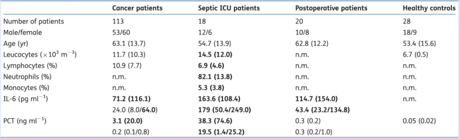
Patient characteristic and laboratory measures are displayed in Table1and details of the cancer patients in Table2. The 18 ICU patients suffered from severe sepsis and nine of them died from sepsis-induced organ failure. From septic patients, a total of 53 blood samples were drawn. Postoperative sub-jects had comparable surgery-related data [ASA I –III, elec-tive major abdominal or urological surgery, duration of anaesthesia: 202.3 (13.1) min, no signs of infection]. Healthy controls were aged 26 –79 yr, which was comparable with the ICU group, but, about 10 yr younger than cancer and postoperative patients. Their medical history did not indicate any severe disease, and they were either taking no drugs at all or at least no drugs for severe systemic co-morbidity.
Markers of inflammation
Laboratory markers of inflammation in ICU patients were elevated in accordance with the severity of their disease (Table1). In cancer patients, a clinically relevant increase in inflammatory markers was observed in some patients, specifi-cally in those with short survival. Correspondingly, IL-6 con-centrations were increased (Table3). PCT as sensitive marker of systemic inflammation due to infection was elevated in
63.9%, 36.0%, and 29.6% of the patients with end of life within 10, 10 –30 days, or after 30 days, respectively.
NOP and pN/OFQ expression in peripheral blood
Expression of NOP differed between patient groups and the healthy controls, with highest measures in cancer patients (Fig. 1A). NOP expression in ICU patients at admission was
also increased. This change was mainly related to an increase in septic non-survivors [8.3 (5.5/10.1); P,0.001], whereas there was no difference in septic survivors [5.0 (4.0/8.2)] compared with healthy controls (HC). Sequential blood samples in ICU patients did not change over time. For pN/OFQ, expression was significantly lower in ICU and cancer patients (P,0.001) but not in postoperative patients (Fig.1B).
Time of survival, daily opioid doses (morphine equiva-lents), and duration of opioid pretreatment (ranging between 0 and 72 weeks) were not related to NOP or pN/ OFQ mRNA in cancer patients. The 16 cancer patients who were not taking any opioid medication did not differ in NOP [8.8 (7.5/15.9)] and pN/OFQ expression [3.4 (2.4/5.9)] com-pared with those receiving opioids [NOP: 10.9 (7.0/17.4); pN/OFQ: 3.7 (1.9/5.6)]. In addition, no difference could be observed in patients with no pain at rest (NRS¼0; n¼29), light pain (NRS¼0 –40; n¼55), or severe pain (NRS≥45; n¼29) at admission to the palliative care unit for NOP or pN/OFQ. Furthermore, there was no difference in expression between different kinds of cancer, existence of bone metastases, or liver metastases [NOP: 11.3 (7.5/23.5) vs 10.9 (7.8/16.9); pN/OFQ: 3.2 (1.4/5.0) vs 3.9 (2.0/6.3)].
For NOP, no significant correlation to plasma concen-trations of PCT and IL-6 was seen. In contrast, patients with increased PCT had lower pN/OFQ mRNA expression com-pared with subjects with normal PCT values (P,0.001) (Fig. 2). These results were also verified in the subgroup of cancer patients [PCT≤0.5 ng ml21: 4.3 (3.3/6.1), 0.5,PCT≤2.0 ng ml21: 3.2 (2.1/6.9), PCT.2 ng ml21: 1.9 (1.0/3.8); P,0.001].
Table 1Patient characteristic and laboratory data from patients suffering from cancer pain, patients with sepsis, postoperative patients, and healthy contols. Data are presented as number of patients, mean (SD), or median (IQR). Laboratory results deviating from the normal range are printed in bold. IL-6, interleukin-6; PCT, procalcitonin; n.m., not measured; ICU, intensive care unit
Cancer patients Septic ICU patients Postoperative patients Healthy controls
Number of patients 113 18 20 28 Male/female 53/60 12/6 10/8 18/9 Age (yr) 63.1 (13.7) 54.7 (13.9) 62.8 (12.2) 53.4 (15.6) Leucocytes (×103m23) 11.7 (10.3) 14.5 (12.0) n.m. 6.7 (0.5) Lymphocytes (%) 10.9 (7.7) 6.9 (4.6) n.m. n.m. Neutrophils (%) n.m. 82.1 (13.8) n.m. n.m. Monocytes (%) n.m. 5.3 (3.8) n.m. n.m. IL-6 (pg ml21) 71.2 (116.1) 163.6 (108.4) 114.7 (154.0) n.m. 24.0 (8.0/64.0) 179 (50.4/249.0) 43.4 (23.2/134.8) PCT (ng ml21) 3.1 (20.0) 38.3 (74.6) 0.3 (0.2) 0.05 (0.02) 0.2 (0.1/0.8) 19.5 (1.4/25.2) 0.3 (0.2/1.0)
pN/OFQ expression also varied depending on IL-6 measures. pN/OFQ expression was 4.1 (2.2/6.1) for individuals with IL-6≤24 pg ml21, 2.5 (1.0/4.0) for those with IL-6 con-centrations of 24 –100 pg ml21, and 1.4 (0.7/2.4) for patients with IL-6.100 pg ml21(P,0.001).
Discussion
The present investigation shows that NOP and pN/OFQ mRNA expression are altered in severely ill patients compared with healthy controls, with highest NOP levels in cancer patients
and lowest pN/OFQ in ICU patients. Although in the present study, there were varying proportions of cell types in patients’ peripheral blood, specifically in the late stage cancer and sepsis, the relative quantification of mRNA content corrects for sample-to-sample variations caused by differences in quality and quantity of nucleic acid. A limitation of our study is the lack of measurement of protein which will be addressed in a future project. However, it is well accepted that peripheral blood mononuclear cells transcribe mRNA encoding NOP.15 16 The receptor protein was detected on the cell surface of all types of white blood cells without any age- or sex-dependent differences.17 The existence of NOP on T-cells and the ability of immune cells to produce N/OFQ5 14suggest an immunomodulatory role. For pN/OFQ, expression in peripheral blood mononuclear cells and
Table 2 Patient characteristic and cancer-related details of cancer patients. Number (%) of patients or mean (SD)
Type of cancer Gastrointestinal tract 27 (23.9%) Lung 23 (20.3%) Breast 17 (15.0%) Cervix, ovary 15 (13.3%) Prostatic 11 (9.7%) Kidney, bladder 10 (8.9%) Others (oropharynx, liver,
pancreas) 10 (8.9%) Metastasis known Bone 43 (38.1%) Liver 44 (38.9%) Lung 21 (18.6%) Visceral 31 (27.4%) Kind of pain Bone 8 (7.1%) Visceral 33 (29.2%) Neuropathic 18 (15.9%) Mixed 36 (31.9%) Other symptoms Nausea/vomiting 37 (32.7%)/19 (16.8%) Tiredness, fatigue/ sedation 40 (35.4%)/9 (7.9%) Constipation 50 (44.2%) Cachexia 35 (30.7%) Oedema 33 (29.2%) Dyspnoea at exertion/at rest 32 (28.3%)/16 (14.2%) Treatment Surgery 104 (92.0%) Chemotherapy 74 (65.5%) Radiation 55 (48.7%) Analgesic drugs at admission no/WHO I/II/III
7 (6.2%)/9 (8.0%)/13 (11.5%)/84 (74.3%) Co-analgesics: antidepressive/gabapentin or pregabalin 35 (31.0%)/5 (4.4%) Cortison 49 (43.4%)
Duration of opioid treatment (months)
7.0 (15.8)
Patients with pain at rest/ movement at admission
84 (74.3%)/99 (87.63%)
Table 3 Comparison of cancer patients allocated to different PCT groups. Data are presented as number of patients, mean (SD), or median (IQR). Laboratory results deviating from normal range are printed in bold. NRS, numeric rating scale 0 –10 for pain scores; CRP, C-reactive protein; IL-6, interleukin-6; PCT, procalcitonin; NRS,
ANOVA, or the Kruskal –Wallis test where applicable; P-values refer
to post hoc analysis; **P≤0.001; *P,0.05 PCT≤0.5 ng ml21 0.5 ng ml21 <PCT≤2.0 ng ml21 PCT>2.0 ng ml21 Number of patients (male/ female) 77 (35/42) 23 (13/10) 13 (5/8) Age (yr) 65.4 (7.6) 65.3 (11.9) 69.3 (8.0) Survival (days) 70.2 (110.8) 38.0 (107.2) 18.9 (25.3) Pain scores at admission (NRS) 20 (0/40) 0 (0/50) 25 (0/40) rest/movement 70 (40/80) 75 (0/90) 45 (30/70) Morphine equivalents/day (mg) 120 (40/180) 30 (0/90) 30 (20/120) NOP (normalized ratio) 10.7 (7.3/16.4) 12.1 (7.4/24.5) 8.5 (5.9/23.1) pN/OFQ (normalized ratio) 4.2 (2.4/6.3) 2.9 (2.1/4.5) 1.4 (0.9/2.6)* Leucocytes (×103m23) 9.9 (11.4) 13.1 (7.9) 24.1 (19.4)* Lymphocytes (%) 14.5 (9.8) 7.9 (5.4)* 7.5 (6.8)* CRP (mg litre21) 8.7 (7.6) 19.0 (22.6)* 25.3 (17.2)** IL-6 (pg ml21) 26.4 (37.2) 78.0 (74.2)** 224.6 (242.1)** 14.2 (5.9/29.0) 47.3 (27.0/100.0) 157.8 (24.0/440.6) PCT (ng ml21) 0.15 (0.13) 1.0 (0.04)** 19.0 (49.8)** 0.1 (0.07/0.17) 0.8 (0.7/1.2) 3.7 (2.7/4.5)
neutrophils has also been described.14 16Ex vivo studies con-firmed that stimulated human polymorphonuclear neutro-phils rapidly secrete N/OFQ by exocytosis with dose-dependent induction of polymorphonuclear chemo-taxis.5 18
Clinical reports have mainly focused on nociceptin protein concentrations, however, no clear-cut correlation between circulating protein levels and pain states could be established in the past.19 Whereas some groups reported increased plasma concentrations in pain states, for example, acute, sub-acute, and chronic pain compared with controls, other publications stated no change in women suffering from labour pain or decreased plasma nociceptin in fibromyalgia syndrome, cluster headache, and migraine.20–22In another trial, cerebrospinal fluid concentrations of nociceptin were linked to opioid medication with lowest levels in the morphine-treated group and highest in the untreated chronic pain group.23
Our study did not reveal an association of daily morphine equivalents or duration of opioid treatment and pain scores, type of cancer, or metastasis in cancer patients with either NOP or pN/OFQ expression. However, low number of patients within the subgroups and the heterogeneity of included cancer diseases may contribute to this lack of association. Furthermore, low peptide levels in plasma do not automati-cally mean that expression of encoding mRNA level is low. Nociceptin might also be released from other sources due to its wide prevalence in multiple organs.8 Furthermore, it has to be considered that pN/OFQ is not only the precursor for nociceptin, but also for nocistatin, which reverses N/OFQ effects, but at a site other than NOP.24
NOP – pN/OFQ and inflammation
There is ample evidence that opioid receptors and the endogenous opioid peptides underlie neuroimmune regu-lation responding to peripheral infectious stimuli.25 The NOP –N/OFQ-system also appears to be integrated in such a neuroimmunomodulatory pathway. N/OFQ significantly con-tributes to endogenous pain control during prolonged nociceptive stimulation.26 Homozygous NOP knockout mice or mice deficient for the N/OFQ precursor experience increased inflammatory hyperalgesia compared with wild-type animals. It is supposed that a prolonged nociceptive input is required to release N/OFQ. The lack of increase in pN/OFQ expression in postoperative patients might confirm these findings. Later time points of blood sampling might be more appropriate to detect any changes in mRNA expression. Activation of spinal NOP prevented acute cutaneous neurogenic inflammation in another trial and an endogenous N/OFQ-mediated decrease in pain hypersensi-tivity during inflammation was discussed.27 Further exper-imental trials revealed divergent results in NOP mRNA expression after an immunological challenge with bacterial superantigen staphylococcal enterotoxin A (SEA).10 Whereas the amount of splenic mRNA for NOP and N/OFQ was significantly reduced after SEA challenge in mice, there was the opposite trend in the thymus. Acosta and Davies28 reported that LPS was a potent inducer of N/OFQ in sensory neurones, involving LPS receptor components of Toll-like receptor 4 and myeloid differentiation protein-1. However, neurones rather than blood cells were investigated. Williams
24 20 16 12 8 4 mRNA expression NOP pN/OFQ 12 8 4 HC PO ICU CA * * ** ** * A B
Fig 1mRNA expression of NOP and pN/OFQ (normalized ratio) in blood cells from healthy controls (HC), septic patients from the ICU, cancer patients (CA), and postoperative patients (PO). Box plots show medians (horizontal lines), IQR (boxes), and 5/95th percentile (error bars). *P,0.05; **P,0.001. (A) Kruskal– Wallis
test P,0.001; post hoc analysis PO vs HC: P¼0.01; ICU vs HC: P¼0.01; CA vs HC: P,0.001. (B) Kruskal– Wallis test P,0.001;
post hoc analysis PO vs HC: P¼0.2; ICU vs HC: P,0.001; CA vs HC: P¼0.005. 30 25 20 15 10 5 5 £0.50.5–2 >2 £0.50.5–2 >2 ng ml–1 PCT mRNA e xpression mRNA e xpression NOP pN/OFQ *
Fig 2 mRNA expression of NOP and pN/OFQ in all participants of the study. For septic patients, multiple blood samples during the course of sepsis were considered. Box plots show medians (horizontal lines), IQR (boxes), and 5/95th percentile (error bars). Kruskal– Wallis test *P,0.001, post hoc analysis P,0.01 for patients with PCT (procalcitonin) ≤0.5 ng ml21 vs Group 0.5,PCT≤2 ng ml21; **P,0.001 for Group PCT≤0.5 ng ml21vs PCT .2.0 ng ml21.
and colleagues14 29 observed higher nociceptin protein measures in four septic non-survivors than in survivors. However, the measured values were low and no control group was included.
The present results add further evidence to the previously published hypothesis that there is a considerable modifi-cation of the NOP –N/OFQ-system in inflammatory states with significantly decreased pN/OFQ in patients with elevated PCT and IL-6. Previous data demonstrated that in contrast to haematological malignancies, solid carcinomas per se did not increase circulating PCT concentrations, regardless of the histotype or stage of the disease.30Increased PCT con-centrations in the terminal phase of a malignancy are dis-cussed as a result of local release of inflammatory mediators from the necrotizing tumour.31 In addition to bacterial toxins, some cytokines like tumour necrosis factor (TNF)-a and IL-6 are able to trigger PCT synthesis.31It has to be emphasized that some of the cancer patients might have suffered from a concomitant infection, for example, pneumonia, urological tract infection, resulting in elevated markers of inflammation. However, only one patient was diagnosed with infection.
In contrast, modification of NOP and pN/OFQ expression was less pronounced in postoperative patients. It might be speculated that the perioperative stress response is a less potent stimulus for the NOP–N/OFQ system than sepsis or inflammatory and immunomodulatory changes in cancer patients. Furthermore, induction of cytokine and chemokine responses might take longer and were not detected in the blood sampled immediately after emergence from anaesthesia.
In the light of increasing interest in the clinical use of NOP as a target for pharmacotherapy in pain states and other dis-eases, our knowledge on the NOP– N/OFQ-pathway is still incomplete and further studies are necessary to define its regulation and the impact of application of exogenous NOP ligands.
Conclusions
The present study underlines the modulation of the NOP–N/ OFQ-pathway in end-stage cancer and patients with sepsis. The results emphasize a possible role of inflammatory and immunosuppressive processes, whereas pain intensities and opioid medication were not associated with the expression in peripheral blood cells of these end-stage cancer patients. Further details of the physiological and pathophysiological response of the NOP–N/OFQ system need to be elucidated in future patients’ studies.
Acknowledgements
We thank Makbule Kobilay and Sabine Mering for excellent technical assistance. Furthermore, we acknowledge the provision of PCT measurement for cancer patients by Brahms Diagnostica, Berlin, Germany. Further thanks go to Prof. E. Klaschik (Centre for Palliative Medicine, Malteser Hospital Bonn Rhein/Sieg, University of Bonn, Germany).
Conflict of interest
None declared.
Funding
The study was supported in part by a grant from the R. Sackler Research Foundation.
References
1 Calo’ G, Guerrini R, Rizzi A, Salvadori S, Regoli D. Pharmacology of nociceptin and its receptor: a novel therapeutic target. Br J Pharmacol 2000; 129: 1261– 83
2 Mollereau C, Parmentier M, Mailleux P, et al. ORL1, a novel member of the opioid receptor family. Cloning, functional expression and localization. FEBS Lett 1994; 341: 33– 8
3 Meunier JC, Mollereau C, Toll L, et al. Isolation and structure of the endogenous agonist of opioid receptor-like ORL1 receptor. Nature 1995; 377: 532– 5
4 Reinscheid RK, Nothacker HP, Bourson A, et al. Orphanin FQ: a neuropeptide that activates an opioidlike G protein-coupled receptor. Science 1995; 270: 792 –4
5 Fiset ME, Gilbert C, Poubelle PE, Pouliot M. Human neutrophils as a source of nociceptin: a novel link between pain and inflam-mation. Biochemistry 2003; 42: 10498–505
6 Taylor F, Dickenson A. Nociceptin/orphanin FQ. A new opioid, a new analgesic? Neuroreport 1998; 9: R65–70
7 Zeilhofer HU, Calo G. Nociceptin/orphanin FQ and its receptor— potential targets for pain therapy? J Pharmacol Exp Ther 2003; 306: 423– 9
8 Lambert DG. The nociceptin/orphanin FQ receptor: a target with broad therapeutic potential. Nat Rev Drug Discov 2008; 7: 694– 710
9 Chen Y, Sommer C. Nociceptin and its receptor in rat dorsal root ganglion neurons in neuropathic and inflammatory pain models: implications on pain processing. J Peripher Nerv Syst 2006; 11: 232– 40
10 Goldfarb Y, Reinscheid RK, Kusnecov AW. Orphanin FQ/nociceptin interactions with the immune system in vivo: gene expression changes in lymphoid organs and regulation of the cytokine response to staphylococcal enterotoxin A. J Neuroimmunol 2006; 176: 76– 85
11 Mogil JS, Pasternak GW. The molecular and behavioral pharma-cology of the orphanin FQ/nociceptin peptide and receptor family. Pharmacol Rev 2001; 53: 381– 415
12 Peluso J, LaForge KS, Matthes HW, Kreek MJ, Kieffer BL, Gaveriaux-Ruff C. Distribution of nociceptin/orphanin FQ receptor transcript in human central nervous system and immune cells. J Neuroimmunol 1998; 81: 184 –92
13 Wick MJ, Minnerath SR, Roy S, Ramakrishnan S, Loh HH. Expression of alternate forms of brain opioid ‘orphan’ receptor mRNA in activated human peripheral blood lymphocytes and lymphocytic cell lines. Brain Res Mol Brain Res 1995; 32: 342– 7 14 Williams JP, Thompson JP, Rowbotham DJ, Lambert DG. Human
peripheral blood mononuclear cells produce pre-pro-nociceptin/ orphanin FQ mRNA. Anesth Analg 2008; 106: 865 –6
15 Williams JP, Thompson JP, McDonald J, et al. Human peripheral blood mononuclear cells express nociceptin/orphanin FQ, but not mu, delta, or kappa opioid receptors. Anesth Analg 2007; 105: 998 –1005
16 McDonald J, Leonard AD, Serrano-Gomez A, et al. Assessment of nociceptin/orphanin FQ and micro-opioid receptor mRNA in the human right atrium. Br J Anaesth 2010; 104: 698 –704
17 Kruger C, Kothe L, Struppert A, Pietruck C, Simm A, Grond S. Expression und function of the ORL-1 receptor on human leuko-cytes. Schmerz 2006; 20: 509 –18
18 Serhan CN, Fierro IM, Chiang N, Pouliot M. Cutting edge: nociceptin stimulates neutrophil chemotaxis and recruitment: inhibition by aspirin-triggered-15-epi-lipoxin A4. J Immunol 2001; 166: 3650– 4 19 Barnes TA, Lambert DG. Editorial III: Nociceptin/orphanin FQ peptide-receptor system: are we any nearer the clinic? Br J Anaesth 2004; 93: 626 –8
20 Anderberg UM, Liu Z, Berglund L, Nyberg F. Plasma levels on noci-ceptin in female fibromyalgia syndrome patients. Z Rheumatol 1998; 57(Suppl 2): 77 –80
21 Ertsey C, Hantos M, Bozsik G, Tekes K. Plasma nociceptin levels are reduced in migraine without aura. Cephalalgia 2005; 25: 261– 6 22 Ertsey C, Hantos M, Bozsik G, Tekes K. Circulating nociceptin levels during the cluster headache period. Cephalalgia 2004; 24: 280– 3 23 Raffaeli W, Samolsky Dekel BG, Landuzzi D, et al. Nociceptin levels in the cerebrospinal fluid of chronic pain patients with or without intrathecal administration of morphine. J Pain Symptom Manage 2006; 32: 372– 7
24 Johnson EE, Connor M. Towards a receptor for nocistatin? Br J Pharmacol 2007; 152: 415– 6
25 Rittner HL, Brack A, Stein C. Pain and the immune system. Br J Anaesth 2008; 101: 40– 4
26 Depner UB, Reinscheid RK, Takeshima H, Brune K, Zeilhofer HU. Normal sensitivity to acute pain, but increased inflammatory hyperalgesia in mice lacking the nociceptin precursor polypep-tide or the nociceptin receptor. Eur J Neurosci 2003; 17: 2381– 7
27 Dong XW, Williams PA, Jia YP, Priestley T. Activation of spinal ORL-1 receptors prevents acute cutaneous neurogenic inflam-mation: role of nociceptin-induced suppression of primary affer-ent depolarization. Pain 2002; 96: 309– 18
28 Acosta C, Davies A. Bacterial lipopolysaccharide regulates noci-ceptin expression in sensory neurons. J Neurosci Res 2008; 86: 1077– 86
29 Williams JP, Thompson JP, Young SP, et al. Nociceptin and urotensin-II concentrations in critically ill patients with sepsis. Br J Anaesth 2008; 100: 810– 4
30 Giovanella L, Suriano S, Ricci R, Ravani P, Ceriani L. Circulating pro-calcitonin in aseptic carcinoma patients: a specificity study with (18)F-fluorodeoxyglucose positron-emission tomography/com-puted tomography as benchmark. Clin Chem Lab Med 2010; 48: 1163– 5
31 Gorisek B, Miksic NG, Krajnc P, Pakiz M, Turk Z. The role of procalcitonin in gynaecological surgery. J Int Med Res 2009; 37: 918– 26