RESEARCH OUTPUTS / RÉSULTATS DE RECHERCHE
Author(s) - Auteur(s) :
Publication date - Date de publication :
Permanent link - Permalien :
Rights / License - Licence de droit d’auteur :
Institutional Repository - Research Portal
Dépôt Institutionnel - Portail de la Recherche
researchportal.unamur.be
University of Namur
Prolyl carboxypeptidase activity in the circulation and its correlation with body weight
and adipose tissue in lean and obese subjects
Kehoe, Kaat; Noels, Heidi; Theelen, Wendy; De Hert, Emilie; Xu, Shenguan; Verijken, An;
Arnould, Thierry; Fransen, Erik; Hermans, Nina; Lambeir, Anne-Marie; Venge, Per; Van Gaal,
Luc; De Meester, Ingrid
Published in: PLoS ONE DOI: 10.1371/journal.pone.0197603 Publication date: 2018 Document Version
Publisher's PDF, also known as Version of record Link to publication
Citation for pulished version (HARVARD):
Kehoe, K, Noels, H, Theelen, W, De Hert, E, Xu, S, Verijken, A, Arnould, T, Fransen, E, Hermans, N, Lambeir, A-M, Venge, P, Van Gaal, L & De Meester, I 2018, 'Prolyl carboxypeptidase activity in the circulation and its correlation with body weight and adipose tissue in lean and obese subjects', PLoS ONE, vol. 13, no. 5, 13(5): e0197603, pp. e0197603. https://doi.org/10.1371/journal.pone.0197603
General rights
Copyright and moral rights for the publications made accessible in the public portal are retained by the authors and/or other copyright owners and it is a condition of accessing publications that users recognise and abide by the legal requirements associated with these rights. • Users may download and print one copy of any publication from the public portal for the purpose of private study or research. • You may not further distribute the material or use it for any profit-making activity or commercial gain
• You may freely distribute the URL identifying the publication in the public portal ?
Take down policy
If you believe that this document breaches copyright please contact us providing details, and we will remove access to the work immediately and investigate your claim.
circulation and its correlation with body
weight and adipose tissue in lean and obese
subjects
Kaat Kehoe1, Heidi Noels2, Wendy Theelen2, Emilie De Hert1, Shenguan Xu3,
An Verrijken4,5, Thierry Arnould6, Erik Fransen7, Nina Hermans8, Anne-Marie Lambeir1,
Per Venge3, Luc Van Gaal4,5, Ingrid De Meester1*
1 Laboratory of Medical Biochemistry, Department of Pharmaceutical Sciences, University of Antwerp,
Antwerp, Belgium, 2 Institute for Molecular Cardiovascular Research, RWTH Aachen University, Aachen, Germany, 3 Section of Clinical Chemistry, Department of Medical Sciences, University of Uppsala, Uppsala, Sweden, 4 Department of Endocrinology, Diabetology and Metabolism, Antwerp University Hospital, Edegem, Belgium, 5 Laboratory of Experimental Medicine and Paediatrics (LEMP), Faculty of Medicine and Health Sciences, University of Antwerp, Antwerp, Belgium, 6 Laboratory of Biochemistry and Cell Biology (URBC), Namur Research Institute for Life Sciences (NARILIS), University of Namur (UNamur), Namur, Belgium, 7 StatUa Center for Statistics, University of Antwerp, Antwerp, Belgium, 8 Natural Products & Food Research and Analysis (NatuRA), Department of Pharmaceutical Sciences, University of Antwerp, Antwerp, Belgium
Abstract
Background
Prolyl carboxypeptidase (PRCP) is involved in the regulation of body weight, likely by hydro-lysing alpha-melanocyte-stimulating hormone and apelin in the hypothalamus and in the periphery. A link between PRCP protein concentrations in plasma and metabolic disorders has been reported. In this study, we investigated the distribution of circulating PRCP activity and assessed its relation with body weight and adipose tissue in obese patients and patients who significantly lost weight.
Methods
PRCP activity was measured using reversed-phase high-performance liquid chromatogra-phy in different isolated blood fractions and primary human cells to investigate the distribu-tion of circulating PRCP. PRCP activity was measured in serum of individuals (n = 75) categorized based on their body mass index (BMI < 25.0; 25.0–29.9; 30.0–39.9; 40.0 kg/ m2) and the diagnosis of metabolic syndrome. Differences in serum PRCP activity were determined before and six months after weight loss, either by diet (n = 45) or by bariatric sur-gery (n = 24). Potential correlations between serum PRCP activity and several metabolic and biochemical parameters were assessed. Additionally, plasma PRCP concentrations were quantified using a sensitive ELISA in the bariatric surgery group.
a1111111111 a1111111111 a1111111111 a1111111111 a1111111111 OPEN ACCESS
Citation: Kehoe K, Noels H, Theelen W, De Hert E,
Xu S, Verrijken A, et al. (2018) Prolyl
carboxypeptidase activity in the circulation and its correlation with body weight and adipose tissue in lean and obese subjects. PLoS ONE 13(5): e0197603.https://doi.org/10.1371/journal. pone.0197603
Editor: Clemens Fu¨rnsinn, Medical University of
Vienna, AUSTRIA
Received: June 6, 2017 Accepted: May 4, 2018 Published: May 17, 2018
Copyright: © 2018 Kehoe et al. This is an open
access article distributed under the terms of the
Creative Commons Attribution License, which permits unrestricted use, distribution, and reproduction in any medium, provided the original author and source are credited.
Data Availability Statement: All relevant data are
within the paper and in the Supporting Information files.
Funding: This work was funded by the Fund for
Scientific Research Flanders (Belgium, FWO-Vlaanderen, grant number 11E4613N and FWO-SB grant number 1S22417N) and a grant from the Interdisciplinary Centre for Clinical Research within the faculty of Medicine at the RWTH Aachen University (IZKF K7-1). Research was supported by
Results
White blood cells and plasma contributed the most to circulating PRCP activity. Serum PRCP activity in lean subjects was 0.83 ± 0.04 U/L and increased significantly with a rising BMI (p<0.001) and decreased upon weight loss (diet, p<0.05; bariatric surgery, p<0.001). The serum PRCP activity alteration reflected body weight changes and was found to be pos-itively correlated with several metabolic parameters, including: total, abdominal and visceral adipose tissue. Plasma PRCP concentration was found to be significantly correlated to serum PRCP activity (0.865; p<0.001). Additionally, a significant decrease (p<0.001) in plasma PRCP protein concentration (mean ± SD) before (18.2 ± 3.7 ng/mL) and 6 months after bariatric surgery (15.7 ± 2.7 ng/mL) was found.
Conclusion
Our novel findings demonstrate that white blood cells and plasma contributed the most to circulating PRCP activity. Additionally, we have shown that there were significant correla-tions between serum PRCP activity and various metabolic parameters, and that plasma PRCP concentration was significantly correlated to serum PRCP activity. These novel find-ings on PRCP activity in serum support further investigation of its in vivo role and involve-ment in several metabolic diseases.
Introduction
Prolyl carboxypeptidase (PRCP, EC 3.4.16.2), a lysosomal carboxypeptidase, changes the bio-logical activity of α-melanocyte-stimulating hormone (α-MSH) [1], as well as angiotensin II and III [2] and des-Arg9-bradykinin [3] by cleaving off one C-terminal amino acid when ala-nine or proline are in the penultimate position [4]. Previous reports have suggested an impor-tant role for PRCP in the regulation of food intake and body weight by truncation of α-MSH [1]. This 13 amino-acid peptide (referred to as α-MSH 1–13) is a critical anorexigenic neuro-modulator in the hypothalamus that suppresses appetite and inhibits food intake by binding target neurons expressing the melanocortin receptors 3 (MC3R) and 4 (MC4R) [5]. Little was known about the inactivation of α-MSH 1–13, until Wallingfordet al. discovered that PRCP is responsible for removing the C-terminal Val of α-MSH 1–13, producing α-MSH 1–12, which does not trigger the MCR and thus is not neuroactive [1].
Furthermore, PRCP knockout (KO) mice are known to have a significantly lower body weight, total fat, BMI and body length, compared to wild-type mice. PRCP KO mice consume significantly less food compared to controls and have been shown to be resistant to diet-induced obesity [1]. Pharmacological inhibition of PRCP, centrally as well as solely in the periphery, decreases food intake in wild-type and obese mice [6,7]. In addition, our group has shown that pyroglutamated apelin-13, an adipokine mostly known for its cardiovascular effects, is also truncated by PRCP at the C-terminus [8]. This peptide has been related to obe-sity and might also play an important role in PRCP’s function in body weight control [9–11]. PRCP is ubiquitously expressed but data concerning the cellular sources of plasma PRCP or PRCP in different circulating and vascular cell types are scarce [12,13]. Previous reports have indicated the presence of PRCP in the lysosomes and in the granular fraction of leukocytes [14,15]. PRCP activity has also been demonstrated in primary human fibroblasts and endothe-lial cells cultured from human pulmonary arteries [16]. In 1995, Jackmanet al. provided
the European Union: FP6 (HEPADIP Contract LSHM-CT-2005-018734). The funders had no role in study design, data collection and analysis, decision to publish, or preparation of the manuscript. We dedicate this paper to our colleague, Prof. Sandra Apers, who passed away much too early on February 5th, 2017.
Competing interests: The authors have declared
evidence that PRCP is active in human alveolar macrophages [17]. More recently, PRCP was purified from human neutrophils, thereby revealing that these cells highly express PRCP [18]. Furthermore, it has been shown that PRCP is not only located in the lysosomes but also on the cell membrane of endothelial cells [19]. Although these data are informative, these do not per-mit us to predict which cell or tissue type(s) most likely contribute(s) to the circulating PRCP activity.
Plasma PRCP concentrations have been shown to be increased in obesity, diabetes, and car-diovascular dysfunction and significant correlations with several metabolic and carcar-diovascular parameters have been determined [18]. More recently, differences in PRCP expression and function have been observed in the heart, kidney and plasma of Zucker diabetic fatty rats fed a high-fat diet compared to Zucker lean controls. Plasma PRCP concentrations have been found to be higher in diabetic rats compared to the controls. Furthermore, PRCP protein has been found to be significantly elevated in plasma of five uncontrolled diabetic patients and treat-ment of these patients with anti-diabetic drugs (such as metformin) returned the PRCP pro-tein levels almost back to baseline. However, the underlying mechanisms by which the elevated plasma levels were reversed have not been fully elucidated and require further investi-gation. It is possible that the decrease in PRCP protein levels is related to simultaneous body weight reduction caused by metformin therapy [20].
Despite information on gene and protein expression, the question regarding the actual PRCP activity in plasma remains unanswered. Indeed, the majority of PRCP’s functions have been assigned to its enzymatic activity and to its role in peptide turnover. These PRCP activity measurements have never been performed in well-characterized larger populations and could provide complementary information regarding PRCP’sin vivo function.
The aims of this study were 1) to investigate PRCP activity in different primary cell types (monocytes, macrophages, lymphocytes, granulocytes, endothelial cells) to determine the dis-tribution of PRCP activity in the circulation and 2) to measure PRCP activity in serum col-lected from individuals categorized based on their BMI and the diagnosis of metabolic syndrome (MS). Additionally, differences in serum PRCP activity were determined before and after significant weight loss, either by diet or by bariatric surgery. Correlations between the serum PRCP activity and several metabolic and biochemical parameters were assessed. In addition, plasma PRCP protein concentrations were also quantified in the bariatric surgery group.
Materials and methods
PRCP activity in human circulation
The study was approved by the local Ethics Committee at the University Clinic of Aachen, Germany (EK191/14) and blood was collected with written consent of the blood donors. Human blood was collected from six healthy volunteers using citrate as an anticoagulant and the first 3 mL was discarded. White blood cells, blood platelets, platelet-rich and platelet-free plasma were isolated for analysis of PRCP activity.
The number of white blood cells and platelets per mL were counted using an automated hematology analyser. After adding prostacyclin (100 ng/mL), the blood sample was centri-fuged for 15 min at 365g without brakes. The cell pellet was used for the isolation of white blood cells, as described below. The supernatant, being platelet-rich plasma, was collected, sup-plemented with acid-citrate-dextrose (ACD) (1:6 Vol.-%) and apyrase (0.1 units/mL), and cen-trifuged for 10 min at 1045g without brakes. The supernatant was used to prepare platelet-free plasma by centrifuging again for 10 min at 2500g. Instead, the platelet-containing pellet was gently resuspended in Hepes pH 6.6 containing glucose (1 mg/mL), bovine serum albumin
(BSA, 1 mg/mL), ACD (1:15 Vol.-%), apyrase (0.1 units/mL) and prostacyclin (100 ng/mL). After centrifugation for 10 min at 1045g without brakes, the platelet pellet was gently resus-pended in Hepes pH 7.45 containing glucose (1 mg/mL) and BSA (1 mg/mL). Isolated cells were counted using a Neubauer chamber. All fractions were frozen at -20 ˚C for analysis of PRCP activity.
Where indicated, isolated platelets were stimulated with thrombin (20 units/mL) in the presence of CaCl2(2 mM) for 45 min at 37 ˚C. Control platelets were kept for 45 min at 37 ˚C
in the presence of prostacyclin (100 ng/mL). After stimulation, platelets were centrifuged for 2 min at 3000g without brakes. The supernatants as well as the platelet pellets were frozen at -20 ˚C for analysis of PRCP activity.
To isolate white blood cells, citrate-anticoagulated human blood was centrifuged for 15 min at 365g. The cell pellet was incubated in red blood cell lysis buffer (0.155 M NH4Cl, 0.01 M
KHCO3and 0.1 mM EDTA in water) for 10 minutes at room temperature. After
centrifuga-tion for 5 min at 300g, the cells were washed with phosphate buffered saline (PBS), counted and the cell pellet was frozen at -20 ˚C for PRCP analysis.
To calculate the relative contribution of white blood cells, platelets and platelet-free plasma to the total PRCP activity in blood, PRCP activity of white blood cell lysates, platelet lysates, platelet-free plasma as well as total blood lysates were first expressed in U/mL blood, after which the relative contribution was calculated. For white blood cells and platelets, PRCP activ-ity in U/mL blood was calculated as U/cell number multiplied by the cell number per mL of blood. The platelet-free plasma volume fraction of total blood was calculated as “1—haemato-crit value/100”, neglecting the volume of white blood cells. Next, the contribution of platelet-free plasma to total circulating PRCP activity (in U/mL blood) was calculated as “PRCP activity (U/mL plasma) x platelet-free plasma volume fraction of blood”.
PRCP activity in peripheral blood mono- and polymorphonuclear cells
Peripheral blood mononuclear cells (PBMCs) were isolated by Ficoll-Paque density gradient centrifugation of buffy coats (Blood Transfusion Centre) [21]. Buffy coats were diluted twice in DPBS (1:1, vol/vol) (Lonza, Verviers, Belgium) and layered on top of the Ficoll-Paque PRE-MIUM (Sigma-Aldrich, Diegem, Belgium) solution and centrifuged for 40 min at 400g (no brakes). PBMCs were collected from the interface and subsequently washed two times with complete RPMI 1640 medium (Life Technologies, Ledeberg, Belgium). PBMCs of one buffy coat were grown overnight in complete RPMI 1640 medium in four T25 culture flasks at 37 ˚C under 5% CO2. On day 2, the non-adherent cells (predominantly lymphocytes) were collected.
The adherent cells (mostly monocytes) were either harvested and collected or differentiated into M1 and M2 macrophages for 6 days in complete RPMI medium supplemented with 100 ng/mL granulocyte macrophage colony stimulating factor (Immunotools, Friesoythe, Ger-many) or 20 ng/mL macrophage colony stimulating factor (Immunotools) according to our in-house protocol [22]. Macrophages were harvested or further activated by incubation of the cells with 20 ng/mL lipopolysaccharide (LPS, Sigma-Aldrich) and 100 U/mL interferon-γ (IFN-γ, Immunotools) for 24 h for M1 activation and with 20 ng/mL interleukin-4 (Immunotools) for M2 activation. To confirm appropriate macrophage activation by these stimulation protocols, we determined tumor necrosis factor-α (TNFα) levels in the 8 h aspirate and interleukin-6 (IL-6), interleukin-1β (IL-1β) and interleukin-10 (IL-10) levels in the 24 h aspirate by using ELISA. All ELISA’s were performed according to the manufacturer’s instructions (Immunotools) [22]. Subsets of lymphocytes (CD4+T cells, CD8+T cells and CD19+B cells) were isolated from PBMCs using Dynabeads FlowComp™ Human CD4, CD8 and CD19 pan B (Life Technolo-gies), according to manufacturer’s instructions.
Granulocytes were isolated from the lower fraction of the Ficoll-Paque centrifugation. A 6% dextran (Sigma-Aldrich) sedimentation and red blood cell lysis was performed to obtain a pure granulocyte population. According to Maqboolet al. > 95% of the isolated cells are neutrophils [23]. For confirmation, hematoxylin and eosin staining was performed. Next, granulocytes were activated by incubating the cells for 30 min with 10μM phorbol 12-myris-tate 13-ace12-myris-tate (PMA, Sigma-Aldrich) at 37 ˚C under 5% CO2. Supernatants were collected
where relevant and all cells described above were lysed for PRCP activity measurement (S1 Appendix).
Human aortic endothelial and smooth muscle cells
Human aortic endothelial (EC) cells and human aortic smooth muscle (SMC) cells (both from Promocell, Heidelberg, Germany) were cultured in Endothelial Cell culture Medium MV, respectively, Smooth Muscle Cell growth medium 2 (Promocell) according to the manufactur-er’s protocol. Cells were plated in 6 well plates and stimulated with 10 ng/mL TNFα for 24 h. Supernatants were collected and the cells were lysed for PRCP activity measurements.
Cell lysates
Washed cells were suspended in a lysis buffer for PRCP activity (1% octylglucoside, 10 mM EDTA, 70μg/mL aprotinin in a 50 mM Tris buffer, pH 8.3). Suspensions were then incubated on ice for 1 h at 4 ˚C and centrifuged for 10 min at 12000g at 4 ˚C. The supernatant was with-held and stored at -20 ˚C until further analysis. The protein content of the cell lysates was determined according to the Bradford method using bovine serum albumin (Sigma-Aldrich) as a standard [24].
PRCP activity and protein measurements
PRCP activity was measured using a previously validated reversed-phase high-performance liquid chromatography assay [25]. The hydrolysis of the substrate N-benzyloxycarbonyl-L-Pro-Phe (Z-Pro-Phe, Bachem, Bubendorf, Switzerland) by PRCP at 37 ˚C leads to the release of Z-Pro and Phe. The quantification of the peak height of either of these two products is pro-portional to the PRCP activity, which is expressed as units per liter (U/L). One unit defines the amount of enzyme needed to hydrolyse 1μmol of substrate per minute. A sensitive in-house polyclonal antibody-based ELISA was used for the measurement of PRCP protein concentra-tions in plasma [18].
Study populations
Patients visiting the obesity clinic of the Antwerp University Hospital (a tertiary referral facil-ity) for a problem of overweight or obesity, presenting at their own initiative or referred by their treating physician, were consecutively recruited. Every patient underwent a standard metabolic work-up approved by the Ethics Committee of the Antwerp University Hospital (reference 6/25/125, Belgian registration number B30020071389) and requiring written informed consent of the patient. All patients were female and predominantly of Western Euro-pean descent. At enrolment none were involved in a weight management program. Subjects had to be 18 years or older. PRCP activity was first measured in baseline serum of female adults categorized based on their BMI (< 25.0; 25.0–29.9; 30.0–39.9; 40.0 kg/m2) and the diagnosis of metabolic syndrome according to Albertiet al [26]. 10–15 patients were included per group (study cohort 1). Serum PRCP activity was also investigated in female adults before and 6 months after weight loss either by diet (n = 45) or by bariatric surgery (n = 24) (study cohort
2). Population characteristics are summarized in Tables1and2. In addition, serum PRCP activity was correlated with several metabolic and biochemical parameters and with plasma PRCP protein concentration. SeeS2 Appendixfor individual data points and more informa-tion about the inclusion criteria of the patients and metabolic work-up.
Statistical analysis
Data were analysed using the statistical SPSS software version 22 (IBM, New York, United States). Differences in PRCP activity between the various blood fractions collected from six healthy volunteers were analysed with Friedman and Wilcoxon Signed-Rank test. Differences in PRCP activity between various cell types and differences in cytokine levels in the superna-tant of monocytes and (activated) M1 macrophages were analysed with Kruskal-Wallis and Mann-Whitney U-tests. P-values were corrected for multiple testing based on scientifically rel-evant pairwise comparisons. A one-way ANOVA, followed by a post-hoc analysis, using Tukey’s HSD correction for multiple comparisons, was carried out to assess differences in serum PRCP activity between the different BMI categories. An ANCOVA was used to assess the influence of both BMI and metabolic syndrome on the PRCP activity in serum. A t-test for paired samples was used to compare the PRCP activity and protein concentration before and after weight loss either by diet or bariatric surgery. The relation between the PRCP activity and several metabolic and biochemical parameters and PRCP protein concentration was described using Pearson’s correlation coefficient. Both baseline levels as well as changes after weight loss (indicated by Δdelta) were investigated. PRCP activity levels are presented as boxplots or as median with interquartile range (GraphPad Prism 7 software, La Jolla, California, USA). Sig-nificant changes are indicated with an asterisk (⇤p<0.05,⇤⇤p<0.01,⇤⇤⇤p<0.001).
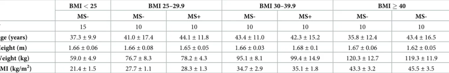
Table 1. Patient characteristics of study cohort 1.
BMI < 25 BMI 25–29.9 BMI 30–39.9 BMI 40
MS- MS- MS+ MS- MS+ MS- MS-N 15 10 10 10 10 10 10 Age (years) 37.3± 9.9 41.0± 17.4 44.1± 11.8 43.4± 11.0 42.3± 15.2 35.8± 12.4 43.4± 16.5 Height (m) 1.66± 0.06 1.66± 0.08 1.65± 0.05 1.66± 0.03 1.68± 0.1 1.67± 0.06 1.62± 0.05 Weight (kg) 59.0± 4.9 76.7± 8.3 78.2± 4.3 95.1± 8.1 99.4± 14.9 120.3± 12.7 119.3± 11.9 BMI (kg/m2) 21.4± 1.5 27.7± 1.1 28.3± 1.3 34.7± 2.9 35.1± 1.8 43.3± 3.2 45.5± 3.5 Mean value± SD is shown for all parameters, except N (absolute number). MS-/+: diagnosis of the metabolic syndrome no (-), yes (+)
https://doi.org/10.1371/journal.pone.0197603.t001
Table 2. Patient characteristics of study cohort 2.
Bariatric surgery Diet
Before After Before After
N 24 45
Age (years) 38.7± 12.6 46.0± 12.8
Height (m) 1.64± 0.05 1.65± 0.06
Weight (kg) 114.6± 17.6 88.1± 15.2 96.2± 16.5 86.2± 16.2
BMI (kg/m2) 42.5± 6.1 32.7± 5.3 35.3± 4.9 31.6± 4.8
Mean value± SD is shown for all parameters, except N (absolute number).
Results
PRCP activity in human circulation
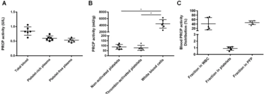
PRCP activity (median with interquartile range) was measured in total lysed blood (12.3 [10.5–13.5] mU/g), platelet-rich (10.3 [9.1–11.4] mU/g) and platelet-free (9.8 [8.9–11.1] mU/ g) plasma of 6 healthy donors. As shown inFig 1A, no significant differences were observed between these groups, indicating that there is little to no PRCP activity found in platelets (p = 0.174). PRCP was also measured in isolated white blood cells, resting platelets and plate-lets activated with thrombin. Again, low PRCP activity was found in resting plateplate-lets and thrombin activation had no effect on the enzyme activity. The specific activity measured in white blood cells was approximately fifty times higher compared to platelets (p = 0.028,Fig 1B). When comparing the relative contribution of blood fractions to the circulating PRCP activity, white blood cells and the platelet-free plasma fraction contributed for 41%, respec-tively, 46% to the total PRCP activity in blood (Fig 1C). Overall, white blood cells and the plasma fraction contribute most to the circulating PRCP activity.
PRCP activity in primary human cells
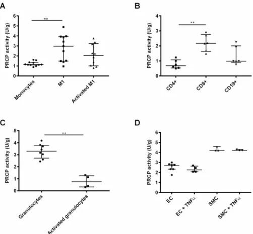
Appropriate activation of M1 macrophages was confirmed by ELISA, since the levels of TNFα, IL-1β and IL-6 were significantly upregulated in the supernatant of activated M1 macrophages (p<0.05, data not shown). PRCP activity is significantly higher in resting M1 macrophages (p = 0.003) compared to monocytes. Even when we take into account that freshly isolated monocytes would be a more suitable comparator as the adhesion to plastic and overnight cul-ture could already alter the monocyte phenotype, these results clearly show that an increase in PRCP activity was found upon monocyte-to-M1-macrophage differentiation. M2 macrophage activation could not be confirmed, since no clear upregulation of IL-10 could be measured in the supernatant of activated M2 macrophages. Therefore, no conclusions could be drawn con-cerning the M2 macrophages. Furthermore, a decrease in PRCP activity was observed after PMA stimulation of granulocytes (p = 0.007). The PRCP activity is higher in CD8+cytotoxic T cells compared to CD4+helper T cells. Human aortic endothelial and smooth muscle cells were stimulated with TNFα for 24 h. However, no significant difference in PRCP activity was found after stimulation. The PRCP activity in human aortic smooth muscle cells is higher than in the human aortic endothelial cells. The results are all shown inFig 2. In addition to human cells, we also isolated and cultured primary cells from the bone marrow of wild-type mice. Similar results as for the human blood cell populations were found (S3 Appendix).
Fig 1. PRCP activity in human circulation. (A) PRCP activity was measured in total lysed blood, platelet-rich and
platelet-free plasma but no significant changes were found (p = 0.174). (B) PRCP activity was significantly higher in lysed white blood cells compared to resting and thrombin-activated blood platelets (p = 0.028). (C) PRCP activity measured in human blood is mainly attributed to white blood cells and the plasma fraction. (⇤p<0.05, n = 4–6 per
group).
Serum PRCP activity in obese patients
PRCP activity was measured in serum of 15 lean and 60 obese female patients, which were fur-ther divided into three subgroups based on their BMI. The mean serum PRCP activity
(mean± SD) in lean subjects (BMI < 25.0 kg/m2) was 0.83± 0.04 U/L, 1.03 ± 0.04 U/L in
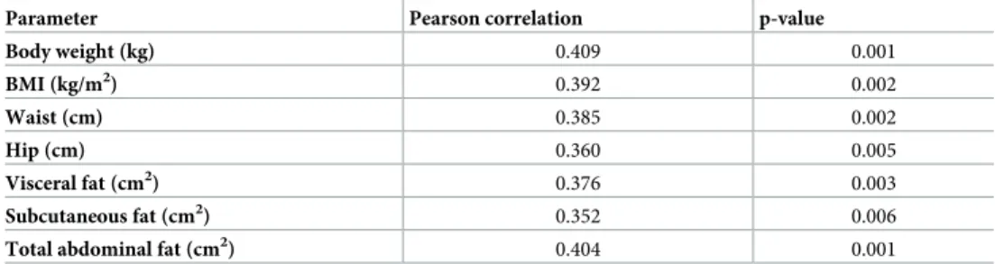
over-weight patients (BMI 25.0–29.9 kg/m2), 1.13± 0.03 U/L in obese patients (BMI 30.0–39.9 kg/ m2) and 1.23± 0.05 U/L in morbidly obese patients (BMI 40.0 kg/m2). Thus, the PRCP activity in serum of the groups with elevated BMI is significantly elevated compared to the PRCP activity in the lean group, as shown inFig 3A. The three subgroups were then further divided based on the diagnosis of metabolic syndrome (MS- or MS+). As presented inFig 3B, no significant differences in mean serum PRCP activity were found between metabolic syn-drome (MS+) patients and those without (MS-) (p > 0.05). In parallel, we studied PRCP activ-ity in serum of rats with metabolic syndrome. Again, no significant differences in serum PRCP activity between the controls and rats with metabolic syndrome were found (S4 Appendix). Correlations between serum PRCP activity in overweight/obese patients (n = 60) and patient age, several metabolic and biochemical parameters were investigated. An overview of the sig-nificant correlations is given inTable 3and correlations between serum PRCP activity and BMI, body weight and total abdominal tissue are shown inFig 4. The correlation with the
Fig 2. PRCP activity in primary human cells. (A) PRCP activity was significantly increased upon
monocyte-to-macrophage (M1 phenotype) differentiation. (B) PRCP activity was measured in different subsets of lymphocytes and was found highest in CD8+cytotoxic T cells. (C) PRCP activity was measured in resting and PMA-activated
granulocytes and (D) in control and stimulated (TNFα, 24h) human aortic endothelial and smooth muscle cells. (⇤⇤p<0.01, n = 3–11 per group).
amount of adipose tissue is intriguing since this suggests that PRCP might be secreted in serum by adipocytes. No correlations were found with systolic and diastolic blood pressure, lipid parameters, percentage glycated haemoglobin A1, oral glucose tolerance test results (baseline glucose and insulin levels and glucose level at 120 min) and high-sensitive C-reactive protein levels (r < 0.25).
Serum PRCP activity before and after weight loss
A group of 69 obese patients were treated with either a hypocaloric diet (n = 45) or bariatric surgery (n = 24). Diet as well as bariatric surgery caused a significant decrease in body weight
Fig 3. Serum PRCP activity in lean and obese patients. (A) PRCP activity was measured in serum of lean and obese
patients categorized based on their BMI and (B) the diagnosis of metabolic syndrome (no (-), yes (+)). (⇤⇤⇤p<0.001,
n = 10–20 per group).
https://doi.org/10.1371/journal.pone.0197603.g003
Table 3. Significant correlations between serum PRCP activity in overweight/obese patients (n = 60) and several metabolic parameters.
Parameter Pearson correlation p-value
Body weight (kg) 0.409 0.001 BMI (kg/m2) 0.392 0.002 Waist (cm) 0.385 0.002 Hip (cm) 0.360 0.005 Visceral fat (cm2) 0.376 0.003 Subcutaneous fat (cm2) 0.352 0.006
Total abdominal fat (cm2) 0.404 0.001
https://doi.org/10.1371/journal.pone.0197603.t003
Fig 4. Correlations between serum PRCP activity and BMI, body weight and total abdominal adipose tissue.
Correlation between serum PRCP activity and (A) BMI (p = 0.002), (B) body weight (p = 0.001) and (C) total abdominal adipose tissue (p = 0.001) in overweight/obese patients (n = 60).
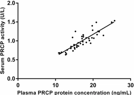
(mean± SD; 10.0 ± 7.2 vs. 26.5 ± 7.9 kg, p < 0.001) and BMI (mean ± SD; 3.6 ± 2.7 vs. 9.8± 3.0 kg/m2, p < 0.001) although more pronounced after surgery. The mean serum PRCP activity (mean± SD) before and 6 months after diet was 0.99 ± 0.25 U/L and 0.90 ± 0.25 U/L, while before and after bariatric surgery 1.09± 0.24 U/L and 0.90 ± 0.19 U/L. In both cases there was a significant decrease in the mean serum PRCP activity (mean± SD; 0.08 ± 0.03 U/L for diet and 0.19± 0.03 U/L for bariatric surgery). Line plots indicating the individual change in serum PRCP activity after diet or bariatric surgery are shown inFig 5. Serum PRCP activity correlated significantly with plasma PRCP protein concentration (0.865; p<0.001) (Fig 6). The mean plasma PRCP protein concentrations (mean± SD) before and 6 months after bariatric surgery were 18.2± 3.7 ng/mL and 15.7 ± 2.7 ng/mL. We also found a significant decrease (p<0.001) in the mean plasma PRCP protein concentration (mean± SD; 2.43 ± 2.26 ng/mL) upon weight loss.
Fig 5. Serum PRCP activity changes after weight loss. Line plots indicating the individual change in PRCP activity in
serum of patients who lost weight either by diet (n = 45, p = 0.015) or by bariatric surgery (n = 24, p < 0.001).
https://doi.org/10.1371/journal.pone.0197603.g005
Fig 6. Correlation between serum PRCP activity and plasma PRCP protein concentration. Correlation between
serum PRCP activity and plasma PRCP protein concentration in the bariatric surgery group (0.865; p < 0.001, n = 24).
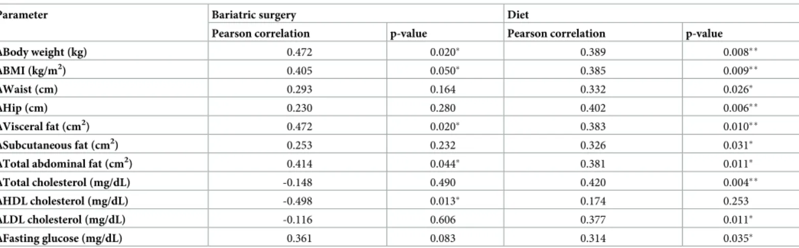
In the bariatric surgery group significant positive correlations were found between the decrease in serum PRCP activity and the decrease in body weight, BMI, total abdominal adi-pose tissue, visceral abdominal adiadi-pose tissue and subcutaneous abdominal adiadi-pose tissue. An inverse correlation was found with the increase in high-density lipoprotein cholesterol. Addi-tional positive correlations with the decrease in waist and hip circumference, total cholesterol, low-density lipoprotein cholesterol and baseline glucose level were found in the diet group. These results are all summarized inTable 4.
Discussion
The presence of PRCP protein and activity in the circulation has previously been reported, but the distribution and possiblein vivo relevance of circulating PRCP remained unclear [18,20,25,27].
The first aim of this study was to unravel the distribution of PRCP activity in human circu-lation. We found that white blood cells and plasma contributed the most to the circulating PRCP activity. In contrast, very low PRCP activity was found in resting and thrombin-acti-vated blood platelets. Our next experiments were focused on finding which cells were poten-tially responsible for the plasma activity and which subsets of white blood cells significantly expressed PRCP. The highest PRCP activity was measured in aortic smooth muscle cells, fol-lowed by M1 macrophages, granulocytes, aortic endothelial cells and lymphocytes. A signifi-cant upregulation of PRCP activity was observed upon monocyte-to-M1-macrophage differentiation. M1 macrophages are considered to be the pro-inflammatory macrophages, which might indicate a function for PRCP in the inflammatory response [28,29]. This enzyme is thought to exert its function in inflammation via activation of the prekallikrein pathway with subsequent bradykinin formation [3,30,31]. Also, previous reports have already linked plasma PRCP to C-reactive protein and several inflammatory cytokines [20,27]. However, in our study populations no significant correlations with serum PRCP activity and C-reactive protein levels were found. Another explanation for the increased PRCP activity in macro-phages could be that monocyte-to-macrophage differentiation is associated with an increase in cytoplasmic volume and more specifically with the development of larger lysosomal and mito-chondrial structures to enhance their degradative capacity [32–34]. Activation of M1 macro-phages through the classical LPS/IFN-γ pathway leads to a decrease in PRCP activity, indicating that PRCP could potentially be secreted. However, PRCP activity levels were
Table 4. Correlations between the decrease in PRCP activity and changes in several metabolic and biochemical parameters after diet or bariatric surgery (⇤p 0.05, ⇤⇤p 0.01).
Parameter Bariatric surgery Diet
Pearson correlation p-value Pearson correlation p-value
ΔBody weight (kg) 0.472 0.020⇤ 0.389 0.008⇤⇤ ΔBMI (kg/m2) 0.405 0.050⇤ 0.385 0.009⇤⇤ ΔWaist (cm) 0.293 0.164 0.332 0.026⇤ ΔHip (cm) 0.230 0.280 0.402 0.006⇤⇤ ΔVisceral fat (cm2 ) 0.472 0.020⇤ 0.383 0.010⇤⇤ ΔSubcutaneous fat (cm2) 0.253 0.232 0.326 0.031⇤
ΔTotal abdominal fat (cm2) 0.414 0.044⇤ 0.381 0.011⇤
ΔTotal cholesterol (mg/dL) -0.148 0.490 0.420 0.004⇤⇤
ΔHDL cholesterol (mg/dL) -0.498 0.013⇤ 0.174 0.253
ΔLDL cholesterol (mg/dL) -0.116 0.606 0.377 0.011⇤
ΔFasting glucose (mg/dL) 0.361 0.083 0.314 0.035⇤
undetectable in the cell culture medium. A more pronounced decrease in PRCP activity was found after degranulation of granulocytes caused by PMA-stimulation. Once more, these results point to a granular localisation of PRCP and a possible function in the immune response [18]. The highest PRCP activity was found in cytotoxic T cells compared to helper T cells and B cells. Furthermore, PRCP activity remained unchanged after TNFα stimulation of human aortic endothelial and smooth muscle cells [35–36].
The development of a sensitive immunoassay by Xuet al. has demonstrated that plasma protein concentrations of PRCP are increased in obese patients and are even more elevated in patients with both obesity and diabetes. Strong correlations with metabolic (BMI, blood glu-cose, etc.) parameters have been reported [18]. Complementary to this study, we investigated PRCP activity in the serum of normal, overweight, obese and morbidly obese patients. We found that serum PRCP activity was increased with a rising BMI the PRCP activity in serum of the groups with elevated BMI is significantly elevated compared to the PRCP activity in the lean group. In addition to correlations with BMI, body weight, waist and hip circumference, we also found strong associations with the amount of total, visceral and subcutaneous abdomi-nal adipose tissue. The ratio of PRCP protein concentration to activity in serum could reflect the severity of obesity and sheds some much needed light on the possible additional source(s) of circulating PRCP. Moreover, this could potentially aid in the determination of PRCP’s potential role in regulating levels of intact apelin and α-MSH, two peptides involved in the reg-ulation of food intake.
Since polymorphisms of the PRCP gene have been related to metabolic syndrome among coronary artery disease patients [37], we also categorized the patients based on metabolic syn-drome. However, no significant difference in PRCP activity between the MS- and MS+ groups were found. Whether the existence of metabolic syndrome affects circulating PRCP activity specifically in patients with coronary artery disease remains to be investigated and will be addressed in future studies.
Aside from investigating PRCP activity in overweight and obese patients, we also explored its profile after weight loss. Thus, PRCP activity was measured in 24 patients who underwent bariatric surgery and 45 patients who were put on a calorie-restricted diet. PRCP activity and body weight decreased significantly after diet but even more pronounced after bariatric surgery, which is also associated with several dietary guidelines and restrictions [38]. Again, significant correlations were found with parameters concerning weight, waist and hip circum-ference and the amount of adipose tissue. The role of PRCP in adipocytes deserves further study, especially because adipocytes produce and secrete apelin-13, a substrate of PRCP. A strong correlation between the serum PRCP activity and plasma protein concentration was found, thus confirming the specificity of our activity measurements. Moreover, we confirmed the influence of weight loss on plasma PRCP protein concentrations. Our results suggested that both PRCP activity, as well as PRCP protein concentration, can be used to investigate the regulation of PRCP in plasma.
On the other hand, endothelial dysfunction, as observed in obese patients [39], may also lie at the basis of the raised PRCP protein concentrations in plasma, since PRCP has been shown to be located on the membrane of endothelial cells and to regulate endothelial cell growth [18,40].
Conclusion
Our study was the first comprehensive investigation of the distribution of PRCP activity in human circulation. We have demonstrated that white blood cells and plasma contribute the most to circulating PRCP activity. Serum PRCP activity was found to correlate with plasma
PRCP protein concentration, body weight and the amount of adipose tissue. These findings, taken together with the recent observation that the adipokine apelin-13 is hydrolysed by PRCP, underscore the importance of further research needed in this field to fully understand its regulation and biological function in body weight control. Gaining a greater understanding of PRCP’s multi-faceted biological functions could open up novel therapeutic avenues for the treatment of obesity.
Supporting information
S1 Appendix. PRCP activity in peripheral blood mono- and polymorphonuclear cells. (DOCX)
S2 Appendix. Study populations. (DOCX)
S3 Appendix. PRCP and DPP2 activity in primary murine cells. (DOCX)
S4 Appendix. PRCP in a rodent model of metabolic syndrome. (DOCX)
Acknowledgments
We thank Ms. Nicole Klein and Mr. Nagabushan Reddy for their technical assistance. We ded-icate this paper to our colleague, Prof. Sandra Apers, who passed away much too early on Feb-ruary 5th, 2017.
Author Contributions
Conceptualization: Kaat Kehoe, Heidi Noels, Anne-Marie Lambeir, Luc Van Gaal, Ingrid De Meester.
Data curation: Heidi Noels, Wendy Theelen, Emilie De Hert, Shenguan Xu, An Verrijken, Thierry Arnould.
Formal analysis: Erik Fransen. Funding acquisition: Heidi Noels.
Investigation: Kaat Kehoe, Heidi Noels, Wendy Theelen, Emilie De Hert, Shenguan Xu, Thierry Arnould, Nina Hermans, Ingrid De Meester.
Methodology: Kaat Kehoe, Heidi Noels, Nina Hermans.
Supervision: Anne-Marie Lambeir, Per Venge, Luc Van Gaal, Ingrid De Meester. Writing – original draft: Kaat Kehoe, Emilie De Hert.
Writing – review & editing: Heidi Noels, Wendy Theelen, Shenguan Xu, An Verrijken, Thierry Arnould, Erik Fransen, Nina Hermans, Anne-Marie Lambeir, Per Venge, Luc Van Gaal, Ingrid De Meester.
References
1. Wallingford N, Perroud B, Gao Q, Coppola A, Gyengesi E, Liu ZW, et al. Prolylcarboxypeptidase regu-lates food intake by inactivating α-MSH in rodents. J Clin Invest. 2009; 119:2291–2303.https://doi.org/ 10.1172/JCI37209PMID:19620781
2. Odya CE, Marinkovic DV, Hammon KJ, Stewart TA, Erdo¨s EG. Purification and properties of prolylcar-boxypeptidase (angiotensinase C) from human kidney. J Biol Chem. 1978; 253:5927–5931. PMID:
28321
3. Chajkowski SM, Mallela J, Watson DE, Wang J, McCurdy CR, Rimoldi JM, et al. Highly selective hydro-lysis of kinins by recombinant prolylcarboxypeptidase. Biochem Biophys Res Commun. 2011; 405:338– 343.https://doi.org/10.1016/j.bbrc.2010.12.036PMID:21167814
4. O’Donoghue AJ, Eroy-reveles AA, Knudsen GM, Ingram J, Zhou M, Statnekov JB, et al. Global identifi-cation of peptidase specificity by multiplex substrate profiling. Nat Methods. 2013; 9:1095–1100.
5. Williams DL, Schwartz MW. The melanocortin system as a central integrator of direct and indirect con-trols of food intake. Am J Physiol Regul Integr Comp Physiol. 2005; 289:R2–3.https://doi.org/10.1152/ ajpregu.00226.2005PMID:15956761
6. Zhou C, Garcia-Calvo M, Pinto S, Lombardo M, Feng Z, Bender K, et al. Design and synthesis of prolyl-carboxypeptidase (PrCP) inhibitors to validate PrCP as a potential target for obesity. J Med Chem. 2010; 53:7251–7263.https://doi.org/10.1021/jm101013mPMID:20857914
7. Jeong JK, Diano S. Prolyl carboxypeptidase and its inhibitors in metabolism. Trends Endocrinol Metab. 2013; 24:61–67.https://doi.org/10.1016/j.tem.2012.11.001PMID:23245768
8. Kehoe K, Van Elzen R, Verkerk R, Sim Y, Van der Veken P, Lambeir AM, et al. Prolyl carboxypeptidase purified from human placenta: its characterization and identification as an apelin-cleaving enzyme. Bio-chim Biophys Acta. 2016; 1864:1481–1488.https://doi.org/10.1016/j.bbapap.2016.07.004PMID:
27449720
9. O’Shea M, Hansen MJ, Tatemoto K, Morris MJ. Inhibitory effect of apelin-12 on nocturnal food intake in the rat. Nutr Neurosci. 2003; 6:163–167.https://doi.org/10.1080/1028415031000111273PMID:
12793520
10. Higuchi K, Masaki T, Gotoh K, Chiba S, Katsuragi I, Tanak K, et al. Apelin, an APJ receptor ligand, regu-lates body adiposity and favors the messenger ribonucleic acid expression of uncoupling proteins in mice. Endocrinology. 2007; 148:2690–2697.https://doi.org/10.1210/en.2006-1270PMID:17347313 11. Castan-Laurell I, Dray C, Attane´ C, Duparc T, Knauf C, Valet P. Apelin, diabetes, and obesity.
Endo-crine. 2011; 40:1–9.https://doi.org/10.1007/s12020-011-9507-9PMID:21725702
12. Tan F, Morris PW, Skidgel RA, Erdo¨s EG. Sequencing and cloning of human Prolylcarboxypeptidase (Angiotensinase C). Biochemistry. 1993; 268:16631–16638.
13. Jeong JK, Diano S. Prolyl carboxypeptidase mRNA expression in the mouse brain. Brain Res. 2014; 1542:85–92.https://doi.org/10.1016/j.brainres.2013.10.031PMID:24161824
14. Kakimoto T, Oshima G, Yeh HSJ, Erdo¨s EG. Purification of lysosomal prolylcarboxypeptidase angioten-sinase C. Biochim Biophys Acta. 1973; 302:178–182. PMID:4692653
15. Odya CE, Erdo¨s EG. Human Prolylcarboxypeptidase. Methods Enzymol. 1981; 80:460–466. PMID:
7341916
16. Kumamoto K, Stewart TA, Johnson AR, Erdo¨s EG. Prolylcarboxypeptidase (angiotensinase C) in human lung and cultured cells. J Clin Invest. 1981; 67:210–215.https://doi.org/10.1172/JCI110015
PMID:7451650
17. Jackman HL, Tan F, Schraufnagel D, Dragović T, Dezso¨ B, Becker RP, et al. Plasma membrane-bound and lysosomal peptidases in human alveolar macrophages. Am J Respir Cell Mol Biol. 1995; 13:196– 204.https://doi.org/10.1165/ajrcmb.13.2.7626287PMID:7626287
18. Xu S, Lind L, Zhao L, Lindahl B, Venge P. Plasma Prolylcarboxypeptidase (Angiotensinase C) is increased in obesity and diabetes mellitus and related to cardiovascular dysfunction. Clin Chem. 2012; 58:1110–1115.https://doi.org/10.1373/clinchem.2011.179291PMID:22539806
19. Shariat-Madar Z, Mahdi F, Schmaier AH. Recombinant prolylcarboxypeptidase activates plasma pre-kallikrein. Blood. 2004; 103:4554–4561.https://doi.org/10.1182/blood-2003-07-2510PMID:14996700 20. Tabrizian T, Hataway F, Murray D, Shariat-Madar Z. Prolylcarboxypeptidase gene expression in the
heart and kidney : effects of obesity and diabetes. Cardiovasc Hemat Agents Med Chem. 2015; 13:113–23.
21. Bo¨yum A. Isolation of mononuclear cells and granulocytes from human blood. Isolation of mononuclear cells by one centrifugation, and of granulocytes by combining centrifugation and sedimentation at 1 g. Scand J Clin Lab Invest. 1968; 97:77–89.
22. Matheeussen V, Waumans Y, Martinet W, Van Goethem S, Van der Veken P, Scharpe´ S, et al. Dipepti-dyl peptidases in atherosclerosis: expression and role in macrophage differentiation, activation and apo-ptosis. Basic Res Cardiol. 2013; 108:350.https://doi.org/10.1007/s00395-013-0350-4PMID:23608773 23. Maqbool M, Vidyadaran S, George E, Ramasamy R. Optimisation of laboratory procedures for isolating
24. Bradford MM. A rapid and sensitive method for the quantitation microgram quantities of protein utilizing the principle of protein-dye Binding. Anal Biochem. 1976; 72:248–254. PMID:942051
25. Kehoe K, Verkerk R, Sim Y, Waumans Y, Van der Veken P, Lambeir AM, et al. Validation of a specific prolylcarboxypeptidase activity assay and its suitability for plasma and serum measurements. Anal Bio-chem. 2013; 443:232–239.https://doi.org/10.1016/j.ab.2013.09.002PMID:24036038
26. Alberti KGMM, Eckel RH, Grundy SM, Zimmet PZ, Cleeman JI, Donato KA, et al. Harmonizing the met-abolic syndrome. Circulation. 2009; 120:1640–1645.https://doi.org/10.1161/CIRCULATIONAHA.109. 192644PMID:19805654
27. Kehoe K, Brouns R, Verkerk R, Engelborghs S, De Deyn PP, Hendriks D, et al. Prolyl carboxypeptidase activity decline correlates with severity and short-term outcome in acute ischemic stroke. Neurochem Res. 2014; 40:81–88.https://doi.org/10.1007/s11064-014-1468-yPMID:25370794
28. Martinez FO, Gordon S. The M1 and M2 paradigm of macrophage activation: time for reassessment. F1000Prime Rep. 2014; 6:13.https://doi.org/10.12703/P6-13PMID:24669294
29. Mosser DM, Edwards JP. Exploring the full spectrum of macrophage activation. Nat Rev Immunol. 2008; 8:958–969.https://doi.org/10.1038/nri2448PMID:19029990
30. Zhu L, Carretero OA, Liao TD, Harding P, Li H, Sumners C, et al. Role of prolylcarboxypeptidase in angiotensin II type 2 receptor-mediated bradykinin release in mouse coronary artery endothelial cells. Hypertension. 2010; 56:384–390.https://doi.org/10.1161/HYPERTENSIONAHA.110.155051PMID:
20606103
31. Ngo ML, Mahdi F, Kolte D, Shariat-Madar Z. Upregulation of prolylcarboxypeptidase (PRCP) in lipopoly-saccharide (LPS) treated endothelium promotes inflammation. J Inflamm. 2009; 6:3.
32. McCullough KC, Basta S, Kno¨tig S, Gerber S, Schaffner R, Kim YB, et al. Intermediate stages in mono-cyte-macrophage differentiation modulate phenotype and susceptibility to virus infection. Immunology. 1999; 98:203–212.https://doi.org/10.1046/j.1365-2567.1999.00867.xPMID:10540219
33. Sokol RJ, Hudson G, James NT, Frost IJ. Human macrophage development: a morphometric study. J Anat. 1987; 151:27–35. PMID:3654357
34. Daigneault M, Preston JA, Marriott HM, Whyte MKB, Dockrell DH. The identification of markers of mac-rophage differentiation in PMA-stimulated THP-1 cells and monocyte-derived macmac-rophages. PLoS One. 2010; 5, e8668.https://doi.org/10.1371/journal.pone.0008668PMID:20084270
35. Couffinhal T, Duplàa C, Labat L, Lamaziere JMD, Moreau C, Printseva O, et al. Tumor necrosis factor-alpha stimulates ICAM-1 expression in human vascular smooth muscle cells. Arterioscler Thromb Vasc Biol. 1993; 13:407–414.
36. Mackay F, Loetscher H, Stueber D, Gehr G, Lesslauer W. Tumor necrosis factor alpha (TNF-alpha)-induced cell adhesion to human endothelial cells is under dominant control of one TNF receptor type, TNF-R55. J Exp Med. 1993; 177:1277–1286. PMID:8386742
37. McCarthy JJ, Meyer J, Moliterno DJ, Newby LK, Rogers WJ, Topol EJ. Evidence for substantial effect modification by gender in a large-scale genetic association study of the metabolic syndrome among cor-onary heart disease patients. Hum Genet. 2003; 114:87–98. https://doi.org/10.1007/s00439-003-1026-1PMID:14557872
38. Shannon C, Gervasoni A, Williams T. The bariatric surgery patient—nutrition considerations. Aust Fam Physician. 2013; 42:547–552. PMID:23971062
39. Avagaro A, De Kreutzenberg SV. Mechanisms of endothelial dysfunction in obesity. Clin Chim Acta. 2005; 360:9–26.https://doi.org/10.1016/j.cccn.2005.04.020PMID:15982646
40. Adams GN, Stavrou EX, Fang C, Merkulova A, Alaiti MA, Nakajima K, et al. Prolylcarboxypeptidase promotes angiogenesis and vascular repair. Blood. 2013; 122:1522–1531.https://doi.org/10.1182/ blood-2012-10-460360PMID:23744584