HAL Id: tel-02953383
https://tel.archives-ouvertes.fr/tel-02953383
Submitted on 30 Sep 2020HAL is a multi-disciplinary open access archive for the deposit and dissemination of sci-entific research documents, whether they are pub-lished or not. The documents may come from teaching and research institutions in France or abroad, or from public or private research centers.
L’archive ouverte pluridisciplinaire HAL, est destinée au dépôt et à la diffusion de documents scientifiques de niveau recherche, publiés ou non, émanant des établissements d’enseignement et de recherche français ou étrangers, des laboratoires publics ou privés.
Synaptotoxicity in Alzheimer’s disease : Influence of
APP processing on excitatory synapses
Rebecca Powell
To cite this version:
Rebecca Powell. Synaptotoxicity in Alzheimer’s disease : Influence of APP processing on excitatory synapses. Neurons and Cognition [q-bio.NC]. Université Grenoble Alpes, 2019. English. �NNT : 2019GREAV051�. �tel-02953383�
THÈSE
Pour obtenir le grade de
DOCTEUR DE LA COMMUNAUTE UNIVERSITE GRENOBLE
ALPES
Spécialité : Neurosciences - Neurobiologie Arrêté ministériel : 25 mai 2016
Présentée par
Rebecca POWELL
Thèse dirigée par Alain BUISSON, Professeur, UGA
Préparée au sein du l’institut des Neurosciences de Grenoble INSERM U1216 – Equipe Neuropathologies et Dysfonctions Synaptiques
Dans l'École Doctorale de Chimie et Sciences du vivant
Synaptotoxicité dans la maladie
d’Alzheimer : Influence du
processing de l’APP sur les synapses
excitatrices
Thèse soutenue publiquement le 6 décembre 2019, devant le jury composé de :
Pr Rémy SADOUL
Professeur - Université Grenoble Alpes - Président du jury Dr Claire MEISSIREL
Chargée de recherche - Université Claude Bernard Lyon 1 - Rapporteur Dr Marc DHENAIN
Directeur de recherche - CNRS, Université Paris -Sud - Rapporteur Dr Montserrat SOLER-LOPEZ
Laboratory Scientist and Manager - ESRF, Grenoble - Examinateur Dr Harold MacGillavry
Assistant Professor - Universiteit Utrect, Netherlands - Examinateur Pr Alain BUISSON
Synaptotoxicity in Alzheimer’s disease:
Influence of APP processing
on excitatory synapses
Acknowledgments
First and foremost, I would like to kindly thank the members of the jury: Claire Meissirel, Montse Soler-Lopez, Marc Dhenain, Harold Mac Gillavry and Rémy Sadoul for accepting to be part of my thesis jury and making time for evaluating my work.
En particulier j’aimerais remercier Claire Meissirel et Marc Dhenain d’avoir accepté d’être rapporteur de ma thèse et d’avoir pris le temps d’analyser mon manuscrit.
A special thanks to Montse, for following the evolution of my thesis project and, now, for being member of my thesis jury. Also, I would like to warmly thank Harold Mac Gillavry for travelling all the way from Utrecht to assess my work.
Je tiens à remercier tout particulièrement Rémy Sadoul pour avoir accepté d’être examinateur dans ce jury de thèse, mais surtout pour m’avoir fait découvrir les neurosciences en L3. C’est en grande partie grâce à toi que j’en suis là où en j’en suis aujourd’hui!
A very special thank you my PhD director, Alain Buisson. Thank you for believing in me and giving me the opportunity to carry out my thesis in your team. During these four years you’ve taught me a lot professionally, scientifically and even on a personal level. By trusting me I’ve learned to trust myself. Thank you for mentoring, advice and inspiration, I couldn’t have asked for a better boss!
Un grand merci à toute mon équipe avec qui nous avons toujours partagé de bons moments, eu de bonnes conversations et de bonnes rigolades! C’était un plaisir de me lever le matin sachant que j’allais à ma « deuxième maison » où il y faisait bon vivre grâce à vous! Merci à Muriel, Mireille et Fabien, mes profs de fac (que je redoutais) qui sont devenus mes collègues de travail (que j’apprécie énormément, les profs sont des humains vraiment gentils en fait!). Merci de m’avoir appris les sciences, en cours et aussi pendant ma thèse! Mais surtout merci pour vos conseils, votre soutien, votre ouverture d’esprit, votre gentillesse et votre bonne humeur! Merci à mes voisines de bureau, Sylvie et Eve (mes deuxièmes mamans), pour m’avoir soutenue dans les bons et les moins bons moments, d’avoir toujours veillées sur moi et pour toutes les barres de rires qu’on s’est payées! Votre bonne humeur (et bon humour) est sans faille et je vous en remercie sincèrement! Et puis, merci au « petit frère » PhD, Adrien, pour tous les moments que nous avons partagé au labo, les discussions de tout et n’importe quoi et les fous rires en tout genre (et surtout pour nos comparaisons de performances sportives qui ne servaient à rien puisque j’étais la plus nulle à chaque fois haha!).
I would like to thank the rest of the members of the 2nd floor of the institute, as well as all the members of the rest of the GIN, for taking part in making these four years absolutely unforgettable!
Un merci tout spécial aux amis. Tout d’abord les amies « labos » Marta, Elé et Perrine! Merci les filles d’avoir été là à mes débuts comme stagiaires M2! Merci à toi Perrine d’avoir été ma camarade de galère pendant le stage M2 et un infini merci pour m’avoir fait comprendre les stats en un temps éclair haha! Merci à vous Elé et Marta, sans vous je crois que je n’en serais pas là aujourd’hui. Je ne vous remercierais jamais assez pour votre bonne humeur, votre gentillesse, votre amitié. Je n’aurais jamais imaginé rencontrer des filles aussi géniales que vous! Don’t change a thing ;)
Un grand merci aussi aux amis « pas labos », Kelly, Tony, Laura, Tam, Pex, Tazo, Tris (et encore d’autres que je ne peux citer par soucis de place)! Merci d’avoir été là, et juste merci d’être vous! Don’t change a thing either ;)
Diolch yn fawr Mammy, Daddy! Thank you for always believing in me, for your continuous support through thick and thin, for your patience especially in the last few months (I know I’ve been a real pain in the back side!) and for everything that you do (I could make a list of things to thank you for as long as this thesis) you’re the best!
And mustn’t forget the bros! Alex, Liam and Jonathan! What a great gang! U ma homies, u da best!
Mes derniers remerciements vont à toi Pup. Toi qui es à mes côtés depuis le tout début. On en aura fait du chemin! On a grandi ensemble et je te remercie du fond du cœur pour tout ce que tu es, tout ce que tu m’as apporté et tout ce que tu as dû endurer! Si j’en suis là aujourd’hui, autant sur le plan professionnel que personnel, c’est grâce à toi! Love you long time <3
Résumé
La maladie d’Alzheimer (MA) est définie comme une maladie neurodégénérative où des altérations synaptiques mènent à la perte neuronale parallèlement à des défauts de mémoire et d’apprentissage. Il est établi que les dysfonctions synaptiques observées dans la MA sont initiées par les formes oligomériques du peptide β-amyloïde (Aβ), un dérivé protéolytique de l’Amyloïd Precursor Protein (APP). Cependant, le chemin qu’empreinte Aβ, selon son origine intra- ou extracellulaire, afin d’induire ces effets délétères et la façon dont ses effets sont maintenus et se propagent dans le cerveau restent encore à définir.
Dans cette étude, nous avons utilisé plusieurs formes mutées de l’APP qui conduisent à des peptides Aβ avec des signatures moléculaires uniques, tel que : la mutation Swedish (K670M/N671L) (APPswe) qui augmentent la sécrétion (extracellulaire) d’Aβ; la mutation Osaka (E693Δ) (APPosa) qui cause une accumulation intraneuronale (intracellulaire) d’Aβ; ainsi que la mutation Icelandic (A673T) (APPice) qui a été établi comme diminuant la production d’Aβ et protégeant contre la MA. Ces formes mutées d’APP ont été surexprimées dans des cultures de neurones corticaux murins et on permit : i) d’étudier la morphologie et fonction des épines dendritiques, l’élément post-synaptique, par microscopie confocale; ii) de tenter de mieux comprendre comment la pathologie se développe et se propage dans le cerveau et iii) d’identifier un nouveau partenaire d’intéraction avec l’Aβ faisant la lumière sur un possible rôle physiologique de ce peptide dans les neurones.
Nous montrons qu’une accumulation pathologique d’Aβ, due à la surexpression d’APPwt, APPswe et APPosa mais pas APPice, induit une diminution significative de la densité synaptique particulièrement celle des épines les plus fonctionnelles, dites « mushroom ». Ses épines mushroom restantes présentent également une augmentation significative de leur volume et il semblerait que l’Aβ intracellulaire soit suffisant pour induire ses effets. Ses épines mushroom élargies présentent également une plasticité structurale altérée puisqu’elles n’ont pas augmenté d’avantage de volume à la suite d’une activation synaptique. Il semblerait que ceci soit la résultante d’un défaut de la dynamique activité-dépendante du cytosquelette d’actine dans les épines. Ces altérations de la morphologie, structure et plasticité synaptique serait dû à une intéraction, nouvellement identifiée, de l’Aβ avec l’actine et pourrait faire lumière sur un possible rôle physiologique de l’Aβ dans la plasticité synaptique activité-dépendante. De plus, nous montrons que le clivage amyloïde de l’APP est aussi activité-dépendant et que la séquence du peptide Aβ généré est aussi importante, dans l’induction de la synaptotoxicité, que sa concentration. En effet, car nous montrons que des concentrations pathologiques du peptide Aβice n’engendrent pas de perte ou de gonflement des épines mushroom. Enfin, nous mettons en lumière que l’Aβ sécrété dans le milieu extracellulaire affecte, non seulement le neurone sécrétant lui-même, mais aussi la densité synaptique des neurones sains avoisinant (qui ne surexpriment pas d’APP) d’une manière APP-dépendante, rappelant un mécanisme de propagation du type prion. L’ensemble de ces données démontrent que le clivage protéolytique de l’APP et la production d’Aβ qui en découle est un processus finement accordé, impliqué dans le remodelage de l’actine dans la plasticité synaptique activité-dépendante et ouvre de nouvelles voies pour le développement de stratégies thérapeutiques contre la MA.
Abstract
Alzheimer’s disease (AD) is defined as a neurodegenerative disorder where synaptic defects lead to neuronal loss and concurrent memory impairments. It is now well-established that synaptic dysfunction in AD is initiated by oligomeric forms of the amyloid-β peptide (Aβ), a proteolytic derivative of Amyloid Precursor Protein (APP). However, the pathway by which Aβ induces its deleterious effects, whether it is due to intra- and/or extracellular Aβ pools, and how these effects are sustained and propagated throughout the brain, are still unclear.
In this study, we used several mutated forms of APP which give rise to Aβ peptides with unique molecular signatures, such as: the Swedish mutation (K670M/N671L) (APPswe) which increases secreted (extracellular) Aβ; the Osaka mutation (E693Δ) (APPosa) which causes intraneuronal (intracellular) accumulation of Aβ; and the Icelandic mutation (A673T) (APPice) which has been reported to decrease Aβ production and protect against AD. These mutated forms of APP were overexpressed in cultured mouse cortical neurons in order to: i) study the morphology and function of dendritic spines, the post-synaptic element of synapses, by confocal microscopy, ii) get a better insight into pathology development and propagation and iii) identify a novel interacting partner bringing to light the possible physiologic role of Aβ in neurons.
We report that pathological Aβ accumulation, due to APPwt, APPswe and APPosa overexpression but not APPice overexpression induces a significant decrease in spine density especially mushroom spines, accompanied by a significantly increased volume of the remaining mushroom spines, and that intracellular Aβ is sufficient to induce these effects. These enlarged mushroom spines have impaired structural plasticity as they did not increase in volume following synaptic activation seemingly as a result of defective activity-dependent actin dynamics in the spines. This alteration of synaptic morphology, structure and plasticity seems to be due to a newly-identified interaction between actin and Aβ, hinting a possible physiological role for Aβ in activity-dependent synaptic plasticity. We also show that synaptic activity modulates amyloïdogenic APP processing which, in pathological conditions, further exacerbates these synaptic defects. Furthermore, we show that Aβ sequence is as important as Aβ concentration in inducing synaptic alterations since pathological concentrations of Aβ harbouring the Icelandic mutation had no effect on spine density or volume. Lastly, we bring to light that secreted Aβ, not only affects the Aβ-secreting neuron itself, but also affects spine density of nearby neurons in an APP-dependent manner, reminiscent of a prion-like mechanism. Together these results demonstrate that APP processing is a finely tuned equilibrium involved in actin-remodelling during activity-dependent synaptic plasticity and opens a new route for AD therapeutic strategies.
List of Abbreviations
4-AP: 4 aminopyridine ABP: Actin-binding protein AC: Adenylate cyclase AChE: Acetylcholinesterase AD: Alzheimer’s disease ADAM: A Desintegrin and
Metalloprotease (α-secretase)
ADDL/Aβ-derived diffusible
ligand/synonym of Aβ peptide
AICD: APP Intracellular domain AMP/ADP/ATP: Adenosine
Mono/Di/Tri-phosphate
AMPA:
α-amino-3-hydroxy-5-methyl-4-isoxazolepropionic acid
AMPAr: AMPA receptor AMPK: AMP-activated protein
kinase
Aph1: Anterior Pharynx
defective 1 homolog (γ-secretase)
APLP1/2: APP-like Protein 1/2 APOE: Apolipoprotein E APP: Amyloid Precursor
Protein
AP-V:
D,L-2-amino-5-phosphonopentanoic acid 5
ARF6: ADP Ribosylation factor
6
Aβ: Amyloid Beta peptide BACE1: Beta-site Cleaving
Enzyme 1 (β-secretase)
Bic: Bicuculline
βsecI: β-secretase Inhibitor C83/α-CTF: α C-terminal Fragment (Non-Amyloïdogenic pathway) C99/β-CTF: β C-terminal Fragment (Amyloïdogenic pathway)
CA1/3: Hippocampal Cornu
Ammonis Region 1/3
CAA: Cerebral Amyloid
Angiopathy
CaMKII:
Calcium/calmodulin-dependent protein kinase II
cAMP: cyclic Adenosine
monophosphate
CCV: Clathrin-coated vesicle
Cdk5: Cyclin-dependent kinase
5
CJD: Creutzfeldt-Jacob disease CREB: cAMP response element
binding protein (Erk activated transcription factor)
CSF: Cerebrospinal Fluid CTE: Chronic Traumatic
Encephalopathy
C-ter: Cterminal DAG: Diacylglycerol EE: Early Endosome EOFAD: Early-onset Familial
Alzheimer disease
EPSP: Excitatory Postsynaptic
potential
ER: Endoplasmic Reticulum ERF: Extracellular signal related
kinase
EZ: Endocytic zone
F- or G-actin: Filamentous or
globular Actin
FADs: Familial Alzheimer’s
disease
GABA(r): α-aminobutyric acid
(receptor)
GSK3β: Glycogen Synthase
kinase 3β
GWAS: Genome-wide
Association Study
HEK: Human Embryonic Kidney
cells
iGluRs: Ionotropic Glutamate
receptor
IP3: Ionsitol-1,4,5-triphosphate KO: Knock-out
LA: Life-act or Life-actin LE: Late Endosome
LFU: Low-frequency Uncaging LOAD: Late-Onset Alzheimer
disease LTD: Long-term Depression LTP: Long-term Potentiation MAP2: Microtubule-associated Protein MAPK: Mitogen-activated protein kinase
MCI: Mild Cognitive
Impairments
mGluRs: metabotropic
Glutamate receptor coupled to G proteins
MMSE: Mini Mental Status
Evaluation
MRI: Magnetic Resonance
Imagery MTs: Microtubules NEP: Neprilysin NFTs: Neurofibrillary Tangles NMDA: N-Methyl-D-Aspartic Acid
NMDAr: NMDA receptor N-ter: N-terminal
PAK: p21-activated kinase Pen2: Presinilin Enhancer 2
homolog (γ-secretase)
PET-FDG: Positon Emission
Tomography
PKA/C: Protein kinase A/C PLC: Phospholipase C PM: Plasma membrane PP1/2(A or B): Protein phosphatase 1/2(A or B) PR: polyribosomes PrPc: Prion protein c PS1/2: Presinilin 1/2 (γ-secretase) PSD: Postsynaptic Density
RE: Recycling Endosome sAPPα/β: soluble APP α/β
(N-terminal fragment)
Ser: Serine
SPECT: Single Photon Emission
Computed Tomography
SSH1: Slingshot protein
phosphatase 1
STEP: Striatal enriched
phosphatase
Tau: Tubulin-associated Unit TGN: Trans-Golgi Network TTX: Tetrodotoxine
Synaptotoxicity in Alzheimer’s disease:
Influence of APP processing on excitatory synapses
I.
Alzheimer’s disease ... 1
A brief bit of history ... 1
A. A worldwide issue for public health ... 2
B. Dementia ... 2
C. Statistics of Alzheimer’s disease ... 2
D. Clinical aspects of Alzheimer’s disease ... 3
E. Symptoms ... 3
1. Diagnostic ... 5
2. Different forms of AD & risk factors ... 7
F. Familial Alzheimer’s disease (FAD) ... 7
1. a) Presinilins PS1 and PS2 ... 8
b) APP ... 9
Sporadic Alzheimer’s disease ... 9
2. a) Apolipoprotein E ... 9
b) Other genetic factors ... 10
c) Environmental factors ... 11
Histopathological aspects of Alzheimer’s disease ... 12
G. Macroscopic lesions ... 12
1. Microscopic lesions... 13
2. a) Intracellular Tau and neurofibrillary tangles ... 14
b) Aβ peptide and extracellular senile plaques ... 17
c) Links between Aβ and Tau ... 19
Propagation of the pathology throughout the brain ... 20
H. Propagation of Tau ... 20 1. Propagation of Aβ ... 22 2. APP-dependent propagation ... 24 3.
II.
Amyloid Precursor Protein processing and Amyloidogenesis
... 25
APP – Background ... 25
A. APP processing and trafficking... 26
B. The amyloïdogenic pathway, β- and γ-secretases ... 26
1. The non-amyloïdogenic pathway, α- and γ-secretases ... 28
2. Other cleavage pathways of APP ... 28
3. Intracellular trafficking of APP ... 29 4.
Aβ production and clearance ... 31
C. Aβ production sites ... 31
1. Aβ peptide degradation and clearance ... 34
2. Toxic and physiologic roles of Aβ ... 35
D. The different forms of Aβ ... 36
E. The different mutations of APP ... 38
F. APP mutations affecting Aβ production ... 38
1. a) Mutations affecting β-cleavage ... 39
b) Mutations affecting γ-cleavage ... 40
APP mutations affecting Aβ sequence ... 41
2. a) The hotspot for Aβ mutations (aa 693 to 694 of APP) ... 41
b) Other ... 42
Not all mutations on APP are toxic ... 43
3. Therapeutic strategies ... 43
G. Decreasing Aβ production ... 44
1. a) Inhibition of γ-secretase ... 44
b) Inhibition of β-secretase ... 45
Immunotherapies ... 47
2. Decreasing Aβ aggregation ... 49
3. Increasing Aβ clearance ... 49
4. Counteracting the toxic effects of Aβ ... 49
5. Aβ and Synaptotoxicity ... 50
H.
III.
The excitatory glutamatergic synapse
... 53
The chemical synapse ... 53
A. Glutamatergic neurotransmission ... 54
B. Glutamate receptors and synaptic transmission ... 55
C. Metabotropic receptors ... 55 1. Ionotropic receptors ... 57 2. a) AMPA receptors ... 58 b) NMDA receptors ... 59 c) Kainate receptors... 60
The Dendritic spine ... 61
D. Background ... 61
1. Dendritic spine morphology ... 62
2. a) Thin spines ... 64
b) Stubby spines ... 64
c) Mushroom spines ... 65
Dendritic spine morphogenesis ... 65
3. Actin cytoskeleton: the scaffold of dendritic spines ... 66 E.
Synaptic plasticity ... 69 F. Long-term Potentiation (LTP) ... 69 1. Long-term Depression (LTD) ... 72 2. Dendritic spine dynamics, the basis of synaptic plasticity ... 73
G. Actin dynamics in dendritic spines ... 73
1. The interplay between the actin cytoskeleton and synaptic plasticity ... 73
2. a) The signalling pathways that regulate F-actin networks ... 73
b) F-actin reorganisation during synaptic plasticity ... 74
IV.
Aβ pathology and excitatory synapses ... 77
The impact of Aβ on synaptic transmission ... 77
B. Alterations of synaptic activity and cognitive function ... 77
3. Alterations of the number and function of synaptic receptors ... 78
4. Alterations of synaptic plasticity ... 78
5. The impact of Aβ on dendritic spine morphology ... 81
C. Alterations of the synapse ... 81
3. Alterations of the actin cytoskeleton... 83
4. Intracellular vs extracellular Aβ... 84
D. Intracellular Aβ accumulation: an early event in AD ... 84
3. Forms of intracellular Aβ oligomers ... 84
4. Intraneuronal localisation of Aβ and consequences of its accumulation ... 85
5. The relationship between intra- and extracellular Aβ ... 85
E. Origin of intracellular Aβ... 86
3. Functional relationship between the intra- and extracellular Aβ pools... 87
4. Aβ secretion and spreading of the disease in the brain ... 88
5.
V.
The research project ... 91
VI.
Results
... 95
Introduction... 95
A. Research article ... 95
B.
VII.
Discussion & Perspectives
... 143
Intracellular Aβ: the instigator of the early cognitive alterations in AD? ... 143
A. Regulation of dendritic spine actin dynamics: a physiological role for Aβ? ... 144
B. Activity-dependent amyloïdogenic processing of APP: a finely tuned C. equilibrium? ... 147
Aβ sequence over Aβ concentration? ... 150 D.
AD pathology propagation in the brain: a prion-like APP-dependent E.
mechanism? ... 151 Take-home messages ... 153 F.
VIII.
Supplementary data ... 157
IX.
List of publications ... 159
X.
References ... 160
1
I.
Alzheimer’s disease
A brief bit of history
A.
On the 25th of November 1901 at Frankfurt Hospital, German medical doctor Aloïs Alzheimer (Figure 1, left panel) receives a new patient. This 51 year-old woman named Auguste Dieter (Figure 1, right panel) has a set of marked cognitive disorders. Dr Alzheimer will note a reduction of memory and the comprehension of language, a very pronounced aphasia, an unpredictable behaviour, paranoia and auditory hallucinations (Maurer et al., 1997).
He observed, for example, that Mrs. Dieter was incapable of remembering the colour or shape of an object presented to her a few minutes before. She also had an Amnestic writing disorder and her spontaneous speech was full of paraphrasic derailments.
In 1906, Dr. Alzheimer will present this particular cognitive pathology that will take on his name in his article “Über einen eigenartigen schweren Erkrankungsprozeß der Hirnrinde” or “About a peculiar serious disease process of the cerebral cortex”. The post-mortem autopsy of Auguste D, in 1911, will allow the description of two particular histological markers, present in her brain, characteristics of her pathology: neurofibrillary tangles and senile plaques.
2
A worldwide issue for public health
B.
With a world population growing older, developed, as well as developing, countries are facing new major social and economic challenges which are neurodegenerative diseases. The emergence of these pathologies is characteristic of aging societies. Between the years 2000 and 2050, the number of people of over the age of 60 will rise from 65 million to 2 billion. Such a rapid increase will require important economic, social and medical measures in certain countries. This rise in the proportion of older people is due to a “demographic transition”, corresponding to a decline in mortality and fecundity.
One of the principal consequences of this worldwide aging is the rise of pathologies such as dementia.
Dementia
C.
Dementia is a chronic and evolving syndrome, where a person’s cognitive capacities are more strongly affected than someone aging normally. These cognitive functions such as memory, learning, reasoning, orientation and attention decline progressively and irreversibly. This will elicit sensory, motor and behavioural impairments, affecting an elderly person’s autonomy, creating a worldwide issue for public health; as this entails major medical and social costs. Several types of dementia exist, some can be classified and differ depending on what causes them.
The rise of Alzheimer’s disease (AD) sparked a strong interest in the scientific and medical community. Unknown to the general public four decades ago, AD is actually at the origin of the majority of dementias encountered in elderly people, roughly 60 to 70 %. This neurodegenerative pathology usually starts with marked memory deficits followed by a progressive decline of all the other cognitive functions like language, judgement and mood, ultimately leading to the death of the patient (the symptoms will be further described in Part I.E.1).
Statistics of Alzheimer’s disease
D.
Since the early 80s, the scientific and medical community has been focusing on the identification of the symptoms, causes and risk factors of AD as well as potential therapeutic strategies. Although
3
the knowledge on this disease is considerably expanding, the biological alterations leading to AD are still unclear.
Amongst all the different types of dementia, 6 out of 10 cases are due to AD. Linked to aging, only 2 % of people under the age of 65 develop the disease and this is usually due to hereditary forms of the pathology which will be discussed further. However, AD affects more than 4 % of people over the age of 65, and 15 % of people over 80.
According to World Alzheimer Report, someone in the world develops dementia every 3 seconds. There were an estimated 46.8 million people worldwide living with dementia in 2015 and this number is believed to be close to 50 million people in 2017. These numbers will almost double every 20 years, reaching 75 million in 2030 and 131.5 million in 2050. Approximately 70 % of these cases will be attributed to AD.
According to “France Alzheimer & maladies apparentées”, in France, more than 850 000 people are affected by AD or a related disease. It is estimated that 1 in 4 people aged over 65 will develop AD by 2020, making this disease a true pandemic.
Clinical aspects of Alzheimer’s disease
E.
Symptoms 1.
Memory disorders
One of the most obvious symptoms of AD is memory loss. Though forgetting someone’s name or not remembering where you put your keys is quite natural, the development of AD is characterised by the appearance of memory deficits. These deficits range from light anterograde amnesia (forgetting recent facts, such as forgetting an appointment or forgetting where an everyday object is in the house) to severe retrograde amnesia (forgetting older facts, such as historical events or the name of a family member…). The person is going to become more and more dependent on their entourage and is going to have to ask several times the same information without being able to retain it (Jahn, 2013; Sultzer et al., 2014).
Temporal-spatial disorientation
Patients can get lost in familiar environments and lose notion of time. They can also have difficulties to understand something if it doesn’t happen immediately or forget where they are or how they got there.
4
Mood disorders
AD patients are prone to mood swings and personality disorders. They can be delusional, get confused, anxious, depressed or even aggressive towards their close environment for no apparent reason (Mograbi and Morris, 2014).
Apathy, indifference
AD patients who suffer from apathy turn in on themselves; lose their interest and their motivation even for activities and hobbies they usually enjoy. They can appear indifferent and depressed, expressing very little or no emotions towards something whether it be good or bad (Mograbi and Morris, 2014; Nobis and Husain, 2018).
Aphasia, language disorders
People affected by AD can have difficulties in participating or following a conversation. They can have a hard time finding their words or sometimes just stop in the middle of a sentence without knowing how to finish it (Kirshner, 2012; Whitwell et al., 2015).
Agnosia, recognition impairments
AD sufferers can have difficulties in recognising objects or people of their close environment without presenting any sensory impairment. Agnosia is often at the root of many behaviour disorders where patients have ill-adapted attitudes towards certain objects which they are no longer capable of recognising (Davis et al., 2012).
Apraxia, gesture impairments
Apraxia, which is difficult to perceive in early stages of AD, is characterised by the patient’s growing difficulties to accomplish gestures which require motor coordination. Over time the AD patient can forget acquired movements, lose dexterity and eventually lose the ability to accomplish elaborate tasks such as hand writing. At advanced stages of the disease, the patient can even forget how to execute the most simple of tasks such as brushing their teeth or chewing their food (Lesourd et al., 2013).
Progression of the disease
Though the appearance of symptoms is gradual, this can greatly vary from one individual to another. In general, AD patients progressively lose their autonomy thus becoming more and more reliant on their entourage. Since the evolution of the pathology can take several decades,
5
establishing a general timeline of the progression of the disease which fits all patients is difficult. However, the medical community have distinguished three phases over the course of which the pathology develops (Sperling et al., 2014).
Firstly, there is the asymptomatic phase which can last more than a decade during which the patient undergoes anatomical as well as biological modifications, such as loss of neuronal density or variations in cerebrospinal fluid (CSF) composition. At this stage, these modifications do not induce any symptoms, more than likely due to compensatory mechanisms. From a clinical stand point, at this stage, the patient could only be distinguished from a healthy individual via elaborate neuropsychological tests.
Then comes the pre-dementia symptomatic phase which lasts 3 to 5 years, called MCI, for Mild Cognitive Impairments, during which the patient can still accomplish everyday tasks although certain cognitive impairments appear but without loss of autonomy. During this phase, the patient suffers from light executive function and memory deficits but not yet dementia (Eshkoor et al., 2015; Popp et al., 2015) (Figure 2).
Finally comes the symptomatic phase of dementia, the most severe stage of AD where the MCIs have progressed into full blown cognitive deficits with the emergence of behaviour disorders (Aisen et al., 2010; Tan et al., 2014).
Diagnostic 2.
In order to identify whether a person is affected by AD or not, doctors firstly need to identify the presence of some form of dementia. For that, they use neurophysiological and behavioural tests. The most common test is the MMSE (for Mini Mental Status Evaluation) which globally evaluates cognitive functions (Derouesne et al., 1999).
Since dementia is not always due to AD, doctors must then investigate for specific signs of the pathology. They usually resort to magnetic resonance imagery (MRI) (Colliot et al., 2013) which allows the following of the evolution of cerebral atrophy. They can also use Positon Emission Tomography with Fluorodeoxyglucose (PET-FDG) (La Joie et al., 2013) in order to obtain functional imagery and bring to light a potential hypo-metabolism of certain areas of the brain. Single Photon Emission Computed Tomography (SPECT) can also be used to detect cerebral hypoperfusion in the temporal areas which is a characteristic lesion of AD (Valotassiou et al., 2010). Lumbar puncture can also be performed, as well as amyloid PET scans, in order to verify for the presence of Amyloid Beta
6
42 peptide (Aβ42) and Tau protein (both are histopathological biomarkers associated with AD which will be described in Part I.G) in the cerebrospinal fluid (CSF) and brain, and confirm the diagnosis for AD (Olsson et al., 2016; Palmqvist et al., 2015). Aβ accumulation starts early on in the development of the pathology and reaches a plateau when clinical symptoms occur. Biomarkers of synaptic dysfunction appear after and are strongly correlated with the severity of clinical symptoms. Tau protein accumulates later in the CSF and is correlated with Neurofibrillary Tangles (NFTs) and neuronal death (Figure 2).
Finally, other exams are performed in order to discard other causes of dementia (vitamin deficiency, hormone imbalance, infection, stroke…). Whilst these sets of exams enable doctors to assess quite specifically whether a patient is affected by AD, to this day there still isn’t a formal diagnosis other than post-mortem brain autopsy.
Figure 2: Timeline of the apparition of AD biomarkers and cognitive impairments at preclinical stages (adapted from Tan et al., 2014). The horizontal axis indicates clinical stages of AD: preclinical AD, Mild Cognitive Impairments (MCI), and
dementia. The vertical axis indicates the relative values of each biomarker. Aβ is identified in cerebrospinal fluid (CSF) Aβ42 ELISA assays or PET amyloid imaging. Synaptic dysfunction is evidenced by functional imagery (FDG-PET or MRI). The horizontal “cut-points” line represents the threshold for the identification of the different stages.
7
Recently, several promising lesser-invasive biomarkers have been brought to light as potential predictors of AD and brain amyloidosis. Cofilin 2 has been shown to be upregulated in the serum of AD animal models, AD patients and patients with MCI compared to controls (Sun et al., 2019). Also, high-precision plasma Aβ42/40 in combination with age and APOE ε4 status (a genetic risk factor for AD, which will be further described in Part I.E.2.a) has been shown to be a very accurate predictor for AD and could be used in prevention trials to screen for individuals likely to be amyloid PET-positive and at risk for AD dementia (Schindler et al., 2019).
Different forms of AD & risk factors
F.
For several decades, AD was classed into two possible clinic cases depending on the age of the onset of the pathology. If a person younger than 65 years of age was diagnosed with AD, it was considered a “presenile dementia”, where as if a similar diagnosis was given to a person over 65 it was considered an “Alzheimer-type senile dementia” (Roth et al., 1967, 1966; Tomlinson et al., 1970). To this day, there isn’t any formal evidence showing that AD is different depending on the age of onset. More recently, neuro-imaging, epidemiology and neuropathology research have highlighted the fact that AD is a multifactorial disease. On one hand, certain genetic factors are responsible for “familial forms of AD” (FADs) and generally result in an early onset of the pathology, some of these genetic factors will be further discussed below. On the other hand, other factors such as environmental factors seem responsible for “sporadic” AD, much more prevalent and with a later onset. Nevertheless, age still seems to be a factor in the development of AD and may reflect the effect of accumulating different risk factors throughout life.
Familial Alzheimer’s disease (FAD) 1.
Familial Alzheimer's disease (FAD) or early-onset familial Alzheimer's disease (EOFAD) is an uncommon form of Alzheimer's disease that usually strikes earlier in life, usually between 40 and 50 years of age and is inherited in an autosomal dominant fashion (Bertram and Tanzi, 2005). Familial AD requires the patient to have at least one first-degree relative with a history of AD. Nonfamilial cases of AD are referred to as "sporadic" AD, and encompass the majority of AD cases where genetic risk factors are minor or unclear. The genetic mutations which induce FADs are all localised on the genes coding for proteins involved in the production of Aβ: the Amyloid Precursor Protein (APP), Presinilin 1 (PS1) and Presinilin 2 (PS2) which are situated on chromosomes 21, 14 and 1 respectively.
8
A mutation on one of these genes will affect the production, the metabolism, the sequence and/or stability of the Aβ peptides found in AD brains (see Part II.F.).
Gene
Symbol Location Function Pathway
APP 21q21.3 Neuronal development, Synaptic formation and repair,
β-Amyloid production APP processing
PS1 14q24.3 γ-Secretase activity, Intracellular signalling, β-Amyloid
production APP processing
PS2 1q42.13 γ-Secretase activity, β-Amyloid production, Synaptic plasticity APP processing
Table 1: Genes implicated in risk of early-onset Alzheimer’s disease (adapted from Giri et al., 2016).
a) Presinilins PS1 and PS2
Presinilins 1 and 2 are part of the γ-secretase complex, essential to Aβ production. Mutations on these genes will alter the formation process of the Aβ peptide in favour of an increased synthesis and aggregation.
PS1 gene is located on chromosome 14q24.3, and it is a vital component of the γ-secretase complex, which cleaves APP into Aβ fragments. To date, 215 pathogenic mutations have been identified in PS1 and account for up to 50% of EOFAD, with early age of onset. Mutant γ-secretase increases Aβ42 level while it decreases Aβ40 level, leading to an increase in the Aβ 42/40 ratio. In addition to their role in γ-secretase activity, PS1 mutations may compromise neuronal function, affecting GSK-3β activity and kinesin-I-based motility, thus leading to neurodegeneration (Pigino et al., 2003).
PS2 gene is located on chromosome 1q31-q42, and it is very similar in structure and function to PS1. PS2 mutations are very rare, and to date only 13 pathogenic PS2 mutations have been detected in 29 families (Cruts et al., 2012a). PS2 is a main component of the γ-secretase complex (Wakabayashi and De Strooper, 2008) and mutation of this protein alters the γ-secretase activity and results in an increase of the Aβ 42/40 ratio in a similar manner to the PS1 mutation (Steiner, 2004; Tanzi and Bertram, 2005). People bearing PS2 mutations have a later onset of the pathology. It has been shown that β-secretase activity is enhanced by PS2 mutation, through reactive oxygen species-dependent activation of extracellular signal-regulated kinase (Park et al., 2012). Although, PS2 shows close homology to PS1, less amyloid peptide is produced by PS2.
9
b) APP
Mutations in the gene coding for APP were first brought to light by the observation that people with Down syndrome (trisomy 21) presented similar neuropathological features as people with AD. This raised the question of the possible existence of genes located on chromosome 21 involved in AD pathogenesis. For the first time in 1991, a region containing the APP gene was identified and enabled the identification of a mutation implicated in an autosomal dominant form of AD (Goate et al., 1991). Currently, out of the 30 mutations observed on the APP gene, 25 of them are pathogenic. Indeed, most of the mutations on the APP gene are situated near or around the secretases’ cleavage sites (α, β and γ). These types of mutations tune APP cleavage and, as a result, modulate not only Aβ peptide production but can also modulate Aβ’s three-dimensional folding and aggregation capacities (Streltsov et al., 2011). These APP mutations will be described in more detail in Part II.F.
While early-onset familial AD is estimated to account for only 3.5% of total Alzheimer's disease (Harvey et al., 2003), it has presented a very useful model in studying various aspects of the disease. Currently, the early-onset familial AD gene mutations guide the vast majority of animal model-based therapeutic discovery and development for AD.
Sporadic Alzheimer’s disease 2.
Sporadic or Late-Onset Alzheimer’s disease (LOAD) is considered multifactorial and is genetically far more complex than EOFAD with the possible involvement of multiple genes and environmental factors. Most cases of LOAD are sporadic with no family history of the disease.
a) Apolipoprotein E
Allelic variations for the gene coding for Apolipoprotein E (APOE, the major cholesterol carrier in the brain), on chromosome 19, represents the main genetic risk factor for LOAD (Strittmatter et al., 1993). The APOE gene has three allelic variations: APOξ2 allele (5-10%), APOξ3 allele (70-75%) and APOξ4 allele (15-20%). It has been shown that the APOξ4 allele increases the risk for AD by 20% (Corder et al., 1994) and is associated with both early- and late-onset AD (Borgaonkar et al., 1993). The presence of one ξ4 allele is enough to increase three-fold the risk for AD whereas the presence of both copies increases the risk 12-fold (Michaelson, 2014). The APOE gene codes for a protein essential to lipid metabolism but also for hepatic clearance of Aβ peptides (Kline, 2012). Notably, a
10
study has shown that patients with AD because of APOξ4 tend to have less Aβ in the Cerebrospinal Fluid (CSF) than healthy people, but this Aβ was found mostly in oligomeric form, suggesting a link between APOξ4 and oligomeric forms of Aβ (Tai et al., 2013). Other studies have highlighted that the APOξ2 allelic variant may have a neuroprotective effect compared to its homolog APOξ4 (Corder et al., 1994).
b) Other genetic factors
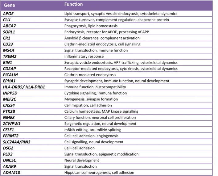
Before the era of large-scale Genome-Wide Association Study (GWAS), ε4 allele of the APOE gene was the only well-established risk factor for the pathogenesis of LOAD, but with technological advances, researchers have identified a number of regions of interest in the genome that may increase a person’s risk for LOAD to varying degrees. It was striking to note that most of the genes identified by GWAS that could be linked with the Aβ cascade or tau pathology roughly cluster within three pathways: Lipid metabolism, Inflammatory response and Endocytosis (Giri et al., 2016) (Figure 3).
11
These 29 or so genes and their role are briefly described in the table below (Table 2).
Gene Function
APOE Lipid transport, synaptic vesicle endocytosis, cytoskeletal dynamics
CLU Synapse turnover, complement regulation, chaperone protein
ABCA7 Phagocytosis, lipid homeostasis
SORL1 Endocytosis, receptor for APOE, processing of APP
CR1 Amyloid β clearance, complement activation
CD33 Clathrin-mediated endocytosis, cell signalling
MS4A Signal transduction, immune function
TREM2 Inflammatory response
BIN1 Synaptic vesicle endocytosis, APP trafficking, cytoskeletal dynamics
CD2AP Receptor-mediated endocytosis, cytokinesis, cytoskeletal dynamics
PICALM Clathrin-mediated endocytosis
EPHA1 Synaptic development, immune function, neural development
HLA-DRB5/ HLA-DRB1 Immune function, histocompatibility
INPP5D Cytokine signalling, immune function
MEF2C Myogenesis, synapse formation
CASS4 Cell migration, cell adhesion
PTK2B Calcium homeostasis, MAP kinase signalling
NME8 Ciliary function, neuronal cell proliferation
ZCWPW1 Epigenetic regulation, neural development
CELF1 mRNA editing, pre-mRNA splicing
FERMT2 Cell–cell adhesion, angiogenesis
SLC24A4/RIN3 Cell signalling, neural development
DSG2 Cell–cell adhesion
PLD3 Signal transduction, epigenetic modification
UNC5C Neural development
AKAP9 Signal transduction
ADAM10 Hippocampal neurogenesis, cell adhesion
Table 2: Common and rare gene variants associated with Alzheimer’s disease identified by GWAS (adapted from Giri et al., 2016).
c) Environmental factors
Despite the fact that sporadic or late-onset AD is the most frequent form of the pathology; its origin is still to be understood. From a clinical stand point the elements which spark LOAD are various and heterogeneous. Nevertheless, several environmental risk factors have been identified and associated to disease development. One of the predominant risk factors is lifestyle. Indeed, an improper diet rich in saturated fats, salt and refined sugars may lead to obesity, hypertension and type II diabetes. These metabolic disorders raise the level of inflammation in the body and increase the incidence of AD. Alcohol and tobacco consumption are also aggravating factors as they are known to accelerate cellular aging. Other factors such as environmental pollution due to neurotoxic metals (lead, mercury, aluminium, cadmium and arsenic) and/or pesticides may alter Aβ production
12
and Tau phosphorylation thus increasing the incidence of AD also (Chin-Chan et al., 2015). Furthermore, social context is also a determining factor in the development of AD. The level of education, physical activity and post-menopause treatments are amongst the parameters susceptible in modulating the risk of developing sporadic forms of the pathology (Dosunmu et al., 2007; Barnes and Yaffe, 2011).
Nonetheless, several neuroprotective environmental factors susceptible to slow down or even decrease the incidence of AD have been brought to light. Performing regular physical activity, consuming foods rich in omega-3 and foods rich in antioxidants (fatty fish, nuts, seeds, fruit, vegetables, green tea…), the use of non-steroid anti-inflammatories and having a cognitively stimulating environment are some of them.
Histopathological aspects of Alzheimer’s disease
G.
Macroscopic lesions 1.
On a macroscopic level, AD is characterised by a cerebral atrophy mainly localised in the entorhinal cortex, hippocampus, amygdala, and in the temporal and frontal gyri. This translates as a weight reduction of the temporal, parietal and frontal lobes. Often a dilation of the cerebral ventricles can also be observed (Perl, 2010) (Figure 4).
Figure 4: Cross section of a normal brain versus AD brain (adapted from Crimins et al., 2013). AD patient brains are
13
Microscopic lesions 2.
Microscopic lesions that appear in the brain are not homogenous. The topography and the kinetics of the apparition of the lesions are crucial to identify the pathology. Two types of lesions can be distinguished, on one hand the ones associated with protein aggregation and on the other hand ones associated with synaptic and neuronal loss (Duyckaerts et al., 2009).
On a tissue level, two main histopathological lesions, hallmarks of AD, can be identified: intracellular neurofibrillary tangles (NFTs) composed of hyperphosporylated Tau protein, and extracellular senile plaques composed of aggregated Aβ peptides that form insoluble antiparallel β-sheets called fibrils (Figure 5).
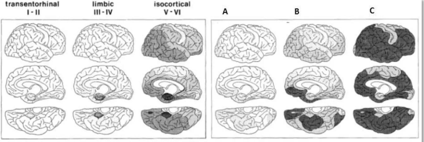
It has been shown that these two hallmarks evolve differently in the brain during AD progression (Braak and Braak, 1991). A model of AD development, in 6 different stages, has been established (Figure 6).
NFTs first accumulate in the entorhinal cortex and hippocampus (Figure 6, stages I and II). During these so called “silent” stages, Mild Cognitive Impairments (MCIs) appear but are not determining signs of AD as these cognitive symptoms may also point towards potential other forms of dementia. Then, NFTs progress to the limbic (Figure 6, stages III and IV) and cortical (Figure 6, stages V and VI) areas of the brain. At this stage, Fully Developed Alzheimer is attained and cognitive deficits are significant.
Figure 5: Hallmarks of AD (adapted from Nixon et al., 2007). AD brain stained with Bielschowsky silver revealing
14
As for amyloid deposits, which constitute senile plaques, they are initially found in the cortical areas (Figure 6, stage A). They then progress to the associated cortical areas such as the hippocampus (Figure 6, stage B) and finally to the rest of the cortex (Figure 6, stage C) including the sensori-motor cortex.
a) Intracellular Tau and neurofibrillary tangles
These lesions were first described by Alois Alzheimer in the early 20th Century, during the study of his patient’s brain (Alzheimer et al., 1907, translated into English in 1995). However, it wasn’t until the 1960s that electron microscopy studies revealed the exact composition of these lesions; namely: neurofilaments, microtubule associated proteins (MAP2), vimentin and tropomyosin proteins, elements of the proteasome, proteoglycans, inflammatory molecules, amyloïdogenic proteins (APP, presinilin, APE) and elements of the cytoskeleton such as Tau protein (Smith et al., 1996).
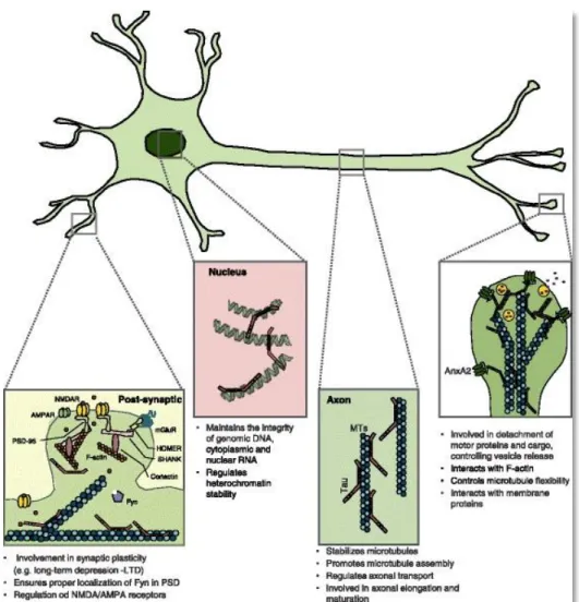
Tau (Tubulin Associated Unit) protein is an organiser and stabiliser of microtubules (MTs). These MTs are, in a sense, the scaffold of cells as they shape and structure them. Tau phosphorylation allows the regulation of its binding to MTs. Unphosphorylated Tau has a higher affinity for MTs and so phosphorylated Tau remains detached from the MTs. The MT stabilisation and assembly role of Tau highlights its importance in axonal growth and cytoskeletal structure stability in the mature neuron. Interestingly, Tau is not the only MT-associated protein. In vivo studies in mice show that knocking out Tau does not significantly alter the stabilisation of the MTs. This suggests early developmental compensation mechanisms by other MAPs, mainly MAP1A which is found to be increased (D Ke et al., 2012). This compensation doesn’t seem to occur in adult brains (D Ke et al., 2012), which might explain why Tau pathology causes neuronal MT instability.
Figure 6: Evolution of the spatial distribution of NFTs and senile plaques during AD (adapted from Braak and Braak, 1991).
15
In immature neurons, Tau is mainly present in axonal and somatodendritic compartments. In mature neurons, however, Tau is mainly found in the axon (Burack and Halpain, 1996) where they allow transport of cargo from the cell body to the axon terminals (Avila et al., 2016) by interacting with transport proteins like dynein and kinesin (Dixit et al., 2008). In addition, it has also been shown that the N-terminal projection of Tau has the ability to bind to the Annexin A2 protein at the level of the axonal plasma membrane, highlighting the ability of Tau to interact with cell membranes (Sotiropoulos et al., 2017). Through this membrane interaction, Tau is believed to play a role in various cell signalling pathways by interacting with transmembrane receptors (Guo et al., 2017). Interestingly, Tau has also been found in the nucleus, where it is believed to bind to DNA and plays a role of protection against stressful stimuli such as hypothermia or hyperthermia which can cause DNA damage (Guo et al., 2017). Additionally, Tau has been described to play a role in the regulation of genetic and epigenetic expression by binding and changing the conformation of DNA and its binding to histones. Furthermore, other roles have been found for Tau, including a role in the insulin signalling pathways (Jolivalt et al., 2008) (Figure 7).
Although Tau is mainly found in axons, it is also present in somatodendritic compartments and post-synaptic densities (Wang and Mandelkow, 2016). In dendrites and dendritic spines, Tau has an important role in synaptic plasticity. Tau acts as a scaffold at the level of the synapses, both in pre- and post-synaptic compartments. Tau is known to play a role in synaptic activity, as it interacts with Fyn tyrosine kinase and binds to PSD-95, which regulates the function of post-synaptic receptors, such as NMDA receptors (NMDAr) (Ittner et al., 2010; Arendt et al., 2016) (Figure 7).
In pathological conditions, it is precisely at the level of the postsynaptic density that Tau has been proposed to exert its deleterious effects. In the case of AD, studies using live microscopy and confocal imaging have demonstrated that Aβ causes the mislocalisation and abnormal phosphorylation of Tau at the postsynaptic level, abnormally increasing its abundance in this compartment (Frandemiche et al., 2014; Amar et al., 2017). This may increase the interaction of Tau with Fyn tyrosine kinase and PSD-95, increasing the synaptic activity through NMDAr, which might lead to excitotoxicity. Furthermore, a study showed that Tau is required to cause Long-term Depression (LTD) in the hippocampus (Kimura et al., 2014). This could mean Tau plays a role in downregulating AMPA receptors (AMPAr), possibly leading to synapse pruning (Figure 7).
16
NFTs appear when Tau is abnormally phosphorylated and starts forming aggregates within the neuron. Studies have also reported that Tau cleavage by caspases generates peptides that are more prone to form aggregates (Gamblin et al., 2003). These peptides aggregate with the non-cleaved Tau around MTs, where Tau gets hyperphosphorylated. This causes Tau to unbind from the MTs and cause their destabilisation whilst simultaneously aggregating into NFTs and accumulating in the cytosol (Rissman et al., 2004).
It is now widely accepted that Tau protein is the major component of NFTs. However, NFTs aren’t a determining factor for AD since they are also found in frontotemporal dementia and other neurodegenerative pathologies known as tauopathies (Delacourte and Buée, 2000).
Figure 7: Identified functions and localisations of Tau in neurons (adapted from Sotiropoulos et al., 2017). Tau protein is
located in several compartments in the neuron. In axons, Tau stabilises MTs and regulates axonal transport. At the synapses, Tau interacts with membrane proteins and plays a role in neuronal activity as well as synapse plasticity. In the nucleus, Tau protects DNA and nuclear RNA, while regulating genetic and epigenetic expression.
17
b) Aβ peptide and extracellular senile plaques
Although described for the first time by Alois Alzheimer in the early 20th Century (Alzheimer et al., 1907, translated into English in 1995) after brain autopsy of his patient, the principal constituent of the extracellular senile plaques wasn’t established until 1984 by Dr. Glenner’s team. These plaques showed to be aggregates of a peptide named Amyloid Beta 42 peptide or Aβ (Glenner and Wong, 1984).
Aβ is a physiological peptide, produced throughout the course of life. Senile plaques, however, are characteristic lesions of AD where Aβ peptides aggregate in an aberrant manner. Amyloid deposits are absent in young individuals and gradually appear with aging. In fact, it is only when these plaques grow considerably in number and size that they induce neuroinflammation, which is characterised by the activation of brain resident immune cells.
Although Aβ is the major constituent of senile plaques, there are other elements that participate in the making of these plaques. Indeed, during the formation of these plaques Aβ binds with ApoJ (or clusterin) (Martin-Rehrmann et al., 2005), ApoE, cholesterol (Lesser et al., 2011), with cathepsin D, with components of the extracellular matrix such as thrombospondin, ICAM1 (Inter-Cellular Adhesion Molecule 1) and heparan sulfate proteoglycans, but also Ca2+, iron (Everett et al., 2018) and other metals (copper, zinc…) (Ha et al., 2007).
Furthermore, these amyloid deposits can aggregate into different forms in the brain (Duyckaerts et al., 2009; LeVine and Walker, 2010).
The first correspond to “diffuse” forms where the deposits are relatively large, up to several hundred micrometres in diameter. These have little immunoreactivity and are difficult to identify by immunohistochemistry. Congo Red and Thioflavine S (two dyes which allow staining of aggregates) do not allow the visualisation of these diffuse plaques as they are mainly composed of Aβ40. However, they can be revealed with antibodies against Aβ (Güntert et al., 2006). These types of plaques can be found in large numbers and sizes in individuals who allegedly have no cognitive deficits, which raises doubts as to their toxicity in the brain. In fact, it has been proposed that these forms of plaques are an early form of senile plaques in certain brain areas (Duyckaerts et al., 2009; LeVine and Walker, 2010) (Figure 8).
The second form of plaques are described as “focal” which are spherical deposits much smaller in size but also much more dense. As they contain a lot more Aβ42 molecules, these focal deposits seem to form the core of senile plaques. They are surrounded by processes coming from nearby
18
neurons and astrocytes (Duyckaerts et al., 2009; LeVine and Walker, 2010)(Figure 8).
Other amyloid deposits, described as “stellar deposits” have also been identified. Smaller and likely associated to astrocytes, these deposits are less studied than the previous aforementioned forms although often observed (Duyckaerts et al., 2009) (Figure 8).
In some cases, amyloid β can also accumulate in the walls of blood vessels, particularly in the ones present in the core of senile plaques, which induces CAA (Cerebral Amyloid Angiopathy) also known as congophilic angiopathy (Yamada and Naiki, 2012).
It’s the form and properties of the amyloid peptides which induce the formation of fibrils and eventually plaques on the outside of neurons. However, some Aβ can be found intracellularly (Bayer and Wirths, 2010; Tomiyama et al., 2010; Thal and Fändrich, 2015). Quite intriguingly, this intracellular accumulation of Aβ42 correlates much more with neuronal cell death than the formation of extracellular senile plaques (Christensen et al., 2008) and the smaller oligomers seem more toxic than fibrillary Aβ deposits.
Figure 8: Macroscopic lesions and the different aspects of amyloid deposits (adapted from Duyckaerts et al., 2009). Left
panel: Image of a normal brain versus an advanced stage of AD brain. Right panels: the different types of amyloid deposits identifiable by immunohistochemistry. (A) Diffuse amyloid peptide deposits. (B) Focal deposits (arrowheads) much denser, which form the core of amyloid plaques, surrounded by a halo of much less dense amyloid peptides. (C) Stellar deposits (arrows).
19
c) Links between Aβ and Tau
The hypothesis of the amyloid cascade originated from the observation that mutations which cause AD are always associated with alterations of the Amyloid Precursor Protein (APP) metabolism, leading to Aβ42 overproduction particularly. On the contrary, Tau mutations do not lead to AD. Subsequently, it has been postulated that it may be the accumulation of neurotoxic amyloid peptides which drives Tau protein modifications leading to MT destabilisation, formation of NFTs, and eventually neuronal loss and memory deficits (Hardy and Higgins, 1992). Indeed, a study showed that Aβ42 fibrils induce NFT formation (Götz et al., 2001). It has also been proposed that Aβ can induce Tau cleavage by caspases, generating Tau fragments prone to aggregate and form NFTs (Gamblin et al., 2003).
Nevertheless, NFTs have been observed in brains in absence of amyloid deposits (Braak and Braak, 1997) which suggests that both lesions occur independently and in parallel of one another, and that NFT formation might even precede amyloid plaque formation (Duyckaerts et al., 2009). Moreover, amongst the different transgenic mouse models of AD used in research, the ones that bare mutations only on APP and PS do not present any NFTs. On the other hand, the triple transgenic 3xTg-AD mouse model, developed by LaFerla’s team, which bares the Swedish double mutation on APP (APPswe), a mutation on PS1 (PS1 M146V) and a Tau mutation (MAPT P301L) develop NFTs. However, NFTs appear after amyloid plaque formation in this 3xTg-AD model (Oddo et al., 2003; Billings et al., 2005). The possibility of a synergistic interaction between both Aβ and Tau pathways in
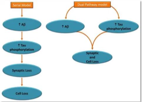
Figure 9: Two Hypothesized Models Linking Core Features of Alzheimer’s Disease (adapted from Small and Duff, 2008). The
amyloid hypothesis assumes a serial model of causality, whereby abnormal elevations in Aβ drive tau hyperphosphorylation and other downstream manifestations of the disease. According to the dual pathway hypothesis, Aβ elevations and tau hyperphosphorylation can be linked by separate mechanisms driven by a common upstream molecular defect.
20
the physiopathology of AD, driving synaptic dysfunction and neuronal degeneration, is widely accepted (Small and Duff, 2008) (Figure 9).
Propagation of the pathology throughout the brain
H.
Extracellular Aβ deposits and intracellular Tau aggregates are both required to make the definite neuropathological diagnosis of AD. However, the interplay between these two proteins and the way they spread throughout the brain is still to be clearly determined. It is known that Tau pathology in the cell body of neurons precedes Aβ plaque formation and the two histopathological markers evolve and spread spatiotemporally differently in the brain. Indeed, Tau seems to spread outwards, from the centre to the cortex where as Aβ deposits do the opposite, spreading from the cortex to the centre (Braak and Braak, 1991). As NFTs appear first, this was long used as an argument to say that Tau was the initiating factor of AD.
Propagation of Tau 1.
From the observations made during brain autopsies of patients with tauopathies, that NFTs and other Tau aggregates progress in a stereotypical neuroanatomical pattern of spreading, came the hypothesis that Tau might spread from neuron to neuron. To support this hypothesis, several studies show that Tau is present in the extracellular compartment, that Tau can be released from donor cells and uptaken by recipient cells (Goedert et al., 2010; Mudher et al., 2017).
The secretion of Tau has been described to occur in multiple ways. Interestingly, the vast majority (90%) of secreted Tau is released as a free protein, whereas only a small fraction is bound to microvesicles such as exosomes or ectosomes (Katsinelos et al., 2018). The mechanisms of this secretion are still not fully understood, though it has been reported that Tau may exit the cell through diffusion or through the formation of nanotubes between cells (Tardivel et al., 2016). It has also been reported that phosphorylation of Tau increases Tau secretion through exosomes (Katsinelos et al., 2018). This makes sense in the light of the fact that phosphorylated Tau is more available for secretion as unphosphorylated Tau would rather tend to bind to MTs.
The uptake mechanism of Tau by recipient cells is not fully described yet but several studies seem to indicate that i) small oligomeric species of Tau, not monomeric, are spontaneously uptaken by the
21
recipient cells in vitro ii) this would occur through simple diffusion or endocytosis (Wu et al., 2013, 2016) (Figure 10).
Interestingly, Tau fibrils bind to transmembrane protein APP and increase its internalisation as well as increasing intracellular aggregation of Tau (Takahashi et al., 2015).
Most convincingly, several studies have showed that intracerebral injection of misfolded Tau (whether it be from brain extracts from transgenic mice with Tau P301S mutation or purified Tau fibrils from P301S brain extracts or preformed recombinant Tau fibrils) into the brain of transgenic mice expressing human wild-type Tau, not only induced Tau pathology but also the spread of this pathology along a path that is neuroanatomically connected (Ahmed et al., 2014; de Calignon et al., 2012; Iba et al., 2013; Dujardin et al., 2014; Mudher et al., 2017).
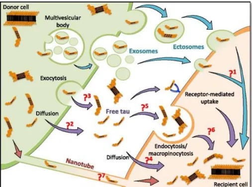
Figure 10: Different pathways identified for Tau propagation (adapted from Mudher et al., 2017). Tau proteins can be
transferred from donor cells (green) to recipient cells (orange) using different routes. Pathway indicated by blue arrows: tau proteins are released in the medium by extracellular vesicles like exosomes and ectosomes. Violet pathway: Around 90% of tau in the extracellular space is found as free protein, passive diffusion facilitated by a membraneous transporter/receptor (?2) or active exocytosis (?3) might contribute to this process. Uptake of free tau species by recipient cells, including APP-mediated endocytosis has been reported. Whether free or aggregated tau is taken up by other mechanisms such as diffusion (?4) or non-receptor mediated endocytosis/macropinocytosis (?5) has not been resolved. Nor is it known how membrane-bound tau can escape from vesicles and enter the cytoplasm of recipient cells (?6). Red pathway: Tau was shown to be present inside nanotubes connecting cells in vitro and to allow its interneuronal transfer. This mechanism could potentially participate in prion-like propagation of tau pathology but whether it is a mode of transcellular transfer of seeding-competent tau species in vivo needs to be investigated.