Article
Vectorial Import via a Metastable Disulfide-Linked
Complex Allows for a Quality Control Step and Import
by the Mitochondrial Disulfide Relay
Graphical Abstract
Highlights
d
IMS import by the disulfide relay proceeds via formation of a
covalent intermediate
d
Import is driven by hydrophobic and disulfide-dependent
interactions
d
The covalent intermediate is also formed by
oxidation-incompetent substrates
d
Redox quality control allows for IMS substrate release and
proteasomal degradation
Authors
Markus Habich, Silja Lucia Salscheider,
Lena Maria Murschall, ..., Hamid Kashkar,
Joern Dengjel, Jan Riemer
Correspondence
[email protected]
In Brief
Disulfide formation in the mitochondrial
intermembrane space (IMS) is an
essential process that is coupled to
protein import. Here, Habich et al. report
on a redox quality control step in which
the import competence of substrates is
tested, and when it fails, substrates are
released to the cytosol for degradation.
Habich et al., 2019, Cell Reports26, 759–774 January 15, 2019ª 2018 The Author(s). https://doi.org/10.1016/j.celrep.2018.12.092
Cell Reports
Article
Vectorial Import via a Metastable Disulfide-Linked
Complex Allows for a Quality Control Step and
Import by the Mitochondrial Disulfide Relay
Markus Habich,1Silja Lucia Salscheider,1Lena Maria Murschall,1Michaela Nicole Hoehne,1Manuel Fischer,1
Fabian Schorn,2Carmelina Petrungaro,1Muna Ali,1Alican J. Erdogan,1Shadi Abou-Eid,3Hamid Kashkar,2
Joern Dengjel,3and Jan Riemer1,4,*
1Institute for Biochemistry, Department of Chemistry, University of Cologne, Zuelpicher Str. 47a/R. 3.49, 50674 Cologne, Germany 2Cologne Excellence Cluster on Cellular Stress Responses in Aging-Associated Diseases (CECAD), Center for Molecular Medicine Cologne
(CMMC) and Institute for Medical Microbiology, Immunology and Hygiene, University of Cologne, CECAD Research Center, Joseph-Stelzmann-Str. 26, 50931 Cologne, Germany
3Department of Biology, University of Fribourg, Chemin du Muse´e 10, 1700 Fribourg, Switzerland 4Lead Contact
*Correspondence:[email protected] https://doi.org/10.1016/j.celrep.2018.12.092
SUMMARY
Disulfide formation in the mitochondrial
intermem-brane space (IMS) is an essential process. It is
cata-lyzed by the disulfide relay machinery, which couples
substrate import and oxidation. The machinery relies
on the oxidoreductase and chaperone
CHCHD4-Mia40. Here, we report on the driving force for IMS
import and on a redox quality control mechanism.
We demonstrate that unfolded reduced proteins,
upon translocation into the IMS, initiate formation
of a metastable disulfide-linked complex with
CHCHD4. If this interaction does not result in
produc-tive oxidation, then substrates are released to the
cytosol and degraded by the proteasome. Based
on these data, we propose a redox quality control
step at the level of the disulfide-linked intermediate
that relies on the vectorial nature of IMS import.
Our findings also provide the mechanistic framework
to explain failures in import of numerous human
disease mutants in CHCHD4 substrates.
INTRODUCTION
In human cells, proteins of the mitochondrial intermembrane space (IMS) fulfil essential functions in calcium signaling, respira-tory chain assembly, hypoxia response, and apoptosis signaling. IMS proteins are synthesized by cytosolic ribosomes and are imported into the IMS by dedicated machineries. Many IMS proteins rely not on classical bipartite mitochondrial targeting sequences (MTSs) for their import but, instead, on conserved cysteines that are arranged in specific patterns (Chatzi et al., 2016; Endo et al., 2010; Erdogan and Riemer, 2017; Harbauer et al., 2014; Wasilewski et al., 2017).
These proteins are substrates of the mitochondrial disulfide relay; more specifically, of the IMS import receptor and
oxidore-ductase CHCHD4 (also human Mia40; in yeast, Mia40) (Banci et al., 2009, 2010; Fischer et al., 2013; Hofmann et al., 2005; Me-secke et al., 2005). CHCHD4 is an unusual oxidoreductase. Un-like oxidoreductases from other oxidizing compartments, Un-like the endoplasmic reticulum or the bacterial periplasm, CHCHD4 does not consist of one or more thioredoxin-like domains but, instead, uses a minimal helix-turn-helix fold containing a hydro-phobic groove with chaperone function for substrate interaction (Banci et al., 2009, 2010; Kawano et al., 2009; Koch and Schmid, 2014a). Connected to this fold is a short helix with the redox-active cysteine-proline-cysteine motif (C53 and C55 of human CHCHD4). The N- and C-terminal regions of CHCHD4 are un-structured. At least in vitro, they are not required for CHCHD4 function, and a truncated human CHCHD4 can functionally replace Mia40 in yeast cells (Chacinska et al., 2008; Sztolsztener et al., 2013). However, in intact mammalian cells, the N-terminal region serves in the interaction with apoptosis-inducing factor (AIF), which recently has been shown to be critical for the mito-chondrial import of CHCHD4 (Hangen et al., 2015; Meyer et al., 2015).
CHCHD4 substrates are synthesized by cytosolic ribosomes as reduced precursors. In the cytosol, members of the glutare-doxin family (in yeast, members of the thioreglutare-doxin family) main-tain them in their reduced state (Banci et al., 2013; Durigon et al., 2012). Additionally, at least in vitro, Zn2+maintains sub-strates in a reduced state (Morgan et al., 2009). In yeast, the proteasome system fulfils a control function and removes parts of newly produced Mia40 substrates immediately after synthesis (Bragoszewski et al., 2013; Kowalski et al., 2018). It is not known whether this is also the case in mammalian cells.
During passage through the translocase of the outer mem-brane (TOM) pore, substrates already interact with CHCHD4 ( Pe-leh et al., 2016; von der Malsburg et al., 2011). The initiation of the interaction between CHCHD4 and its substrates therefore depends on two hydrophobic motifs. These motifs are placed in an amphipathic helix that contains a conserved cysteine residue. The first motif consists of hydrophobic residues posi-tioned directly adjacent to this specific substrate cysteine.
In vitro experiments indicated that they initiate a loose
non-cova-lent interaction with the hydrophobic groove of CHCHD4 (Koch and Schmid, 2014b). Subsequently, a hydrophobic patch in the amphipathic helix of the substrate (also termed mitochondrial IMS sorting signal [MISS] or IMS targeting signal [ITS]) that serves as the second motif strengthens this non-covalent inter-action (Koch and Schmid, 2014b; Milenkovic et al., 2009; Sideris et al., 2009). Both non-covalent interactions initiate helix folding and orient the substrate and, therefore, allow nucleophilic attack of the thiolate anion of the substrate cysteine residue on CHCHD4. This results in the formation of an intermolecular disul-fide bond, likely via C55 of CHCHD4 (Banci et al., 2009; Koch and Schmid, 2014a; Terziyska et al., 2009). This intermolecular disul-fide bond is long-lived in vitro and in vivo compared with intermo-lecular disulfide bonds that are formed during oxidative protein folding in other compartments (Chacinska et al., 2004; Fischer et al., 2013; Koch and Schmid, 2014a; Mesecke et al., 2005; Pet-rungaro et al., 2015; Sideris et al., 2009). It has been proposed that this is an adaptation to the coupling of vectorial import and oxidative folding in the IMS that is not found in other oxidizing compartments (Koch and Schmid, 2014a). The inter-molecular disulfide is resolved by a second nucleophilic attack from a cysteine of the substrate, which leads to the formation of a disulfide bond in the substrate. The importance of CHCHD4 for substrate import is illustrated by the fact that depletion of CHCHD4 results in loss of its substrates (Fischer et al., 2013; Hangen et al., 2015; Hofmann et al., 2005; Meyer et al., 2015). Likewise, also in yeast, depletion of Mia40 prevents efficient mitochondrial substrate accumulation (Chacinska et al., 2004; Mesecke et al., 2005).
The precise mechanism of vectorial disulfide relay-mediated IMS import remains debated. Initial models based mainly on
in vitro import experiments in yeast suggested a pathway in
which disulfide formation between the substrate and Mia40 leads to directed diffusion of the substrate into the IMS ( Mile-nkovic et al., 2009; Sideris et al., 2009; von der Malsburg et al., 2011). However, these experiments neglected the reducing influences of the cytosol and IMS, the competition of different substrates for import, the chaperone function of Mia40, and the extremely low import efficiency of in organello import ap-proaches compared with the highly efficient IMS import in intact cells (Fischer et al., 2013; Kojer et al., 2015). An alternative model proposed that disulfide formation is negligible for protein import and only a consequence of a preceding chaperone-like ‘‘pulling’’ of the substrate by Mia40 that solely relies on hydrophobic inter-actions (Baker et al., 2012; Peleh et al., 2016; Weckbecker et al., 2012). The latter ‘‘trans-site receptor’’ model is based on the fact that the MISS/ITS signal of disulfide relay substrates is both necessary and sufficient for targeting proteins into yeast mito-chondria in vitro, although, again, the import efficiencies of the respective approaches were very low (Milenkovic et al., 2009; Sideris et al., 2009). Moreover, genetic separation of the oxido-reductase and chaperone function of Mia40 suggested that the oxidoreductase function might not be essential, although the respective experiments were performed in the presence of high amounts of oxidant (Peleh et al., 2016).
The trans-site receptor model is contradicted by work in intact cells. Expression of a redox-inactive CHCHD4 mutant
(C53S,C55S) in human cells abolished the import and oxida-tion of selected substrate precursors, although the hydropho-bic interaction between the substrate and CHCHD4 was not targeted (Fischer et al., 2013). Additionally, disease mutants like NDUFB10C107S (Friederich et al., 2017) and TIMM8C66W
(Hofmann et al., 2002) demonstrated the importance of sub-strate cysteines because they also fail to accumulate in mitochondria.
To reconcile these apparently exclusive findings and address the important question of how the disulfide relay pop-ulates the IMS in intact cells as well as to provide a framework for why disease mutants of CHCHD4 substrates fail to accu-mulate in the IMS, we revisited the mechanisms of disulfide relay-dependent import in intact mammalian cells. A series of dynamic pulse-chase and proteomic approaches allowed observation of vectorial transport with the correct stoichiom-etry between the substrate and CHCHD4, the correct spatio-temporal setting of events, in the endogenous redox milieu of the IMS and the cytosol. With our approaches, we demon-strated the formation of a metastable disulfide-linked interme-diate between CHCHD4 and the translocating substrate. This intermediate is also formed by substrate variants, including patient mutants, that do not become oxidized and do not accumulate in mitochondria and by CHCHD4 variants that fail to oxidize their substrates. At the level of this intermediate, a decision is made for either productive mitochondrial protein import and oxidation or—if oxidation does not proceed effi-ciently—substrate release to the cytosol and proteasomal degradation of the unfolded precursor. Thus, the disulfide relay itself provides a layer of quality control for its substrates and determines whether they are competent for folding and import.
RESULTS
In Intact Cells, Cysteine Variants of COX19 Fail to Become Oxidized Despite Formation of Mixed Disulfide Intermediates with CHCHD4
First we explored the spatiotemporal importance of cysteines and hydrophobic interactions in the human disulfide relay by analyzing cysteine mutants in disulfide relay substrates. We therefore mainly focused on selected model twin CX9C substrate
proteins (COX19, COA4, and NDUFB10;Figure S1A). Although IMS proteins that rely on different cysteine motifs for import and oxidative folding have been identified (Barchiesi et al., 2015; Petrungaro et al., 2015; Weckbecker et al., 2012), the ma-jority of known CHCHD4 substrates are members of the twin CX9C family (Cavallaro, 2010; Fischer et al., 2013; Petrungaro et al., 2015). We generated different HEK293 cell lines that express, in a stable and inducible fashion, hemagglutinin (HA)-tagged substrate variants of COX19, COA4, and NDUFB10. This ensures comparatively low expression levels that lead to full accumulation of wild-type proteins in mitochondria (Fischer et al., 2013). For COX19, we mutated the cysteines that, in the mature protein, form the outer disulfide (C30S,C61S) or the inner disulfide (C40S,C51S). For COA4, we generated a quadruple cysteine mutant to alanine that renders the protein even more hydrophobic, and for NDUFB10, we generated the C107S
mutant, which corresponds to a patient mutation (Friederich et al., 2017;Figure S1A).
Mechanistically, disulfide-linked interaction between CHCHD4 and substrates is the first step in oxidative folding. To test whether COX19 variants form disulfide-linked inter-mediates with CHCHD4 during their biogenesis in intact cells, we employed a pulse-chase assay followed by denaturing immunoprecipitation (IP) against CHCHD4 and a subsequent denaturing re-IP against the different COX19 variants ( Fig-ure 1A). Notably, except for the cysteine-free quadruple mutant of COX19, all variants of COX19 did covalently interact with CHCHD4 (Figure 1B). Wild-type NDUFB10 and the C107S mutant also interacted with CHCHD4 (Figure S1B). Thus, sub-strates, even when they do not contain the full set of cysteines, traverse the TOM pore and initiate interaction with CHCHD4. It also indicates that not only the cysteine with an adjacent MISS/ ITS is capable of interaction with CHCHD4, but, instead, that substrates exhibit a certain plasticity with respect to their CHCHD4 interaction site. In line with this, we found that, for most human twin CX9C proteins, both helices possess
hydro-phobic stretches that could serve as alternative CHCHD4 inter-action sites (Figure S1C).
Does interaction of substrate cysteine variants with CHCHD4 result in productive oxidation? To test this, we assessed the oxidation kinetics of newly synthesized COX19. To this end, we employed an oxidation assay in which we coupled a pulse-chase assay to redox state determination and IP of the protein of interest (Fischer et al., 2013; Figure 1C). Although wild-type COX19 quickly became oxidized, all cysteine variants remained completely reduced over the course of the experi-ment (Figure 1D). This is contradictory to in vitro experiments in which COX19 cysteine variants did become oxidized by CHCHD4 and also formed mixed disulfide intermediates (Bien
et al., 2010;Figures S1D–S1I). Thus, in intact cells, interaction of the substrate and CHCHD4 is not predictive of oxidative folding.
Additionally, we tested whether forced import of COX19 into the IMS by equipping it with an MTS would allow oxidation of COX19 cysteine variants. To this end, we generated stable cell lines expressing COX19C30S,C61S, COX19C40S,C51S, or COX19C30S,C40S,C51S,C61S fused to the bipartite MTS of the soluble protein SMAC (COX19 variants). Like MTS-COX19WT, all MTS cysteine variants co-localized with
mito-chondria in immunofluorescence (Figure 1E). As shown previously, oxidation of wild-type COX19 was slightly acceler-ated when fused to this MTS (Fischer et al., 2013;Figure 1F). However, when we performed the oxidation assay with both MTS cysteine variants, we observed that these proteins did not become oxidized (Figures 1G and 1H). Instead, they ap-peared to be destabilized (compare withFigure 1D).
Taken together, COX19 cysteine variants, even when effi-ciently targeted to the IMS, are not oxidized in intact cells.
In vitro, the proteins still have the capacity to interact
with CHCHD4 and become oxidized by it (Figure S1). How-ever, in the in vivo situation with its vectorial import and reducing redox milieu, non-productive oxidation is prevented despite an initial covalent interaction between the substrate and CHCHD4 (Fischer et al., 2013; Kojer et al., 2012; Figure 1B).
Interaction with CHCHD4 Does Not Result in
Mitochondrial Retention of COX19 Cysteine Variants
Do substrates that interact with CHCHD4 but fail to become oxidized still accumulate in mitochondria? In other words, is the hydrophobic interaction with CHCHD4 sufficient to pull sub-strates into the IMS? To test this, we analyzed the mitochondrial
Figure 1. Despite Interaction with CHCHD4, COX19 Cysteine Variants Fail to Become Oxidized in Intact Cells (A) In vivo CHCHD4-substrate interaction assay. HEK293 cells stably expressing COX19-HA variants were pulse-labeled with35
S-methionine for 4 h. Then thiol exchange reactions were inhibited by addition of n-ethylmaleimide (NEM) to preserve mixed disulfides. Cells were lysed in SDS, and a denaturing immuno-precipitation (IP) against CHCHD4 enriched monomeric CHCHD4 and mixed disulfides of COX19-HA variants bound to CHCHD4. The latter were isolated by a second IP (re-IP) against the HA tag of COX19-HA variants. This mixed disulfide was analyzed in non-reducing or reducing SDS-PAGE and visualized by autoradiography.
(B) Interaction between CHCHD4 and COX19 cysteine variants, performed as described in (A). 1% of the IP against CHCHD4 was loaded as a control. Expression was induced during the 4-h pulse with 1mg/mL doxycycline. CHCHD4 interacts covalently with different COX19 cysteine variants in vivo. In silico analysis suggests that many twin CX9C substrate proteins contain multiple sites for interaction with CHCHD4 (Figure S1C). Red, reducing SDS-PAGE; red,
non-reducing SDS-PAGE; red arrow, disulfide-linked CHCHD4-COX19 dimer; blue arrow, COX19; asterisk, background band; #, disulfide-linked CHCHD4-COX19 dimer, possibly with truncated CHCHD4; +, COX19 not in the disulfide-linked complex. n = 2 biological replicates.
(C) In vivo oxidation assay to follow oxidative folding in intact cells. Cells are pulse-labeled for 5 min with35
S-methionine and chased with cold methionine for different times. The chase is stopped by trichloroacetic acid (TCA) precipitation, and then the lysate is treated with 4-acetamido-4-maleimidylstilbene-2,2-di-sulfonic acid (AMS) to determine protein redox states, followed by IP against the HA tag. Eluates are analyzed by Tris-Tricine-PAGE and autoradiography. Reduced COX19 is modified with four AMS, whereas oxidized COX19 remains unmodified.
(D) In vivo oxidation assay with HEK293 cells stably expressing different cysteine variants of COX19-HA. COX19 cysteine variants are not oxidized in vivo. Performed as described in (C). Expression was induced 1 h before the experiment with 1mg/mL doxycycline. n = 2 biological replicates.
(E) Localization of MTS-COX19 cysteine variants. COX19 cysteine variants carrying the MTS of SMAC localize to mitochondria. HEK293 cells stably expressing the indicated MTS-COX19-HA variants were incubated for 24 h with 1mg/mL doxycycline to induce expression. Cells were fixated, permeabilized, and stained using primary antibodies against the HA epitope (aHA, green) and cytochrome c (acyt c, red). Nuclei were stained with DAPI (blue). Cells were analyzed by fluorescence microscopy. Scale bar, 10mm. n = 6–9 cells, 2 biological replicates.
(F) Oxidation kinetics of MTS-COX19-HA wild-type protein. Addition of a cleavable MTS allows oxidation of processed wild-type COX19-HA. Performed as described in (C). n = 2 biological replicates.
(G) Oxidation kinetics of MTS-COX19-HA cysteine variants. Addition of a cleavable MTS does not result in oxidation of COX19 cysteine variants. Performed as described in (C). n = 2 biological replicates.
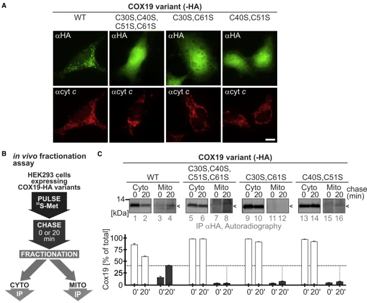
accumulation of COX19, NDUFB10, and COA4 cysteine variants in stable inducible HEK293 cells. When we tested the cellular localization of these proteins by immunofluorescence, we found that, at steady state, cysteine variants of COX19, NDUFB10, and COA4 failed to co-localize with the mitochon-drial marker protein cytochrome c. Instead, these variants exhibited cytosolic staining (Figures 2A and S2A; Friederich et al., 2017).
Next we assessed the fate of newly synthesized COX19 and COA4 variants in a dynamic fractionation experiment. With
this approach, we coupled a radioactive pulse-chase approach to rapid cell fractionation and IP of the protein of in-terest (Fischer et al., 2013; Figure 2B). Although wild-type COX19 and COA4 appeared in the mitochondrial fraction after 20 min of chase time, the cysteine variants of both proteins re-mained in the cytosolic fraction (Figures 2C and S2B). Although the hydrophobic MISS/ITS motif was present in the cysteine variants, and despite their initial interaction with CHCHD4, substrate cysteine mutants failed to accumulate in mitochondria.
A
B C
Figure 2. Interaction with CHCHD4 Does Not Result in Mitochondrial Retention of COX19 Cysteine Variants
(A) Immunofluorescence analyses of COX19-HA cysteine variants stably expressed in HEK293 cells. Performed as described inFigure 1E. Although COX19WT
-HA accumulates in mitochondria, neither of the cysteine variants does. Instead, they remain in the cytosol. Scale bar, 10mm. n = 7–10 cells, 2 biological replicates. (B) In vivo fractionation assay to assess post-translational protein import. Expression was induced by doxycycline treatment for 1 h before the pulse-chase experiment. Cells were first pulse-labeled for 5 min with35
S-methionine and then chased with cold methionine for 0 or 20 min. At the end of the chase, cells were transferred to ice and incubated with a low concentration of digitonin for 10 min to selectively permeabilize the plasma membrane and leave the OMM intact. To separate the mitochondrial from the cytosolic fraction, lysates were centrifuged, and COX19-HA variants were immunoprecipitated. Eluates were analyzed by SDS-PAGE and autoradiography.
(C) Pulse-chase fractionation experiment of COX19-HA cysteine variants expressed in HEK293 cells. Although COX19WT
-HA accumulates in mitochondria, neither of the cysteine variants does so. Instead, they remain in the cytosol. Performed as described in (B); n = 2 biological replicates.
Redox-Active Cysteines and the Hydrophobic Groove in CHCHD4 Are Required for Formation of Mixed Disulfide Intermediates with Substrates
The presence of all cysteines is required for efficient oxidation and substrate accumulation in mitochondria. Therefore, it is conceivable that formation of a covalent substrate-CHCHD4 complex also requires the presence of cysteines in CHCHD4. However, recent studies emphasized the importance of the hy-drophobic groove in CHCHD4 for substrate import (Banci et al., 2010; Peleh et al., 2016). We thus next dissected ‘‘redox’’ and ‘‘chaperone’’ contributions of CHCHD4 for substrate interaction and oxidation. To this end, we employed HEK293 cell lines stably expressing two sets of C-terminally Strep-tagged CHCHD4 variants. The first set included the ‘‘redox-incompetent’’ variants CHCHD4C53S, CHCHD4C55S, and CHCHD4C53S,C55S. The second set encompassed the chap-erone variants CHCHD4F68Eand CHCHD4C53S, C55S, F68Ewith a mutation in the hydrophobic binding groove of CHCHD4 ( Fig-ure 3A). In yeast, different mutations of the hydrophobic binding groove of CHCHD4 have been investigated. The F315E,F318E Mia40 mutant has been shown to be strongly impaired in sub-strate interaction, oxidation, and import (Kawano et al., 2009; Peleh et al., 2016). However, these mutations change the imme-diate surroundings of a structural cysteine of Mia40. We deemed it possible that the corresponding mutant might abrogate mito-chondrial import of CHCHD4, which, in human cells, depends on its own machinery and the protein AIF and not on the MTS pathway as in yeast (Hangen et al., 2015; Sztolsztener et al., 2013). For our studies, we instead employed the equivalent of the yeast Mia40F311Emutant, CHCHD4F68E(Figure 3A). This
mu-tation is far enough from the structural disulfides in CHCHD4 so that it is not likely to affect CHCHD4 folding. Yeast Mia40F311E also strongly hampered substrate accumulation but impaired viability in Mia40 complementation experiments less severely (Wrobel et al., 2013). All CHCHD4 variants were expressed to similar levels. Exceptions were the F68E-based variants that were present at lower levels, indicating that even this ‘‘mild’’ mutant might be affected in efficient folding (Figure 3B).
With these CHCHD4 variants in hand, we tested the interaction between CHCHD4 and its substrates in radioactive pulse-chase assays, followed by denaturing or native IP of CHCHD4 variants (Figures 3C and 3D). This assay allows testing the transient
inter-action between CHCHD4 and its substrates that is lost upon folding of proteins (Figure S3A). In denaturing IP with the redox-incompetent variants of CHCHD4, we observed a strong accumulation of proteins on wild-type CHCHD4 (Figure 3C, lanes 2 and 7) compared with mock and CHCHD4C53S,C55S.
Like-wise, CHCHD4C53Sco-precipitated many substrates (Figure 3C, lanes 3 and 8). CHCHD4C55Sdid so to a much lesser extent ( Fig-ure 3C, lanes 4 and 9). This indicates that cysteine residue 55 is likely the more reactive cysteine residue in the redox-active CPC motif of CHCHD4. This is also in line with our finding that, in intact cells, interaction between CHCHD4 and its reoxidizing enzyme ALR (augmenter of liver regeneration) takes place via C55 ( Fig-ure S3B). We therefore provide in vivo evidence for previous
in vitro data (Banci et al., 2009, 2011; Koch and Schmid, 2014a). Next we explored the interactome of the chaperone variants of CHCHD4 in native IP with detergent concentrations we success-fully employed before to analyze the interaction of CHCHD4 and substrates (Petrungaro et al., 2015). We found that substrates co-precipitated preferentially with wild-type CHCHD4 and not with the redox-inactive or chaperone mutants (Figure 3D). CHCHD4 variants were precipitated in similar amounts under these conditions. Substrates likely interacted covalently via di-sulfide bonds as they migrated at higher molecular weight than CHCHD4 under non-reducing conditions but at lower molecular weight after treatment with a reductant (compare Figure 3D, lanes 2 and 7). These data indicate that the hydrophobic groove in CHCHD4 is critical for initiation of disulfide-linked interactions. This is also in line with previous in vitro data (Koch and Schmid, 2014b) but somewhat contradictory to an exclusive role of hy-drophobic interactions in substrate import.
To further mechanistically dissect the role of chaperone versus redox function, we determined the redox state of CHCHD4F68E using an inverse redox state determination assay (Erdogan et al., 2018;Figure S3C). We found the CPC motif to be over-whelmingly reduced (Figure S3D). This might be explained by the mechanism of CHCHD4 reoxidation. It relies on ALR, which has to interact with the hydrophobic groove in CHCHD4 to perform reoxidation (Banci et al., 2011). Interfering with this inter-action might prevent reoxidation and lead to a reduced CPC motif. The presence of a partially reduced CPC motif might also explain at least some of the experimental outcomes with hydrophobic groove mutants of yeast Mia40 in the past that
Figure 3. Redox-Active Cysteines and the Hydrophobic Groove in CHCHD4 Are Required for Formation of Mixed Disulfide Intermediates with Substrates
(A) Structure of CHCHD4 (PDB: 2K3J) and helix 2 of human COX17 (PDB: 2L0Y). Color-coding represents the hydrophobicity of the amino acids according to the Tanford hydrophobicity scale. The substrate is positioned on the hydrophobic groove of CHCHD4, close to the redox-active cysteines C53 and C55. Phenyl-alanine F68 is localized in the hydrophobic groove. Its mutation should disturb the CHCHD4-substrate interaction.
(B) Steady-state levels of CHCHD4 variants. Cells stably expressing CHCHD4-Strep variants were induced for 24 h with 1mg/mL doxycycline. Cells were lysed, and lysates were analyzed by SDS-PAGE and immunoblot with the indicated antibodies. n = 4 biological replicates.
(C and D) Pulse-chase analyses of CHCHD4-substrate interactions. HEK293 cells stably expressing CHCHD4-Strep variants were pulse-labeled with
35
S-methionine for 4 h. Then thiol exchange reactions were inhibited by addition of NEM to preserve mixed disulfides. Cells were lysed in 2% SDS for denaturing IP (C, n = 2 biological replicates) or in 1% Triton X-100 for native IP (D, n = 1). Then lysates were subjected to IP against Strep, analyzed by non-reducing or reducing SDS-PAGE, and visualized by autoradiography. Disulfide-linked substrate-CHCHD4 complexes and CHCHD4 substrates are indicated. Mainly CHCHD4WT
and CHCHD4C53S
interact with CHCHD4 substrates. CHCHD4 chaperone mutants fail to enrich substrates.
(E) Summary of quantitative proteomics analysis (see Figures S3E–S3H and Table S1 for detailed data). For substrate accumulation, C55 and the hydrophobic binding groove are required. When comparing the C53S variant and the wild-type, it appears that some substrates are more enriched on C53S, whereas most substrates appear to interact with both proteins to equal extent. The experiment was reproduced with inverted isotope labeling (n = 2 biological replicates).
A B
C D
emphasized the importance of the hydrophobic groove over the redox-active cysteine motif (Peleh et al., 2016; Wrobel et al., 2013). Thus, both impaired reoxidation by ALR (and thus less oxidized CHCHD4) and decreased substrate interaction appear to underlie the lower efficiency of the CHCHD4F68Evariant in the
formation of the metastable disulfide-linked CHCHD4-substrate complex.
To specify our analyses, we combined different quantitative proteomics approaches (Figures 3E and S3E–S3H;Table S1). We compared differential accumulation of substrates on CHCHD4 variants. These results supported the findings with pulse-chase assays: cysteine 55 appeared to be most active in substrate interaction (accumulation of substrates on C53S compared with C55S), and the hydrophobic groove in CHCHD4 was required for accumulation of substrates compared with the wild-type (accumulation of substrates on wild-type CHCHD4 compared with the F68E variant). Comparison of CHCHD4C53S and CHCHD4WT indicated that most substrates were not en-riched; i.e., both variants bound them to a similar extent. How-ever, some proteins were enriched on CHCHD4C53S. A special case is ALR, which, for CHCHD4 reoxidation, is targeted in a nucleophilic attack by C55. Taken together, these results imply that, in intact human cells, both the redox function and the chaperone function of CHCHD4 are critical for interaction with substrates.
CHCHD4F68Ebut Not Redox-Inactive CHCHD4 Variants Partially Complement Lack of Endogenous CHCHD4
We next analyzed CHCHD4 redox and chaperone variants in complementation experiments (Figure 4). To this end, we first aimed to generate CHCHD4 knockout cells by employing CRISPR-Cas9 gene editing using guide RNAs targeting different sequences within the CHCHD4 gene. Transfected cells were subsequently sorted, and CHCHD4 expression was examined by western blotting (Figure 4A). Two clonal cell lines (clones #2 and #3) showed a marked reduction in CHCHD4 expression ( Fig-ure 4A), and genomic sequence analysis confirmed CRISPR-Cas9-induced insertion or deletion (indel) mutations in CHCHD4. Incomplete ablation of CHCHD4 expression in these cells may rely on an inefficient cell sorting procedure or hemizygous gene mutation in some sorted cells. Independently, reduced CHCHD4 expression in these cells was associated with altered substrate abundance (Figures 4B, 4C, and S4A). Some
sub-strates like COX17 were almost completely lost, whereas others like NDUFB10 were mildly affected. We then complemented the cells with different CHCHD4 variants and tested again for sub-strate levels as proxy for CHCHD4 function (Figures 4D and S4B). We found that wild-type CHCHD4 complemented loss of CHCHD4. Substrate levels were even slightly increased compared with HEK293T cells (Figure 4D, compare lanes 1 and 3). Conversely, CHCHD4C53S(which we chose because of its strong interaction with substrates) and CHCHD4C53S,C55S could not complement CHCHD4 loss. Instead, it even appeared that expression of CHCHD4C53S further decreased levels of substrates (Figure 4D, compare lanes 2 and 4). Of note, the expression of CHCHD4F68Ecould at least partially complement the loss of CHCHD4. This indicates that, although in this mutant interaction with substrates was impaired (Figure 3), the capacity for mitochondrial import was still sufficient to partially rescue substrate levels. Collectively, these experiments underline the important role of redox-active cysteines in substrate import.
Mutations of Cysteines in CHCHD4 Are Deleterious for Substrate Oxidation, Import, and Growth
CHCHD4C53Sappeared to further decrease substrate levels in the CHCHD4-CRISPR clone. To test whether this mutant exerts a dominant-negative effect, we next investigated oxidation ki-netics, substrate levels, and viability in wild-type cells overex-pressing CHCHD4 variants. Under these conditions, substrate oxidation kinetics were indeed affected (Figure 5A). Although, for COX19, oxidation was extremely delayed in the presence of all cysteine variants of CHCHD4, the behavior of endogenous NDUFB7 was more graded. Expression of CHCHD4C55Sand
CHCHD4C53S,C55S delayed NDUFB7 oxidation by about half compared with CHCHD4WT. CHCHD4C53Sexpression exhibited the strongest delay in oxidation. The differences in oxidation delay caused by CHCHD4 variants indicated that CHCHD4C53S and, to a somewhat lower extent, CHCHD4C55S and
CHCHD4C53S,C55S suppress the function of endogenous CHCHD4.
We corroborated and extended our findings with an orthog-onal experiment and assessed whether decreased substrate oxidation rates result in lower steady-state substrate levels. Therefore, we expressed different cysteine variants of CHCHD4 in wild-type HEK293 cells for 5 days and tested for levels of diverse substrates. Again, we found that different substrates
Figure 4. The CHCHD4F68EMutant, but Not CHCHD4 Cysteine Variants, Can Complement Lack of Endogenous CHCHD4
(A) Schematic of the CRISPR-Cas9 strategy to target the CHCHD4 gene. Two guides (#2 and #3) directed against the fourth exon of CHCHD4 (targeting both forms of CHCHD4, CHCHD4.1 and CHCHD4.2) gave rise to clones #2 and #3, respectively. Successful targeting of the gene by both guides was confirmed by sequencing. Use of both guides led to incomplete loss of CHCHD4, as confirmed by immunoblot. n = 3 biological replicates. TCE staining indicates equal loading of proteins.
(A–C) Levels of CHCHD4 substrates in CHCHD4 CRISPR-Cas9 clones. Cell lysates were analyzed by immunoblot against the indicated proteins (B, n = 2 bio-logical replicates) and quantitative mass spectrometry (C, n = 3 biobio-logical replicates). Levels of CHCHD4 substrates are differentially affected by depletion of CHCHD4. Many are present at strongly decreased levels. An asterisk indicates the NDUFA8 signal from a preceding decoration of the membrane. TCE staining indicates equal loading of proteins. (A) and (B) are derived from the same experiment and therefore have the same TCE loading control.
(D) Complementation of CHCHD4 CRISPR-Cas9 clones with different CHCHD4 variants. Cell lines were stably transfected with inducible plasmids harboring the indicated variants of CHCHD4 or with the empty vector (Mock). Expression of CHCHD4 variants was induced for 5 days in glucose-containing medium. Cells were lysed, and protein levels were analyzed by SDS-PAGE and immunoblot against the indicated proteins. Expression of wild-type CHCHD4, but not CHCHD4C53S and CHCHD4C53S,C55S
, complemented loss of CHCHD4. CHCHD4F68E
partially complemented compared with wild-type CHCHD4. Notably, wild-type CHCHD4 overexpression resulted in an increase in levels of selected substrates, and expression of CHCHD4C53S
appeared to decrease the levels of some substrates (e.g., COX17 and NDUFS5) even further. n = 2 biological replicates.
A
B C
D
E
were affected to very different degrees by the expression of CHCHD4 variants (Figures 5B and 5C). Although we found that expression of wild-type CHCHD4 generally led to increased steady-state substrate levels compared with the mock control, some substrates, like NDUFB10 and CHCHD3, hardly changed. This supports the notion that, for many substrates, CHCHD4 is normally present in amounts limiting import. Heterogeneity might therefore stem from differences in substrate hydrophobicity, expression levels, and translation and turnover rates. Overex-pression of CHCHD4C53S,C55S, but also CHCHD4C55S, resulted
in decreased substrate levels for many proteins; some sub-strates, like COX17, were almost completely depleted. Upon overexpression of CHCHD4C53S, substrate levels were even slightly more decreased (Figures 5B and 5C). Interestingly, expression of the chaperone variant CHCHD4F68Edid not result in massively changed substrate levels (i.e., also not increased levels; Figures S5A and S5B). Conversely, expression of CHCHD4C53S, C55S, F68E showed decreased substrate levels similar to the ones found for expression of CHCHD4C53S,C55S.
This also implies that the slightly decreased levels of this variant compared, e.g., with the CHCHD4C53S,C55Svariant, do not affect
its functional effect (Figure 3B). The findings that redox mutants are deleterious for substrate accumulation and chaperone mutants are not are in line with our observations in the comple-mentation experiments (Figure 4).
To examine the degree of the dominant effect of CHCHD4C53S overexpression in a complimentary unbiased approach, we further employed quantitative proteomics (Figure 5D). Again, CHCHD4WT overexpression resulted in increased levels of multiple substrates, whereas CHCHD4C53Sexpression reduced
substrate levels. Of note, members of the twin CX3C family of
proteins (TIMM9, TIMM10, and TIMM13) were also affected by expression of CHCHD4 variants (Figure 5D).
Expression of CHCHD4 cysteine mutants for 5 days on glucose affected the steady-state levels of its substrates, some of which are subunits or assembly factors of the respira-tory chain. Therefore, we tested whether cell growth on galac-tose, a carbon source requiring respiration, was affected. We induced CHCHD4 variant expression for up to 7 days in cells grown on galactose-containing medium and assessed cell viability using the PrestoBlue assay. We found that overexpres-sion of wild-type CHCHD4 and CHCHD4C53S,C55Sdid not affect
cell growth compared with non-induced cells (Figure 5E). Inter-estingly, overexpression of the single mutants CHCHD4C53S
and CHCHD4C55Sled to a strong decrease in cell growth after
7 days compared with non-induced cells. Expression of CHCHD4C53S showed the strongest decrease in growth, mirroring the effect on substrate steady-state levels. Cell growth was not affected upon expression of CHCHD4F68Eor CHCHD4C53S, C55S, F68E(Figure S5C).
Loss of wild-type CHCHD4 oxidoreductase properties resulted in deleterious dominant-negative effects for protein oxidation and cell viability. How could CHCHD4 cysteine mu-tants suppress the function of endogenous CHCHD4? First, these mutants might displace endogenous CHCHD4 from TOM pores (Fischer et al., 2013), and because they are not capable of substrate oxidation (Figures S5D–S5F), substrates would fail to accumulate in the IMS. However, this does not explain the strong differences between the three mutants (C53S, C55S, and C53S/C55S). The single mutants might still be redox-active and act (especially CHCHD4C53S) as nucleo-philes that attack the mixed disulfide bond between endogenous CHCHD4 and the substrate or already partially oxidized sub-strates. To this end, they had to be in their reduced state. Using the inverse redox state determination assay (Erdogan et al., 2018;Figure S5G), we found that, indeed, both CHCHD4C53S
and CHCHD4C55Swere completely reduced. We thus propose that both C55 and C53 act as nucleophiles in the mutant back-grounds that attack and destroy the metastable complex of sub-strate and endogenous CHCHD4 or attack partially oxidized substrates. In both scenarios, disulfide relay efficiency is compromised. This is reflected by slower oxidation rates, lower substrate levels, and decreased respiratory growth. C55 there-fore constitutes the more dominant cysteine (also indicated by the strong interaction with substrates of the CHCHD4C53S
mutant).
Collectively, based on our results with substrate and CHCHD4 mutants, we propose that substrates can follow two paths from the initially formed metastable covalent substrate-CHCHD4 complex, one leading to productive disulfide formation and another one resulting in the degradation of substrates and, thus, decreased steady-state levels.
Failure of Productive Oxidation Results in IMS Release and Proteasomal Degradation in the Cytosol
If productive substrate oxidation fails to proceed after formation of the mixed disulfide intermediate between substrates and CHCHD4, then substrates are apparently degraded. This is the case for substrate cysteine variants (Figure 1G) but also for sub-strates in the setting of CHCHD4 cysteine variant expression
Figure 5. CHCHD4 Cysteine Variants that Cannot Oxidize Their Substrates Are Deleterious for Oxidation, Import, and Growth
(A) In vivo oxidation assay of endogenous COX19 and NDUFB7 in HEK293 cells stably expressing different cysteine variants of CHCHD4. COX19 and NDUFB7 oxidation is impaired to different extents in the presence of different CHCHD4 cysteine variants. Performed as described inFigure 1C. n = 2 biological replicates. (B) Steady-state levels of CHCHD4 substrates in HEK293 cells stably expressing different cysteine variants of CHCHD4. Expression of CHCHD4 variants was induced for 5 days in glucose-containing medium. Cells were lysed, and protein levels were analyzed by SDS-PAGE and immunoblot against the indicated proteins. n = 2–3 biological replicates.
(C) Quantification of (B), showing substrate-specific differences in substrate levels.
(D) Steady-state levels of CHCHD4 substrates in HEK293 cells stably expressing wild-type CHCHD4 and CHCHD4C53S
. Expression of CHCHD4 was induced for 5 days in glucose-containing medium. Cells lysates were analyzed by quantitative mass spectrometry. In CHCHD4WT
-expressing cells, substrate levels were generally increased, whereas in CHCHD4C53S-expressing cells, levels were decreased. n = 3 biological replicates.
(E) Viability of cells upon overexpression of CHCHD4 cysteine variants. CHCHD4C53S
and CHCHD4C55S
exhibit the most severe effects on cell viability. Expression of CHCHD4 variants was induced for 4 and 7 days in galactose-containing medium. Cells were analyzed by PrestoBlue cell viability reagent. Fold viability is presented as the viability data of induced cells divided by the data of non-induced cells of the same cell line. n = 3 biological replicates.
(Figures 4and5). Thus, we next investigated the hypothesis that this degradation takes place in the cytosol (Figure 6). To this end, we first tested whether the proteasome, as the major cytosolic degradation pathway, targets an MTS-COX19 cysteine variant (Figure 6A). MTS-COX19 cysteine variants are processed in the IMS, as indicated by cleavage of its MTS, and then are rapidly degraded (Figures1G and6A). Treatment with the proteasomal inhibitor MG132 strongly stabilized the processed form of the MTS-COX19 cysteine variant, which is indicative of release of the processed form from the IMS to the cytosol and subsequent cytosolic degradation. In line with this, MG132-mediated stabili-zation also resulted in the appearance of cytosolic COX19, further supporting that processed MTS-COX19 is released from the IMS and rapidly degraded in the cytosol (Figures 6B andS6A).
Stabilization upon inhibition of proteasomal degradation also took place for COX19, COA4, and NDUFB10 cysteine variants without MTS (Figures 6C, 6D, and S6B–S6D). This was not the case for the respective wild-type proteins (Figures 6C, 6D, and S6B–S6E), indicating that, in human cells, proteasomal degradation is indeed a mechanism removing proteins that failed the ‘‘test’’ for efficient protein oxidation in the IMS.
How could substrate release from the IMS be initiated? In the IMS, reducing enzymes and the highly reducing gluta-thione pool might contribute to resolving the mixed disulfide bond between the substrate and CHCHD4 (Fischer et al., 2013; Kojer et al., 2012). Although work in yeast has shown that the IMS harbors glutaredoxins and thioredoxins, in the mammalian IMS, only the glutaredoxin GRX1 was found (Pai et al., 2007). If GRX1 influenced disulfide relay import, then its overexpression should be deleterious for substrate accu-mulation. Indeed, expression of IMS-targeted GRX1 led to decrease of the CHCHD4 substrate COX17 (Figure S6F), the substrate also most affected in assays with the dominant CHCHD4C53S.
It is noteworthy that the degradation pattern appears to be very different for the same COX19 variants with and without MTS (compare Figures1G and6A with Figures1D and6C). Substrates with an MTS appear to be degraded much faster. Possibly, switching of the import pathway (MTS versus disul-fide relay) might explain these more rapid degradation kinetics of the MTS variants. MTS variants are more rapidly imported (Fischer et al., 2013). One might speculate that faster import and, therefore, loss of cytosolic chaperones results in faster IMS release and degradation. By comparison, ‘‘normal’’ CHCHD4 substrates might stay longer in the cytosol before entry into the IMS, resulting in increased apparent stability. In our experiments, this would translate into less stable MTS-COX19 variants.
Taken together, substrates that initially enter the IMS and form a complex with CHCHD4 undergo cytosolic degradation when they fail to become efficiently oxidized. This process is likely facilitated by the vectorial nature of IMS protein import. Release of substrates from CHCHD4 would take place at a time when parts of the incoming substrate are still exposed to the cytosol and, thus, could be extracted much more easily.
DISCUSSION
A Redox Quality Control Step in Vectorial Oxidation-Dependent Protein Import into the IMS
Here we present evidence for a redox-dependent ‘‘IMS import quality control step’’ (Figure 6E). We propose that the vectorial nature of substrate import and the capacity for productive disul-fide formation in combination with the specific redox environ-ment of the IMS form important determinants in this quality control step. This redox quality control is likely embedded in a network of pathways that maintain IMS protein homeostasis, including the unfolded protein response activated by the mistar-geting of proteins (UPRam) (Wrobel et al., 2015), mitochondrial precursor overaccumulation stress (mPOS) (Wang and Chen, 2015), mitochondrial compromised protein import response (mito-CPR) (Weidberg and Amon, 2018), and YME1 pathways (Baker et al., 2012; Schreiner et al., 2012).
In our model of the redox-dependent IMS import quality control step, a substrate coming from the cytosol partially traverses the outer mitochondrial membrane (OMM), enters the IMS, and initiates interaction with CHCHD4. Hydrophobic interactions therefore orient the substrate for subsequent for-mation of the mixed disulfide intermediate between CHCHD4 and its substrate. This intermediate has to persist until further substrate cysteines enter the IMS. We propose that this time span allows the intermediate to serve as a point for decision-making toward either productive disulfide formation or cyto-solic substrate degradation. This decision likely depends on the IMS redox environment and IMS redox enzymes like gluta-redoxins and thiogluta-redoxins (GRX1 is present in the human IMS [Pai et al., 2007, and Trx1 is present in the yeast IMS proteome [Vo¨gtle et al., 2012]). Alternatively, (premature) dissolution of the disulfide in the intermediate might be driven by the second (free) cysteine in CHCHD4 (e.g., C53). This is the reverse reaction of the mixed disulfide bond formation leading to the metastable CHCHD4 substrate intermediate. When the CHCHD4-substrate complex is resolved without productive oxidation, the substrate likely is still in transit across the OMM, allowing it to slide back to the cytosol and undergo degradation. This retrotranslocation probably occurs with the help of cytosolic factors that prevent clogging of TOM pores such as, in yeast, the protein Msp1 (in humans, ATAD1) (Chen et al., 2014; Weidberg and Amon, 2018). Importantly, Msp1/ATAD1 cannot remove substrates from mitochondria that, in the IMS, covalently interact with a folded protein like CHCHD4. Thus, disulfide reduction is a prerequisite for the subsequent removal. The redox-dependent IMS import quality control step therefore avoids accumulation of dead-end CHCHD4 substrate intermediates in the TOM pore and mis-folding-prone proteins (e.g., disease mutants lacking individual cysteines) in the IMS. Moreover, such a mechanism would pre-vent IMS accumulation of (mislocalizing) cytosolic proteins that contain cysteines in a helical context. Thus, the disulfide relay itself provides a layer of quality control for its substrates and determines whether they are competent for folding and import. With our study, we provide insights into the role of the protea-some in IMS protein import. Previous studies in yeast proposed the degradation of a significant share of proteins before even
Figure 6. Failure of Productive Oxidation Results in IMS Release and Proteasomal Degradation in the Cytosol
(A) Pulse-chase stability assay of MTS-COX19-C30S,C40S,C51S,C61S-HA in the presence of a proteasomal inhibitor. The destabilized MTS variant C30S,C40S,C51S,C61S is stabilized by MG132 treatment. For proteasome inhibition, 5mM MG132 was present throughout the pulse-chase experiment. Performed as described inFigure 1C without maleimide modification. Expression was induced 1 h before the experiment with 1mg/mL doxycycline. n = 2 biological replicates.
(B) Localization of MTS-COX19 cysteine variants in the presence and absence of proteasomal inhibitor. The experiment was performed as described inFigure 1E, except that cells were cultured in the presence or absence of the proteasomal inhibitor MG132 at 5mM for 4 h. Upon incubation with MG132, the cysteine variant, but not the wild-type of COX19, accumulates in the cytosol. MitoTrackerRed was used to stain mitochondria (mito). Scale bar, 10mm. WT, n = 35 cells; C30,40,51,61S, n= 10 cells; technical replicates.
(C) Pulse-chase stability assay of COX19 cysteine variants. Cysteine variants, but not the wild-type, are stabilized by MG132 treatment. Performed as described in (A). For proteasome inhibition, 5mM MG132 was present throughout the pulse-chase experiment. n = 2 biological replicates.
(D) Steady-state levels of COX19 wild-type and cysteine variants. Cysteine variants are stabilized by MG132 treatment. For proteasome inhibition, cells were treated with 5mM MG132 for 4 h prior to cell lysis. (n = 2 biological replicates)
(E) Model for vectorial protein import by the mitochondrial disulfide relay and the redox quality control step that targets substrates for proteasomal degradation after unsuccessful probing for oxidative folding (seeDiscussionfor details).
reaching the IMS and identified the required E2 and E3 machin-ery for proteasomal targeting (Ubc4 and Rsp5) (Bragoszewski et al., 2013; Kowalski et al., 2018). In mammalian cells, however, such a massive influence on cytosolic CHCHD4 substrates en
route to mitochondria appears not to take place (Figures 6C, 6D, andS6B–S6E). Instead, we propose that the proteasome only comes into action after substrates interact with CHCHD4 and fail to be oxidized and imported (Figure 6E). In this scenario, proteasomal degradation is likely also supported by E2 and E3 ligases. These might well be the homologs of Ubc4 and Rsp5 (Kowalski et al., 2018).
Another layer of proteasomal surveillance of the IMS proteome includes reduction and retrotranslocation of mature oxidized proteins followed by their proteasomal degradation ( Bragoszew-ski et al., 2015). This process appeared to take place under very specific conditions. It might well complement the Yme1-depen-dent control of the mature IMS proteome that has been proposed earlier (Baker et al., 2012; Schreiner et al., 2012), but it is not connected to our proposed IMS import quality control step and would take place at a later time point in the life of a protein.
The Driving Force of CHCHD4-Dependent Import in Intact Cells
Our work also provides insights into the driving force for IMS import by the disulfide relay. Knowledge from structural and
in vitro experiments points to a mixture of hydrophobic and
co-valent interactions that initiate oxidative folding. Some models emphasize rapid oxidation of proteins to retain them in the IMS (‘‘folding trap model’’). Alternative models focus on the receptor function of CHCHD4. They emphasize non-covalent interactions to drive protein import and suggest that hydrophobic inter-actions are sufficient for translocation (‘‘trans-site receptor model’’). In this model, oxidation is a mere consequence of IMS import and not required for the import process (Peleh et al., 2016). However, we think that such models overstress
in vitro import experiments and neglect the lack of import
effi-ciency observed with these experiments. For example, the import efficiency of cysteine-free Mia40 substrate mutants into yeast mitochondria (Baker et al., 2012; Weckbecker et al., 2012) is usually very low (<10%) compared with the efficient protein import in intact cells. Likewise, import of substrates into yeast mitochondria containing Mia40 only capable of hydro-phobic interaction yielded very low import efficiencies (Peleh et al., 2016). Moreover, in vitro import assays were performed under oxidizing conditions (i.e., in a buffer under ambient oxygen with redox conditions much more oxidizing than the conditions in the highly reducing cytosol).
Our data, obtained mainly by experiments in intact cells, indi-cate that hydrophobic interactions alone are, in general, not capable of complementing the lack of disulfide formation. Instead, we propose that, although hydrophobic interactions guide substrate and CHCHD4 toward each other, the covalent interactions in a dynamic metastable complex between CHCHD4 and substrate and the following disulfide formation are the main drivers of efficient protein import in intact cells. This is also in line with the concepts proposed by previous studies (Banci et al., 2009; Sideris et al., 2009; Koch and Schmid,
2014b). Hydrophobic interactions alone might contribute to import of very low amounts of proteins; e.g., proteins that have to be dually localized and that otherwise would become targets of the proposed IMS import quality control step.
Implications for Disease-Causing Substrate Mutants in Human Patients
Our model is also in line with increasing evidence of hampered import of disease mutants of CHCHD4 substrates (e.g., NDUFB10 [Friederich et al., 2017], TIMM8 [Hofmann et al., 2002]), CHCHD10 [Lehmer et al., 2018], and COA6 [Baertling et al., 2015]). Indeed, the patient mutant NDUFB10C107Sfails to accumulate in mitochondria despite interacting with CHCHD4 to the same extent as the wild-type protein (Figure S1B). Conse-quently, we also observed stabilization of this mutant by protea-somal inhibition (Figure S6D). Thus, we propose that the mentioned patient mutants of CHCHD4 substrates fail at the ‘‘redox quality control step’’ presented in this study.
Notably, tissue heterogeneity with respect to mitochon-drial accumulation, which we observed, for example, for NDUFB10C107S (Friederich et al., 2017), might indicate dif-ferences in the IMS redox milieu, CHCHD4 levels, and redox state and/or quality control and proteasomal activity. This might also point to potential treatment strategies like stimulation of CHCHD4 activity, increase in CHCHD4 levels, suppression of the ubiquitin proteasome system, or rendering the IMS a more oxidizing environment.
STAR+METHODS
Detailed methods are provided in the online version of this paper and include the following:
d KEY RESOURCES TABLE
d CONTACT FOR REAGENT AND RESOURCE SHARING
d EXPERIMENTAL MODEL AND SUBJECT DETAILS
d METHOD DETAILS
B Pulse Chase Biogenesis assay B In vitro oxidation kinetics B Immunofluorescence
B Stable Isotope Labeling in Cell Culture and Mass Spec-trometric Analysis
B Assay to address inverse and direct redox states of protein thiols
B Generation of CHCHD4 CRISPR clones B Complementation of CHCHD4 CRISPR clones B Western blot analysis for CHCHD4-substrates B Quantitative label-free proteomics
B Viability assay B Chemical treatments B Structure modeling
B Hydrophobicity scaling and hierarchical clustering
d QUANTIFICATION AND STATISTICAL ANALYSIS
SUPPLEMENTAL INFORMATION
Supplemental Information includes six figures and two tables and can be found with this article online athttps://doi.org/10.1016/j.celrep.2018.12.092.
ACKNOWLEDGMENTS
The Deutsche Forschungsgemeinschaft (DFG; RI2150/1-2, RI2150/2-2, and CRC1218 / TP B02) funds research in the laboratory of J.R. M.H. was a PhD fellow of the Carl Zeiss Stiftung. The CECAD imaging facility provided the mi-croscope setup, and the CECAD proteomics facility supported label-free quantitative proteomics.
AUTHOR CONTRIBUTIONS
J.R. and M.H. designed the study and planned experiments. M.H., S.L.S., L.M.M., M.N.H., M.F., M.A., A.J.E., and C.P. performed experiments and analyzed data. S.A.-E. and J.D. performed the SILAC-based proteomics anal-ysis. H.K. and F.S. helped with generating CRISPR-Cas9 clones. M.H., S.L.S., L.M.M., M.H., and J.R. designed the figures. J.R. and M.H. wrote the manu-script with critical input from C.P. and S.L.S.
DECLARATION OF INTERESTS
The authors declare no competing interests. M.F. is currently employed at Uni-versita¨tsspital Basel, Switzerland. C.P. is currently employed at ETH Zurich, Switzerland. M.A. is currently employed at PromoCell, Heidelberg, Germany.
Received: August 15, 2018 Revised: November 15, 2018 Accepted: December 20, 2018 Published: January 15, 2019
REFERENCES
Baertling, F., A M van den Brand, M., Hertecant, J.L., Al-Shamsi, A., P van den Heuvel, L., Distelmaier, F., Mayatepek, E., Smeitink, J.A., Nijtmans, L.G., and Rodenburg, R.J. (2015). Mutations in COA6 cause cytochrome c oxidase deficiency and neonatal hypertrophic cardiomyopathy. Hum. Mutat. 36, 34–38.
Baker, M.J., Mooga, V.P., Guiard, B., Langer, T., Ryan, M.T., and Stojanovski, D. (2012). Impaired folding of the mitochondrial small TIM chaperones induces clearance by the i-AAA protease. J. Mol. Biol. 424, 227–239.
Banci, L., Bertini, I., Cefaro, C., Ciofi-Baffoni, S., Gallo, A., Martinelli, M., Side-ris, D.P., Katrakili, N., and Tokatlidis, K. (2009). MIA40 is an oxidoreductase that catalyzes oxidative protein folding in mitochondria. Nat. Struct. Mol. Biol. 16, 198–206.
Banci, L., Bertini, I., Cefaro, C., Cenacchi, L., Ciofi-Baffoni, S., Felli, I.C., Gallo, A., Gonnelli, L., Luchinat, E., Sideris, D., and Tokatlidis, K. (2010). Molecular chaperone function of Mia40 triggers consecutive induced folding steps of the substrate in mitochondrial protein import. Proc. Natl. Acad. Sci. USA
107, 20190–20195.
Banci, L., Bertini, I., Calderone, V., Cefaro, C., Ciofi-Baffoni, S., Gallo, A., Kallergi, E., Lionaki, E., Pozidis, C., and Tokatlidis, K. (2011). Molecular recog-nition and substrate mimicry drive the electron-transfer process between MIA40 and ALR. Proc. Natl. Acad. Sci. USA 108, 4811–4816.
Banci, L., Barbieri, L., Bertini, I., Luchinat, E., Secci, E., Zhao, Y., and Aricescu, A.R. (2013). Atomic-resolution monitoring of protein maturation in live human cells by NMR. Nat. Chem. Biol. 9, 297–299.
Barchiesi, A., Wasilewski, M., Chacinska, A., Tell, G., and Vascotto, C. (2015). Mitochondrial translocation of APE1 relies on the MIA pathway. Nucleic Acids Res. 43, 5451–5464.
Bien, M., Longen, S., Wagener, N., Chwalla, I., Herrmann, J.M., and Riemer, J. (2010). Mitochondrial disulfide bond formation is driven by intersubunit electron transfer in Erv1 and proofread by glutathione. Mol. Cell 37, 516–528.
Bragoszewski, P., Gornicka, A., Sztolsztener, M.E., and Chacinska, A. (2013). The ubiquitin-proteasome system regulates mitochondrial intermembrane space proteins. Mol. Cell. Biol. 33, 2136–2148.
Bragoszewski, P., Wasilewski, M., Sakowska, P., Gornicka, A., Bo¨ttinger, L., Qiu, J., Wiedemann, N., and Chacinska, A. (2015). Retro-translocation of
mitochondrial intermembrane space proteins. Proc. Natl. Acad. Sci. USA
112, 7713–7718.
Cavallaro, G. (2010). Genome-wide analysis of eukaryotic twin CX9C proteins. Mol. Biosyst. 6, 2459–2470.
Chacinska, A., Pfannschmidt, S., Wiedemann, N., Kozjak, V., Sanjua´n Szklarz, L.K., Schulze-Specking, A., Truscott, K.N., Guiard, B., Meisinger, C., and Pfan-ner, N. (2004). Essential role of Mia40 in import and assembly of mitochondrial intermembrane space proteins. EMBO J. 23, 3735–3746.
Chacinska, A., Guiard, B., M€uller, J.M., Schulze-Specking, A., Gabriel, K., Ku-tik, S., and Pfanner, N. (2008). Mitochondrial biogenesis, switching the sorting pathway of the intermembrane space receptor Mia40. J. Biol. Chem. 283, 29723–29729.
Chatzi, A., Manganas, P., and Tokatlidis, K. (2016). Oxidative folding in the mitochondrial intermembrane space: A regulated process important for cell physiology and disease. Biochim. Biophys. Acta 1863 (6 Pt A), 1298–1306.
Chen, Y.C., Umanah, G.K., Dephoure, N., Andrabi, S.A., Gygi, S.P., Dawson, T.M., Dawson, V.L., and Rutter, J. (2014). Msp1/ATAD1 maintains mitochon-drial function by facilitating the degradation of mislocalized tail-anchored proteins. EMBO J. 33, 1548–1564.
Cox, J., and Mann, M. (2008). MaxQuant enables high peptide identification rates, individualized p.p.b.-range mass accuracies and proteome-wide pro-tein quantification. Nat. Biotechnol. 26, 1367–1372.
Crooks, G.E., Hon, G., Chandonia, J.M., and Brenner, S.E. (2004). WebLogo: a sequence logo generator. Genome Res. 14, 1188–1190.
Durigon, R., Wang, Q., Ceh Pavia, E., Grant, C.M., and Lu, H. (2012). Cytosolic thioredoxin system facilitates the import of mitochondrial small Tim proteins. EMBO Rep. 13, 916–922.
Endo, T., Yamano, K., and Kawano, S. (2010). Structural basis for the disulfide relay system in the mitochondrial intermembrane space. Antioxid. Redox Signal. 13, 1359–1373.
Erdogan, A.J., and Riemer, J. (2017). Mitochondrial disulfide relay and its sub-strates: mechanisms in health and disease. Cell Tissue Res. 367, 59–72.
Erdogan, A.J., Ali, M., Habich, M., Salscheider, S.L., Schu, L., Petrungaro, C., Thomas, L.W., Ashcroft, M., Leichert, L.I., Roma, L.P., and Riemer, J. (2018). The mitochondrial oxidoreductase CHCHD4 is present in a semi-oxidized state in vivo. Redox Biol. 17, 200–206.
Fischer, M., Horn, S., Belkacemi, A., Kojer, K., Petrungaro, C., Habich, M., Ali, M., K€uttner, V., Bien, M., Kauff, F., et al. (2013). Protein import and oxidative folding in the mitochondrial intermembrane space of intact mammalian cells. Mol. Biol. Cell 24, 2160–2170.
Friederich, M.W., Erdogan, A.J., Coughlin, C.R., 2nd, Elos, M.T., Jiang, H., O’Rourke, C.P., Lovell, M.A., Wartchow, E., Gowan, K., Chatfield, K.C., et al. (2017). Mutations in the accessory subunit NDUFB10 result in isolated com-plex I deficiency and illustrate the critical role of intermembrane space import for complex I holoenzyme assembly. Hum. Mol. Genet. 26, 702–716.
Hangen, E., Fe´raud, O., Lachkar, S., Mou, H., Doti, N., Fimia, G.M., Lam, N.V., Zhu, C., Godin, I., Muller, K., et al. (2015). Interaction between AIF and CHCHD4 Regulates Respiratory Chain Biogenesis. Mol. Cell 58, 1001–1014.
Harbauer, A.B., Zahedi, R.P., Sickmann, A., Pfanner, N., and Meisinger, C. (2014). The protein import machinery of mitochondria-a regulatory hub in metabolism, stress, and disease. Cell Metab. 19, 357–372.
Hofmann, S., Rothbauer, U., M€uhlenbein, N., Neupert, W., Gerbitz, K.D., Brun-ner, M., and Bauer, M.F. (2002). The C66W mutation in the deafness dystonia peptide 1 (DDP1) affects the formation of functional DDP1.TIM13 complexes in the mitochondrial intermembrane space. J. Biol. Chem. 277, 23287–23293.
Hofmann, S., Rothbauer, U., M€uhlenbein, N., Baiker, K., Hell, K., and Bauer, M.F. (2005). Functional and mutational characterization of human MIA40 acting during import into the mitochondrial intermembrane space. J. Mol. Biol. 353, 517–528.
Kawano, S., Yamano, K., Naoe´, M., Momose, T., Terao, K., Nishikawa, S., Wa-tanabe, N., and Endo, T. (2009). Structural basis of yeast Tim40/Mia40 as an oxidative translocator in the mitochondrial intermembrane space. Proc. Natl. Acad. Sci. USA 106, 14403–14407.
Koch, J.R., and Schmid, F.X. (2014a). Mia40 is optimized for function in mito-chondrial oxidative protein folding and import. ACS Chem. Biol. 9, 2049–2057.
Koch, J.R., and Schmid, F.X. (2014b). Mia40 targets cysteines in a hydropho-bic environment to direct oxidative protein folding in the mitochondria. Nat. Commun. 5, 3041.
Kojer, K., Bien, M., Gangel, H., Morgan, B., Dick, T.P., and Riemer, J. (2012). Glutathione redox potential in the mitochondrial intermembrane space is linked to the cytosol and impacts the Mia40 redox state. EMBO J. 31, 3169–3182.
Kojer, K., Peleh, V., Calabrese, G., Herrmann, J.M., and Riemer, J. (2015). Kinetic control by limiting glutaredoxin amounts enables thiol oxidation in the reducing mitochondrial intermembrane space. Mol. Biol. Cell 26, 195–204.
Kowalski, L., Bragoszewski, P., Khmelinskii, A., Glow, E., Knop, M., and Chacinska, A. (2018). Determinants of the cytosolic turnover of mitochondrial intermembrane space proteins. BMC Biol. 16, 66.
Lehmer, C., Schludi, M.H., Ransom, L., Greiling, J., Jungha¨nel, M., Exner, N., Riemenschneider, H., van der Zee, J., Van Broeckhoven, C., Weydt, P., et al. (2018). A novel CHCHD10 mutation implicates a Mia40-dependent mitochon-drial import deficit in ALS. EMBO Mol. Med. 10, e8558.
Mesecke, N., Terziyska, N., Kozany, C., Baumann, F., Neupert, W., Hell, K., and Herrmann, J.M. (2005). A disulfide relay system in the intermembrane space of mitochondria that mediates protein import. Cell 121, 1059–1069.
Meyer, K., Buettner, S., Ghezzi, D., Zeviani, M., Bano, D., and Nicotera, P. (2015). Loss of apoptosis-inducing factor critically affects MIA40 function. Cell Death Dis. 6, e1814.
Milenkovic, D., Ramming, T., M€uller, J.M., Wenz, L.S., Gebert, N., Schulze-Specking, A., Stojanovski, D., Rospert, S., and Chacinska, A. (2009). Identifi-cation of the signal directing Tim9 and Tim10 into the intermembrane space of mitochondria. Mol. Biol. Cell 20, 2530–2539.
Morgan, B., Ang, S.K., Yan, G., and Lu, H. (2009). Zinc can play chaperone-like and inhibitor roles during import of mitochondrial small Tim proteins. J. Biol. Chem. 284, 6818–6825.
Pai, H.V., Starke, D.W., Lesnefsky, E.J., Hoppel, C.L., and Mieyal, J.J. (2007). What is the functional significance of the unique location of glutaredoxin 1 (GRx1) in the intermembrane space of mitochondria? Antioxid. Redox Signal.
9, 2027–2033.
Peleh, V., Cordat, E., and Herrmann, J.M. (2016). Mia40 is a trans-site receptor that drives protein import into the mitochondrial intermembrane space by hydrophobic substrate binding. eLife 5, e16177.
Petrungaro, C., Zimmermann, K.M., K€uttner, V., Fischer, M., Dengjel, J., Bogeski, I., and Riemer, J. (2015). The Ca(2+)-Dependent Release of the Mia40-Induced MICU1-MICU2 Dimer from MCU Regulates Mitochondrial Ca(2+) Uptake. Cell Metab. 22, 721–733.
Ran, F.A., Hsu, P.D., Wright, J., Agarwala, V., Scott, D.A., and Zhang, F. (2013). Genome engineering using the CRISPR-Cas9 system. Nat. Protoc. 8, 2281– 2308.
Rappsilber, J., Mann, M., and Ishihama, Y. (2007). Protocol for micro-purifica-tion, enrichment, pre-fractionation and storage of peptides for proteomics using StageTips. Nat. Protoc. 2, 1896–1906.
Schindelin, J., Arganda-Carreras, I., Frise, E., Kaynig, V., Longair, M., Pietzsch, T., Preibisch, S., Rueden, C., Saalfeld, S., Schmid, B., et al. (2012). Fiji: an open-source platform for biological-image analysis. Nat. Methods 9, 676–682.
Schreiner, B., Westerburg, H., Forne´, I., Imhof, A., Neupert, W., and Mokranjac, D. (2012). Role of the AAA protease Yme1 in folding of proteins in the intermembrane space of mitochondria. Mol. Biol. Cell 23, 4335–4346.
Shevchenko, A., Tomas, H., Havlis, J., Olsen, J.V., and Mann, M. (2006). In-gel digestion for mass spectrometric characterization of proteins and proteomes. Nat. Protoc. 1, 2856–2860.
Sideris, D.P., Petrakis, N., Katrakili, N., Mikropoulou, D., Gallo, A., Ciofi-Baffoni, S., Banci, L., Bertini, I., and Tokatlidis, K. (2009). A novel inter-membrane space-targeting signal docks cysteines onto Mia40 during mitochondrial oxidative folding. J. Cell Biol. 187, 1007–1022.
Suzuki, Y., Ali, M., Fischer, M., and Riemer, J. (2013). Human copper chap-erone for superoxide dismutase 1 mediates its own oxidation-dependent import into mitochondria. Nat. Commun. 4, 2430.
Sztolsztener, M.E., Brewinska, A., Guiard, B., and Chacinska, A. (2013). Disul-fide bond formation: sulfhydryl oxidase ALR controls mitochondrial biogenesis of human MIA40. Traffic 14, 309–320.
Terziyska, N., Grumbt, B., Kozany, C., and Hell, K. (2009). Structural and functional roles of the conserved cysteine residues of the redox-regulated import receptor Mia40 in the intermembrane space of mitochondria. J. Biol. Chem. 284, 1353–1363.
Tyanova, S., Temu, T., Sinitcyn, P., Carlson, A., Hein, M.Y., Geiger, T., Mann, M., and Cox, J. (2016). The Perseus computational platform for comprehen-sive analysis of (prote)omics data. Nat. Methods 13, 731–740.
Vo¨gtle, F.N., Burkhart, J.M., Rao, S., Gerbeth, C., Hinrichs, J., Martinou, J.C., Chacinska, A., Sickmann, A., Zahedi, R.P., and Meisinger, C. (2012). Inter-membrane space proteome of yeast mitochondria. Mol. Cell. Proteomics 11, 1840–1852.
von der Malsburg, K., M€uller, J.M., Bohnert, M., Oeljeklaus, S., Kwiatkowska, P., Becker, T., Loniewska-Lwowska, A., Wiese, S., Rao, S., Milenkovic, D., et al. (2011). Dual role of mitofilin in mitochondrial membrane organization and protein biogenesis. Dev. Cell 21, 694–707.
Wang, X., and Chen, X.J. (2015). A cytosolic network suppressing mitochon-dria-mediated proteostatic stress and cell death. Nature 524, 481–484.
Wasilewski, M., Chojnacka, K., and Chacinska, A. (2017). Protein trafficking at the crossroads to mitochondria. Biochim Biophys Acta Mol Cell Res 1864, 125–137.
Waterhouse, A., Bertoni, M., Bienert, S., Studer, G., Tauriello, G., Gumienny, R., Heer, F.T., de Beer, T.A.P., Rempfer, C., Bordoli, L., et al. (2018). SWISS-MODEL: homology modelling of protein structures and complexes. Nucleic Acids Res. 46 (W1), W296–W303.
Weckbecker, D., Longen, S., Riemer, J., and Herrmann, J.M. (2012). Atp23 biogenesis reveals a chaperone-like folding activity of Mia40 in the IMS of mitochondria. EMBO J. 31, 4348–4358.
Weidberg, H., and Amon, A. (2018). MitoCPR-A surveillance pathway that protects mitochondria in response to protein import stress. Science 360, eaan4146.
Whitney, P.L., and Tanford, C. (1962). Solubility of amino acids in aqueous urea solutions and its implications for the denaturation of proteins by urea. J. Biol. Chem. 237, 1735–1737.
Wrobel, L., Trojanowska, A., Sztolsztener, M.E., and Chacinska, A. (2013). Mitochondrial protein import: Mia40 facilitates Tim22 translocation into the inner membrane of mitochondria. Mol. Biol. Cell 24, 543–554.
Wrobel, L., Topf, U., Bragoszewski, P., Wiese, S., Sztolsztener, M.E., Oelje-klaus, S., Varabyova, A., Lirski, M., Chroscicki, P., Mroczek, S., et al. (2015). Mistargeted mitochondrial proteins activate a proteostatic response in the cytosol. Nature 524, 485–488.