ORIGINAL ARTICLE
TGF-
βRI kinase activity mediates Emdogain-stimulated
in vitro osteoclastogenesis
Reinhard Gruber&Gilles Roos&Jordi Caballé-Serrano& Rick Miron&Dieter D. Bosshardt&Anton Sculean
Received: 17 July 2013 / Accepted: 17 October 2013 / Published online: 13 November 2013 # Springer-Verlag Berlin Heidelberg 2013
Abstract
Objectives Emdogain, containing an extract of fetal porcine enamel matrix proteins, is a potent stimulator of in vitro osteoclastogenesis. The underlying molecular mechanisms are, however, unclear.
Material and methods Here, we have addressed the role of transforming growth factor-beta receptor type 1 (TGF-βRI) kinase activity on osteoclastogenesis in murine bone marrow cultures.
Results Inhibition of TGF-βRI kinase activity with SB431542 abolished the effect of Emdogain on osteoclastogenesis in-duced by receptor activator of nuclear factor kappa-B ligand or tumor necrosis factor-alpha. SB431542 also suppressed the Emdogain-mediated increase of OSCAR, a co-stimulatory protein, and dendritic cell-specific transmembrane protein and Atp6v0d2, the latter two being involved in cell fusion. Similar to transforming growth factor-beta1 (TGF-β), Emdogain could not compensate for the inhibition of IL-4 and IFNγ on osteoclast formation. When using the murine macrophage cell line RAW246.7, SB431542 and the smad-3 inhibitor SIS3 blocked Emdogain-stimulated expression of the transcription factor NFATc1.
Conclusions Taken together, the data suggest that TGF-βRI kinase activity is necessary to mediate in vitro effects of Emdogain on osteoclastogenesis.
Clinical relevance Based on these in vitro data, we can spec-ulate that at least part of the clinical effects of Emdogain on osteoclastogenesis is mediated via TGF-β signaling. Keywords Emdogain . Enamel matrix derivative . Osteoclast . TGF-β . Differentiation . SB431542 . SIS3 . Bone marrow
Introduction
Osteoclasts, the exclusive bone resorbing cells, originate from hematopoietic progenitors [1, 2]. Under physiologic condi-tions, osteoclasts contribute to calcium-phosphate homeosta-sis and bone remodeling [1, 2]. Bone regeneration also in-volves osteoclastogenesis [3]. Under chronic inflammatory conditions, osteoclasts cause bone destruction, for example in periodontal disease, rheumatoid arthritis, and colitis [4]. It is thus of clinical relevance to understand the process of osteo-clastogenesis and how it is modulated by local and systemic factors, including pharmacological therapies. In vitro models have traditionally provided insights into the process of osteo-clastogenesis [5].
Osteoclastogenesis is controlled by the key factor, receptor activator of nuclear factor kappa-B ligand (RANKL), also known as tumor necrosis factor ligand superfamily member 11 [5]. Osteoclasts generated from bone marrow are characterized by histochemical staining of tartrate-resistant acid phosphatase (TRAP) and their multinucleated morphology [1,2]. Moreover, these cells express other functional genes such as cathepsin K (CathK) and the calcitonin receptor (CTR). Osteoclasts express co-stimulatory molecules activating the immunoreceptor tyrosine-based activation motif (ITAM)-dependent pathway
R. Gruber
:
R. Miron:
D. D. Bosshardt:
A. Sculean Department of Periodontology, School of Dental Medicine, University of Bern, Bern, SwitzerlandR. Gruber
:
J. Caballé-Serrano:
D. D. BosshardtDepartment of Oral Surgery and Stomatology, School of Dental Medicine, University of Bern, Bern, Switzerland
R. Gruber (*)
:
G. Roos:
J. Caballé-SerranoLaboratory of Oral Cell Biology, School of Dental Medicine, University of Bern, Freiburgstrasse 7, 3010 Bern, Switzerland e-mail: reinhard.gruber@zmk.unibe.ch
D. D. Bosshardt
Robert K. Schenk Laboratory of Oral Histology, School of Dental Medicine, University of Bern, Bern, Switzerland
[6]. Osteoclast-associated receptor (OSCAR) and triggering receptor expressed in myeloid cells (TREM2) are receptors that are associated with the respective adaptor molecules Fc receptor common gamma chain (FcRγ) and DNAX-activating protein 12 kDa (DAP12). Downstream signaling pathways culminate in the expression and activation of the master regulator nuclear factor of activated T cells c1 (NFATc1), and genes regulating cell fusion such as dendrocyte expressed seven transmembrane protein (DC-STAMP) [7] and ATPase, H+transporting, lysosomal 38 kDa, V0 subunit d2 (Atp6v0d2) [8]. The expression levels of the respective genes, consequently, provide insights into osteoclastogenesis in vitro.
Emdogain is the trade name for the combination of enamel matrix derivative (EMD) isolated from the tooth germs of piglets and propylene glycol alginate (Institut Straumann, Basel, Switzerland, formerly Biora, Malmö, Sweden) [9, 10]. Emdogain can support periodontal tissue regeneration [11], however, also root resorption following surgical debride-ment was reported [12]. Emdogain can prevent root resorption after tooth replantation in rats [13,14], with certain clinical translation [15–18]. In vitro, Emdogain clearly stimulates the differentiation of the mouse monocytic cell line RAW 264.7 and primary bone marrow cells into osteoclast-like cells [19,20]. Chromatography further revealed fractions of enamel matrix derivatives responsible for the pro-osteoclastogenic activity of Emdogain, however, the molecular details have not been discovered so far [20]. Therefore, it is relevant to better understand the details on how Emdogain supports os-teoclastogenesis in vitro.
Transforming growth factor-beta1 (TGF-β) signaling is among the main mechanisms that mediate at least part of the cellular response to EMD and Emdogain [21–26]. In vitro, TGF-β can increase osteoclastogenesis in the presence of RANKL or tumor necrosis factor-alpha (TNFα) [27]. TGF-β binds to the type II receptor, which in turn activates the type I receptor (TGF-βRI). TGF-β signaling supports osteoclasto-genesis for example, by increasing the master regulator NFATc1 [28]. However, Emdogain contains not only TGF-β and TGF-β-like substances [10]. It is therefore not clear if TGF-β signaling mediates the effect of Emdogain on osteo-clastogenesis [19, 20]. The mechanism through which Emdogain acts to stimulate osteoclastogenesis remains to be determined.
We therefore tested the hypothesis that osteoclastogenesis in the presence of Emdogain involves TGF-β signaling. To support this assumption, we blocked the TGF-βRI kinase with the pharmacologic compound SB431542 and studied osteo-clastogenesis in murine bone marrow cultures. RAW 264.7 murine monocytic cells served as a model to study impact of SB431542 and SIS3, the latter being a smad-3 signaling inhibitor, on the regulation of NFATc1. Based on this
in vitro setting, we report that TGF-βRI kinase signaling mediates the pro-osteoclastogenic effects of Emdogain at the level of cell morphology, expression of differentiation and fusion markers, and the master regulator NFATc1.
Material and methods
In vitro osteoclastogenesis in bone marrow cultures
Bone marrow cells were prepared by flushing the femur and tibiae of 4- to 6-week-old female mice (strain Balb/c,) and seeded at one million bone marrow cells per square centimeter in Eagle's Minimum Essential Medium—Alpha Modification (aMEM) supplemented with 10 % fetal calf serum (FCS), antibiotics. For osteoclastogenesis, medium was supplement-ed with macrophage colony-stimulating factor (M-CSF) at 30 ng/ml and soluble RANKL at 30 ng/ml. Cells were addi-tionally exposed to Emdogain (Institut Straumann AG, Basel, Switzerland; 100μg EMD/ml), human transforming growth factor-beta1 (TGF-β1) or human TNFα, both at 5 ng/ml. For indicated experiments, Emdogain from four different batches and reconstituted (0.1 % acetic acid) lyophilized EMD was used. In addition, Emdogain (10 mg/ml) was heat treated at 96 °C for 3 min as TGF-β resists high temperatures [29]. Also experiments with murine IL-4 and murine IFNγ were per-formed. Recombinant proteins were purchased from Prospec (Ness-Ziona, Israel). SB431542 was used at 10μM (Santa Cruz Biotechnology, Santa Cruz, CA). After 5 days, histo-chemical staining for TRAP (Sigma Aldrich, St. Louis, MO) was performed.
Expression of marker genes in bone marrow cultures Total RNA was isolated using the High Pure RNA Isolation Kit (Roche Applied Science, Rotkreuz, Switzerland). Reverse transcription (RT) was performed with Transcriptor Universal cDNA Master and PCR was done with TaqMan universal PCR Master Mix (Applied Biosystems, Carlsbad, CA) or the FastStart Universal Probe Master Rox on a 7500 Real-Time PCR System (Roche). Probes for CTR, TRAP, CathK, OSCAR, TREM2, FcRγ, DAP12, and beta actin were obtain-ed from the TaqMan Gene Expression Assays service (Applied Biosystems). The FastStart Universal SYBR Green Master Rox (Roche) was used for DC-STAMP (forward: aagctccttgagaaacgatca; reverse: cag gac tgg aaa cca gaa atg) and Atp6v0d2 (forward: aag cct ttg ttt gac gct gt; reverse: gcc agc aca ttc atc tgt acc). Primers were designed with the online Universal ProbeLibrary System. The mRNA levels were cal-culated by normalizing to the housekeeping gene beta actin using theΔCt method.
Expression of NFATc1 in RAW 264.7
RAW 264.7, macrophage-like cells, were kindly provided by Jürg Gertsch (Institute of Biochemistry and Molecular Medicine, University of Bern). Cells were exposed to growth medium containing RANKL at 10 ng/ml with various combi-nations of Emdogain, TGF-β and SB431542 for 24 h. One experiment was performed with a TGF-β pan specific polyclonal Ab (AB-100-NA; R&D Systems, McKinley Place NE, MN) as reported recently [26]. Also the Smad3 inhibitor SIS3 at 10μM (Calbiochem) was used in this setting. Total RNA was isolated and RT-PCR was performed for NFATc1 (forward: tccaaagtcattttcgtgga; reverese: ctttgcttccatctcccaga) according to the SYBR Green protocol.
Statistical analysis
Experiments were repeated in triplicates at least twice. Data are reported as mean and standard deviation of all data points. Statistical analysis was performed with ANOVA and post-hoc testing. p values less than 5 % were considered significant.
Results
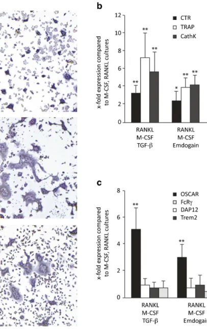
Emdogain stimulates RANKL-induced osteoclastogenesis To investigate the impact of Emdogain on osteoclastogenesis, we determined the formation of multinucleated cells staining positive for TRAP. RANKL and M-CSF induced the forma-tion of osteoclasts. As expected [19, 20], Emdogain and TGF-β increased the number and size of osteoclast-like cells in vitro (Fig.1a). Similar to recombinant TGF-β [29], heat-treatment of Emdogain [30] maintained its activity on osteo-clastogenesis (data not shown). Emdogain considerably (greater than twofold) increased the mRNA level of TRAP, CathK and CTR, being in line with the morphological changes (Fig.1b). Emdogain also increased OSCAR, while the other co-stimulatory molecules TREM2, FcRγ, and DAP12, remained unchanged (Fig.1c). Together, the findings show that similar to TGF-β, Emdogain is a potent enhancer of RANKL-induced osteoclastogenesis.
Emdogain stimulates TNF-induced osteoclastogenesis Besides RANKL, TNFα can also induce osteoclastogenesis in the presence of TGF-β [31]. Thus, we determined if Emdogain serves as cofactors for TNFα. Multinucleated cells staining positive for TRAP were found in cultures containing TNFα and TGF-β (Fig.2a). When TGF-β was replaced by Emdogain, osteoclasts developed even though they were less
in number and had fewer nuclei. These findings demonstrate that Emdogain can serve as a cofactor for TNFα-induced osteoclastogenesis, again, analogous to TGF-β.
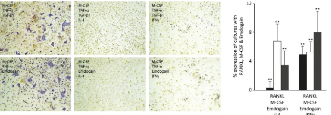
Emdogain cannot overcome the inhibition of IL-4 and IFNγ on osteoclastogenesis
To further learn how Emdogain exerts its effect on osteoclas-togenesis, we performed the bone marrow cultures in the presence of the potent inhibitors IL-4 and IFNγ (Fig.3). As expected, IL-4 and IFNγ substantially diminished the forma-tion of osteoclasts in vitro. Neither TGF-β nor Emdogain could compensate for the suppression of osteoclastogenesis, further suggesting a functional similarity of the two pro-osteoclastogenic factors.
SB431542 abolished osteoclastogenesis in the presence of Emdogain
Having shown that the effects of TGF-β and Emdogain on osteoclastogenesis are comparable, we went on to investigate if the cellular response to Emdogain involves TGF-β signaling. To do this, we performed the experiments in the pres-ence of SB431542, an inhibitor of TGF-βRI kinase activity. Osteoclastogenesis was markedly decreased in the presence of SB431542 (Fig.4a). These morphologic changes were accom-panied by a reduction in the expression of the osteoclastogenic marker genes TRAP, CathK, and CTR (Fig.4b). SB431542 also blocked the effects of TGF-β and Emdogain on the expression of DC-STAMP and Atp6v0d2 (Fig.4c). However, SB431542 also blocks osteoclastogenesis in basic cultures containing RANKL and M-CSF, supporting the role of endog-enous TGF-β in osteoclastogenesis (data not shown). Together, the data suggest Emdogain cannot overcome the essential role of the TGF-βRI kinase in osteoclastogenesis.
SB431542 and SIS3 suppressed the effects of Emdogain on NFATc1 expression
In the bone marrow culture, SIS3 also blocked osteoclasto-genesis in the presence of TGF-β and Emdogain (Fig.5a). We next took advantage of a murine macrophage cell line RAW246.7 and NFATc1, the latter being the master regulator of osteoclastogenesis, which is strongly increased by TGF-β [28]. RAW246.7 cells responded with increased mRNA levels of NFATc1 when activated with Emdogain (Fig. 5b). Importantly, SB431542 and SIS3 both blocked the effects of Emdogain on the expression of NFATc1. Together, these data further support the assumption that Emdogain mediates its activity via TGF-βRI kinase and smad3 signaling, targeting the key transcription factor of osteoclastogenesis, NFATc1.
Discussion
The present study was based on two previous observations: First, similar to TGF-β [27], enamel matrix derivative can support in vitro osteoclastogenesis [19,20]. Second, TGF-β can mediate at least part of the cellular responses to Emdogain
in vitro [21–24]. Together, these data have raised the possibil-ity that the stimulatory effects of Emdogain on osteoclasto-genesis also involve TGF-β. The present in vitro study supports this hypothesis as blocking TGF-βRI kinase coun-teracts all supportive effects of Emdogain on in vitro osteoclastogenesis.
Fig. 1 Emdogain stimulates RANKL-induced
osteoclastogenesis. Multinucleated cells staining positive for TRAP were considered osteoclast-like cells. a Emdogain and TGF-β
substantially increased the number and size of osteoclasts. b Emdogain and TGF-β similarly increased the mRNA level of TRAP, CathK, and CTR. c Likewise, Emdogain and TGF-β increased OSCAR, while the other co-stimulatory molecules remained unchanged. Data represent the triplicate values of one out of two independent experiments. **p <0.01 compared to cultures with RANKL and M-CSF
Fig. 2 Emdogain stimulates TNFα -induced osteoclastogenesis. Multi-nucleated cells which are TRAP positive (violet) were observed in bone marrow cultures containing TNFα and TGF-β. When TGF-β was
replaced by Emdogain, osteoclasts developed, even though they were less in number and had fewer nuclei. Experiments were performed twice with similar results
Our findings extend previous observations that Emdogain has effects similar to TGF-β on osteoclastogenesis induced with RANKL and TNFα [27] and the regulation of NFATc1 [28]. Furthermore, the strong upregulation of genes involved in cell fusion, DC-STAMP and ADP6, is in line with the
existing knowledge on TGF-β [32]. Together, our study adds to the current understanding that Emdogain causes a cellular response, similar to TGF-β1.
The question arises, if Emdogain mediates its activity on osteoclastogenesis exclusively via TGF-β signaling? This
Fig. 3 Emdogain cannot overcome the inhibition of IL-4 and IFNγ on osteoclastogenesis. Osteoclastogenesis was performed in the presence of IL-4 and IFNγ. a Both factors diminished osteoclastogenesis in vitro. Emdogain and TGF-β could NOT compensate for the suppression of
osteoclastogenesis. b Gene expression was reduced by IL-4 and IFNγ to less than 10 % of the respective controls. The data represent the triplicate data of one out of two experiments. **p <0.01 compared to cultures with RANKL, M-CSF, and Emdogain
Fig. 4 SB431542 abolished osteoclastogenesis in the presence of Emdogain. Osteoclastogenesis was suppressed in the presence of SB-431542, an inhibitor of TGF-βRI kinase activity (a). These microscopic changes were accompanied by a reduction in the expression of the osteoclastogenic marker genes TRAP, CathK, and CTR (b). Moreover, also the genes that regulate cell fusion dendrocyte expressed seven transmembrane protein (DC-STAMP ) and ATPase, H+ transporting,
lysosomal 38 kDa, V0 subunit d2 (Atp6) included in the analysis (c). SB431542 also suppresses osteoclastogenesis in the presence of various batches of Emdogain and EMD (d). The findings shown were conformed by another independent experiment. Expression data represent the mean of triplicate values. **p <0.01 compared to cultures with RANKL, M-CSF, and TGF-β1/Emdogain, respectively
question is hard to answer because TGF-βRI kinase is oblig-atory for osteoclastogenesis, also when no extra TGF-β1 is added to the in vitro system. For example, in the presence of SB431542, RANKL-induced osteoclastogenesis is almost completely suppressed [33]. Nevertheless, we provide evi-dence that Emdogain mediates its activity via TGF-βRI ki-nase, e.g., SB431542 abolished the stimulatory effect of Emdogain on DC-STAMP, ADP6, and NFATc1 expression. We also show that blocking smad-3 signaling with SIS3 blunted NFATc1 expression and osteoclastogenesis. In sup-port of these findings, smad3 is crucial for TGF-β1-induced osteoclast differentiation in giant cell tumor of bone [34]. Moreover, smad3 overexpression can reverse the inhibitory effect of SB431542 on in vitro osteoclastogenesis [33]. Emdogain also caused smad-3 phosphorylation in epithelial cell and mesenchymal cells, respectively [25,35]. Together, these data support a direct involvement of TGF-βRI kinase signaling in the Emdogain-mediated cellular actions presented here.
Further support for the hypothesis comes from findings that Emdogain, similar to TGF-β, maintains its activity when heated to 96 °C [29,30]. It remains however open if TGF-β or other factors that require the TGF-βRI kinase cause the effects of Emdogain on osteoclastogenesis. Emdogain pre-sumably contains TGF-β1 or analogous molecules as sug-gested by studies with neutralizing antibodies raised against TGF-β1 [21–24] and the respective immunoassays [25,26]. We also have data that a TGF-β1 neutralizing antibody re-duced the potential of Emdogain to enhance NFATc1 expres-sion in RAW246.7 cells (data not shown). Yet, others failed to show positive binding of a TGF-β1 antibody to Emdogain [36]. It thus remains a controversial subject if Emdogain contains TGF-β1. Also, other explanations for an involve-ment of TGF-β1 are possible. Emdogain can increase the expression of TGF-β1 in various cell types, pointing towards an autocrine mechanism [9,10]. Overall, our data together
with those of others support the assumption that Emdogain contains TGF-β1 and/or analogous molecules that requires the TGF-βRI kinase to support osteoclastogenesis in the murine bone marrow culture.
There remains the discrepancy with the in vivo data show-ing that Emdogain can prevent root resorption after tooth replantation [13,14]. However, also in vitro, TGF-β1 inhibits osteoclastogenesis in the presence of stromal cells, which are forced to produce the key inhibitor of osteoclastogenesis, osteoprotegerin [37,38]. On the other hand, in vivo inhibition of TGF-β1 by neutralizing antibody [39] and TGF-βRI kinase inhibitors [40] can reduce osteoclast differentiation. In vitro, Emdogain and TGF-β1 can also indirectly modulate osteo-clastogenesis by stimulating cells to produce osteolytic factors such as IL-11 [41, 42]. Our ongoing studies indicate that Emdogain-induced IL-11 expression in oral fibroblasts also requires TGF-β signaling (Stähli et al.; manuscript in prepa-ration). Thus, the in vitro data cannot be easily translated into the clinical scenario. Future in vitro studies should consider the possibility that Emdogain can decrease osteoclastogenesis in a co-culture model of hematopoietic progenitors and mes-enchymal cells. The hypothesis is supported by data showing that Emdogain decreases the RANKL/OPG ratio in mesen-chymal cells [43]. It will thus be worth investigating if the changes in the RANKL/OPG ratio caused by Emdogain also involve the TGF-βRI and the downstream smad-3 kinase.
Emdogain is a mixture of proteins with different peptides being responsible for its different biologic properties. The main component amelogenin has a role in osteoclastogenesis. Recombinant amelogenin inhibits in vitro osteoclastogenesis and root resorption [13], and in line with this finding, amelogenin-null mice experience elevated osteoclastogenesis [44]. Nevertheless, Emdogain supports in vitro osteoclastogen-esis as indicated by the present study and recent observations [19,20]. The data thus suggest that, at least in vitro, amelogenin cannot overcome the pro-osteoclastogenic activity of
Fig. 5 SB431542 and SIS3 suppressed the effects of Emdogain on NFATc1 expression. SIS3, the inhibitor of smad3 signaling, abolished osteoclastogenesis in the presence of TGF-β1 and Emdogain in the bone marrow culture (a). NFATc1 is increasingly expressed when the murine
macrophage cell line RAW246.7 is exposed to Emdogain. SB431542 and SIS3 both blocked the effects of Emdogain on the expression of NFATc1 (b). This experiment was performed two times with similar results. **p <0.01 compared to cells with RANKL and Emdogain
Emdogain. The next steps would be to further characterize the possible pro-osteoclastogenic molecules previously ba-sically purified by chromatography [19,20]. Once the pro-osteoclastogenic activity is identified, Emdogain can be selec-tively modulated to control the respective in vitro properties. However, it should not be overlooked that the early transient osteoclastogenesis is part of the physiologic regeneration se-quence. For example, TGF-β can enhance the osteoinductive activity of BMP-2 in vivo [45] and fracture healing is associ-ated with strong expression of pro-osteoclastogenic genes [3]. The present study puts another piece into the mosaic to better understand the cellular response to Emdogain.
Acknowledgments We thank Catherine Solioz for her skillful technique assistance. This work was supported in part by the Straumann Institute. Conflict of interest The authors declare to have no conflict of interest related to this study.
References
1. Boyle WJ, Simonet WS, Lacey DL (2003) Osteoclast differentiation and activation. Nature 423:337–342. doi:10.1038/nature01658
2. Teitelbaum SL (2000) Bone resorption by osteoclasts. Science 289: 1504–1508
3. Kon T, Cho TJ, Aizawa T, Yamazaki M, Nooh N, Graves D, Gerstenfeld LC, Einhorn TA (2001) Expression of osteoprotegerin, receptor activator of NF-kappaB ligand (osteoprotegerin ligand) and related proinflammatory cytokines during fracture healing. J Bone Miner Res 16:1004–1014. doi:10.1359/jbmr.2001.16.6.1004
4. Braun T, Schett G (2012) Pathways for bone loss in inflammatory disease. Curr Osteoporos Rep 10:101–108. doi: 10.1007/s11914-012-0104-5
5. Suda T, Takahashi N, Udagawa N, Jimi E, Gillespie MT, Martin TJ (1999) Modulation of osteoclast differentiation and function by the new members of the tumor necrosis factor receptor and ligand fam-ilies. Endocr Rev 20:345–357
6. Koga T, Inui M, Inoue K, Kim S, Suematsu A, Kobayashi E, Iwata T, Ohnishi H, Matozaki T, Kodama T, Taniguchi T, Takayanagi H, Takai T (2004) Costimulatory signals mediated by the ITAM motif cooperate with RANKL for bone homeostasis. Nature 428:758–763. doi:10.1038/nature02444
7. Kukita T, Wada N, Kukita A, Kakimoto T, Sandra F, Toh K, Nagata K, Iijima T, Horiuchi M, Matsusaki H, Hieshima K, Yoshie O, Nomiyama H (2004) RANKL-induced DC-STAMP is essential for osteoclastogenesis. J Exp Med 200:941–946. doi:10.1084/jem. 20040518
8. Lee SH, Rho J, Jeong D, Sul JY, Kim T, Kim N, Kang JS, Miyamoto T, Suda T, Lee SK, Pignolo RJ, Koczon-Jaremko B, Lorenzo J, Choi Y (2006) v-ATPase V0 subunit d2-deficient mice exhibit impaired osteoclast fusion and increased bone formation. Nat Med 12:1403– 1409. doi:10.1038/nm1514
9. Bosshardt DD (2008) Biological mediators and periodontal regener-ation: a review of enamel matrix proteins at the cellular and molecular levels. J Clin Periodontol 35:87–105. doi:10.1111/j.1600-051X. 2008.01264.x
10. Grandin HM, Gemperli AC, Dard M (2012) Enamel matrix deriva-tive: a review of cellular effects in vitro and a model of molecular arrangement and functioning. Tissue Eng Part B Rev 18:181–202. doi:10.1089/ten.TEB.2011.0365
11. Sculean A, Alessandri R, Miron R, Salvi GE, Bosshardt DD (2011) Enamel matrix proteins and periodontal wound healing and regener-ation. Clin Adv Periodontics 101–117. doi:10.1111/j.1600-9657. 2008.00559.x
12. St George G, Darbar U, Thomas G (2006) Inflammatory external root resorption following surgical treatment for intra-bony defects: a report of two cases involving Emdogain and a review of the literature. J Clin Periodontol 33:449–454. doi:10.1111/j.1600-051X.2006.00926.x
13. Yagi Y, Suda N, Yamakoshi Y, Baba O, Moriyama K (2009) In vivo application of amelogenin suppresses root resorption. J Dent Res 88: 176–181. doi:10.1177/0022034508329451
14. Hamamoto Y, Kawasaki N, Jarnbring F, Hammarstrom L (2002) Effects and distribution of the enamel matrix derivative Emdogain in the periodontal tissues of rat molars transplanted to the abdominal wall. Dent Traumatol 18:12–23
15. Schjott M, Andreasen JO (2005) Emdogain does not prevent progres-sive root resorption after replantation of avulsed teeth: a clinical study. Dent Traumatol 21:46–50. doi:10.1111/j.1600-9657.2004.00295.x
16. Poi WR, Carvalho RM, Panzarini SR, Sonoda CK, Manfrin TM, Rodrigues Tda S (2007) Influence of enamel matrix derivative (Emdogain) and sodium fluoride on the healing process in delayed tooth replantation: histologic and histometric analysis in rats. Dent Traumatol 23:35–41. doi:10.1111/j.1600-9657.2006.00481.x
17. Filippi A, Pohl Y, von Arx T (2006) Treatment of replacement resorption by intentional replantation, resection of the ankylosed sites, and Emdogain—results of a 6-year survey. Dent Traumatol 22:307–311. doi:10.1111/j.1600-9657.2005.00363.x
18. Fridstrom M, Schollin J, Crossner CG (2008) Evaluating Emdogain and healing of replanted teeth using an intra-individual experimental-control study design. Dent Traumatol 24:299–304. doi:10.1111/j. 1600-9657.2008.00559.x
19. Itoh N, Kasai H, Ariyoshi W, Harada E, Yokota M, Nishihara T (2006) Mechanisms involved in the enhancement of osteoclast for-mation by enamel matrix derivative. J Periodontal Res 41:273–279. doi:10.1111/j.1600-0765.2005.00868.x
20. Otsuka T, Kasai H, Yamaguchi K, Nishihara T (2005) Enamel matrix derivative promotes osteoclast cell formation by RANKL production in mouse marrow cultures. J Dent 33:749–755. doi:10.1016/j.jdent. 2005.02.006
21. Kawase T, Okuda K, Yoshie H, Burns DM (2002) Anti-TGF-beta antibody blocks enamel matrix derivative-induced upregulation of p21WAF1/cip1 and prevents its inhibition of human oral epithelial cell proliferation. J Periodontal Res 37:255–262
22. Hama H, Azuma H, Seto H, Kido J, Nagata T (2008) Inhibitory effect of enamel matrix derivative on osteoblastic differentiation of rat calvaria cells in culture. J Periodontal Res 43:179–185. doi:10. 1111/j.1600-0765.2007.01010.x
23. Wada Y, Yamamoto H, Nanbu S, Mizuno M, Tamura M (2008) The suppressive effect of enamel matrix derivative on osteocalcin gene expression of osteoblasts is neutralized by an antibody against TGF-beta. J Periodontol 79:341–347. doi:10.1902/jop.2008.070197
24. Heng NH, N'Guessan PD, Kleber BM, Bernimoulin JP, Pischon N (2007) Enamel matrix derivative induces connective tissue growth factor expression in human osteoblastic cells. J Periodontol 78:2369– 2379. doi:10.1902/jop.2007.070130
25. Gruber R, Bosshardt DD, Richard JM, Gemperli AC, Buser D and Sculean A (2013) Enamel matrix derivative inhibits adipocyte differ-entiation of 3T3-L1 cells via activation of TGF-βRI kinase activity. PLoS One
26. Sakoda K, Nakajima Y, Noguchi K (2012) Enamel matrix derivative induces production of vascular endothelial cell growth factor in human gingival fibroblasts. Eur J Oral Sci 120:513–519. doi:10. 1111/j.1600-0722.2012.00999.x
27. Fox SW, Fuller K, Bayley KE, Lean JM, Chambers TJ (2000) TGF-beta 1 and IFN-gamma direct macrophage activation by TNF-alpha to osteoclastic or cytocidal phenotype. J Immunol 165:4957–4963
28. Fox SW, Evans KE, Lovibond AC (2008) Transforming growth factor-beta enables NFATc1 expression during osteoclastogenesis. Biochem Biophys Res Commun 366:123–128. doi:10.1016/j.bbrc. 2007.11.120
29. Miyazono K, Hellman U, Wernstedt C, Heldin CH (1988) Latent high molecular weight complex of transforming growth factor beta 1. Purification from human platelets and structural characterization. J Biol Chem 263:6407–6415
30. Nagano T, Iwata T, Ogata Y, Tanabe T, Gomi K, Fukae M, Arai T, Oida S (2004) Effect of heat treatment on bioactivities of enamel matrix derivatives in human periodontal ligament (HPDL) cells. J Periodontal Res 39:249–256. doi:10.1111/j.1600-0765.2004.00733.x
31. Kim N, Kadono Y, Takami M, Lee J, Lee SH, Okada F, Kim JH, Kobayashi T, Odgren PR, Nakano H, Yeh WC, Lee SK, Lorenzo JA, Choi Y (2005) Osteoclast differentiation independent of the TRANCE-RANK-TRAF6 axis. J Exp Med 202:589–595. doi:10. 1084/jem.20050978
32. Cicek M, Vrabel A, Sturchio C, Pederson L, Hawse JR, Subramaniam M, Spelsberg TC, Oursler MJ (2011) TGF-beta in-ducible early gene 1 regulates osteoclast differentiation and survival by mediating the NFATc1, AKT, and MEK/ERK signaling path-ways. PLoS One 6:e17522. doi:10.1371/journal.pone.0017522
33. Yasui T, Kadono Y, Nakamura M, Oshima Y, Matsumoto T, Masuda H, Hirose J, Omata Y, Yasuda H, Imamura T, Nakamura K, Tanaka S (2011) Regulation of RANKL-induced osteoclastogenesis by TGF-beta through molecular interaction between Smad3 and Traf6. J Bone Miner Res 26:1447–1456. doi:10.1002/jbmr.357
34. Lou Z, Yang Y, Ren T, Tang S, Peng X, Lu Q, Sun Y, Guo W (2013) Smad3 is the key to transforming growth factor-beta1-induced oste-oclast differentiation in giant cell tumor of bone. Med Oncol 30:606. doi:10.1007/s12032-013-0606-8
35. Kawase T, Okuda K, Momose M, Kato Y, Yoshie H, Burns DM (2001) Enamel matrix derivative (EMDOGAIN) rapidly stimulates phosphorylation of the MAP kinase family and nuclear accumulation of smad2 in both oral epithelial and fibroblastic human cells. J Periodontal Res 36:367–376
36. Saito K, Konishi I, Nishiguchi M, Hoshino T, Fujiwara T (2008) Amelogenin binds to both heparan sulfate and bone morphogenetic protein 2 and pharmacologically suppresses the effect of noggin. Bone 43:371–376. doi:10.1016/j.bone.2008.03.029
37. Quinn JM, Itoh K, Udagawa N, Hausler K, Yasuda H, Shima N, Mizuno A, Higashio K, Takahashi N, Suda T, Martin TJ, Gillespie
MT (2001) Transforming growth factor beta affects osteoclast differ-entiation via direct and indirect actions. J Bone Miner Res 16:1787– 1794. doi:10.1359/jbmr.2001.16.10.1787
38. Thirunavukkarasu K, Miles RR, Halladay DL, Yang X, Galvin RJ, Chandrasekhar S, Martin TJ, Onyia JE (2001) Stimulation of osteo-protegerin (OPG) gene expression by transforming growth factor-beta (TGF-factor-beta). Mapping of the OPG promoter region that mediates TGF-beta effects. J Biol Chem 276:36241–36250. doi:10.1074/jbc. M104319200
39. Edwards JR, Nyman JS, Lwin ST, Moore MM, Esparza J, O'Quinn EC, Hart AJ, Biswas S, Patil CA, Lonning S, Mahadevan-Jansen A, Mundy GR (2010) Inhibition of TGF-beta signaling by 1D11 anti-body treatment increases bone mass and quality in vivo. J Bone Miner Res 25:2419–2426. doi:10.1002/jbmr.139
40. Mohammad KS, Chen CG, Balooch G, Stebbins E, McKenna CR, Davis H, Niewolna M, Peng XH, Nguyen DH, Ionova-Martin SS, Bracey JW, Hogue WR, Wong DH, Ritchie RO, Suva LJ, Derynck R, Guise TA, Alliston T (2009) Pharmacologic inhibition of the TGF-beta type I receptor kinase has anabolic and anti-catabolic effects on bone. PLoS One 4:e5275. doi:10.1371/journal.pone.0005275
41. Brett PM, Parkar M, Olsen I, Tonetti M (2002) Expression profiling of periodontal ligament cells stimulated with enamel matrix proteins in vitro: a model for tissue regeneration. J Dent Res 81:776–783 42. Elias JA, Zheng T, Whiting NL, Trow TK, Merrill WW, Zitnik R,
Ray P, Alderman EM (1994) IL-1 and transforming growth factor-beta regulation of fibroblast-derived IL-11. J Immunol 152:2421– 2429
43. Takayanagi K, Osawa G, Nakaya H, Cochran DL, Kamoi K, Oates TW (2006) Effects of enamel matrix derivative on bone-related mRNA expression in human periodontal ligament cells in vitro. J Periodontol 77:891–898. doi:10.1902/jop.2006.050244
44. Hatakeyama J, Sreenath T, Hatakeyama Y, Thyagarajan T, Shum L, Gibson CW, Wright JT, Kulkarni AB (2003) The receptor activator of nuclear factor-kappa B ligand-mediated osteoclastogenic pathway is elevated in amelogenin-null mice. J Biol Chem 278:35743–35748. doi:10.1074/jbc.M306284200
45. Tachi K, Takami M, Sato H, Mochizuki A, Zhao B, Miyamoto Y, Tsukasaki H, Inoue T, Shintani S, Koike T, Honda Y, Suzuki O, Baba K, Kamijo R (2011) Enhancement of bone morphoge-netic protein-2-induced ectopic bone formation by transforming growth factor-beta1. Tissue Eng Part A 17:597–606. doi:10.1089/ ten.TEA.2010.0094