Initiated by CREB: Resolving Gene Regulatory Programs in
Learning and Memory
Switch in Cofactors and Transcription Regulators between Memory Consolidation
and Maintenance Network
Jenifer C. Kaldun and Simon G. Sprecher*
Consolidation of long‐term memory is a highly and precisely regulated multistep process. The transcription regulator cAMP response element‐binding protein (CREB) plays a key role in initiating memory consolidation. With time processing,first the cofactors are changed and, secondly, CREB gets
dispensable. This ultimately changes the expressed gene program to genes required to maintain the memory. Regulation of memory consolidation also requires epigenetic mechanisms and control at the RNA level. At the neuronal circuit level, oscillation in the activity of CREB and downstream factor define engram cells. Together the combination of all regulation mechanisms allows correct memory processing while keeping the process dynamic andflexible to adjust to different contexts. Also see the video abstract here https://youtu.be/ BhSCSmorpEc.
1. Introduction
The ability to form and store memories requires lasting changes within defined neural circuits. These modifications are assumed to occur by regulating the strength of specific synaptic connec-tions.[1,2] The molecular mechanisms that underlie learning
processes are evolutionarily conserved and shared between invertebrates and vertebrates.[3,4] Much of the molecular and
genetic understanding stems from studies in model organisms such as the sea snail Aplysia californica, the fruit fly Drosophila melanogaster (D. melanogaster), the nematode Caenorhabditis elegans (C. elegans), as well as mice and rats.[5–11]Different forms and phases of memories can be roughly divided into short‐term memories (STMs) and long‐term memories (LTMs).[6,12]STM is
based on transient post‐translational modifications at synapses, whereas LTM typically requires de novo protein biosynthesis.[4,6] This so‐called long‐term synaptic plasticity can either strengthen
synapses (long‐term potentiation [LTP]) or weaken synapses (long‐term depression [LTD]).[13] It is well established that the consolidation of STM to LTM requires gene transcription and RNA translation.[4,6,14] Thus, changes in the gene regulatory program are a critical process for proper LTM formation. While the early mechan-isms that link neuronal activity with changes of gene expression are well studied, little is as yet known about the changes in the transcriptional program that allows long‐lasting memories to be formed and maintained.
Important studies in Aplysia and D. melanogaster led to the discovery of the adenyl kinase (AC)→cAMP→protein ki-nase A (PKA) pathway as a key player in synaptic plasticity, learning, and memory. These results were also confirmed in mouse and rat models, emphasizing how evolutionarily conserved the mechanism is.[3,15,16] PKA is known to activate the cAMP response element‐binding proteins (CREB), a family of conserved transcription factors (TFs).[16] Indeed, manipulations of CREB lead to memory defects. Strikingly, downregulation of CREB impairs memory whereas overexpression enhances memory.[3,15,16] Therefore, the cyclic adenosine monophosphate (cAMP) signaling pathway is considered a key player in learning and memory. Ca2+ ions from voltage‐gated ion channels also act as a messenger.[17]
Interestingly, CREB can also be activated by Ca2+
signal-ing.[18,19]As a consequence, CREB is able to integrate multiple
types of information to regulate gene expression.[19]
Synaptic plasticity may be accompanied by structural plasticity, changing the size and shape of the synaptic ending. In the presynapse, synaptic plasticity changes the amount of released transmitter, whereas in the postsynapse the amount of neurotransmitter receptors is adapted.[13,20]
Even though CREB is well studied in different model animals, the downstream processes leading to LTM are not understood completely. Recent studies show that molecular processes involved in LTM consolidation and plasticity involve additional gene regulatory mechanisms. Epigenetic mechan-isms such as DNA methylation and histone acetylation[21,22]as well as the role of cofactors[23,24] are gaining importance. Moreover, recent evidence suggests that CREB initiates a gene
J. C. Kaldun, Dr. S. G. Sprecher Department of Biology University of Fribourg 1700, Fribourg, Switzerland E-mail: simon.sprecher@unifr.ch
The ORCID identification number(s) for the author(s) of this article can be found under https://doi.org/10.1002/bies.201900045.
http://doc.rero.ch
regulatory cascade, which appears to be critical for long‐lasting memories.[23,25]Thus, CREB provides an early entry point and a
critical link to a gene regulatory program in LTM. In this review, wefirst focus on how CREB is regulated and how it functions and then provide an overview of direct and down-stream CREB targets. In the second part, we discuss the role of other gene regulatory mechanisms and how the gene regulatory networks may control memories.
2. Main Text
2.1. Manifold Mechanisms Fine‐Regulate CREB Activity The conserved transcription regulator CREB is involved in the development, cell survival, plasticity, and learning and
mem-ory.[3,11,15,26]CREB belongs to the bZIP family of TFs together
with cAMP responsive element modulator (CREM) and activating transcription factor 1 (ATF1). These function as homodimers or heterodimers with other bZIP TFs, allowing the formation of different gene regulatory complexes.[15,26,27]CREB
binds to its recognition sequence, the cAMP response element (CRE) on promotors of target genes (Figure 1). The CRE sequence is palindromic, but also half‐canonical CRE sites and variations are recognized with different affinities.[26,28–30]This
suggests that variations in the recognition strength affect the expression of target genes. Interestingly, CREB genes are spliced in different isoforms, which can function as
transcription activators or repressors. Repressor and activator isoforms seem to compete for CRE sequences, so the ratio between them is a part of their gene regulation func-tion.[5,15,26,31–34] An important step to regulate CREB activity is by phosphorylation and dephosphorylation.[19,26,35] Thefirst described activator of CREB was the cAMP–PKA signaling pathway, which phosphorylates CREB1 at Ser‐133.[33,36] The
cAMP–PKA pathway is the main downstream effector of G‐ protein coupled receptors (GPCR), such as dopamine receptors, which respond to a wide array of ligands[37,38](Figure 1). This signaling mechanism is well recognized for its role in learning and memory from invertebrates to mammals.[16]Besides PKA, CREB1 is also phosphorylated by Ca2+‐signaling through Ca2+‐ calmodulin‐dependent kinases (CamK).[18,19,34] Ca2+is
impor-tant in neuronal processes, e.g., through voltage‐gated channels or glutamate receptors.[17]Another upstream effector of CREB
activation is growth signaling through receptor‐tyrosine kinases (RTK) and their downstream kinases such as PKB/AKT, extracellular‐signal‐regulated kinase (ERK), or mitogen‐acti-vated protein kinase (MAPK)[19,34](Figure 1). All the mentioned
kinases and other kinases can either phosphorylate CREB at Ser‐133 or other phosphorylation sites on the protein. In fact, over 300 stimuli have been reported to affect CREB phosphor-ylation state.[11,15] Moreover, CREB is subject to further post‐ transcriptional modifications.[19] CREB is described to be
acetylated, for example, by CREB‐binding protein (CBP), which is suggested to regulate the duration of phosphorylation.[39]
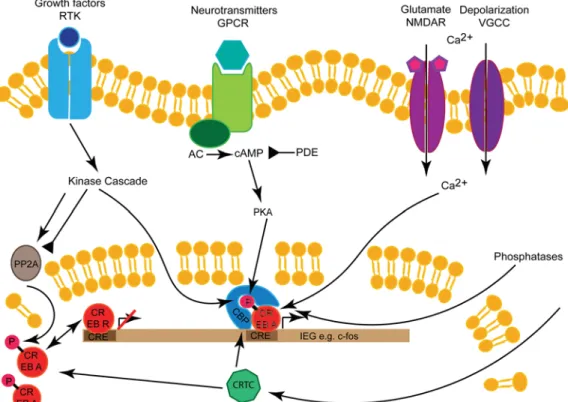
Figure1. Regulation of CREB: the CREB TF is activated and phosphorylated by growth factors through RTK, neurotransmitters through GPCRs, or
Ca2+via glutamatergic NMDA receptors or VGCCs. Downstream of GPCRs is the AC→cAMP→PKA pathway, which can be inhibited by PDEs. The
signaling pathways also regulate phosphatases like PP2A, which can inactivate CREB (PDE). CREB activator isoforms (CREB A) and repressor
isoforms (CREB B) compete for CRE sequence in promotor regions. CREB dimers initiate target gene transcription together with the cofactors CBP
and CRTC. NMDA,N‐methyl‐D‐aspartate; VGCC, voltage‐gated cation channel.
Another modification is glycosylation. It was shown that glycosylation reduces the transcriptional activity of CREB as well as affecting its phosphorylation by different PKA iso-forms.[40] In conclusion, post‐transcriptional modifications allow thefine‐tuning of CREB by reducing its activity.
Besides becoming phosphorylated by different kinases, dephosphorylation by phosphatases is also involved in regulat-ing CREB, hence ensurregulat-ing that target genes are not expressed constitutively. Both protein phosphatase 1 (PP1) and PP2A have been reported to dephosphorylate CREB, causing reduced CREB activity.[15,16,19,41,42]The phosphatases are also involved
in regulating the signaling cascades upstream of CREB, thereby influencing it indirectly. CREB activity can also be restricted by inactivating the signaling cascades that lead to its activation. The cAMP–PKA signaling pathway can be counteracted by phosphodiesterases (PDEs) (Figure 1), which break the phosphodiester bound in the second messenger thereby inactivating it.[43]Thus, two principles are involved in
regulat-ing CREB activity: balancregulat-ing phosphorylation and dephosphor-ylation on CREB and regulating upstream signaling cascades.
Phosphorylation of Ser‐133 enables the recruitment of the cofactor CBP or its paralog p300.[15,44,45] Besides CBP/p300, transducer of CREB/CREB‐regulated transcription coactivator (TORC/CRTC), which consists of three members, can function
as a cofactor for CREB without the requirement of CREB Ser‐ 133 phosphorylation for the interaction[46,47](Figure 1). CREB
requires a cofactor for the activation of gene expression and formation of the gene regulatory complex. Therefore, the cofactors and their recruitment are an important step in regulating transcription, a concept that will be discussed later. Taken together, CREB regulate the expression of target genes in response to a broad range of stimuli. The activity of CREB and the transcription rate of the target genes are fine‐tuned by regulating CREB itself and the respective signaling cascades. As a consequence, CREB‐mediated transcription is not merely “on” or “off” but displays various degrees in between. 2.2. The Discovery of CREB Target Genes
CREB is an early transcriptional regulator in LTM, hence it is critical to understand which genes it regulates directly. In the past, various approaches and models have been used to gain insight into the target genes of CREB (Figure 2). The assembly of human and other genomes allowed a genome‐wide search for CRE sites. By using hidden Markov models on human, mouse, and rat genomes for cross‐species comparison, about 4100 putative conserved CRE sites close to transcription starts
Figure2. Screening approaches for CREB targets: schematic representation of the major approaches to identify CREB targets in different animal
species. Left: represents in silico analysis of genomes, middle: transcriptomics‐based methods, and right: ChIP methods.
were identified[48,49](Figure 2). The limitations of this approach
are that it does not reveal whether the genes are regulated in vivo by CREB, or at which developmental stage, or in which tissue.
Another widely used approach for the identification of genes downstream of CREB, or involved in learning, is transcrip-tomics, in particular microarrays (Figure 2). In mammals, both cell cultures[50–52]and dissected brain regions[53–56]have been used to analyze the transcriptomic profile after stimulation of CREB. A variety of stimuli have been used to activate the CREB signaling pathway, for example, forskolin treatment,[51,52]which
activates cAMP‐signaling,[57] constitutive active CREB,[50,53]
electrostimulation,[55,56] or conditioning training.[54] Similar approaches have also been used in Drosophila,[58,59]Aplysia,[60] and C. elegans.[61] In Drosophila, selected cells involved in learning and memory were picked either by patch clamp[62]or
tagged nuclei (INTACT)[63] to obtain cell‐type‐specific tran-scriptomic profiles. Although these approaches can identify targets involved in these contexts, it has its limitations. First, it only captures the temporal transcription profile, whereas the induced processes are dynamic and show changes in the gene expression over time. Hence, the choice of timepoint for analysis can affect the result. Second, this analysis does not reveal whether the found hit is a direct target or an indirect target. Last, smaller changes in gene expression might not be captured.
Chromatin immunoprecipitation (ChIP) has also been used to discover possible target genes in the cell culture or dissected brain tissue (Figure 2). In this technique, DNA‐binding proteins are precipitated with the DNA to which they are bound. By using an antibody against CREB, the promoters that it might occupy were identified.[55,64]Again, it still has to be
verified that these promotors are truly regulated by CREB. Besides looking for CREB occupancy, antibodies against the cofactor CRTC or histone modifications[23]have also been used
tofind putative‐active genes in specific cells.
Recently, another approach was applied to find memory‐ relevant genes after LTM induction, namely “targeted DNA adenine methyltransferase identification” (TaDa).[65] For this
approach, a DNA adenine methyltransferase (DAM) is fused to the RNA polymerase. When the RNA polymerase binds to DNA the DAM methylates nearby nucleotides, allowing the visualiza-tion of genes that were transcribed.[65,66]
Together these multiple approaches in different model organisms with different conditions, produce a list of putative CREB targets. The mainfindings are summarized in Table 1. Reassuringly, all studies found known CREB targets or memory genes such as somatostatin[29]or c‐fos,[74]hence supporting the
validity of the techniques.
Furthermore, in these studies, neuronal genes like neuro-transmitter receptor subunits or vesicle fusion proteins are found, consistent with CREB’s role in the neuronal pro-cesses.[58–61] Moreover, CREB’s reported role in
develop-ment[11,15]is recapitulated by hits such as CDKs (involved in
cell‐cycle regulation) or hox genes. All screens discovered transcription regulators, for example, egr1, as putative CREB targets. These seem to be involved in orchestrating the CREB‐ mediated response, as we discuss later. Another group of targets that was found was one related to signaling, such as
MAPK or PDEs. CREB might, therefore, be involved in regulating downstream or upstream signaling cascades.
Additionally, these screens also recover genes whose func-tions are unknown or not associated with neuronal processes. Other hits have homologs associated with learning in other species. Hence, testing these hits provides a good basis for a better understanding of CREB‐regulated processes.
Moreover, similar genes have been found in different species, verifying the conserved role of CREB. Thesefindings support the notion that the downstream transcriptional programs are similar, despite different conditions in the respective screens. This leads to the speculation that there might be a conserved core program induced by CREB upon activation. There is still work that needs to be done to verify that these hits are indeed true CREB targets in vivo. Nevertheless, there are also differences, which can partly be attributed to the approaches but also speak for a more specific downstream program depending on species, tissues, and conditions. More-over, these screens also uncovered putative hits that should be analyzed in the future.
2.3. What Is Happening Downstream of CREB?
The transcriptional changes initiated by the cAMP pathway appear not to be static but rather the initiation of a cascade of events (Figure 3A). It has been shown that there are at least two waves of gene transcription.[12]Neuronal activity triggers CREB‐ mediated transcription of the so‐called immediate early genes (IEGs). These IEGs are then responsible for the second wave of transcription.[12,75,76] Well‐known IEGs that are CREB targets
are the transcription regulators c‐fos,[74] c‐jun,[35] and egr1/
zif268[71](Table 1). Furthermore, these genes are also used as
markers for neuronal activity.[75]Therefore, the IEGs might be part of the core program downstream of CREB. Besides neuronal activity, CREB responds to other signals as well:[19] it can be speculated that it might be a switch that turns LTM formation and plasticity on when the context and cellular state allows it. The IEGs may then regulate specific changes needed to establish long‐term plasticity and memory depending on the state of the neuron. However, it cannot be ruled out that CREB is also required for the second wave because plasticity‐ associated genes that are more likely to be a target of the second wave possess CRE promotor sequences as well[48,49] (Table 1). One possibility would be that CREB repressor isoforms might inhibit these genes when memory formation is not happening. A second possibility would be that CREB requires one of the TFs of the first transcription wave as a partner to activate these genes.
Support for this mechanism comes from the observation that CREB activity is biphasic, the first peak coming shortly after neuronal activity and the second around 3–6 h later when the second wave starts.[12] Accordingly, in the first round, CREB turns on the plasticity transcription program context‐specific, whereas in the second wave it regulates the genes together with the IEGs.
Another TF that was found is CCAAT/enhancer binding protein (C/EBP). In Aplysia, C/EBP plays a role in long‐term
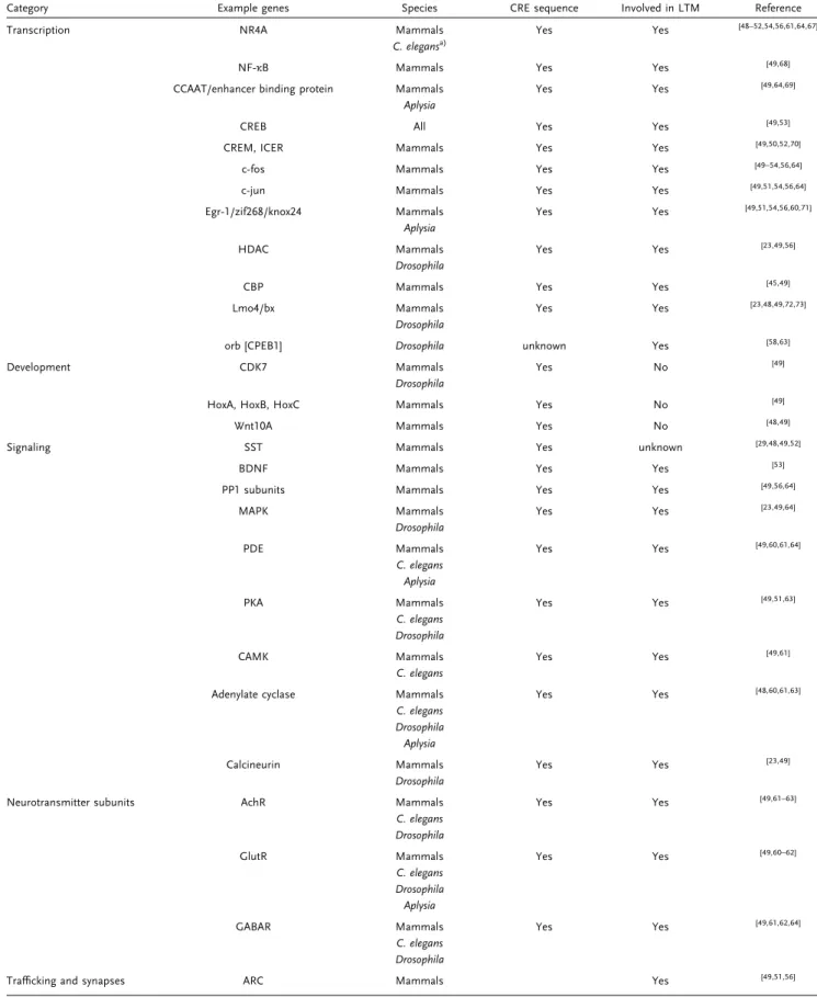
Table1. Summary of the discovered CREB target genes: an overview of putative CREB targets from the screen described in Section 2.2.
Category Example genes Species CRE sequence Involved in LTM Reference
Transcription NR4A Mammals Yes Yes [48–52,54,56,61,64,67]
C. elegansa)
NF‐κB Mammals Yes Yes [49,68]
CCAAT/enhancer binding protein Mammals Yes Yes [49,64,69] Aplysia
CREB All Yes Yes [49,53]
CREM, ICER Mammals Yes Yes [49,50,52,70]
c‐fos Mammals Yes Yes [49–54,56,64]
c‐jun Mammals Yes Yes [49,51,54,56,64]
Egr‐1/zif268/knox24 Mammals Yes Yes [49,51,54,56,60,71] Aplysia
HDAC Mammals Yes Yes [23,49,56]
Drosophila
CBP Mammals Yes Yes [45,49]
Lmo4/bx Mammals Yes Yes [23,48,49,72,73]
Drosophila
orb [CPEB1] Drosophila unknown Yes [58,63]
Development CDK7 Mammals Yes No [49]
Drosophila
HoxA, HoxB, HoxC Mammals Yes No [49]
Wnt10A Mammals Yes No [48,49]
Signaling SST Mammals Yes unknown [29,48,49,52]
BDNF Mammals Yes Yes [53]
PP1 subunits Mammals Yes Yes [49,56,64]
MAPK Mammals Yes Yes [23,49,64]
Drosophila
PDE Mammals Yes Yes [49,60,61,64]
C. elegans Aplysia
PKA Mammals Yes Yes [49,51,63]
C. elegans Drosophila
CAMK Mammals Yes Yes [49,61]
C. elegans
Adenylate cyclase Mammals Yes Yes [48,60,61,63]
C. elegans Drosophila Aplysia
Calcineurin Mammals Yes Yes [23,49]
Drosophila
Neurotransmitter subunits AchR Mammals Yes Yes [49,61–63]
C. elegans Drosophila
GlutR Mammals Yes Yes [49,60–62]
C. elegans Drosophila Aplysia
GABAR Mammals Yes Yes [49,61,62,64]
C. elegans Drosophila
Trafficking and synapses ARC Mammals Yes [49,51,56]
(Continued)
facilitation, and impairment of the gene in mammals causes memory defects but the exact role of it is not clear.[4,69]
The nuclear receptor subfamily 4 group A (NR4A) is also listed as putative CREB target[48–52,54,56,61,64,67](Table 1). The NR4A group has three members and is known for its role in development;[77] however, there are also reports that these transcription regulators are required in memory.[4,67] It is possible that NR4As are activated in development as well as in plasticity events by CREB. This raises a question: do the target genes change between the two processes or are genes required in development also required in plasticity and memory? Regardless, a key question is how the transcription regulators work together, i.e., whether each of them turns on a specific transcription program or whether they are required for a common program.
In conclusion, CREB seems to have at least two roles in memory consolidation: first, it functions as an on‐switch for plasticity depending on a broad range of signals; second, it is a TF for memory genes along with the IEGs. This duality could ensure that consolidation occurs and allows aflexible response by the combination of the different translation regulators.
2.4. CREB Regulates Neuronal Genes
The ultimate aim of memory consolidation is to create long‐ lasting changes in the synapses. Hence, it is not surprising that synaptic and neuronal genes are among the putative CREB target genes (Table 1). Synaptotagmin, for example, is a calcium sensor regulating the formation of the complex required for the release of neurotransmitter vesicles,[78] whereas Shank and Homer are scaffolding proteins in the postsynapse and therefore involved in the structure and organization of the synapse.[79] Changes in these proteins are believed to modulate synaptic plasticity by adjusting the response of the synapse to neuronal activity.[13,20] There are two points to be considered in the
regulation of these genes. First, synaptic plasticity happens at the pre‐ and the postsynaptic membrane.[13,20] Whereas
plasticity on the presynaptic side is characterized by a change in the neurotransmitter release, the postsynaptic membrane responds by adjusting the number of receptors, a process involving different proteins from the presynaptic side.[13,20]This
means that the cell has to“know” at which synaptic location the neuronal activity is happening to respond accordingly. The molecules that signal from the synapses to the nucleus[80]most
likely already contain the information about the synaptic site, so they might directly regulate kinases/phosphatases, transcrip-tion regulators, or epigenetic processes. Second, plasticity can either enhance or weaken a synapse.[13,20]This means that two opposite responses are exploited. Again, this suggests modulat-ing of upstream signalmodulat-ing pathways, the TFs themselves, or their accessibility to genes. A further observation is that the transcriptional program downstream of CREB is not always identical but varies.[51,56]
Among the putative CREB targets are subunits of the major neurotransmitter receptors such as acetylcholine, glutamate, or γ‐aminobutyric acid (GABA). However, neurons differ in the neurotransmitter system that they use. This proposes that neuronal genes are differentially regulated in specific neuron types. The differences between neuronal cell types might be determined during neuronal differentiation via epigenetic mechanisms, making unused genes inaccessible to transcrip-tion. On the other hand, recent single‐cell RNAseq studies have shown that there could be neurons that use more than one neurotransmitter system in parallel,[81] so the downstream program of CREB must be able to discriminate which system is in use at a given time.
CREB and its transcriptional regulatory network have to ensure the proper consolidation of memory and appropriate changes for synaptic plasticity while adjusting the response to neuron type and synaptic site. This seems to be realized by the synapse‐to‐nucleus signaling mechanism adjusting upstream signaling pathways, transcription regulators, and epigenetic processes accordingly.
2.5. How to Update a Memory Trace
Section 2.4 described how CREB and the IEGs could regulate neuronal genes. However, CREB and its downstream factors could also be involved in regulating the upstream processes. Both CREB family members and components of CREB‐ regulating signaling cascades are putative targets (Table 1). The most prominent example is the CREM isoform inducible cAMP early repressor (ICER). ICER is an IEG and functions as a transcriptional repressor. As ICER also uses CRE sequences it could inhibit CREB‐mediated transcription.[15,70,82] However,
CREB activator isoforms seem to be required for the second wave of transcription,[12] so repressor isoforms might be required for selecting the target genes by inhibiting those that Table1. (Continued)
Category Example genes Species CRE sequence Involved in LTM Reference
In rodents, not in humans
Synaptotagmin Mammals Yes Yes [49,52,60,63,64]
Aplysia
Homer Mammals Yes Yes [49]
Shank Mammals Yes Yes [49,61,64]
C. elegans
a)The found gene is a homolog to the mammalian gene.
are not required. Members of the cAMP–PKA signaling pathway or other upstream pathways could be regulated during consolidation, for example, negatively by promoting PDEs to restrict CREB activity, or positively by promoting the expression of cascade members to sustain or amplify CREB activity. The upregulation of signaling pathways might further allow updating or halting of the memory consolidation process when new information is relayed to the neuron. Furthermore, this suggests that the signaling pathways could also be required to fine‐tune the second wave by regulating the respective TFs and cofactors, allowing modulation or cessation of LTM formation as well. This might be dependent on which training paradigm is
used. In Drosophila, classical olfactory conditioning (where an odor is paired with an unconditioned stimulus) is used for training. Aversive conditioning requires multiple training session with breaks in between to generate LTM.[83] Hence,
LTM might require a switch between active and inactive CREB signaling, which could be achieved by changing between repressor and activator forms or turning signaling pathways on and off. Appetitive conditioning requires only one training session.[84]However, appetitive LTM is only formed when the sugar has nutritional value. This information was shown to be relayed to the mushroom body (MB) after ingestion, so a few hours after the original association was made.[85,86]It, therefore,
Figure3. TFs and cofactors of CREB: A) Depending on the synaptic activity and the context, CREB will activate or not transcription. IEGs like c‐fos,
jun, egr1, and so on are TFs themselves and turn on their target genes in combination with CREB. B) The formation of LTM requires CREB and its
cofactor CBP. Early maintenance (day4 in Drosophila) still depends on CREB but requires CRTC instead of CPB. Later maintenance (day 7 in
Drosophila) is completely independent of CREB and requires Bx.
appears that neurons involved in the memory trace need to stay prepared to continue the consolidation process when the sugar is nutritious or stop the process when it is not.
CREB could regulate itself and the upstream signaling pathway positively and negatively, thereby fine‐tuning the cellular response by allowing updates or stops of the consolidation process.
2.6. ARC Could Transfer Messenger RNA (mRNA) between Neurons
A quite interesting CREB target is activity‐regulated cytoskele-ton‐associated protein ARC/ARG3.1. The protein is involved in the trafficking of AMPA receptors and impairing ARC leads to LTM defects.[87] ARC seems to be involved in both LTD and LTP and is localized to dendrites.[87,88] However, ARC might
have another role during plasticity. ARC has viral group‐specific antigen (Gag) polyprotein elements and with these GAG elements, ARC is able to form capsids.[89,90] These capsids can then be exocytosed in exosomes (Figure 4), which is a communication method between neurons and between
neurons and glia.[91] Most interestingly, the ARC capsids contain ARC RNA but they are also able to encapsidate other mRNAs.[89,90] In the Drosophila neuromuscular junction, the trans‐synaptic transport of ARC is required for synaptic plasticity.[89] Interestingly, the mRNA in the capsids can be translated in the destination neuron.[89,90] Local synaptic translation of mRNA in neurons allows synaptic changes to be started at a very early timepoint, while in the meantime proteins are newly synthesized in the cell body. In summary, there seems to be a conserved mechanism of trans‐synaptic communication by viral‐like proteins, but it remains unclear whether specific mRNAs are shuffled between neurons and how the process is regulated.
2.7. BDNF and Feedback Loop Define Engram Cells
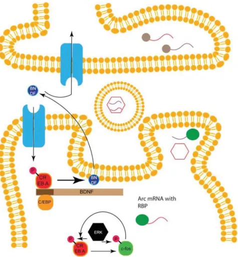
A further prominent CREB target is the brain‐derived neurotrophic factor (BDNF), which is involved in the develop-ment of the nervous system and is also a key regulator in synaptic plasticity.[34,53,92]BDNF is secreted and may act in an autocrine and paracrine fashion[93] (Figure 4). Interestingly,
Figure4. Feedback loops, BDNF and ARC: binding of BDNF to its receptor activates CREB‐mediated transcription. BDNF is one target gene of CREB
and can initiate a feedback loop. BDNF can also bind to receptors in the paracrine area. Another feedback loop exists between CREB and its target
c‐fos and depends on phosphorylation by the kinase ERK. The mRNA of the CREB target ARC is transported to the dendrites where it can form
capsids, which are loaded into exosomes. Exosomes can be uptaken by the synapse and the encapsid mRNA can be translated.
BDNF is both upstream and downstream of CREB.[34,93,94] Whereas BDNF expression is mediated by CREB, binding of BDNF to its receptor tropomyosin receptor kinase B (TrkB) triggers CREB activity. In fact, there seems to be a positive feedback loop involving BDNF, CREB, and C/EPB.[95]Training leads to the release of BDNF, which can activate CREB‐ mediated gene expression via its receptor TrkB. The CREB target C/EBP leads to BDNF transcription[34,95](Figure 4). This loop may have the function of sustaining CREB activity for later integration of information while keeping CREB activity restricted. In fact, studies in rodents show that consolidation is still notfinished 24 h after memory formation.[12]
BDNF can also activate the AKT–TOR signaling pathway, which is involved in translation regulation, thereby affecting CREB downstream processes.[96,97]
In Drosophila there seems to be a feedback loop between CREB and the fly homolog of c‐fos, kayak. The expression of c‐fos is dependent on CREB, whereas c‐fos is required for CREB expression (Figure 4). However, this cycling only occurs in a subset of MB cells, which were described as being important for LTM consolidation.[98]Thus, cells with the CREB‐c‐fos loop seem to be the engram cells for the formation of a memory. In mammals, it was shown that cells with higher CREB activity are preferably recruited as engram cells in the amygdala.[99–101] Moreover, CREB activity seems to cycle for autoregulatory feedback.[102] In conclusion, the loop between CREB and c‐fos or CREB and BDNF may keep CREB active during the consolidation process without allowing the constitutive activity of CREB. Furthermore, because BDNF is secreted it might also inhibit neighboring neurons from being part of the engram or it might recruit them by signaling pathways downstream of TrkB.
Because only a few engram cells encode a memory,[1,102]
studying only a small population of cells will be beneficial for understanding the memory consolidation process. Recently, a conditional knockout line for CREB was established in the fly.[25]To this end, the coding sequence of CREB wasflanked by
flippase recognition target (FRT) sites. Using a flippase under the control of the powerful Gal4–UAS system, CREB can be knocked out in specific cells. With these knockout flies, it was confirmed that CREB in the MB is important for LTM formation. It was also shown that only a subset of MB neurons is required for LTM. Furthermore, the downstream neurons of these cells require CREB for LTM.[25]Thus, thisfly‐line allows
the study of memory‐relevant cells in greater detail.
In summary, it seems that CREB is cycling with a downstream factor during LTM consolidation in engram cells.
2.8. Cofactors Allow Switching between Memory Phases CREB can use both CBP/p300 and CRTC as cofactors for gene transcription. Which cofactor is required for which process or context is thus a critical distinction in the regulation of CREB activity.[15,44–47] Interestingly, CBP can function as a histone acetyltransferase (HAT), and therefore activate promotor regions.[39]
CBP furthermore only binds to CREB after it has been phosphorylated at Ser‐133.[34,45] This speaks more for a role of
CBP in mediating thefirst transcription wave after CREB becomes activated. CRTC, however, can bind to CREB independent of its
phosphorylation status.[46,47,103] Moreover, upon an increase in cAMP or Ca2+, CRTC is dephosphorylated and transported from synapses to the nucleus.[24,103,104]One kinase that phosphorylates CRTC to keep it in the cytoplasm is the 5′‐AMP‐activated protein kinase (AMPK), which is involved in regulating energy metabolism depending on adenosine triphosphate/adenosine monophosphate (ATP/AMP) levels.[103,105]This would be another pathway to allow
or continue consolidation when the cell is under no particular pressure. Nevertheless, both CRTC and CBP are required for
LTM.[23,24,45]Recently, the requirement of the cofactors during LTM
formation and maintenance was analyzed in the fruitfly.[23]LTM
formation seems to require CREB and CBP. However, early maintenance requires CRTC, thus suggesting a switch in the cofactor between the two states of LTM[23](Figure 3B).
Interestingly, CRTC1 in mammals is also reported to be required for maintenance, so this change in the cofactor during memory consolidation seems to be conserved.[24]This switch in the cofactor might be a simple mechanism for changing gene expression programs. Furthermore, because CRTC can be shuffled from the synapse to the nucleus,[24,103,104]it may relay
further information required for maintenance.
The maintenance for even longer timepoints is completely independent of CREB and requires the TF Beadex (bx), a homolog to mammalian Lim domain only transcription factors (LMO)[106](Figure 3B). It seems that CREB and CRTC induce
bx expression during memory consolidation, whereas bx autoregulates itself and other maintenance genes for a long‐ lasting memory.[23]The separation of regulatory networks and maintenance networks by exchanging the involved factors could be a conserved mechanism in LTM consolidation.
Interestingly, LMO4 has a CRE site within its promotor, making it a possible CREB target.[49]In fear memories, LMO4
might act as a repressor, whereas in reward learning it was associated with motivational significance.[72,73]Future studies of
LMO4 will provide insight into how these different functions may be achieved.
Both cofactors have also been reported to interact with IEG TFs such as c‐jun or fos.[35,45,103]By using different
combina-tions between CREB, the IEG transcription regulators and the cofactors, the downstream transcription program could be dynamically and variably adjusted. Different cofactors and transcription regulators might differ in their promotor pre-ference or in the ability to recruit the polymerase complex or histone modifiers to start transcription. Understanding how these factors interact by using immune precipitation and other proteomics techniques will help us untangle the gene regulatory network.
2.9. CREB Is Not the Only Transcription Regulator
CREB is considered a crucial TF in learning and memory. Although the regulation of CREB activity provides different mechanisms to regulate gene expression, TFs usually work in combination. Surprisingly, little is known of other TFs in learning and memory. CREB belongs to the bZIP class of TFs, which can form heterodimers between each other, hence the combination of bZIPs could alter the expressed genes by changing affinity to promotor elements (PEs).[15,26,27]
Furthermore, it is known from mouse models that upon downregulation of CREB there is compensation by CREM and possibly other transcription regulators.[35,47,82] In mouse, the way in which memories are impaired seems to be related to the genetic background and severity of the CREB mutation.[82] There are reports from mouse and fly that CREB might be dispensable for at least some memory forms, or brain regions as knockdown of CREB did not impair LTM.[107–109]However, in Drosophila, recent data truly speak for the role of CREB in memory consolidation.[25]In mammals, we cannot exclude that there might be other factors that could initiate memory consolidation. But it is also possible that loss of CREB is compensated by CREM and other TFs as well as reconfigura-tion of the neuronal network circumventing neurons lacking CREB. Therefore, analyzing other TFs might shed some light on these conflicting observations.
One possible further transcription regulator might be serum response factor (SRF), given that its promotor recognition sequence is found in the c‐fos promotor, other IEGs, and CamKII. Furthermore, cytoskeleton genes are downstream of SRF so this transcription regulator could be responsible for structural plasticity.[35,51,110]
It was shown that manipulation of SRF affects memory,[35]
and furthermore that SRF could regulate its own expression and interact with different cofactors for activation or repression of gene expression.[35,111]In this regard, SRF has many features in common with CREB.
Another candidate for regulating gene expression is nuclear factor‐κB (NF‐κB), mostly known for its role in immune‐related processes.[112]There are reports of memory impairment in NF‐κB
mutants, and moreover, NF‐κB seems to be localized at synapses, so it may be transported activity‐dependent to the nucleus to affect gene expression.[12,68]Taken together, these observations suggest that NF‐κB might have a dual role as a signaling molecule and as a transcription regulator. Moreover, CREB could work in combina-tion with tissue‐ or cell‐type‐specific TFs to regulate the genes required in a cell upon stimulation.
It would be useful to see whether known promotor sequences appear together with the CRE sequence in mem-ory‐related genes to get a better understanding of the gene regulatory system. However, the question is, how these transcription regulators interact with each other, i.e., whether they regulate the genes together and are redundant or whether each of them controls a specific subset. Monitoring their activity or comparing ChIP data and transcriptomic data could help to shed some light on this.
2.10. Epigenetic Mechanisms Are Gaining Importance
Gene expression is not only regulated at the transcriptional level but also by mechanisms controlling the chromatin state. DNA is packed in a compact structure by histone proteins. Whereas histone deacetylases (HDACs) are tightening DNA packing, making it less accessible for the RNA polymerase, HATs loosen it to allow transcription of the respective genes.[21,113] As shown in Table 1 histone‐modifying genes
are likely targets of CREB, indicating a role of epigenetic mechanisms in memory consolidation.[23,49,56] Furthermore,
the CREB cofactor CBP is a known HAT, thereby strengthening this idea.[39] It has been shown that inhibitors for HDACs
enhance memory, confirming the important role of histone modification in learning and memory.[21]Furthermore, histone
modifications at memory genes affect their expression, thereby influencing consolidation.[21,114]The CREB cofactors CBP and
p300 have the ability to acetylate histones, so they may play a role in making genes accessible for transcription.[115–117] Interestingly, depending on the training intensity, different HATs are recruited. Weak training involved CBP, whereas in strong training CBP was replaced at a later timepoint by KAT5/ Tip60.[24]This result alsofits with a recent study in Drosophila. The LTM formation seems to require CBP, but LTM maintenance at an early point requires GCN5 and Tip60; at even later timepoints only Tip60 is required.[23]Recently, it was shown that CCCTC‐binding factor (CTCF) is required for the correct expression of memory genes: in CTCF mutants ARC and BDNF expression is misregulated. This happens because, without CTCF DNA loops between distinct gene regulatory elements are not established.[118]The importance of chromatin remodeling is further highlighted by the observation in Drosophila that distinct members of the SWI/SNF affect memory differently.[119]
Besides histone modifications and chromatin remodeling, gene expression can also be regulated by DNA methylation, which inhibits RNA expression.[22] Interestingly, the CRE element can be methylated because it has CpG islands.[49,64]
Hence, DNA methylation can directly regulate CREB‐depen-dent gene expression. This could affect CREB target genes, such as c‐fos, indicating that methylation might be a common way to regulate learning genes.[22,120]
An additional level of protein expression modulation is regulation at the mRNA level. The stability of mRNA determines how much protein is synthesized from a given transcript. Especially in neurons, mRNAs are bound by RNA‐binding proteins (RBPs), and the so‐called RNA granula made of RNA and associated proteins are transported to synaptic regions.[13,121] In synapses, these mRNAs can be translated directly where the protein will be required and influence synaptic plasticity.[122]These early‐translated
proteins might be a quick way to modulate a synapse before the newly synthesized proteins from the cell body arrive. Sequences in the 3′‐untranslated region (3′‐UTR) of mRNAs act as a kind of postal zip code, defining which RBP proteins can bind and where the mRNA will be localized: different polyadenylation signals can regulate mRNA localization in different subcellular compart-ments.[122]In Drosophila, the RBPs pumillo, orb, and staufen are implicated in learning and memory, highlighting the importance of local synaptic translation for memory formation.[58,123,124]Moreover,
data from different model organism show that noncoding RNAs such as microRNAs (miRNAs) are required for the regulation of plasticity‐involved genes. It is known that miRNA can repress the translation of target mRNAs or even lead to mRNA cleavage, thus reducing the amount of translated proteins.[125–128] Given that neuronal communication happens at a much faster rate and is more dynamic than protein biosynthesis, miRNAs could help to adjust protein levels in this ever‐changing environment. However, miRNAs are also found at synapses, where they repress local synaptic translation until neuronal activity requires it.[125–128]Some miRNAs seem to be directly expressed in response to CREB,
highlighting their role in memory formation.[126]Moreover, mRNA can be regulated by further mechanisms: Drosophila orb2A mRNA retains an intron and is not translated; however, neuronal activity leads to removal of the intron and translation of orb2A.[129] Interestingly, orb2A belongs to the cytoplasmic polyadenylation element‐binding proteins (CPEB), which regulate the polyadenyla-tion of mRNAs, thereby regulating their translapolyadenyla-tion.[130]A further
mechanism to regulate mRNA is the nonsense‐mediated decay with the exon‐junction complex, leading to mRNA degradation.[131]
In summary, mRNA stability, localization, and translation rate are regulated by multiple mechanisms, leading to a diverse pool of mRNAs, while adjusting synapses independent of transcription.
2.11. Multilevel Gene Regulatory Network in LTM Consolidation The consolidation of memory to stable and long‐lasting memories is a tightly regulated multistep process. The consolidation of LTM is very energy‐intensive process, requir-ing protein biosynthesis.[132] Therefore, memory storage only occurs in favorable conditions. The transcription regulator CREB is a master regulator of the expression of memory genes. It integrates signaling from neuronal pathway as well as stress signaling and other processes[19] (Figure 5). However, the transcriptional regulators SRF and NF‐κB as well as yet unknown players might also contribute to the induction of LTM formation.[35,68] The main group of CREB targets comprises other transcription regulators that turn on the next level of transcription (Figure 5). Comparisons between species, brain regions, and different methods to induce LTM formation suggest that there is a conserved core program of CREB targets similar to the immediately early genes c‐fos, egr1, ARC, as well as neurotransmitter subunits and other synaptic proteins. However, there might also be CREB targets specific for memory type and the underlying synaptic changes or cell type. The
transcription programs arefinely regulated by the interaction of multiple transcription regulators, post‐translational modifica-tions, the usage of cofactors and epigenetic mechanisms. This allows the system to respond flexibly to changes or different contexts as well as being adaptable to different neuron types. Moreover, it allows the integration of multiple or conflicting memory traces by gradually adapting the activity of CREB and other transcription regulators.
Feed‐forward loops between CREB and c‐fos and/or
BDNF[95,98] define the engram cell and allow the integration
of delayed information, e.g., the nutritional value of food or during spaced training. Nevertheless, later timepoints of maintenance seem to require, as a first step, a change in cofactors and later also a change in transcriptional regulators. In thefly, bx seems to be the regulator of the maintenance gene regulatory network[23](Figure 5). Thus, there are two regulatory
networks for the different time phases of memory. This allows changing the transcriptional program from genes inducing LTM formation to genes for maintenance, making CREB available for the next memory events. However, what the maintenance network looks like in mammals is not yet known. Recent studies suggested the atypical PKMζ as well as the CAMKII as important factors in this process.[133–135] The bx homolog Lmo4 might be a good starting point for a TF.
Secreted factors such as BDNF and ARC could define the neurons participating within a memory trace and coordi-nate them.
Epigenetic mechanisms, as well as tight regulation of mRNA maturation, stability, localization, and translation, add to the dynamics and diversity required in fast‐acting neurons. There-fore, memory consolidation is regulated on all steps of protein biosynthesis.
Although many possible memory genes are known, their detailed function is not resolved. Because a memory trace is encoded only by a few cells, techniques that can manipulate
Figure5. Simplified gene regulatory network in LTM formation and maintenance: the molecular switch CREB turns on the LTM regulatory network
depending on the context. CREB targets include TFs that induce the second round of transcription and neuronal genes associated with plasticity.
Components of signaling pathways will influence both CREB and CREB target genes. Bx is then responsible for the maintenance genes.
small groups of cells are required to untangle the gene regulatory network.
3. Conclusion and Outlook
Consolidation of LTM is a highly regulated yet dynamic andflexible process. Moreover, it is also strongly conserved, as demonstrated by similar conclusions resulting from diverse scientific approach studies in different species and brain regions. At least two hierarchical levels of gene transcription exist. The first one is mainly regulated by CREB along with other transcription regulators such as SRF or NF‐κB and is a permissive signal to start memory consolidation. The second level is a combination of TFs and other proteins enacting the synaptic changes and memory storage, e.g., the IEGs c‐fos, egr1, or ARC. A variety of principles are required to carry out the task of memory consolidation: signaling pathways, feedback mechanisms between the key players CREB, c‐fos, and BDNF, the cofactors CBP and CRTCs, epigenetic mechanisms and mRNA processing, and synaptic translation. Progressing from LTM formation to long‐lasting maintenance requires a switch in cofactors and transcription regulators. Long‐lasting memories seem to become independent from the consolidation gene regulatory network centered on CREB and exploit a different maintenance network. Although many genes are described to be involved in LTM formation and/or maintenance, it remains a mystery how they function and how they act together to manage the whole process. Comparing and testing putative CREB targets and memory genes discovered in the various screenings might help to untangle the LTM consolidation program. Newer techniques allowing analysis of only a few neurons or even single neurons might help to shed light on the processes that consolidate and maintain memories.
Acknowledgements
The authors thank their colleagues of the Sprecher lab for fruitful discussions and also thank Dr. C. Fritsch for helpful comments on the manuscript. This work was supported by the Swiss National Science
Foundation (grant number31003A_149499) and the Novartis
Founda-tion for Biomedical Research (grant number18A017) to S.G.S.
Conflict of Interest
The authors declare no conflict of interest.
Keywords
cAMP response element‐binding proteins (CREB), epigenetic, gene
regulatory networks, immediate early genes (IEGs), long‐term memories (LTMs), transcription
Received: March3, 2019
Revised: April29, 2019
Published online: June25, 2019
[1] S. Tonegawa, M. Pignatelli, D. S. Roy, T. J. Ryan, Curr. Opin.
Neurobiol.2015, 35, 101.
[2] M. Mayford, S. A. Siegelbaum, E. R. Kandel, Cold Spring Harbor
Perspect. Biol.2012, 4.
[3] A. J. Silva, J. H. Kogan, P. W. Frankland, S. Kida, Annu. Rev.
Neurosci.1998, 21, 127.
[4] E. R. Kandel, Y. Dudai, M. R. Mayford, Cell 2014, 157, 163. [5] C. Pittenger, E. R. Kandel, Philos. Trans. R. Soc., B 2003, 358, 757. [6] S. E. McGuire, M. Deshazer, R. L. Davis, Prog. Neurobiol. 2005,
76, 328.
[7] E. L. Ardiel, C. H. Rankin, Learn. Mem. 2010, 17, 191. [8] H. Sasakura, I. Mori, Curr. Opin. Neurobiol. 2013, 23, 92. [9] C. V. Vorhees, M. T. Williams, ILAR J. 2014, 55, 310. [10] E. B. Han, C. F. Stevens, Learn. Mem. 1999, 6, 539.
[11] A. Barco, H. Marie, Mol. Neurobiol. 2011, 44, 330.
[12] C. M. Alberini, Physiol. Rev. 2009, 89, 121.
[13] V. M. Ho, J. A. Lee, K. C. Martin, Science 2011, 334, 623.
[14] H. P. Davis, L. R. Squire, Psychol. Bull. 1984, 96, 518.
[15] B. E. Lonze, D. D. Ginty, Neuron 2002, 35, 605.
[16] E. R. Kandel, Mol. Brain 2012, 5, 14.
[17] A. Ghosh, M. Greenberg, Science 1995, 268, 239.
[18] M. Sheng, G. McFadden, M. E. Greenberg, Neuron 1990, 4, 571. [19] M. Johannessen, M. P. Delghandi, U. Moens, Cell. Signal. 2004,
16, 1211.
[20] H. R. Monday, P. E. Castillo, Curr. Opin. Neurobiol. 2017, 45, 106. [21] C. Schmauss, Neurosci. Biobehav. Rev. 2017, 83, 63.
[22] C. Kerimoglu, M. S. Sakib, G. Jain, E. Benito, S. Burkhardt, V. Capece, L. Kaurani, R. Halder, R. C. Agís‐Balboa, R. Stilling,
H. Urbanke, A. Kranz, A. F. Stewart, A. Fischer,Cell Rep.2017,
20, 538.
[23] Y. Hirano, K. Ihara, T. Masuda, T. Yamamoto, I. Iwata, A. Takahashi, H. Awata, N. Nakamura, M. Takakura, Y. Suzuki,
J. Horiuchi, H. Okuno, M. Saitoe,Nat. Commun.2016, 7, 13471.
[24] S. Uchida, B. J. W. Teubner, C. Hevi, K. Hara, A. Kobayashi,
R. M. Dave, T. Shintaku, P. Jaikhan, H. Yamagata, T. Suzuki,
Y. Watanabe, S. S. Zakharenko, G. P. Shumyatsky,Cell Rep.2017,
18, 352.
[25] Y. F. Widmer, C. Fritsch, M. M. Jungo, S. Almeida, B. Egger,
S. G. Sprecher,eLife2018, 7.
[26] B. Mayr, M. Montminy, Nat. Rev. Mol. Cell Biol. 2001, 2, 599. [27] T. Hai, T. Curran, Proc. Natl. Acad. Sci. U. S. A. 1991, 88, 3720. [28] M. Comb, N. C. Birnberg, A. Seasholtz, E. Herbert,
H. M. Goodman,Nature1986, 323, 353.
[29] M. R. Montminy, K. A. Sevarino, J. A. Wagner, G. Mandel,
R. H. Goodman,Proc. Natl. Acad. Sci. U. S. A.1986, 83, 6682.
[30] J. C. Craig, M. A. Schumacher, S. E. Mansoor, D. L. Farrens,
R. G. Brennan, R. H. Goodman,J. Biol. Chem.2001, 276, 11719.
[31] J. C. Yin, J. S. Wallach, E. L. Wilder, J. Klingensmith, D. Dang,
N. Perrimon, H. Zhou, T. Tully, W. G. Quinn,Mol. Cell. Biol.1995,
15, 5123.
[32] T. C. Tubon Jr., J. Zhang, E. L. Friedman, H. Jin, E. D. Gonzales,
H. Zhou, D. Drier, J. R. Gerstner, E. A. Paulson, R. Fropf,
J. C. P. Yin,J. Neurosci.2013, 33, 7475.
[33] D. De Cesare, G. M. Fimia, P. Sassone‐Corsi, Trends Biochem. Sci.
1999, 24, 281.
[34] H. Wang, J. Xu, P. Lazarovici, R. Quirion, W. Zheng, Front. Mol.
Neurosci.2018, 11, 255.
[35] C. J. J. Cole, S. A. Josselyn, In Learning and Memory: A Comprehensive Reference (Ed: J. H. Byrne), Elsevier Academic
Press, Cambridge, Massachusetts2008, p. 547.
[36] G. A. Gonzalez, M. R. Montminy, Cell 1989, 59, 675. [37] S. R. Neves, Science 2002, 296, 1636.
[38] N. X. Tritsch, B. L. Sabatini, Neuron 2012, 76, 33.
[39] Q. Lu, A. E. Hutchins, C. M. Doyle, J. R. Lundblad, R. P. S. Kwok, J.
Biol. Chem.2003, 278, 15727.
[40] N. Jin, D. Ma, J. Gu, J. Shi, X. Xu, K. Iqbal, C. X. Gong, F. Liu,
D. Chu,Biochem. Biophys. Res. Commun.2018, 497, 194.
[41] M. Hagiwara, A. Alberts, P. Brindle, J. Meinkoth, J. Feramisco,
T. Deng, M. Karin, S. Shenolikar, M. Montminy,Cell1992, 70, 105.
[42] B. E. Wadzinski, W. H. Wheat, S. Jaspers, L. F. Peruski, R. L. Lickteig,
G. L. Johnson, D. J. Klemm,Mol. Cell. Biol.1993, 13, 2822.
[43] B. S. Moorthy, Y. Gao, G. S. Anand, Mol. Cell. Proteomics 2011, 10, M110.002295.
[44] J. R. Lundblad, R. P. S. Kwok, M. E. Laurance, M. L. Harter,
R. H. Goodman,Nature1995, 374, 85.
[45] J. C. Chrivia, R. P. S. Kwok, N. Lamb, M. Hagiwara,
M. R. Montminy, R. H. Goodman,Nature1993, 365, 855.
[46] M. D. Conkright, G. Canettieri, R. Screaton, E. Guzman, L. Miraglia,
J. B. Hogenesch, M. Montminy,Mol. Cell2003, 12, 413.
[47] S. Kida, T. Serita, Brain Res. Bull. 2014, 105, 17.
[48] M. D. Conkright, E. Guzmán, L. Flechner, A. I. Su, J. B. Hogenesch,
M. Montminy,Mol. Cell2003, 11, 1101.
[49] X. Zhang, D. T. Odom, S. H. Koo, M. D. Conkright, G. Canettieri,
J. Best, H. Chen, R. Jenner, E. Herbolsheimer, E. Jacobsen, S. Kadam, J. R. Ecker, B. Emerson, J. B. Hogenesch, T. Unterman,
R. A. Young, M. Montminy,Proc. Natl. Acad. Sci. U. S. A.2005,
102, 4459.
[50] D. M. Fass, J. E. F. Butler, R. H. Goodman, J. Biol. Chem. 2003, 278, 43014.
[51] E. Benito, L. M. Valor, M. Jimenez‐Minchan, W. Huber, A. Barco, J.
Neurosci.2011, 31, 18237.
[52] L. Pardo, L. M. Valor, A. Eraso‐Pichot, A. Barco, A. Golbano,
G. E. Hardingham, R. Masgrau, E. Galea,Sci. Rep.2017, 7, 6390.
[53] A. Barco, S. Patterson, J. M. Alarcon, P. Gromova, M. Mata‐Roig,
A. Morozov, E. R. Kandel,Neuron2005, 48, 123.
[54] M. B. Keeley, M. A. Wood, C. Isiegas, J. Stein, K. Hellman,
S. Hannenhalli, T. Abel,Learn. Mem.2006, 13, 135.
[55] K. Q. Tanis, R. S. Duman, S. S. Newton, Biol. Psychiatry 2008,
63, 710.
[56] J. E. Ploski, K. W. Park, J. Ping, M. S. Monsey, G. E. Schafe, J.
Neurochem.2010, 112, 636.
[57] R. H. Alasbahi, M. F. Melzig, Pharmazie 2012, 67, 5.
[58] J. Dubnau, A. S. Chiang, L. Grady, J. Barditch, S. Gossweiler, J. McNeil, P. Smith, F. Buldoc, R. Scott, U. Certa, C. Broger,
T. Tully,Curr. Biol.2003, 13, 286.
[59] J. Bozler, B. Z. Kacsoh, H. Chen, W. E. Theurkauf, Z. Weng,
G. Bosco,PLoS Genet.2017, 13, e1007054.
[60] C. Conte, S. Herdegen, S. Kamal, J. Patel, U. Patel, L. Perez, M. Rivota, R. J. Calin‐Jageman, I. E. Calin‐Jageman, Learn. Mem. 2017, 24, 502.
[61] V. Lakhina, R. N. Arey, R. Kaletsky, A. Kauffman, G. Stein,
W. Keyes, D. Xu, C. T. Murphy,Neuron2015, 85, 330.
[62] A. Crocker, X. J. Guan, C. T. Murphy, M. Murthy, Cell Rep. 2016,
15, 1580.
[63] S. G. Jones, K. C. J. Nixon, M. C. Chubak, J. M. Kramer, G3: Genes,
Genomes, Genet.2018, 8, 3433.
[64] S. Impey, S. R. McCorkle, H. Cha‐Molstad, J. M. Dwyer,
G. S. Yochum, J. M. Boss, S. McWeeney, J. J. Dunn, G. Mandel, R. H. Goodman, S. Impey, S. Mccorkle, H. Chamolstad, J. Dwyer, G. Yochum, J. Boss, S. Mcweeney, J. Dunn, G. Mandel,
R. Goodman,Cell2004, 119, 1041.
[65] Y. F. Widmer, A. Bilican, R. Bruggmann, S. G. Sprecher, Genetics 2018, 209, 1167.
[66] T. D. Southall, K. S. Gold, B. Egger, C. M. Davidson, E. E. Caygill,
O. J. Marshall, A. H. Brand,Dev. Cell2013, 26, 101.
[67] J. D. Hawk, T. Abel, Brain Res. Bull. 2011, 85, 21.
[68] B. Kaltschmidt, C. Kaltschmidt, Front. Mol. Neurosci. 2015, 8, 69. [69] A. A. Arguello, X. Ye, O. Bozdagi, G. Pollonini, S. Tronel, D. Bambah‐Mukku, G. W. Huntley, D. Platano, C. M. Alberini, J.
Neurosci.2013, 33, 3646.
[70] C. A. Molina, N. S. Foulkes, E. Lalli, P. Sassone‐Corsi, Cell 1993, 75, 875.
[71] M. W. Jones, M. L. Errington, P. J. French, A. Fine, T. V. P. Bliss,
S. Garel, P. Charnay, B. Bozon, S. Laroche, S. Davis,Nat. Neurosci.
2001, 4, 289.
[72] R. Maiya, V. Kharazia, A. W. Lasek, U. Heberlein, PLoS One 2012, 7, e34559.
[73] R. Maiya, R. A. Mangieri, R. A. Morrisett, U. Heberlein,
R. O. Messing,J. Neurosci.2015, 35, 9638.
[74] S. Ahn, M. Olive, S. Aggarwal, D. Krylov, D. D. Ginty, C. Vinson,
Mol. Cell. Biol.1998, 18, 967.
[75] D. N. Barry, A. N. Coogan, S. Commins, Neurobiol. Learn. Mem.
2016, 128, 46.
[76] K. Minatohara, M. Akiyoshi, H. Okuno, Front. Mol. Neurosci. 2015,
8, 78.
[77] R. A. DeYoung, J. C. Baker, D. Cado, A. Winoto, J. Biol. Chem. 2003,
278, 47104.
[78] S. L. Jackman, J. Turecek, J. E. Belinsky, W. G. Regehr, Nature 2016, 529, 88.
[79] M. K. Hayashi, C. Tang, C. Verpelli, R. Narayanan, M. H. Stearns,
R. M. Xu, H. Li, C. Sala, Y. Hayashi,Cell2009, 137, 159.
[80] A. F. Y. Lim, W. L. Lim, T. H. Ch’ng, Neurobiol. Learn. Mem. 2017, 138, 78.
[81] V. Croset, C. D. Treiber, S. Waddell, eLife 2018, 7. [82] E. Benito, A. Barco, Trends Neurosci. 2010, 33, 230.
[83] T. Tully, T. Preat, S. C. Boynton, M. Del Vecchio, Cell 1994, 79, 35. [84] M. J. Krashes, S. Waddell, J. Neurosci. 2008, 28, 3103.
[85] C. J. Burke, S. Waddell, Curr. Biol. 2011, 21, 746.
[86] P. Y. Musso, P. Tchenio, T. Preat, Cell Rep. 2015, 10, 1023.
[87] S. Chowdhury, J. D. Shepherd, H. Okuno, G. Lyford, R. S. Petralia,
N. Plath, D. Kuhl, R. L. Huganir, P. F. Worley, Neuron 2006,
52, 445.
[88] O. Steward, S. Farris, P. S. Pirbhoy, J. Darnell, S. J. V. Driesche,
Front. Mol. Neurosci.2015, 7, 101.
[89] J. Ashley, B. Cordy, D. Lucia, L. G. Fradkin, V. Budnik, T. Thomson,
Cell2018, 172, 262.
[90] E. D. Pastuzyn, C. E. Day, R. B. Kearns, M. Kyrke‐Smith, A. V. Taibi, J. McCormick, N. Yoder, D. M. Belnap, S. Erlendsson,
D. R. Morado, J. A. G. Briggs, C. Feschotte, J. D. Shepherd,Cell
2018, 172, 275.
[91] V. Budnik, C. Ruiz‐Cañada, F. Wendler, Nat. Rev. Neurosci. 2016, 17, 160.
[92] P. Kowiański, G. Lietzau, E. Czuba, M. Waśkow, A. Steliga, J. Moryś, Cell. Mol. Neurobiol. 2018, 38, 579.
[93] X. Tao, S. Finkbeiner, D. B. Arnold, A. J. Shaywitz, M. E. Greenberg,
Neuron1998, 20, 709.
[94] Y. Zhang, P. Smolen, C. M. Alberini, D. A. Baxter, J. H. Byrne,
Learn. Mem.2016, 23, 714.
[95] D. Bambah‐Mukku, A. Travaglia, D. Y. Chen, G. Pollonini,
C. M. Alberini,J. Neurosci.2014, 34, 12547.
[96] L. Slipczuk, P. Bekinschtein, C. Katche, M. Cammarota,
I. Izquierdo, J. H. Medina,PLoS One2009, 4, e6007.
[97] M. Miron, N. Sonenberg, J. Nutr. 2001, 131, 2988s.
[98] T. Miyashita, E. Kikuchi, J. Horiuchi, M. Saitoe, Cell Rep 2018, 25, 2716.
[99] J. H. Han, S. A. Kushner, A. P. Yiu, C. J. Cole, A. Matynia, R. A. Brown, R. L. Neve, J. F. Guzowski, A. J. Silva, S. A. Josselyn,
Science2007, 316, 457.
[100] Y. Zhou, J. Won, M. G. Karlsson, M. Zhou, T. Rogerson, J. Balaji,
R. Neve, P. Poirazi, A. J. Silva,Nat. Neurosci.2009, 12, 1438.
[101] J. H. Han, S. A. Kushner, A. P. Yiu, H. L. Hsiang, T. Buch, A. Waisman, B. Bontempi, R. L. Neve, P. W. Frankland,
S. A. Josselyn,Science2009, 323, 1492.
[102] A. J. Rashid, C. Yan, V. Mercaldo, H. L. Hsiang, S. Park, C. J. Cole, A. De cristofaro, J. Yu, C. Ramakrishnan, S. Y. Lee, K. Deisseroth,
P. W. Frankland, S. A. Josselyn,Science2016, 353, 383.
[103] C. A. Saura, J. R. Cardinaux, Trends Neurosci. 2017, 40, 720. [104] M. Nonaka, R. Kim, H. Fukushima, K. Sasaki, K. Suzuki, M. Okamura,
Y. Ishii, T. Kawashima, S. Kamijo, S. Takemoto‐Kimura, H. Okuno,
S. Kida, H. Bito,Neuron2014, 84, 92.
[105] S. Herzig, R. J. Shaw, Nat. Rev. Mol. Cell Biol. 2017, 19, 121. [106] J. Thurmond, J. L. Goodman, V. B. Strelets, H. Attrill, L. S. Gramates,
S. J. Marygold, B. B. Matthews, G. Millburn, G. Antonazzo, V. Trovisco, T. C. Kaufman, B. R. Calvi, N. Perrimon, S. R. Gelbart, J. Agapite, K. Broll, L. Crosby, G. Santos, D. Emmert, L. S. Gramates, K. Falls, V. Jenkins, B. Matthews, C. Sutherland, C. Tabone, P. Zhou, M. Zytkovicz, N. Brown, G. Antonazzo, H. Attrill, P. Garapati,
A. Holmes, A. Larkin, S. Marygold, G. Millburn, C. Pilgrim,Nucleic
Acids Res.2019, 47, D759et al.
[107] D. Balschun, D. P. Wolfer, P. Gass, T. Mantamadiotis, H. Welzl,
G. Schütz, J. U. Frey, H. P. Lipp,J. Neurosci.2003, 23, 6304.
[108] M. A. Vogt, D. Inta, A. Luoni, H. Elkin, N. Pfeiffer, M. A. Riva,
P. Gass,Front. Behav. Neurosci.2014, 8, 407.
[109] C. C. Chen, J. K. Wu, H. W. Lin, T. P. Pai, T. F. Fu, C. L. Wu,
T. Tully, A. S. Chiang,Science2012, 335, 678.
[110] S. Bahrami, F. Drabløs, Adv. Biol. Regul. 2016, 62, 37.
[111] N. S. Pulimood, W. S. Rodrigues, D. A. Atkinson, S. M. Mooney,
A. E. Medina,J. Neurosci.2017, 37, 6628.
[112] M. S. S. Ghosh Hayden, Genes Dev. 2012, 26, 203.
[113] M. Haberland, R. L. Montgomery, E. N. Olson, Nat. Rev. Genet. 2009, 10, 32.
[114] J. S. Guan, S. J. Haggarty, E. Giacometti, J. H. Dannenberg,
N. Joseph, J. Gao, T. J. F. Nieland, Y. Zhou, X. Wang, R. Mazitschek, J. E. Bradner, R. A. DePinho, R. Jaenisch,
L. H. Tsai,Nature2009, 459, 55.
[115] A. J. Bannister, T. Kouzarides, Nature 1996, 384, 641.
[116] V. V. Ogryzko, R. L. Schiltz, V. Russanova, B. H. Howard,
Y. Nakatani,Cell1996, 87, 953.
[117] H. M. Chan, N. B. La Thangue, J. Cell Sci. 2001, 114, 2363.
[118] D. S. Sams, S. Nardone, D. Getselter, D. Raz, M. Tal, P. R. Rayi,
H. Kaphzan, O. Hakim, E. Elliott,Cell Rep.2016, 17, 2418.
[119] M. C. Chubak, K. C. J. Nixon, M. H. Stone, N. Raun, S. L. Rice, M. Sarikahya, S. G. Jones, T. A. Lyons, T. E. Jakub,
R. L. M. Mainland, M. J. Knip, T. N. Edwards, J. M. Kramer,Dis.
Models Mech.2019, 7, 12.
[120] S. M. Iguchi‐Ariga, W. Schaffner, Genes Dev. 1989, 3, 612. [121] C. Glock, M. Heumüller, E. M. Schuman, Curr. Opin. Neurobiol.
2017, 45, 169.
[122] K. C. Martin, R. S. Zukin, J. Neurosci. 2006, 26, 7131.
[123] R. Ortiz, M. V. Georgieva, S. Gutiérrez, N. Pedraza,
S. M. Fernández‐Moya, C. Gallego, Cell Rep. 2017, 20, 13.
[124] S. Krüttner, L. Traunmüller, U. Dag, K. Jandrasits, B. Stepien,
N. Iyer, L. G. Fradkin, J. N. Noordermeer, B. D. Mensh,
K. Keleman,Cell Rep.2015, 11, 1953.
[125] Z. Z. Li Hu, Curr. Opin. Neurobiol. 2017, 45, 24.
[126] K. F. Hansen, K. Sakamoto, S. Aten, K. H. Snider, J. Loeser, A. M. Hesse, C. E. Page, C. Pelz, J. S. C. Arthur, S. Impey,
K. Obrietan,Learn. Mem.2016, 23, 61.
[127] B. B. Kaplan, A. N. Kar, A. E. Gioio, A. Aschrafi, Front. Cell.
Neurosci.2013, 7, 126.
[128] C. W. Wei, T. Luo, S. S. Zou, A. S. Wu, Life Sci. 2017, 188, 118. [129] J. Gill, Y. Park, J. P. McGinnis, C. Perez‐Sanchez, M. Blanchette,
K. Si,Cell2017, 169, 836.
[130] M. Ivshina, P. Lasko, J. D. Richter, Annu. Rev. Cell Dev. Biol. 2014, 30, 393.
[131] O. Mauger, P. Scheiffele, Curr. Opin. Neurobiol. 2017, 45, 162.
[132] P. Y. Plaçais, É. de Tredern, L. Scheunemann, S. Trannoy,
V. Goguel, K. A. Han, G. Isabel, T. Preat,Nat. Commun.2017, 8,
15510.
[133] S. B. Baltaci, R. Mogulkoc, A. K. Baltaci, Neurochem. Res. 2019,
44, 281.
[134] T. Rossetti, S. Banerjee, C. Kim, M. Leubner, C. Lamar, P. Gupta,
B. Lee, R. Neve, J. Lisman,Neuron2017, 96, 207.
[135] S. Wang, T. Sheng, S. Ren, T. Tian, W. Lu, Cell Rep. 2016, 16, 1954.