CLINICAL ARTICLE
Motor-evoked potentials (MEP) during brainstem surgery
to preserve corticospinal function
Johannes Sarnthein&Oliver Bozinov&
Angelina Graziella Melone&Helmut Bertalanffy
Received: 21 April 2011 / Accepted: 25 May 2011 / Published online: 10 June 2011 # Springer-Verlag 2011
Abstract
Background Brainstem surgery bears a risk of damage to the corticospinal tract (CST). Motor-evoked potentials (MEPs) are used intraoperatively to monitor CST function in order to detect CST damage at a reversible stage and thus impede permanent neurological deficits. While the method of MEP is generally accepted, warning criteria in the context of brainstem surgery still have to be agreed on. Method We analyzed 104 consecutive patients who under-went microsurgical resection of lesions affecting the brainstem. Motor grade was documented prior to surgery, early postoperatively and at discharge. A baseline MEP stimulation intensity threshold was defined and intraoperative testing aimed to keep MEP response amplitude constant. MEPs were considered deteriorated and the surgical team was notified whenever the threshold was elevated by≥20 mA or MEP response fell under 50%.
Findings On the first postoperative day, 18 patients experienced new paresis that resolved by discharge in 11. MEPs deteriorated in 39 patients, and 16 of these showed new postoperative paresis, indicating a 41% risk of new
paresis. In the remaining 2/18 patients, intraoperative MEPs were stable, although new paresis appeared postoperatively. In one of these patients, intraoperative hemorrhage caused postoperative swelling, and the new motor deficit persisted until discharge. Of all 104 patients, 7 deteriorated in motor grade at discharge, 92 remained unchanged, and 5 patients have improved.
Conclusions Adjustment of surgical strategy contributed to good motor outcome in 33/39 patients. MEP monitoring may help significantly to prevent motor deficits during demanding neurosurgical procedures on the brainstem. Keywords Brainstem . Posterior fossa . Corticospinal tract . Motor-evoked potential . Intraoperative neuromonitoring
Introduction
Brainstem surgery may be associated with substantial risk of damaging the corticospinal tract (CST). The extirpation of lesions at precarious sites like the brainstem requires intraoperative neuromonitoring (IOM) for mapping and monitoring of nerve function. The value of IOM is twofold. (1) During surgery IOM serves as a warning system to monitor impending nerve damage at an early stage so that action can be taken to prevent permanent neurological deficits. (2) Changes in IOM parameters during surgery contribute to the prediction of the neurological status of the patient after surgery.
To date, only few publications have described monitoring of CST function by motor-evoked potentials (MEPs) during surgery for intra- and extraparenchymal brainstem lesions [3, 12]. Although MEPs are well established for the early detection of impending motor deficits during cranial [10,11, 14,17,19] and spinal cord [1,5,15,16] surgical procedures,
J. Sarnthein (*)
:
O. Bozinov:
A. G. MeloneKlinik für Neurochirurgie, UniversitätsSpital Zürich, 8091 Zürich, Switzerland
e-mail: [email protected] J. Sarnthein
Center for Integrative Human Physiology, Universität Zürich, Zürich, Switzerland
A. G. Melone
Dipartimento di Scienze Neurologiche, Università La Sapienza, Roma, Italy
H. Bertalanffy
International Neuroscience Institute, Hannover, Germany
for brainstem surgery a warning criterion has not yet been generally accepted.
In this study we focus on the immediate consequences after surgery to identify intraoperative parameters associated with postoperative motor weakness of patients. We analyzed our series of intraoperative MEP recordings during brainstem surgery, which constitutes, to our knowledge, the largest consecutive series to date. We were interested in how motor outcome correlates with MEP and, in particular, which MEP changes indicate impending motor damage.
Materials and methods Patient selection
We included all consecutive patients from July 2007 to October 2010 who underwent direct surgical resection of lesions with significant involvement of the brainstem. Two surgical procedures were performed by O.B., and all others by the senior author (H.B.). This selection criterion resulted in a series of 104 surgical procedures in 98 patients (60 female, median age 34 years, range 1–78 years).
Lesion location and histology
The lesions affected the brainstem mainly at the level of the mesencephalon in 23 cases, at the level of the pons in 53 cases, and in 28 cases at the level of the medulla oblongata. Among the 60 intra-axial lesions there were 38 cavernomas, 13 low grade gliomas, 6 high grade gliomas and 1 cyst, and 2 exophytic intrinsic lesions (a low grade glioma and a papillar carcinoma). The 44 extra-axial lesions consisted of 11 ependymomas, 6 low grade gliomas, 5 chordomas, 5 meningiomas, 4 haemangioblastomas and 4 medulloblastomas; 2 each of germ cell tumors, plexus papillomas and metastases; and 1 each of craniopharygiomas, high grade gliomas and schwannomas.
In our terminology, intra-axial means that the entire lesion is located within the brainstem and primarily originates from neural or vascular tissue of the brainstem itself, even though part of the lesion may sometimes bulge exophytically beyond the limits of the brainstem. In these lesions, intrinsic brainstem structures such as fiber tracts, nuclei or intrinsic blood vessels are involved directly and to a greater extent than in extra-axial lesions. The extra-axial lesions we describe are topographically located predominantly outside the brainstem and originate from neural or non-neural tissue other than the brainstem, such as cerebellum, cranial nerves, meninges, choroid plexus, etc. Some of these lesions may invade the brainstem, and others are just compressing and displacing it. This differentiation was certainly made for
anatomical and morphological reasons, but mainly because of the existing difference in establishing the indication for surgery. There is more agreement among neurosurgeons about deciding to operate on an extra-axial lesion than on a lesion strictly confined to the brainstem.
Neurological assessment
The motor aspect of the clinical status was documented from patient charts prior to surgery, in the early postoperative stage and at discharge according to the British Medical Research Council motor grade (range 1–5; grade 5: normal muscle contraction). We did not assess long-term motor outcome on a regular basis. Starting from May 2010, in the last 22 patients the neurological status was assessed on a daily basis postoperatively. Worsened postoperative motor grade was considered a new deficit.
Anesthesia management
Following the standard protocol for neurosurgical interven-tions, anesthesia was induced with intravenous application of the sedative drug Propofol (4 to 8 mg/kg/min), the opioid analgesic Remifentanil (1–2 μg/kg/min) and the skeletal muscle relaxant Atracurium (0.5 mg/kg). After intubation, the neuromuscular blocking drug, Atracurium, was omitted because of its interference with electrophysiological monitor-ing and mappmonitor-ing.
Electrophysiological technique
The stimulation and recording of evoked potentials was performed using the ISIS system (www.inomed.com). Transcranial electrical stimulation (TES) current was delivered through two corkscrew electrodes (XOMED, www.medtronic.com) placed at electrode sites C3 and C4. The C3/C4 montage is optimal for low MEP stimulation thresholds [18]. A bite block was placed in the patient's mouth to prevent bite injuries of the tongue resulting from motor stimulation of the jaw. For TES, a stimulus pulse train of 5 to 9 pulses (pulse width 0.5 ms) was applied with an interstimulus interval (ISI) of 4 ms [18]. Stimulus polarity was reversed between stimulations to activate target muscles on both sides. Muscle MEP responses were recorded with pairs of noninsulated needle electrodes placed under the skin (XOMED twisted pair, www. medtronic.com), typically overlying the target muscle belly. Thenar and hypothenar muscles served as target muscles to monitor upper extremity responses. MEP responses were amplified and filtered (100–4,000 Hz) before display.
The baseline MEP stimulation threshold was determined before skin incision. To obtain the baseline MEP threshold
we started by a fixed pattern of stimulus intensity at 30 mA, and then increased it by 5-mA increments until either one of the target muscles on one side responded reliably to stimulation. At least 2 s elapsed from one stimulus train to another. An evoked MEP response as low as 20μV with appropriate response latency qualified as a reliable MEP response [1], although responses were typically >100 μV. The testing was repeated after dura opening and when brainstem function was assumed to be at greatest risk.
Electrophysiological data analysis
In the case of reduced MEP amplitude responses, first technical failures were ruled out, and anesthesia parameters were checked. Then the number of stimulating pulses and subsequently the MEP stimulation intensity were elevated with the aim of a constant MEP response amplitude [1]. Stimulus intensity was limited either by the machine limit (220 mA) or by evoked neck twitches, which disturbed the surgeon. After dura opening, the MEP threshold was tested and adjusted if indicated. Gradually progressive threshold elevations were attributed to anesthetic fade and the baseline MEP threshold was adjusted [7, 8]. Rapid threshold elevations were analyzed in the context of the surgical manipulations and were considered possibly pathological.
For the interpretation of MEP responses, in the last 22 patients we adopted the warning criterion established by Szelenyi et al. [19] on the basis of preliminary observations in the earlier cases of the series. MEPs were considered deteriorated and the surgical team was notified whenever one of the following two possibilities occurred: (1) The MEP intensity threshold had to be elevated by≥20 mA; (2) in individual patients, MEP intensity could not be elevated to the machine limit because of neck twitches disturbing to the surgeon. In these cases, an individual MEP intensity limit was chosen. MEPs were considered deteriorated if the response amplitude fell under 50% at the individual MEP intensity limit. Occurrence of either possibility was considered a significant MEP deterioration and communicated to the surgical team as such.
Statistical analysis
The outcomes of MEP and neurological examinations were dichotomized for statistical treatment wit Fisher’s exact test. Distributions were compared with the Mann-Whitney U test; 95% confidence intervals (CI) were obtained on the basis of the binomial distribution. Statistical analyses were performed using Matlab R2010a (www.Mathworks.com) and SPSS 19 (www.spss.com). Statistical significance was established as p<0.05.
Results
MEP results in an illustrative case
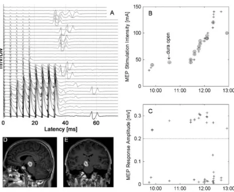
Figure 1 shows the TES intensity during surgery on a patient with a midbrain cavernoma (male, 38 years). Before skin incision, a MEP stimulation threshold of 40 mA was established with five stimulating pulses and MEP response amplitude of 220 μV. After dura opening the stimulation threshold was adjusted to 45 mA. The subsequent exposure of the surgical site involved relatively low risk to the brainstem, and MEPs remained stable. During manipulation of the brainstem from 11:30 on, the MEP intensity had to be elevated. At 12:05 the number of stimulating pulses was increased to nine. From 12:20 on, elevating stimulation intensity up to a self-imposed limit of 140 mA was not sufficient to elicit MEP responses. At dura closing (12:54) the MEPs had recovered with a stimulation intensity of 100 mA. The MEP threshold elevation thus amounted to 55 mA. The patient showed a slight hemiparesis (grade 4) on the first postoperative day, but recovered before discharge.
MEP results in all patients
Initial MEPs were successful on at least one side in all patients, but in four patients (aged 6, 23, 30 and 45 years) MEPs could be elicited only on one side at their individual MEP threshold. None of these four patients developed a new weakness postoperatively. The baseline MEP intensity threshold had a median of 80 mA. In the group of 25 children <18 years, the median was 100 mA and significantly higher than in the group of adults (p = 0.019, Mann-Whitney U-test).
During surgery, MEPs remained stable in 65 patients, and in 39 patients the warning criterion was reached. The warning was based on MEP threshold elevation in 33 patients and on response amplitude reduction in 6 patients. While one might suspect an effect of subdural air collection, the surgery in sitting position was not significantly associated with MEP deterioration (p = 0.7). The MEP threshold elevation varied widely over patients. Figure 2shows the distribution for all 104 surgical cases; the distribution is skewed to high values.
Clinical outcome
Preoperative weakness was documented in 24 surgical cases. In two of them, a new weakness appeared postoperatively, and five patients were discharged with improved motor grade. At the first postoperative exam, new weakness was evident and presumably incurred intraoperatively in 18 of 104 cases. Ten of them had
slight (grade 4) motor deficits that resolved by dis-charge; one returned to preoperative motor grade 4. The other seven had a deficit that did not resolve by discharge; it was slight (grade 4) in four and moderate (grade 3) in three. Their lesions involved the ventral brainstem in five cases. Detailed testing in the last 22 patients
of the series revealed that the new motor deficit was evident at the first exam within 24 h after surgery and—if transient—had resolved on the 2nd postoperative day.
Relationship between MEP results and clinical outcome Of all 104 cases, 65 had no warning and no postoperative motor deficit. Sixteen patients had MEP warnings and new postoperative motor deficits; 23 had warnings but no new deficit. Thus, MEP deterioration to the warning criterion predicts a 41% risk of new weakness [95% CI (26%– 58%)]. The 16 warnings with new postoperative deficits were based on threshold elevation in 11 cases and on amplitude reduction in 5 cases (2 cases with total loss of MEP). The 23 warnings without new deficit were based on threshold elevation in 22 cases and MEP loss in 1 case. Of these 23 cases, 11 were children <18 years.
We next analyzed the effect of threshold elevation (Fig. 2) on the incidence of new deficits. For the range 0–19 mA, in 7/71 patients (10%) new weakness appeared. Above 20 mA threshold elevation, 5/14 cases showed new paresis in the range 20–39 mA, 4/13 in the range 40-59 mA and 2/6 in the range 60/120 mA. While there was no linear increase with threshold
0 20 40 60 80 100 120 0 2 4 6 8 10 12
MEP threshold elevation [mA]
Number of surgical cases
Fig. 2 MEP stimulation threshold elevation during surgery in all 104 cases. There were 42 surgical cases where the MEP stimulation threshold remained unchanged (y-axis cropped). In 33 cases, the MEP
stimulation threshold was elevated≥20 mA
Fig. 1 a MEP monitoring during surgery on a patient with midbrain cavernoma (male, 38 years). b MEP stimulation intensity over time,
electrodes C3–C4. c Muscle MEP response on left thenar muscles,
peak-to-peak amplitude. Stimulation events are circled in b if muscle response amplitude exceeded 0.2 mV. The baseline value was obtained before skin incision (10:00) and adjusted to 45 mA at dura opening (10:30–10:45). During manipulation of the brainstem, the number of stimulating pulses and MEP stimulation intensity were increased.
Upon warning, brain retractors were released. During the critical stage of the surgery (from 12:20 on), MEP stimulation intensity reached a self-imposed threshold maximum of 140 mAwithout muscle response. During dura closing, the muscle response reappeared at a MEP threshold of 100 mA, resulting in a MEP threshold increase of 55 mA compared to baseline. The patient showed a transient slight hemi-paresis (grade 4) postoperatively, which had resolved completely at discharge. d, e Preoperative MR images
elevation, the incidence of new deficits was significant-ly higher above the threshold elevation≥20 mA (Fisher’s exact test p<0.005).
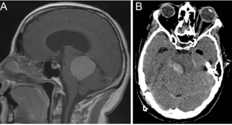
In two patients, a new postoperative deficit oc-curred without MEP deterioration. One patient (male, 61 years, WHO grade II ependymoma, dorsal medulla oblongata, extra-axial) recovered from a slight unilat-eral arm weakness (grade 4) on the 2nd postoperative day; we suspect a transient edema. The second patient (female, 47 years) was operated on for an extra-axial meningioma of the tentorium (Fig. 3a). In the final stage of surgery, a tumor-draining vein was occluded, leading to intra- and postoperative swelling of the brainstem. The MEPs were stable until the end of surgery. The postoperative CT (Fig.3b) shows hemorrhage in the dorsal part of the brainstem, and a severe permanent paresis (grade 1) developed. It must be noted that the latency for MEP deterioration is directly related to the type of injury: mechanical causes evoke more immediate changes, whereas vascular insult may become measurable in the MEP only with significant delay.
For all patients, the occurrence of deteriorated MEPs and a new paresis showed a high degree of association (Fisher’s exact test, p<0.001). However, preoperative deficit was not more frequently associated with MEP deterioration than with a stable MEP (18/47 vs. 6/33, p=0.229). For intra-axial lesions, the MEP warning criterion was met more frequently (43% vs. 30%), and also new paresis occurred more frequently (22% vs. 11%) compared to extra-axial lesions, but these differences did not reach statistical significance (p=0.2). MEPs deteriorated in 10 of the 15 young children <10 years, whereas only one of them showed a new motor deficit at discharge.
Discussion
MEP warning criteria in spinal and supratentorial surgery Although MEP is a well-established component of intraoper-ative neuromonitoring, one of the major problems with the use of MEPs is to determine criteria for when to issue warnings on the basis of changes in the muscle responses [1–5,8–17,19, 20]. In intramedullary spinal cord tumor surgery, total MEP loss was shown to be a valid warning criterion [5]. For supra-tentorial surgery, a 50% reduction of MEP response amplitude has been proposed as a warning criterion [10,11,17].
However, criteria based on MEP response amplitude have some drawbacks. First, there is inherent variability in the amplitude of the muscle responses; MEP responses are often polyphasic and extended over time so that they are difficult to quantify. Second, high MEP stimulation intensity is needed to achieve maximal MEP response already at baseline so that subsequent response deterioration can be assessed. High MEP stimulation intensity may cause movements such as neck twitches, which are disturbing to the surgeon. As a different approach, the “threshold-level” method operates with lower MEP stimulation intensity at onset and may provide earlier warning of an event of deterioration of corticospinal tract function [1]. In a combined approach, it has been proposed to elevate the MEP stimulation threshold and to monitor MEP response amplitude as well [14,19]. This latter approach has not yet been applied to MEP monitoring in brainstem surgery. MEP warning criteria in brainstem surgery
In our study we investigated the practicability of the combined approach proposed by Szelenyi et al. [19] for
Fig. 3 Images from the patient (female, 47 years) with stable MEP and severe new motor deficit postoperatively. a Meningioma of the tentorium, WHO grade II. b In the final stage of surgery, a tumor-draining vein was occluded, leading to intra- and postoperative swelling of the brainstem. The MEPs were stable until the end of
surgery. The postoperative CT shows hemorrhage in the dorsal part of the brainstem. The patient showed a severe hemiparesis (grade 1) postoperatively. The patient needed extended rehabilitation and was ambulatory at 6 months follow-up postoperatively with residual hemiparesis
our series of brainstem surgeries. It was our aim to relate intraoperative changes in the MEP threshold to postoperative changes in muscle strength. In testing the MEP threshold, the increase of MEP stimulation intensity was, however, limited in some cases by appreciable neck twitches that were disturbing to the surgeon. We therefore also monitored MEP response amplitudes and issued a warning at 50% response reduction.
The warning criterion was deliberately defined to emphasize sensitivity, since reactions to warnings were temporary in nature, as outlined in the methods section. Furthermore, it has been shown that sensitive warning criteria trigger surgical re-evaluation earlier [1, 12]. The criterion adopted in our study led to a warning issued in 39 surgical interventions (38%). This rate of significant MEP changes is in-between those of two previous studies on MEPs in brainstem surgery (46% [12], 22% [3]). The warning rate may partly depend on the number of children in the patient population, as children require higher MEP thresholds [6]. The 23 instances of warnings without ensuing paresis can, however, not qualify as “false positives,” since action was taken intraoperatively with the aim to prevent paresis.
Consequences of MEP deterioration for the surgeon The surgery was performed in close contact with the monitoring team. In case a warning of MEP deterioration was issued, the surgical strategy was re-evaluated in the context of all available information and the whole surgical field was inspected. The surgeon reacted to MEP warnings in several ways. Especially in intra-axial lesions, brain retractors, if present, were released. Cottonoids, which could compress the brainstem or adjacent vessels, were removed; systemic venous and arterial blood pressures were checked by contacting the anesthetist; and surgical manipu-lation was sometimes interrupted for a few minutes. A low blood pressure with possible impaired brainstem perfusion was ruled out or corrected. Similarly, a high venous pressure was ruled out or corrected by reducing the positive end-expiratory pressure or elevating the patient's head. In some instances these measures alone have proved to be quite efficient. Conversely, when MEPs remained stable during surgery, the surgeon felt encouraged to continue with surgical dissection, and this contributed to reducing the time of surgery and the associated risks.
Conclusions
MEP deterioration pointed to postoperative weakness in 16/ 39 cases (41%). MEPs were more likely to deteriorate in children, but not in patients with preoperative motor
deficits. Cases with MEP deterioration and no postoperative weakness were regarded such that impairment of motor function was detected while still in a reversible stage. Adjustment of the surgical strategy contributed to good motor outcome: 33/39 patients had no new motor deficits at discharge. Our estimates of the extent to which MEP deterioration is associated with new paresis may thus help to prevent permanent motor deficits during demanding neurosurgical procedures at the brainstem.
Acknowledgements We thank Dr. M. Bjeljac and E. Ciessynna for
their help with MEP recordings in this case series.
Conflicts of interest None.
References
1. Calancie B, Molano MR (2008) Alarm criteria for motor-evoked potentials: what's wrong with the "presence-or-absence"
approach? Spine 33:406–414
2. Deletis V, Rodi Z, Amassian VE (2001) Neurophysiological mechanisms underlying motor evoked potentials in anesthetized humans. Part 2. Relationship between epidurally and muscle
recorded MEPs in man. Clin Neurophysiol 112:445–452
3. Kang DZ, Wu ZY, Lan Q, Yu LH, Lin ZY, Wang CY, Lin YX (2007) Combined monitoring of evoked potentials during microsurgery for lesions adjacent to the brainstem and
intracranial aneurysms. Chin Med J (Engl) 120:1567–1573
4. Kombos T, Süss O (2009) Neurophysiological basis of direct cortical stimulation and applied neuroanatomy of the motor cortex: a review. Neurosurg Focus 27:E3
5. Kothbauer KF, Deletis V, Epstein FJ (1998) Motor-evoked potential monitoring for intramedullary spinal cord tumor surgery: correlation of clinical and neurophysiological data in a series of 100 consecutive procedures. Neurosurg Focus 4:e1
6. Lieberman JA, Lyon R, Feiner J, Diab M, Gregory GA (2006) The effect of age on motor evoked potentials in children under
propofol/isoflurane anesthesia. Anesth Analg 103:316–321
7. Lyon R, Feiner J, Lieberman JA (2005) Progressive suppression of motor evoked potentials during general anesthesia: the phenomenon of "anesthetic fade". J Neurosurg Anesthesiol
17:13–19
8. MacDonald DB (2006) Intraoperative motor evoked potential
monitoring: overview and update. J Clin Monit Comput 20:347–
377
9. Møller AR (2006) Intraoperative Neurophysiological Monitoring. Humana Press Totowa, NJ
10. Neuloh G, Pechstein U, Cedzich C, Schramm J (2004) Motor evoked potential monitoring with supratentorial surgery. Neurosurgery 54:1061–1070, discussion 1070–1062
11. Neuloh G, Pechstein U, Schramm J (2007) Motor tract monitoring
during insular glioma surgery. J Neurosurg 106:582–592
12. Neuloh G, Bogucki J, Schramm J (2009) Intraoperative preservation of corticospinal function in the brainstem. J Neurol Neurosurg
Psychiatry 80:417–422
13. Neuloh G, Schramm J (2009) Are there false-negative results of motor evoked potential monitoring in brain surgery? Cen Eur
Neurosurg 70:171–175
14. Quinones-Hinojosa A, Alam M, Lyon R, Yingling CD, Lawton MT (2004) Transcranial motor evoked potentials during basilar
artery aneurysm surgery: technique application for 30 consecutive
patients. Neurosurgery 54:916–924, discussion 924
15. Sala F, Palandri G, Basso E, Lanteri P, Deletis V, Faccioli F, Bricolo A (2006) Motor evoked potential monitoring improves outcome after surgery for intramedullary spinal cord tumors: a historical control study. Neurosurgery 58:1129–1143, discussion 1129–1143
16. Sutter M, Eggspühler A, Grob D, Jeszenszky D, Benini A, Porchet F, Mueller A, Dvorak J (2007) The diagnostic value of multimodal intraoperative monitoring (MIOM) during spine surgery: a prospective study of 1,017 patients. Eur Spine J 16
(Suppl 2):S162–S170
17. Szelenyi A, Langer D, Kothbauer K, De Camargo AB, Flamm ES, Deletis V (2006) Monitoring of muscle motor evoked potentials during cerebral aneurysm surgery: intraoperative changes and
postoperative outcome. J Neurosurg 105:675–681
18. Szelenyi A, Kothbauer KF, Deletis V (2007) Transcranial electric stimulation for intraoperative motor evoked potential monitoring: Stimulation parameters and electrode montages. Clin
Neuro-physiol 118:1586–1595
19. Szelenyi A, Hattingen E, Weidauer S, Seifert V, Ziemann U (2010) Intraoperative motor evoked potential alteration in intracranial tumor surgery and its relation to signal alteration in postoperative magnetic resonance imaging. Neurosurgery 67:302–313 20. Weinzierl MR, Reinacher P, Gilsbach JM, Rohde V (2007)
Combined motor and somatosensory evoked potentials for intra-operative monitoring: intra- and postintra-operative data in a series of
69 operations. Neurosurg Rev 30:109–116, discussion 116
Comment
In this article the authors report their experience with the use of IOM with MEPs in the resection of brainstem (BS) lesions. Surgery of brainstem lesions is certainly not free of potential ominous consequences, which makes the use of any intra-operative real time monitoring of nervous function located within the stem all the more important. The authors are therefore to be commended for their effort in trying to come up with clearer and more objective clinical rules to be pursued during these operations. The series is significant, although it includes a wide mixture of lesions differently affecting the BS.
The distinction between extra- and intra-axial lesions follows criteria that are not strictly related to the intrinsic nature of the lesion
itself (i.e., ependymoma in the extra-axial group), but also according to the topography of the pathological growth to be excised.
As expected, lesions that grow outside the BS evoke fewer intra-operative MEP changes than those intimately arising from the cellular patrimony of the stem.
Reasons for neuroelectric MEP changes are several. The latency period between the initial aggression and the establishment of the predictable changes in MEPs is variable, as is the mechanism of the insult. Also, some changes may be severe but reversible, whereas others are not.
As mentioned in the text, mechanical causes may tend to evoke more immediate changes, whereas vascular (especially venous) insult or 'edema' may have a significant delay despite the fact that the consequences may end up being worse. The authors clearly demonstrate that they are well aware of the current limitations and caveats involved with the translation of the neurophysiological information into intraoperative surgical decisions.
The heart of the matter is that this technology is no longer dispensable given the possible adverse effects of the surgical manipulation of the BS and the need to minimize it. On the other hand we need clearer indications of what the intraoperative neuro-phsysiologic changes really mean in terms of determining transient or permanent morbidity so that we can act accordingly, either continuing to pursue the resection of the lesion or stopping short of doing so. The third alternative is to alter the surgical strategy, which is what the authors allude to in their text.
This issue is not without importance because there is little one can change in a pristine microsurgical technique itself; what can be altered is, essentially, the location of the dissection or the degree of the resection.
The results are very clearly documented. All patients who deteriorated postoperatively had intra-operative changes signaled to the surgeon. This means that despite the warning and the eventual change in strategy, not all consequences could be avoided.
What advice is there from the surgeon's standpoint taking into consideration the fact that in only 41% of cases did the deterioration of the warning signs predict a new motor deficit? This in fact is the bottom line: Should these warnings forbid us from pursuing a more radical resection or should one still carry on with it?
Of course, experience is foremost and the key to the question, albeit subjective, before we can come up with absolute boundaries for these IOM changes (MEP or others) more independent from the eye and soul of the surgeon, and the monitoring equipment as well. Manuel Cunha e Sa