DNA damage, various insults from environmental and endogenous sources continuously affect DNA integrity. Over time this can lead to the accumulation of somatic mutations, which is thought to be one of the major causes of aging. We have previously found that overexpression of the essential human DNA repair and splicing factor SNEV, also called PRP19 or hPso4, extends replicative life span of cultured human endothelial cells and impedes accumulation of DNA damage. Here, we show that adult-specific overexpression of dPrp19, the D. melanogaster ortholog of human SNEV/PRP19/hPso4, robustly extends life span in female fruitflies. This increase in life span is accompanied by reduced levels of DNA damage and improved resistance to oxidative and genotoxic stress. Ourfindings suggest that dPrp19 plays an evolutionarily conserved role in aging, life span modulation and stress resistance, and support the notion that superior DNA maintenance is key to longevity.
npj Aging and Mechanisms of Disease (2017) 3:5 ; doi:10.1038/s41514-017-0005-z
INTRODUCTION
Aging is characterized by a time-progressive decline of physiolo-gical function at the level of cells, tissues, organs, and ultimately affects the whole organism. According to the“disposable soma” hypothesis of aging, this functional decline results from the accumulation of stochastic damage, for example, due to somatic mutations, and is counteracted by investment into somatic maintenance and repair.1 Accumulation of DNA damage due to decreased repair can accelerate aging, as is observed in segmental progeroid syndromes including the Werner or Hutchinson-Gilford syndromes in humans2and mouse models.3 Similarly, increased exposure to DNA damaging agents, for instance during che-motherapy, can lead to a phenotype of acquired premature progeroid syndrome.4Accelerated accumulation of DNA damage
and premature aging phenotypes are typically well correlated, but whether improved DNA damage repair (DDR) can extend organismal life span remains largely unclear.
In the fruitfly (Drosophila melanogaster), a well-studied model for dissecting the mechanisms of aging, spontaneous somatic mutations accumulate with age, and defective DNA repair is associated with reduced life span.5,6However, overexpression of
DNA repair factors in the fly seems to have highly variable, sometimes contradictory effects that depend on sex, develop-mental stage, and the tissue of intervention. For instance, poly (ADP-ribose) polymerase-1 (PARP-1) modifies histones, transcrip-tion factors and repair enzymes in response to DNA breaks, and its endogenous activity is well correlated with life span in several mammalian species.7 In Drosophila, overexpression of PARP-1
prolongs life span in both sexes, yet only when restricted to the adult nervous system.8 Similarly, overexpression of Gadd45 (growth arrest and DNA damage 45) (ref.9), a regulator of DNA
repair and cellular stress responses, in the nervous system increases fly life span but ubiquitous expression is lethal.10, 11
Indeed, a recent study by Shaposhnikov et al.12 has found that
DNA repair factors can affect Drosophila life span and stress resistance either positively or negatively, depending on the sex and on whether overexpression is ubiquitous or limited to the nervous system. Interestingly, all repair factors that were expressed throughout the adultfly body were found to shorten life span. In another study, Barclay and colleagues examined the effects of overexpression of several known D. melanogaster homologs of human DNA repair genes on Drosophila life span in a genetic model of spinocerebral ataxia (SCA), aiming to identify repair pathways that might be relevant for SCA pathology. They found that an extension of life span and improvement of SCA symptoms could not be attributed to a single repair pathway; instead, each pathway included factors that had either detrimental, beneficial, or no effects on life span.13 Yet, these results—obtained in a diseased mutant background—do not necessarily reflect possible life span effects of DNA repair factors in healthy wild-type flies. Thus, to date, the relationship between DNA damage, repair and organismal aging still remains poorly understood.
Here, we examine the role of adult-specific overexpression of the DNA repair factor Prp19 (pre-messenger RNA (mRNA) processing factor 19) in affecting life span, stress resistance, and DNA damage in Drosophila. PRP19 (also called senescence evasion factor, SNEV, or hPso4) wasfirst characterized in a yeast mutant exhibiting increased sensitivity to DNA interstrand crosslinking induced by treatment with psoralen and ultraviolet (UV) radiation.14 Biochemically, PRP19 acts as an E3 ubiquitin ligase15,
16
and interacts with multiple players in the DNA repair pathways, including each of the two core kinases, ATM (ataxia telangiectasia mutated) and ATR (ataxia telangiectasia related).17,18Apart from
Received: 26 September 2016 Revised: 15 February 2017 Accepted: 15 February 2017 1
Department of Ecology and Evolution, University of Lausanne, Lausanne, Switzerland;2
Department of Biotechnology, BOKU– University of Natural Resources and Life Sciences, Vienna, Vienna, Austria;3Institut für Populationsgenetik, Vetmeduni Vienna, Vienna, Austria;4Department of Developmental Molecular Biology, Max Planck Institute for Biophysical Chemistry, Göttingen, Germany;5
Christian Doppler Laboratory on Biotechnology of Skin Aging, Dept. of Biotechnology, BOKU– University of Natural Resources and Life Sciences, Vienna, Vienna, Austria and6
Evercyte GmbH, Vienna, Austria
Correspondence: Markus Schosserer ([email protected]) and Thomas Flatt (thomas.fl[email protected])
its role in the DNA damage response, an intriguing aspect of PRP19 function is its concomitant and essential involvement in co-transcriptional splicing, where the PRP19 complex regulates the rearrangement of the spliceosome to a catalytically active state through ubiquitination of several factors.19–21 The dual role of PRP19 in DNA repair and transcriptional control is further exemplified by its association with transcription-coupled repair, which is activated when DNA damage blocks elongation,22 but
which has not yet been characterized as a DNA repair mechanism in Drosophila.23
In support of a major role of PRP19/SNEV/hPSO4 in the aging process, it has previously been shown that decreased levels of PRP19 accelerate the induction of cellular senescence in mouse embryonic fibroblasts,24 reduce self renewal of mouse hemato-poietic stem cells,25increase psoralen/UV-A-induced skin aging in mice26 and decrease differentiation of human adipose-derived stromal cells.27Conversely, increased levels of PRP19 extend the replicative potential and total life span of cultured human endothelial cells.17,28 However, the role of PRP19 in organismal life span is unknown. Here, we show that ubiquitous over-expression of the Drosophila ortholog of PRP19, dPrp19 (http:// flybase.org/reports/FBgn0261119.html), reduces DNA damage and extends organismal life span of adult female flies. Our results suggest that PRP19 plays an evolutionarily conserved role in DDR, aging, and stress resistance.
RESULTS
dPrp19 is thefly ortholog of human PRP19/SNEV/Pso4 and is regulated in an age-dependent manner
In support of an evolutionarily conserved role of PRP19/SNEV/Pso4 in aging and DNA repair21in Drosophila, we found a high degree
of amino acid identity (66%) and similarity (81%) between human
PRP19 and fly dPrp19 (Fig. 1a). To determine whether dPrp19 expression is regulated during the fly’s life span, we performed quantitative reverse transcription PCR (qRT-PCR) in whole-body samples of adultflies and found that dPrp19 transcript levels are rather constant in adult males, whereas they decrease with age in adult females (Fig.1b). Over the course of 28 days, we observed a ~2.5-fold reduction in dPrp19 mRNA levels in females; in contrast, male expression was lower and largely unaffected by age (Fig.1b). This observation prompted us to test whether we could—similar to our previous observations in human cells17,28—extend life span and promote stress resistance in Drosophila by overexpressing dPrp19.
dPrp19 overexpression extends Drosophila female life span and prevents DNA damage accumulation
To examine the life span effects of dPrp19 in the fly, we drove adult-specific, ubiquitous overexpression of dPrp19 in three independent transgenic strains carrying a UAS expression cassette (UAS-dPrp19) with the inducible GeneSwitch (GS)-Gal4 driver system.29 By supplementing the fly food medium with the antiprogestin compound RU486 (mifepristone), the GeneSwitch (GS) system can be used to induce overexpression in the F1 of a cross between the UAS responder and the Gal4 driver construct. This allows for spatio-temporal control of candidate gene expression and is commonly used in aging studies.30,31To drive
overexpression of dPrp19 from the UAS lines, we used two different ubiquitously expressing GS-Gal4 driver lines: a Tubulin–GeneSwitch-Gal4(TubGS-Gal4) line and a daughterless-GeneSwitch-Gal4 (daGS-Gal4) line.32 In females, overexpression of dPrp19 resulted in approximately sevenfold upregulation of mRNA (Fig.2b, d, and h) and in males in approximately 12- to 22-fold induction (Fig.2f, j).
Fig. 1 dPrp19 is the Drosophila melanogaster ortholog of human PRP19/SNEV/hPso4. a Prp19 is well-conserved from D. melanogaster to humans, as shown in an alignment of the protein sequences of D. melanogaster dPrp19 and human PRP19/SNEV/hPso4. The two orthologs have an amino acid identity of 66% and a sequence similarity of 81%. The human site of ATM phosphorylation, Ser149, is substituted for Gln in Drosophila(indicated by the black arrow). b dPrp19 expression is higher in female D. melanogaster and decreases with age. Comparison of dPrp19mRNA levels in male (gray) and female (black) wild-typeflies at 6, 14, 21, and 28 days of adulthood shows that dPrp19 levels decrease in females between days 21 and 28. Values represent means across three biological replicates (±1 standard deviation), normalized to Gapdh2 and Tubulinand averaged
Across independent experiments in our laboratories in Vienna and Lausanne, three different UAS responder constructs, and two different Gal4 drivers we observed significant and robust dose-dependent extension of female life span (Fig.2, Supplementary Fig.1, Supplementary Table1). At the highest level of induction (food supplemented with 300 µg/ml RU486) median female life span was increased by between 9.6 and 25%. In one of these assays, dPrp19 overexpression was carried out on food medium containing 2% yeast (Fig. 2a, b), whereas all other assays were performed on a diet containing 5% yeast (Fig. 2c–j). Although mean and maximum life span of controls were strongly affected by the two dietary conditions, dPrp19 overexpression robustly extended female life span in all cases (Fig. 2a, c, and g, Supplementary Table 1). In contrast, male life span was not consistently affected by dPrp19 overexpression in any of our assays (Fig.2e, i, Supplementary Table2), despite higher levels of dPrp19 mRNA than in females.
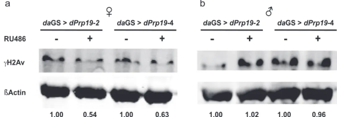
Next, to examine whether ubiquitously increased expression of dPrp19 affects DDR in thefly, we analyzed the abundance of the histone variant γH2Av, a well-established marker for DNA damage.33 Upon dPrp19 overexpression in females, we observed a clear-cut decrease in the levels ofγH2Av (Fig.3a), suggesting a general reduction in the number of DNA double-strand breaks under basal conditions when dPrp19 is upregulated. Interestingly,
we did not observe any reduction of γH2Av signal in males (Fig. 3b), indicating that in contrast to females dPrp19 over-expression does not improve DNA repair capacity and probably thereby does not extend life span.
dPrp19 overexpression increases resistance to DNA damaging compounds
Since human PRP19 is induced by various DNA damaging agents17, 34 and conveys resistance to genotoxic and cytotoxic stress,17,26–28,34,35we next asked whether dPrp19 overexpression
increases resistance against two DNA damaging agents, paraquat (methyl viologen), a herbicide known to induce reactive oxygen species,36 and the genotoxic compound cisplatin, which causes crosslinks between adjacent nucleosomes and which has pre-viously been used to study DNA damage in Drosophila.37Indeed, overexpression of dPrp19 significantly improved the adult survival of females exposed to both paraquat (Fig. 4a; Supplementary Table3) and cisplatin (Fig.4b; Supplementary Table4).
Since in the above experiments dPrp19 overexpression was induced by adding RU486 to the food medium before-but not during-cisplatin exposure, it is possible that transgenic induction of dPrp19 might have decreased during treatment with cisplatin. To rule out this possibility, we examined the adult survival of F1 Fig. 2 Overexpression of dPrp19 leads to dose-dependent extension of female but not male life span. Effects of the induction of the dPrp19 UAS cassette in three independent chromosomal insertions of the same transgenic construct on adult survival and dPrp19 mRNA levels: a, b TubGS-Gal4> UAS-dPrp19-1 (females only), c–f daGS-Gal4 > UAS-dPrp19-2 (females and males), and g–j daGS-Gal4 > UAS-dPrp19-4 (females and males). a, c, e, g, and i show survival curves of experimentalflies at three concentrations of the inducer drug RU486; b, d, f, h, and j show quantification of dPrp19 expression levels relative to the Rp49 control after 72 h of exposure to 300 µg/ml RU486. For all three overexpression constructs, wefind a significant dose-dependent extension of female life span. Overall, we did not find any life span extension in males (for daGS-Gal4> UAS-dPrp19-2 males at 100 µg/ml RU486 we observed a slight reduction in survival, possibly due to inadvertent ‘‘setup mortality” that might have occurred when the assay was set up). For details of life span statistics see Supplementary Table 1 (females) and Supplementary Table2(males); for experimental details see Materials and Methods
flies from a cross between a constitutively active (non-inducible) Tub-Gal4 driver and the UAS overexpression lines. Transgenic F1 offspring carrying both the driver and the overexpression cassette had strongly improved survival over sibling F1 controls lacking the driver construct, thus clearly confirming the protective effects of dPrp19 upon exposure to genotoxic stress (Fig.4c; Supplementary Table4).
DISCUSSION
Here, we have shown that ubiquitous, adult-specific overexpres-sion of dPrp19, an evolutionarily conserved DNA repair factor, significantly and robustly extends female life span in Drosophila.
Overexpression of dPrp19 further reduces DNA damage levels and enhances survival upon oxidative and genotoxic stress. The effects we observed in our experiments are independent of the details of chromosomal insertion position of the UAS-dPrp19 cassette, the Gal4 driver system, and laboratory (food) conditions.
Our data clearly support previous results from cultured human endothelial cells, where PRP19/SNEV/hPso4 overexpression strongly extends replicative life span, lowers basal levels of DNA double-strand breaks, and increases resistance to pro-oxidants as well as to the DNA crosslinker cisplatin.17, 28 In line with these findings, female flies overexpressing dPrp19 exhibited decreased accumulation of DNA double-strand breaks, as indicated by reduced levels of the histone variant γH2Av, the fly homolog of Fig. 4 Overexpression of dPrp19 promotes resistance to genotoxic agents in female flies. a Induction of the dPrp19 cassette with either 100 (gray line) or 300 µg/ml RU486 (black, solid line) significantly increases resistance to 20 mM paraquat (methyl viologen) in females (TubGS-Gal4 > UAS-dPrp19-1) as compared to the uninduced control (black, dashed line; see Supplementary Table3for statistics). b Resistance of females (daGS-Gal4> UAS-dPrp19-2) to genotoxic stress induced by treatment with 400 µg/ml cisplatin was improved significantly by dPrp19 induction at 300 µg/ml RU486; the intermediate induction level of 100 µg/ml RU486 was not significant after Bonferroni correction (see Supplementary Table4for statistics). c Females with constitutively active dPrp19 expression (T2: Tub-Gal4> UAS-dPrp19-2 in gray; T4: Tub-Gal4 > UAS-dPrp19-4 in black) showed significantly improved resistance to cisplatin relative to controls (C2: TM6Sb; dPrp19-2, in dashed gray; C4: TM6Sb; UAS-dPrp19-4, in dashed black line; see Supplementary Table4for statistics)
Fig. 3 Overexpression of dPrp19 decreases γH2Av as marker for DNA double-strand breaks in females but not males. Overexpression of dPrp19 (daGS-Gal4> UAS-dPrp19-2/4) induced by 300 µg/ml RU486 decreases the accumulation of γH2Av by approximately 40–50% in female flies a as compared to the uninduced controls. In males b, we did not observe an effect of dPrp19 induction on DNA damage levels. Numbers represent protein signals ofγH2Av normalized to βActin. The blot shown is representative; qualitatively identical results were obtained with at least two biological and two technical replicates
dPrp19 might improve DNA repair in females and thereby promote stress survival and longer life span: dPrp19 might either directly recruit other DNA repair factors in order to promote fast and efficient repair, or it might act via the regulation of pre-mRNA processing. Human PRP19, for example, is known to interact with proteins involved in DNA repair such as Werner syndrome helicase (WRN),41 Metnase,42 ATR18 and ATM.17 In response to DNA
damage, ATM phosphorylates human PRP19, thereby possibly contributing to an arrest of the cell cycle in order to allow for repair. However, signaling from ATM to PRP19 is not solely responsible for this effect since overexpression of a non-phosphorylatable PRP19 protein still extends replicative potential in human cells, albeit to a lesser extent.17 Since the ATM target phosphorylation site is not conserved in thefly homolog dPrp19, extension of female life span under dPrp19 overexpression could be a consequence of its essential role in pre-mRNA splicing.4,15,21 In a large short interfering RNA screen for factors affecting γH2AX levels in human HeLa cells, Paulsen et al.43
found strong evidence for the importance of mRNA processing factors, such as PRP19, in maintaining genome stability. They observed that the γH2AX signal for double strand breaks was strongly increased upon knockdown of RNA-processing and attributed this at least partly to an increased formation of R-loops. R-loops occur when the transcription machinery is slowed down, for instance by encountering a site of DNA damage, and the transcript folds back on the template duplex DNA strand.44 The resulting DNA–RNA hybrid structure activates non-canonical ATM signaling45and can become the source of mutation and DNA breaks double strand itself.44
Interestingly, PRP19 has also been described as being a sensor of DNA damage during replication stress: It binds and ubiquiti-nates replication protein A, a key factor in the response DNA damage which protectively binds single-stranded DNA, which causes recruitment of ATR and thus initiates activation of further DNA repair factors.18 In a Drosophila mutant model of SCA, overexpression of Drosophila RpA had the largest effect on life span of all assayed DNA repair factors,13indicating a possible joint role in promoting longevity.
In conclusion, overexpression of dPrp19, a protein that functionally interacts with many important DNA repair factors, robustly extends life span of female fruit flies. Importantly, our results demonstrate that this female-specific life span extension is accompanied by decreased DNA damage and by significantly increased resistance to damage caused by paraquat and the DNA crosslinking agent cisplatin. Our data therefore strongly suggest that improved DNA repair capacity can extend the life span of a healthy organism, a view that has so far been almost exclusively based on the study of premature aging phenotypes induced by impaired DNA repair.
MATERIALS AND METHODS Drosophila stocks and maintenance
A D. melanogaster complementary DNA (cDNA) clone of dPrp19 in pBluescript (clone LD02793, stock 13414) was obtained from the
sion (for the purpose of continuous dPrp19 expression during cisplatin treatment) was achieved by crossing the UAS-dPrp19 lines to a constitutive balanced Tubulin-Gal4 (Tub-Gal4) driver obtained from the Bloomington Drosophila Stock Center (BDSC, stock #5138). We used the F1 offspring carrying both the driver and dPrp19 expression cassette (Tub-Gal4 > UAS-dPrp19) as experimentalflies and F1 flies carrying the balancer and one copy of dPrp19 cassette (UAS-dPrp19; TM6Sb) as controls. Experiments involving the UAS line dPrp19-1 were performed in Vienna on a cornmeal–agar diet containing 2% inactivated yeast (a food level corresponding to life span extending dietary restriction); experiments using the lines UAS-dPrp19-2 and UAS-dPrp19-4 were performed in Lausanne on a cornmeal-sucrose-agar medium with 5% yeast. In both laboratories,flies were maintained and assayed at 25 °C on a 12 h:12 h light:dark cycle at approx. 60–70% humidity.
Sequence alignment
Protein sequences of PRP19/SNEV/Pso4 from Homo sapiens [GenBank ID: NP_055317.1] and of dPrp19 from D. melanogaster [GenBank ID: NP_523783.1] were aligned with T-Coffee.46We plotted the alignment
with the help of Jalview (version 2.7).47 Similarities and identities were derived from a pBLAST alignment of both sequences.
Quantitative reverse transcription-PCR
Ten flies per treatment were suspended in TRI reagent (Sigma-Aldrich, St Louis, USA) and mechanically homogenized by using a pellet pestle (Sigma-Aldrich, St Louis, USA). RNA extraction was performed following the manufacturer’s instructions and reverse-transcribed into cDNA using the DyNAmo cDNA Synthesis Kit (Finnzymes, Vantaa, Finland). We assayed mRNA levels of target genes relative to housekeeping control genes were assayed with SYBR Green-based qRT-PCR on a Rotorgene Q thermal cycler (Qiagen, Hilden, Germany) using the 5× HOT FIREPol EvaGreen® qPCR Mix Plus (Solis BioDyne, Tartu, Estonia) and the following primer pairs:(dPrp19: 5′-GTCATACCGGTCCCATTT-3′, and 5′-TGAATACCTTCAGTTCCTGC-3′;
Gapdh2: 5′-GCGGTAGAATGGGGTGAGAC-3′, and 5′-TGAAGAGCGAAAAC AGTAGC-3′; Tubulin: 5′-CGCTCTCTGAGTCAGACCTCGAAA-3′, and 5′-GACA CCAGCCTGACCAACATGGA-3′; Rp49: 5′-CCCACCGGATTCAAGAAGTT-3′, and 5′-AATGTGTATTCCGACCACGTTAC-3′). Since Rp49 levels, as compared to the two other“housekeeping” genes, changed with age (data not shown), Gapdh2, and Tubulin were used for normalization of samples fromflies of different ages. To test for transgene induction by RU486 (mifepristone; Beta Pharma, Princeton, NJ, USA), we collected a 24 h cohort offlies and kept them on food containing 0, 100, or 300 µg/ml RU486 for 3 days. For each sample, we harvested 10 female or maleflies and processed them as described above; data were normalized to Rp49.
Sodium dodecyl sulfate polyacrylamide gel electrophoresis (SDS-PAGE) and western blotting
For SDS-PAGE, we used young (3–4 days old) flies that had been exposed to 300μg/ml RU486 for 3 days. For each sample, we homogenized 5 female or 8 maleflies in 150 μl 2× SDS loading dye (1 M Tris-Clat pH 6.8, 4% SDS, 20% glycerol, 0.025% bromophenol blue, 2.5% ß-mercapto-ethanol), heated to 95 °C for 5 min, cooled on ice and spun down. We separated 30 μl of supernatant, corresponding to the protein content of one fly, on a NuPAGE 4–12% Bis/Tris polyacrylamide gel (Invitrogen, Carlsbad, CA, USA) in MOPS buffer at 200 V. Electrophoresis and blotting to polyvinylidene fluoride membrane (Roth, Karlsruhe, Germany) were performed using the Biorad TGX Gel + TransTurbo Blotting system (Biorad, Hercules, CA, USA) in accordance with the manufacturer’s protocol. After incubation with blocking buffer (3% skim milk powder in phosphate-buffered saline
(PBS) with 0.1% Tween-20) on an orbital shaker for 1 h at room temperature, membranes were incubated overnight with a mix of both primary antibodies diluted in blocking buffer, followed by 1 h incubation with a mix of the secondary antibodies. Antibody incubations were followed by three washes with PBS with 0.1% Tween-20. Membranes were scanned and signal intensity was quantified using the Odyssey infrared imaging system (LI-COR, Lincoln, NE, USA). We used primary antibodies against Histone H2AvD p137 (rabbit, diluted 1:1000; Rockland, Limerick, PA, USA) and βActin (mouse, diluted 1:2500; Abcam, Cambridge, UK). Secondary antibodies were anti-Rabbit-IR-Dye 800 and anti-Mouse-Alexa 680 (LI-COR, Lincoln, NE, USA), both diluted 1:10,000. Each sample was blotted in at least two biological (lysates from a different set offlies) and two technical (same lysates loaded, blotted and analyzed independently) replicates.
Life span assays
Adult survival was determined using standard methods. For experiments in Vienna we collected 24-h cohorts of F1 of reciprocal crosses between TubGS-Gal4 and dPrp19-1. In Lausanne, we collected the F1 of reciprocal crosses between daGS-Gal4 and the dPrp19-2 and dPrp19-4 lines as well as w1118within 24 h of eclosion. Following sorting of genotypes under mild CO2anesthesia, we initiated two replicate cages per RU486 concentration (0, 100, or 300 µg/ml food) with 75 females and 75 males for each genotype. Flies were kept in incubators at 25 °C, 60% humidity and a 12:12 light/dark cycle; deadflies were scored every 24 h, blind with respect to genotype identity. We censoredflies that escaped or got stuck in the food medium and replaced food vials every second day with fresh ones. To assess pairwise differences in survivorship within each genotype and across RU486 treatments, we used log-rank tests implemented in JMPv.10.0 (SAS Institute Inc., Cary, NC, USA). Effects of dPrp19 over-expression on survival were considered significant if P-values passed a Bonferroni-corrected threshold of P < 0.0167. For details of cohort (sample) sizes see Supplementary Tables1and2.
Paraquat resistance assay
Paraquat resistance assays were performed as previously described,48with
minor modifications. Briefly, newly eclosed flies (TubGS-Gal4 > UAS-dPrp19-1) were allowed to mate in bottles containing 100 µg/ml, 300 µg/ml, or no RU486. After 3 days of mating,flies were sexed and 20 females were flipped to vials containing a solution consisting of 2% agar, 5% glucose, and 20 mM paraquat (Sigma-Aldrich, St Louis, USA). Vials were checked for deadflies every 24 h until the last fly was dead. Significant differences in survival between the different RU486 treatments were tested using log-rank tests implemented in JMPv.10.0 (SAS Institute Inc., Cary, NC, USA). For details of cohort (sample) size see Supplementary Table3.
Cisplatin resistance assays
Resistance to cisplatin was assayed on F1 cohorts of experimental (daGS-Gal4 > UAS-dPrp19-2) and (no construct) controlflies from the same cross (daGS-Gal4; w1118), collected within 24 h of eclosion on standard food. We selected and counted femaleflies under mild CO2exposure and housed them on medium supplemented with different concentrations of RU486 (0, 100, or 300 µg/ml food) for 3 days. For each genotype, we used 20 females per vial (10 replicate vials per genotype-RU486 combination), the vials containing 2 ml of an agar (0.5%)-sucrose (5%) solution supplemented with 400 µg/ml cisplatin (P4394, Sigma-Aldrich, St Louis, USA). Since the cisplatin medium did not contain any RU486, we expected induction of dPrp19 expression to decrease over time. To rule out this possibility, we also includedflies from a cross between a constitutively active Tub-Gal4 driver and the overexpression lines to compare experi-mentalflies with an activated dPrp19 cassette (Tub-Gal4 > UAS-dPrp19-2 / UAS-dPrp19-4) to controlflies lacking the driver (dPrp19-2 / UAS-dPrp19-4; TM6Sb). Scoring was performed every 12 h until the lastfly died. We tested for pairwise significant differences in survival between different RU486 concentrations or between pairs of experimental flies and their respective controls with log-rank tests in JMPv.10.0 (SAS Institute Inc., Cary, NC, USA). For details of cohort (sample) sizes see Supplementary Table4.
ACKNOWLEDGEMENTS
We wish to thank Gregor Beitel and Alistair P. McGregor for their generous donation of the pUAST-C5 plasmid; the Drosophila Genomics Resource Center (DGRC) at
Indiana University (Bloomington, IN, USA) for the dPrp19 cDNA clone; Genetic Services Inc. (Cambridge, MA, USA) for generating the UAS–dPrp19 lines; Scott Pletcher for providing the TubGS-Gal4 line; Veronique Monnier and Herve Tricoire for providing the daGS-Gal4; and Akila Weerasekera, Clemens Heissenberger, and Daria Martynow for technical assistance in the laboratory. This work was supported by the Swiss National Science Foundation (SNF Grant PP00P3 133641 to T. F.) and the Austrian Science Fund (FWF: P24498 and I2514 to J.G.; P21498-B11 to T.F.). Additional support was provided by the Austrian Federal Ministry of Science, Research and Economy, the National Foundation for Research, Technology and Development, the Christian Doppler Research Society, and from Chanel Research and Technology.
AUTHOR CONTRIBUTIONS
K.G., H.D., M.S., T.F., and J.G. designed experiments; K.G., H.D., M.S., and M.G. performed experiments; K.G., H.D., and M.S. designed figures and wrote the manuscript; T.F. and J.G. supervised the writing of the manuscript. All authors read, edited, and approved thefinal manuscript.
COMPETING INTERESTS
J.G. is a co-founder of Evercyte GmbH. All other authors declare no competing interests.
REFERENCES
1. Kirkwood, T. B. L. & Melov, S. On the programmed/non-programmed nature of ageing within the life history. Curr. Biol. 21, R701–R707 (2011).
2. Martin, G. M. & Oshima, J. Lessons from human progeroid syndromes. Nature 408, 263–266 (2000).
3. Schumacher, B., Hoeijmakers, J. H. & Garinis, G. A. Sealing the gap between nuclear DNA damage and longevity. Mol. Cell Endocrinol. 299, 112–117 (2009). 4. Grillari, J., Katinger, H. & Voglauer, R. Contributions of DNA interstrand cross-links
to aging of cells and organisms. Nucleic Acids Res. 35, 7566–7576 (2007). 5. Garcia, A. M. et al. Age- and temperature-dependent somatic mutation
accumulation in Drosophila melanogaster. PLoS Genet. 6, e1000950 (2010). 6. Whitehead, I. & Grigliatti, T. A. A correlation between DNA repair capacity and
longevity in adult Drosophila melanogaster. J. Gerontol. 48, B124–B132 (1993). 7. Bürkle, A., Brabeck, C., Diefenbach, J. & Beneke, S. The emerging role of
poly(ADP-ribose) polymerase-1 in longevity. Int. J. Biochem. Cell Biol. 37, 1043–1053 (2005).
8. Shaposhnikov, M. V., Moskalev, A. A. & Plyusnina, E. N. Effect of PARP-1 over-expression and pharmacological inhibition of NF-kB on the lifespan of Drosophila melanogaster. Adv. Gerontol. 24, 405–419 (2011).
9. Moskalev, A. et al. The role of D-GADD45 in oxidative, thermal and genotoxic stress resistance. Cell Cycle 11, 4222–4241 (2012).
10. Plyusnina, E. N., Shaposhnikov, M. V. & Moskalev, A. A. Increase of Drosophila melanogaster lifespan due to D-GADD45 overexpression in the nervous system. Biogerontology 12, 211–226 (2011).
11. Peretz, G., Bakhrat, A. & Abdu, U. Expression of the Drosophila melanogaster GADD45 homolog (CG11086) affects egg asymmetric development that is mediated by the c-Jun N-terminal kinase pathway. Genetics 177, 1691–1702 (2007).
12. Shaposhnikov, M., Proshkina, E., Shilova, L., Zhavoronkov, A. & Moskalev, A. Lifespan and stress resistance in drosophila with overexpressed DNA repair genes. Sci. Rep. 5, 15299 (2015).
13. Barclay, S. S. et al. Systems biology analysis of Drosophila in vivo screen data elucidates core networks for DNA damage repair in SCA1. Hum. Mol. Genet. 23, 1345–1364 (2014).
14. Henriques Pêgas, J. A., JoséVicente, E., Correia Leandro da Silva, K. V. & Guerrini Schenberg, A. C. PSO4: a novel gene involved in error-prone repair in Sacchar-omyces cerevisiae. Mutat. Res. Repair 218, 111–124 (1989).
15. Löscher, M. et al. Interaction of U-box E3 ligase SNEV with PSMB4, the beta7 subunit of the 20 S proteasome. Biochem. J. 388, 593–603 (2005). 16. Hatakeyama, S., Yada, M., Matsumoto, M., Ishida, N. & Nakayama, K. I. U
box proteins as a new family of ubiquitin-protein ligases. J. Biol. Chem. 276, 33111–33120 (2001).
17. Dellago, H. et al. ATM-dependent phosphorylation of SNEVhPrp19/hPso4 is involved in extending cellular life span and suppression of apoptosis. Aging (Albany NY) 4, 290–304 (2012).
18. Maréchal, A. et al. PRP19 transforms into a sensor of RPA-ssDNA after DNA damage and drives ATR Activation via a ubiquitin-mediated circuitry. Mol. Cell. 53, 235–246 (2014).
24. Fortschegger, K. et al. Early embryonic lethality of mice lacking the essential protein SNEV. Mol. Cell. Biol. 27, 3123–3130 (2007).
25. Schraml, E. et al. Haploinsufficiency of SNEV causes defects of hematopoietic stem cells functions. Stem Cells Dev. 17, 355–366 (2008).
26. Monteforte, R. et al. SNEVPrp19/PSO4 deficiency increases PUVA-induced senescence in mouse skin. Exp. Dermatol. 25, 212–217 (2016).
27. Khan, A. et al. SNEVhPrp19/hPso4 regulates adipogenesis of human adipose stromal cells. Stem Cell Rep. (2016). doi:10.1016/j.stemcr.2016.12.001
28. Voglauer, R. et al. SNEV overexpression extends the life span of human endo-thelial cells. Exp. Cell Res. 312, 746–759 (2006).
29. Osterwalder, T., Yoon, K. S., White, B. H. & Keshishian, H. A conditional tissue-specific transgene expression system using inducible GAL4. Proc. Natl. Acad. Sci. 98, 12596–12601 (2001).
30. Slack, C., Giannakou, M. E., Foley, A., Goss, M. & Partridge, L. dFOXO-independent effects of reduced insulin-like signaling in Drosophila. Aging. Cell. 10, 735–748 (2011).
31. Ford, D. et al. Alteration of Drosophila life span using conditional, tissue-specific expression of transgenes triggered by doxycyline or RU486/Mifepristone. Exp. Gerontol. 42, 483–497 (2007).
32. Tricoire, H. et al. The steroid hormone receptor EcRfinely modulates Drosophila lifespan during adulthood in a sex-specific manner. Mech. Ageing Dev. 130, 547–552 (2009).
33. Park, J.-S. et al. Age- and oxidative stress-induced DNA damage in Drosophila intestinal stem cells as marked by Gamma-H2AX. Exp. Gerontol. 47, 401–405 (2012).
34. Mahajan, K. N. & Mitchell, B. S. Role of human Pso4 in mammalian DNA repair and association with terminal deoxynucleotidyl transferase. Proc. Natl Acad. Sci. U.S.A. 100, 10746–10751 (2003).
35. Lu, X. & Legerski, R. J. The Prp19/Pso4 core complex undergoes ubiquitylation and structural alterations in response to DNA damage. Biochem. Biophys. Res. Commun. 354, 968–974 (2007).
WRN for processing of DNA interstrand cross-links. J. Biol. Chem. 280, 40559–40567 (2005).
42. Beck, B. D., Lee, S. S., Hromas, R. & Lee, S.-H. Regulation of Metnase’s TIR binding activity by its binding partner, Pso4. Arch. Biochem. Biophys. 498, 89–94 (2010). 43. Paulsen, R. D. et al. A Genome-wide siRNA screen reveals diverse cellular pro-cesses and pathways that mediate genome stability. Mol. Cell 35, 228–239 (2009).
44. Aguilera, A. & García-Muse, T. R Loops: from transcription byproducts to threats to genome stability. Mol. Cell 46, 115–124 (2012).
45. Tresini, M. et al. The core spliceosome as target and effector of non-canonical ATM signalling. Nature 523, 53–58 (2015).
46. Di Tommaso, P. et al. T-Coffee: a web server for the multiple sequence alignment of protein and RNA sequences using structural information and homology extension. Nucleic Acids Res. 39, W13–W17 (2011).
47. Waterhouse, A. M., Procter, J. B., Martin, D. M. A., Clamp, M. & Barton, G. J. Jalview Version 2--a multiple sequence alignment editor and analysis workbench. Bioinformatics 25, 1189–1191 (2009).
48. Minois, N. et al. Spermidine promotes stress resistance in Drosophila melano-gaster through autophagy-dependent and -independent pathways. Cell Death Dis. 3, e401 (2012).
This work is licensed under a Creative Commons Attribution-NonCommercial-ShareAlike 4.0 International License. The images or other third party material in this article are included in the article’s Creative Commons license, unless indicated otherwise in the credit line; if the material is not included under the Creative Commons license, users will need to obtain permission from the license holder to reproduce the material. To view a copy of this license, visit http:// creativecommons.org/licenses/by-nc-sa/4.0/
© The Author(s) 2017