DOI 10.1007/s00405-011-1626-7
R H I N O L O G Y
Limitations of balloon sinuplasty in frontal sinus surgery
S. Heimgartner · J. Eckardt · D. Simmen · H. R. Briner ·A. Leunig · M. D. Caversaccio
Received: 23 November 2010 / Accepted: 29 April 2011 / Published online: 11 May 2011 © Springer-Verlag 2011
Abstract Balloon sinuplasty is a tool that is used to treat selected patients with paranasal sinus pathologies. No studies have investigated the aetiology of failed access to the frontal sinus. The aim of our study was to specify the intra-operative technical failure rate and to analyse the aetiology of the failed access to predict potential technical diYculties before surgery. We retrospectively analysed the charts of patients who underwent balloon sinuplasty from November 2007 to July 2010 at three diVerent ENT-Centres. CT-analysis of the patients with failed access was performed. Of the 104 frontal sinuses, dilation of 12 (12%) sinuses failed. The anatomy of all failed cases revealed variations in the frontal recess (frontoethmoidal-cell, frontal-bulla-cell or agger-nasi-cell) or osteoneogenesis. In one patient, a lymphoma was overlooked during a balloon only procedure. The lym-phoma was diagnosed 6 months later with a biopsy during functional endoscopic sinus surgery. In complex anatomical situations of the frontal recess, balloon sinuplasty may be challenging or impossible. In these situations, it is essential to have knowledge of classical functional endoscopic sinus surgery of the frontal recess area. The drawbacks of not including a histopathologic exam should be considered in balloon only procedures.
Keywords Frontal sinus · Balloon · Sinuplasty · Dilation · Sinus surgery
Introduction
Functional endoscopic sinus surgery is recommended for patients with chronic rhinosinusitis who have failed maximal medical therapy, including appropriate antibiotics and anti-inXammatory therapy, according to the EPOS guidelines [1]. One of the recent technical developments in endoscopic sinus surgery is balloon sinuplasty.
The concept of balloon dilation of a stenosed human organ was Wrst reported in 1977, when the Wrst cardiac angioplasty was successfully performed on a patient. Balloon dilation has become an established procedure in medical specialties such as cardiology, gastroenterology and urology [2].
In 2002, California-based engineers developed the tech-nique of balloon dilation for sinus surgery [3]. It became well recognised in the worldwide media as a new procedure that oVered more treatment options for patients with chronic rhinosinusitis [4, 5]. Balloon sinuplasty is described as a less invasive technique to open the paranasal sinus ostias without injuring the surrounding mucosa, which results in reduced intraoperative bleeding and mini-mal mucosal damage [4]. Due to anatomical complexity and variations, functional endoscopic sinus surgery of the frontal recess is technically demanding, and the risk for re-stenosis is high, an estimated 29% 6 years after surgery [6].
The medical literature has mainly concentrated on clini-cal and radiologiclini-cal outcome analysis of patients who underwent balloon sinuplasty [7–11]. Weiss [8] showed, in his 2-year-follow up study, a signiWcant and stable improvement in the Lund–MacKay CT score and the S. Heimgartner · J. Eckardt · M. D. Caversaccio (&)
Department of Otorhinolaryngology, Head and Neck Surgery, University Hospital Bern, Switzerland, Freiburgstrasse, 3010 Bern, Switzerland
e-mail: marco.caversaccio@insel.ch
D. Simmen · H. R. Briner
ORL-Zentrum Klinik Hirslanden, Zurich, Switzerland
A. Leunig
Department of Otorhinolaryngology, Head and Neck Surgery, Ludwig Maximilian University of Munich,
SNOT-20 score. There have been limited reports on the intraoperative technical failure rate of achieving a dilation of the frontal recess. This failure rate is between 6 and 19% [2, 10, 12]. To date, no studies have investigated the aetiol-ogy of these failures. The aim of our retrospective, multi-center study was to analyse the use of balloon sinuplasty on diVerent sinuses, to characterise the intraoperative technical failure rate and to predict potential technical diYculties before surgery.
One of the drawbacks of the balloon only sinuplasty is that no samples are obtained for histopathological examina-tion. We illustrate this disadvantage with a clinical case.
Materials and methods
We retrospectively analysed the charts of 64 patients who underwent balloon sinuplasty from November 2007 to July 2010 at the following three clinical locations: the Department of Otorhinolaryngology, Head and Neck Surgery, University Hospital, Berne, Switzerland; the ENT-Centre, Hirslanden Clinic, Zurich, Switzerland; and the Department of Otorhinolaryngology, Klinikum Grosshadern, Munich. The data analysed included patient demographics, the use of the balloon sinuplasty on the diVerent sinuses and the intraoperative technical failure rate. CT-analysis of the patients with the failed balloon sinuplasty was done. All patients who underwent balloon sinuplasty during the speciWed period were included in the study.
Results
A total of 64 patients were included in the study. There were 34 male and 30 female patients within the study popu-lation, and the average age was 45 (18–74) years.
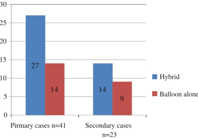
Balloon catheters were used in 136 sinuses (maxillary, frontal and sphenoid) for an average of 2.1 sinuses per patient. The distribution of the diVerent sinuses is detailed in Fig.1. Twenty three patients (36%) were treated with a balloon only procedure, and 41 patients (64%) were treated with a balloon in combination with conventional functional endoscopic sinus surgery (hybrid operation). All hybrid procedures were performed under general anaesthesia. Of the 23 patients treated with the balloon only procedure, 21 were under general anaesthesia and 2 had local anaesthesia with intravenous sedation. The indications based on pri-mary or secondary cases are depicted in Fig.2. Primary cases were the main indication for balloon sinuplasty (n = 41), and 23 were revision cases. 27 (66%) of the pri-mary cases and 14 (61%) of the revision cases were treated with a hybrid operation.
No adverse events occurred during the study period Dilation was achieved in all maxillary and sphenoid sinuses. Of the 104 frontal sinuses in which balloon sinupl-asty was attempted, dilation of 12 (12%) sinuses in 10 patients failed (Fig.3). Of these cases, seven (8 sinuses) were primary cases, and three (4 sinuses) were revision cases. The aetiology of the failed dilations is depicted in Table1 and Figs.4, 5. No statistically signiWcant diVerence
in failure rate was found between the two methods used: Xuoroscopy (63 frontal sinuses, 7 failed) and LUMA (41 frontal sinuses, 5 failed).
The CT-scan of one patient (Fig.6a–d) showed signs of chronic rhinosinusitis and no suspicion of a malignancy. Fig. 1 Distribution of balloon sinuplasty based on the sinuses
9% Maxillary Sinus n=20 Sphenoid Sinus n=12 Frontal Sinus n=104 Total Sinuses n=136
Fig. 2 Use of balloon only or hybrid operation based on primary or
revision cases 0 5 10 15 20 25 30
Pirmary cases n=41 Secondary cases n=23 9 Hybrid Balloon alone 27 14 14
Fig. 3 Success rate for the dilation of the frontal recess
Dilatation possible n=92 Dilatation not possible n=12 Total Frontal Sinuses n=104
The left frontal sinus was opened by conventional endo-scopic frontoethmoidectomy. Balloon sinuplasty of the right frontal sinus failed. 6 months later, the patient pre-sented with a right-sided frontal headache. The CT-scan at that time showed an air-Xuid level in the right frontal sinus. The right frontal sinus was then opened with conventional sinus surgery. Surprisingly, histopathological investiga-tions revealed a lymphoma in the frontal recess.
Discussion
Since the introduction of balloon sinuplasty, the medical literature has mainly focused on follow up analysis. Weiss et al. [8] described a signiWcant and stable improvement in
the SNOT-20 symptom score and the Lund–MacKay CT score in his 2-year-follow up study on patients who under-went balloon catheter sinusotomy. There have been limited studies on the technical failure rate of balloon dilation of the frontal recess, and to date, no study has focused on the aetiology of such failures. In the literature, the failure rate of balloon dilation of the frontal recess is estimated at 6–19% [2, 11, 12]. In our study, the technical failure rate for bal-loon sinuplasty of the frontal recess was 12%, whereas the technical failure rate for the procedure of the maxillary and sphenoid sinus was 0%. As we showed in our failed cases, balloon dilation may be diYcult in patients with anatomic variations of the frontal recess, such as agger nasi cell, frontoethmoidal cell or frontal bulla cell or in cases with signiWcant osteoneogenesis. Leunig [13] studied the Table 1 Aetiology of the
technical failure of balloon dilation in 12 frontal recesses in 10 patients
Patient Number
Failed side Pre-surgery Failed dilation due to Figures
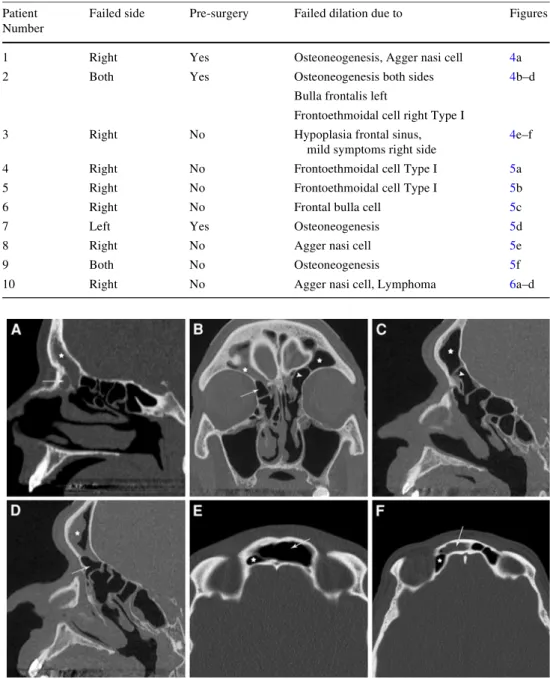
1 Right Yes Osteoneogenesis, Agger nasi cell 4a
2 Both Yes Osteoneogenesis both sides 4b–d
Bulla frontalis left
Frontoethmoidal cell right Type I
3 Right No Hypoplasia frontal sinus,
mild symptoms right side
4e–f
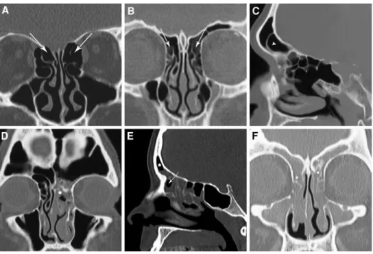
4 Right No Frontoethmoidal cell Type I 5a
5 Right No Frontoethmoidal cell Type I 5b
6 Right No Frontal bulla cell 5c
7 Left Yes Osteoneogenesis 5d
8 Right No Agger nasi cell 5e
9 Both No Osteoneogenesis 5f
10 Right No Agger nasi cell, Lymphoma 6a–d
Fig. 4 a (Patient 1): severe
osteoneogenesis of the frontal sinus/frontal recess (star) with an agger nasi cell (arrow). b–d (Patient 2): chronic rhinosinus-itis of the frontal sinus (star) with osteoneogenesis of the frontal recess on both sides, a frontal bulla cell on the left side (arrowhead) and a fronto-ethmoidal cell on the right side (arrow). e–f (Patient 3): pneu-mosinus dilatans with chronic rhinosinusitis of the left frontal sinus (arrow) crossing the midline to the right side and hypoplasia of the right frontal sinus (star). Because patients’ symptoms were most likely related to the successfully dilated left side, further eVort to dilate the right side was stopped after three failed attempts
anatomical variations of the frontal recess in 1,282 sepa-rately assessed sides of 641 patients who were referred for CT-scans due to chronic rhinosinusitis. In that patient pop-ulation, the incidence of anatomical variations of the frontal recess were as follows: agger nasi cell, 80%; frontoethmoidal
cell, 36.4% (Type I, 17%; Type II 6.8%; Type III, 12.5%; and Type IV, 0.1%) and frontal bulla cell, 16%. All of our failed cases showed this anatomic variations, in addition to frontal sinus hypoplasia or severe osteoneogenesis. No patient with failed balloon dilation had conventional anat-omy of the frontal recess. Therefore, the analysis of the anatomic region of the frontal recess on the preoperative CT-scan for existing anatomic variations in each patient is mandatory and allows the prediction of the potential diY-culties in achieving a dilation of the frontal recess. A pro-found understanding of the anatomy of the frontal recess in individual patients can be only achieved by analysing the CT-scans in multiplanar reconstructions. A discordance of up to 40% has been reported if the frontal recess is analysed in the coronal plane only versus analysis of the recess in multiplanar CT-reconstructions that include the sagittal, coronal and axial planes [14].
Wexler [15] suggests the image-guided frontal mini-trephination approach for guide-wire insertion in a retro-grade fashion to introduce the balloon catheter when cannulation of the frontal recess fails. This method maxi-mises the potential for frontal cannulation and dilation while minimising the Xuoroscopic search time. Leventhal [16] performed balloon dilation of 31 sinuses using an image-guided sinus guiding catheter. This may lead to increased accuracy of device placement and a reduction in the duration of Xuoroscopy. Whether the mini-trephina-tion approach or image-guided catheter placement would have helped in our cases is unknown. Nevertheless, these techniques have the potential to help when balloon dilation with the conventional balloon sinuplasty technique fails.
Fig. 5 a, b (Patients 4 and 5):
frontoethmoidal cell both sides (arrow). c (Patient 6): frontal bulla cell on the right side (arrowhead). d (Patient 7): osteoneogenesis on the left side (star). e (Patient 8): Agger nasi cell (arrow) with obstruction of the frontal recess/frontal sinus (star). f (Patient 9): osteoneo-genesis on both sides (stars)
Fig. 6 (Patient 10) a Preoperative CT scan showing chronic
rhinosi-nusitis of the frontal recess (star). b CT scan 6 months after conven-tional frontoethmoidectomy on the left (arrowhead) and failed balloon dilation of the frontal recess on the right due to an agger nasi cell (ar-row) and missed lymphoma (star). c Obstruction and secretion (star) in the frontal recess due to an agger nasi cell (arrowhead). d After frontal sinusotomy on the right, histopathological exam showed a lym-phoma of the frontal recess
Doubtless factors other than basic anatomic variations of the frontal recess are relevant for the technical failure for balloon sinuplasty. The size of the agger nasi cells, the frontoethmoidal cell and the frontal bulla cell and the angu-lation of these cells probably play a role in cannuangu-lation of the recess. Further anatomic studies are needed to answer the speciWc inXuence to this factors. Furthermore, the learn-ing curve plays a role in the failure rate of balloon sinupl-asty. As this is a multicenter study with diVerent surgeons and the patient number is limited, statistical analysis of this factor was not reasonable. Our failure with 12% is in con-cordance with the failure rate in the literature speciWed with 6–19% [2, 11, 12]. Therefore, we can speculate that the learning curve was not the major factor for failure rate in our study.
It is not common practice to obtain a sample for histopa-thological examination during the balloon only procedure, and we present a case of an initially overlooked lymphoma of the frontal recess during one of these procedures. The lymphoma was conWrmed following endoscopic frontoeth-moidectomy, which was performed 6 months after the orig-inal balloon procedure. To our best knowledge, this is the Wrst reported case of an overlooked malignancy in a case that involved a balloon only procedure. This drawback of not including a histopathological exam in a balloon only procedure should be considered.
Conclusion
Particularly in complex anatomical situations of the frontal recess, such as with an agger nasi cell, frontoethmoidal cell, frontal bulla cell or combinations of these, or in cases of severe osteoneogenesis, dilation of the frontal recess may be challenging or impossible. Therefore, despite the elegance of balloon sinuplasty, the surgeon should have a profound understanding of the underlying anatomy in each individual case and should know how to perform classical functional endoscopic sinus surgery in the frontal recess area in the event of a technical failure of the balloon dilation.
Because it is not common practice to obtain a sample for histopathological examination during the balloon only procedure, there is a potential risk of overlooking a pathol-ogy, which may require additional treatment. Therefore,
conventional surgical techniques with the option of obtain-ing biopsies are recommended if there is any suspicion of neoplastic disease.
Acknowledgments Prof. Marco Domenico Caversaccio is supported by the Swiss National Research Foundation for the project Computer-Aided and Image-guided Medical Interventions (http:// www.co-me.ch) [51 NF 40-111383].
References
1. Fokkens W, Lund V, Mullol J (2007) European position paper on rhinosinusitis and nasal polyps. Rhinol Suppl 20:1–136
2. Vaughan W (2008) Review of ballon sinuplasty. Otolaryngol Head Neck Surg 16:2–9
3. Brown C, Bolger W (2006) Safety and feasilbility of ballon cathe-ter dilation of paranasal sinus ostia. A preliminary investigation. Ann Otol Rhino Laryngol 115:293–299
4. Siow J, Kadah B, Werner J (2008) Balloon sinuplasty: a current hot topic in rhinology. Eur Arch Otorhinolaryngol 265:509–511 5. Melroy C (2008) The balloon dilating catheter as an instrument in
sinus surgery. Otolaryngol Head Neck Surg 139:S23–S26 6. Friedman M, Bliznikas D, Vidyasagar R, Joseph NJ, Landsberg R
(2006) Long-term results after endoscopic sinus surgery involving the frontal recess dissection. Laryngoscope 116:573–579 7. Bolger W, Brown C, Church C et al (2007) Safety and outcomes
of the balloon catheter sinusotomy: a mulicenter 24-week analysis in 115 patients. Otolaryngol Head Neck Surg 137:10–20 8. Weiss R, Church C, Kuhn F, Levine H, Sillers M, Vaughan W
(2008) Long-term outcome analysis of balloon catheter sinustomy: two year follow-up. Otolaryngol Head Neck Surg 139:S38–S46 9. Kuhn F, Church C, Goldberg A et al (2008) Ballon catherter
sinus-tomy: one year follow-up-outcomes and role in functional endo-scopic sinus surgery. Otolaryngol Head Neck Surg 139:S27–S37 10. Catalano P, Payne S (2009) Balloon dilation of the frontal recess
in patients with chronic frontal sinusitis and advanced sinus disease. Ann Otol Rhinol Laryngol 118:107–112
11. Levine H, Sertich A, Holsington D, Weiss R, Pritikin J (2008) A multi-center registry of balloon catheter sinustomy outcomes of 1, 036 patients. Ann Otol Rhinol Laryngol 117:263–270 12. Friedmann M, Wilson M (2008) Illumination guided balloon
sinuplasty. Laryngoscope 119:1399–1402
13. Leunig A, Betz CS, Sommer B, Sommer F (2008) Anatomic variations of the sinuses; multiplanar CT-analysis in 641 patients. Laryngo-Rhino-Otol 87:482–489
14. Leunig A, Sommer B, Betz CS, Sommer F (2008) Surgical anatomy of the frontal recess—is there a beneWt in multiplanar CT-reconstruction? Rhinology 46(3):188–194
15. Wexler DB (2008) Frontal balloon sinuplasty via minitrephin-ation. Otolaryngol Head Neck Surg 139(1):156–158
16. Leventhal D, HeVelWnger R, Rosen M (2007) Using image guid-ance tracking during balloon catheter dilation of sinus ostia. Otolaryngol Head Neck Surg 137(2):341–342
![[PDF] Cours complet pour apprendre Ruby | Formation informatique](data:image/gif;base64,R0lGODlhAQABAIAAAP///wAAACH5BAEAAAAALAAAAAABAAEAAAICRAEAOw==)