HAL Id: hal-01119333
https://hal.archives-ouvertes.fr/hal-01119333
Submitted on 23 Feb 2015HAL is a multi-disciplinary open access archive for the deposit and dissemination of sci-entific research documents, whether they are pub-lished or not. The documents may come from teaching and research institutions in France or abroad, or from public or private research centers.
L’archive ouverte pluridisciplinaire HAL, est destinée au dépôt et à la diffusion de documents scientifiques de niveau recherche, publiés ou non, émanant des établissements d’enseignement et de recherche français ou étrangers, des laboratoires publics ou privés.
Structural defects in layered structures: Their
determination and their impact on reactivity
Sylvain Grangeon, Francis Claret, Christophe Tournassat, Yannick Linard,
Bruno Lanson
To cite this version:
Sylvain Grangeon, Francis Claret, Christophe Tournassat, Yannick Linard, Bruno Lanson. Structural defects in layered structures: Their determination and their impact on reactivity. Clays in natural and engineered barriers for radioactive waste confinement: 6th international conference, Mar 2015, Bruxelles, Belgium. �hal-01119333�
Structural defects in layered structures: Their
determination and their impact on reactivity
Sylvain Grangeon
1, Francis Claret
1, Christophe Tournassat
1, Yannick Linard
2and
Bruno Lanson
31
BRGM, 3 avenue Claude Guillemin, 45060 Orléans cedex 2, France.
2
Andra, Centre de Meuse/Haute Marne, 55290, France.
3
CNRS and Univ. Grenoble Alpes, ISTerre, F-38041, Grenoble, France.
Clay minerals are layered structures that suffer from various and frequent crystallization faults, including point defects (e.g. layer vacancies and/or isomorphic cationic and anionic substitutions) and stacking defects (e.g. interstratification, well-defined or random stacking faults) that complicate the study of their structure, compared to most of other minerals. Determination of their crystal-structure is even further complicated by the small size of these minerals that possibly result from a "poisoning effect" induced by the presence of foreign cations in their layers.
The determination of the actual crystal structure of clay minerals, in particular the nature and density of structural defects, is however of paramount importance to understand and model clay minerals reactivity1. X-ray diffraction patterns of clay minerals often only exhibit 00l reflections and a few asymmetrical hk bands, while the hkl reflections are absent owing to the systematic occurrence of random stacking faults2 (i.e. turbostratism; random translations in the layer plane and/or random rotation about the normal). To overcome this problem, a mathematical formalism was developed that allows the calculation of X-ray diffraction patterns from structures affected by various density and nature of structural defects, including turbostratism3. This development led to a better understanding of the structure from many clay minerals, including layer structure (e.g. nontronite4) and, using a complementary analysis of 00l reflections, of interlayer water and cations organization 5, 6.
It is however obvious that not only clays suffers from the here above mentioned crystallization defects. Rather, nanocrystalline (i.e. particles smaller than 100 nm) and defective lamellar structures affected by turbostratism are ubiquitous in natural and man-made environments. One can for example cite manganese7 and iron oxides8 or nanocrystalline calcium silicate hydrates9 (C-S-H). The former two are reported to control the fate of many trace elements in the environment, including metals and actinides (in the case of vernadite, a manganese oxide) or oxyanions (in the case of fougèrite, an iron hydroxide). These minerals also often exhibit redox properties (e.g. Mn3+/Mn4+ or Fe2+/Fe3+) that enables for the degradation of organic compounds10 11. Contrastingly, C-S-H is not found in natural environments, but is the main hydration product from various types of cement, including ordinary Portland cement, controls main cement chemical and physical properties12, 13, and exerts a strong control on trace elements mobility. All these minerals and phases have close structural similarities with clays, as already noticed decades ago14, and thus methods used for the analysis of clay mineral structure are transposable to these phases. As for clays, a sound understanding of the crystal structure from these minerals and phases is a fundamental basis for many applications. First, the nature of point defects determines the type of elements that can be sorbed (typically, in the case of oxides, isomorphic substitutions create a limited layer charge deficit and favour the sorption of alkali and alkali-Earth elements as hydrated outer-sphere complexes, whereas layer vacancies favour the sorption of multivalent elements as inner-sphere complexes) and, second, the density of these defects dictate the overall reactivity.
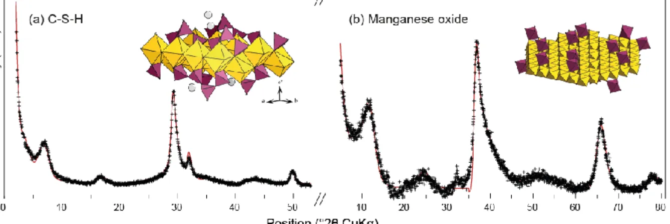
The present work will review recent applications of the specific method used to model X-ray diffraction patterns from nanocrystalline and defective lamellar structures, and will demonstrate that modelling can be used to retrieve accurate structural information, such as crystallite sizes and interlayer and layer structure, including nature and density of structural defects (Figure 1). The consistency between this method and other spectroscopic (e.g.
synchrotron X-ray absorption spectrometry) or microscopic (e.g. transmission electron microscopy) techniques will be illustrated on nanocrystalline manganese (vernadite) and iron (fougèrite) oxides and nanocrystalline calcium silicate hydrates (C-S-H).
Figure 1. Exemple of refined X-ray diffraction patterns from two nanocrystalline and turbostratic structures: C-S-H8 (a) and a biogenic manganese oxide15 (b). In both patterns, crosses are the experimental points and solid lines the calculated patterns. The two insets at the top right of each pattern are sketches of refined layer structures. In C-S-H, layers are built of Ca polyhedra (yellow) with
ribbons of wollastonite-like Si tetrahedra (purple) running at the surface. In the manganese oxide, layers are built of Mn octahedra (yellow) and layer vacancies capped by interlayer Mn sorbed as
inner-sphere complexes (purple). In both cases, typical crystallite size is 10 nm in the layer plane and 3 to 5 nm perpendicular.
References
1. Lanson, B., Modelling of X-ray diffraction profiles: Investigation of defective lamellar structure crystal chemistry. In Layered Mineral Structures and their application in Advanced Technologies, Brigatti, M. F.; Mottana, A., Eds. The European Mineralogical Union and the Mineralogical Society of Great-Britain & Ireland: London, 2011; Vol. 11.
2. Warren, B. E., X-Ray Diffraction in Random Layer Lattices. Physical Review 1941, 59, (9), 693.
3. Drits, V. A.; Tchoubar, C., X-ray diffraction by disordered lamellar structures : theory and
applications to microdivided silicates and carbons. Springer-Verlag: Berlin, 1990; p 371.
4. Gates, W. P.; Slade, P. G.; Manceau, A.; Lanson, B., Site occupancies by iron in nontronites.
Clays and Clay Minerals 2002, 50, (2), 223-239.
5. Ferrage, E.; Lanson, B.; Michot, L. J.; Robert, J. L., Hydration Properties and Interlayer Organization of Water and Ions in Synthetic Na-Smectite with Tetrahedral Layer Charge. Part 1. Results from X-ray Diffraction Profile Modeling. Journal of Physical Chemistry C 2010, 114, (10), 4515-4526.
6. Claret, F.; Sakharov, B. A.; Drits, V. A.; Velde, B.; Meunier, A.; Griffault, L.; Lanson, B., Clay minerals in the Meuse-Haute marne underground laboratory (France): Possible influence of organic matter on clay mineral evolution. Clays and Clay Minerals 2004, 52, (5), 515-532.
7. Bargar, J. R.; Fuller, C. C.; Marcus, M. A.; Brearley, A. J.; Perez De la Rosa, M.; Webb, S. M.; Caldwell, W. A., Structural characterization of terrestrial microbial Mn oxides from Pinal Creek, AZ.
Geochimica et Cosmochimica Acta 2009, 73, (4), 889-910.
8. Trolard, F.; Bourrié, G.; Abdelmoula, M.; Refait, P.; Feder, F., Fougerite, a new mineral of the pyroaurite-iowaite group: description and crystal structure. Clays and Clay Minerals 2007, 55, (3), 323-334.
9. Grangeon, S.; Claret, F.; Lerouge, C.; Warmont, F.; Sato, T.; Anraku, S.; Numako, C.; Linard, Y.; Lanson, B., On the nature of structural disorder in calcium silicate hydrates with a calcium/silicon ratio similar to tobermorite. Cement and Concrete Research 2013, 52, 31-37.
10. Ruby, C.; Upadhyay, C.; Géhin, A.; Ona-Nguema, G.; GÉnin, J.-M. R., In Situ Redox Flexibility of FeII-III Oxyhydroxycarbonate Green Rust and Fougerite. Environmental Science & Technology 2006, 40, (15), 4696-4702.
11. Nasser, A.; Buchanovsky, N.; Gerstl, Z.; Mingelgrin, U., Mineral induced mechanochemical degradation: The imazaquin case. Chemosphere 2009, 75, (1), 20-27.
12. Blanc, P.; Bourbon, X.; Lassin, A.; Gaucher, E. C., Chemical model for cement-based
materials: Temperature dependence of thermodynamic functions for nanocrystalline and crystalline C– S–H phases. Cement and Concrete Research 2010, 40, (6), 851-866.
13. Manzano, H.; Dolado, J. S.; Guerrero, A.; Ayuela, A., Mechanical properties of crystalline calcium-silicate-hydrates: comparison with cementitious C-S-H gels. physica status solidi (a) 2007, 204, (6), 1775-1780.
14. Brunauer, S.; Greenberg, S. A. In The Hydration of Tricalcium Silicate and 8-Dicalcium Silicate
at Room Temperature, Chemistry of Cement: Proceedings of the Fourth International Symposium,
Washington, 1960; US National Bureau of Standards, Ed. Washington, 1960.
15. Grangeon, S.; Lanson, B.; Miyata, N.; Tani, Y.; Manceau, A., Structure of nanocrystalline phyllomanganates produced by freshwater fungi. American Mineralogist 2010, 95, (11-12), 1608-1616.