HAL Id: tel-02496882
https://tel.archives-ouvertes.fr/tel-02496882
Submitted on 3 Mar 2020HAL is a multi-disciplinary open access
archive for the deposit and dissemination of sci-entific research documents, whether they are pub-lished or not. The documents may come from teaching and research institutions in France or abroad, or from public or private research centers.
L’archive ouverte pluridisciplinaire HAL, est destinée au dépôt et à la diffusion de documents scientifiques de niveau recherche, publiés ou non, émanant des établissements d’enseignement et de recherche français ou étrangers, des laboratoires publics ou privés.
Structuring factors of microbial communities in the
atmospheric boundary layer
Romie Tignat-Perrier
To cite this version:
Romie Tignat-Perrier. Structuring factors of microbial communities in the atmospheric boundary layer. Ocean, Atmosphere. Université Grenoble Alpes, 2019. English. �NNT : 2019GREAU034�. �tel-02496882�
THÈSE
Pour obtenir le grade de
DOCTEUR DE LA COMMUNAUTE UNIVERSITE GRENOBLE ALPES
Spécialité : Sciences de la Terre et Univers, Environnement Arrêté ministériel : 25 mai 2016
Présentée par
Romie TIGNAT-PERRIER
Thèse dirigée par Aurélien DOMMERGUE, Maître de Conférences,
Institut des Géosciences de l’Environnement, Université Grenoble Alpes, et
Catherine LAROSE, Chargée de Recherche, Laboratoire Ampère de l’Ecole Centrale de Lyon
Thèse préparée au sein de l’Institut des Géosciences de
l’Environnement à Grenoble
dans l'École Doctorale Terre Univers Environnement
Facteurs de structuration des communautés
microbiennes de la couche limite atmosphérique
Thèse soutenue publiquement le 22 novembre 2019, devant le jury composé de :
Dr. Viviane DESPRES
HDR, Max Planck Institute for Chemistry Biochemistry Department – Mainz (Allemagne), Rapportrice
Dr. Andrea FRANZETTI
HDR, University of Milano Department of Earth and Environmental Sciences – Milan (Italie), Rapporteur
Dr. Barbara D’ANNA
Directrice de Recherche, Laboratoire Chimie Environnement Université Aix Marseille – Marseille (France), Examinatrice
Dr. Pierre AMATO
Chargé de Recherche, Institut de Chimie de Clermont-Ferrand – Clermont-Ferrand (France), Examinateur
Dr. Jean MARTINS
Directeur de Recherche, Institut des Géosciences de l’Environnement Université Grenoble Alpes – Grenoble (France), Président du jury
Pr. Timothy M. VOGEL
Professeur, Laboratoire Ampère Université Lyon 1 – Lyon (France), Examinateur Dr. Catherine LAROSE
Chargée de Recherche, Laboratoire Ampère Ecole Centrale de Lyon – Lyon (France), Directrice de thèse
Dr. Aurélien DOMMERGUE
Maître de conférences, Institut des Géosciences de l’Environnement Université Grenoble Alpes – Grenoble (France), Directeur de thèse
PHD THESIS
Toward the title of
DOCTOR OF SCIENCE OF THE GRENOBLE ALPES UNIVERSITY COMMUNITY
Speciality: Earth and Universe Sciences Arrêté ministériel : 25 mai 2016
Presented by
Romie TIGNAT-PERRIER
PhD thesis supervised by Aurélien DOMMERGUE, Maître de
Conférences, Institut des Géosciences de l’Environnement, Université Grenoble Alpes and
Catherine LAROSE, Chargée de Recherche, Laboratoire Ampère de l’Ecole Centrale de Lyon
PhD thesis prepared in the Institut des Géosciences de
l’Environnement in Grenoble
and the Terre Univers Environnement doctoral school
Structuring factors of microbial communities in
the atmospheric boundary layer
PhD defense on the 22th of November, 2019, in Grenoble In front of the following jury members:
Dr. Viviane DESPRES
HDR, Max Planck Institute for Chemistry Biochemistry Department – Mainz (Germany), Reviewer
Dr. Andrea FRANZETTI
HDR, University of Milano Department of Earth and Environmental Sciences – Milan (Italy), Reviewer
Dr. Barbara D’ANNA
DR, Laboratoire Chimie Environnement Université Aix Marseille – Marseille (France), Examinator
Dr. Pierre AMATO
CR, Institut de Chimie de Clermont-Ferrand – Clermont-Ferrand (France), Examinator Dr. Jean MARTINS
DR, Institut des Géosciences de l’Environnement Université Grenoble Alpes – Grenoble (France), President of the jury
Pr. Timothy M. VOGEL
Professor, Laboratoire Ampère Université Lyon 1 – Lyon (France), Examinator Dr. Catherine LAROSE
Chargée de Recherche, Laboratoire Ampère Ecole Centrale de Lyon – Lyon (France), Supervisor
Dr. Aurélien DOMMERGUE
Maître de conférences, Institut des Géosciences de l’Environnement Université Grenoble Alpes – Grenoble (France), Supervisor
2
Acknowledgements
First of all, I wish to thank my supervisors Catherine Larose and Aurélien Dommergue, as well as my adviser Timothy M. Vogel. I thank you Tim for being one of my best university lecturer. I am grateful to you for telling me about this project on atmospheric microbial communities. Thank you Cath and Aurélien for being supporting and trusting me. I loved discussing with you three about my work, I thank you for everything I learnt from you. These three years have been intense and the PhD a real training work where I developed many skills. I really appreciated to have the opportunity to teach at the University of Lyon 1, to supervise a master student (Myriam Spajer), to do oral and poster presentations in international conferences and to develop English oral and written skills.
I would like to thank the members of my annual comities, i.e. Pierre Amato (Institut de Chimie de Clermont-Ferrand), Barbara d’Anna (LCE de Marseille), Alexandre Poulain (University of Ottawa) and Eric Bapteste (IBPS de Paris) for having accepted to discuss about my work and for giving me advices. I thank the members of my PhD jury, Jean Martins (IGE Grenoble), Barbara d’Anna, Pierre Amato, Andrea Franzetti (University of Milano, Italy) and Viviane Després (Max Planck Institute for Chemistry in Mainz, Germany) for having accepted to evaluate my work. I thank the Région Auvergne-Rhône Alpes for having financed my PhD. I thank the team of the IGE in Grenoble for having welcomed me. I thank particularly Jean-Luc Jaffrezo, Jean Martins and the PhD student Abdoulaye for our discussions on atmospheric chemistry and atmospheric microorganisms. It was nice to be in regular contact with you Abdoulaye.
I could really not have hoped for better coworkers at the Laboratoire Ampère, Ecole Centrale de Lyon: Mia, Concepcion, Rose, Benoît, Arthur and Adrien (all PhD students) as well as Christoph, Cécile, Graeme, Christina, Laure and Pascal. It was so nice to work with you and I would like to keep you all and bring you with me to my next lab. To PhD students: it was really nice to share this PhD experience with you, you were a real every day motivation and by far an important element that made my PhD succeed. I would like to thank particularly Mia: we spent so much time together in the lab during these three years, your presence was so motivating and reassuring, you helped me a lot.
It was really nice to meet Nora from Innsbruck and Frederik from Copenhagen, both PhD students invited in our lab. We also spent good times in conferences.
I thank Laurent Pouilloux, a competent and nice computer scientist from the ECL, for helping me on some of my scripts and for always answering me rapidly when I got computing problems.
I will finish by thanking my family as well as my life partner Robin who made big concessions to be at my sides in Lyon and gave me endless support.
4 “All models are wrong, but some are useful.”
6
Résumé (French)
La couche limite planétaire est la couche atmosphérique la plus basse qui est en interaction directe et constante avec les surfaces terrestres et marines sur lesquelles se concentrent les activités humaines, les cultures et divers écosystèmes. Comprendre l’origine de sa composition à la fois chimique et microbiologique est fondamental dans notre étude approfondie de la biosphère. Alors que les microorganismes de la couche limite planétaire – retrouvés jusqu’à 106 cellules par mètre cube d’air – semblent varier significativement à
l’échelle spatiale et temporelle en termes de concentration et de diversité, ils restent largement méconnus. L’objectif principal de cette thèse est de comprendre comment se structurent les communautés microbiennes dans la troposphère, et en particulier dans la couche limite planétaire, ainsi que d’identifier les facteurs de contrôle majeurs. En travaillant sur des échantillons collectés pendant plusieurs semaines sur neuf sites répartis sur la planète, et en utilisant les technologies de séquençage ADN haut-débit, nous avons étudié la composition taxonomique et fonctionnelle des communautés microbiennes de la phase gazeuse et solide de l’atmosphère (c’est-à-dire non associés aux nuages).
Nos premiers résultats sur la taxonomie des communautés microbiennes révèlent que les surfaces proches des sites sont les contributeurs principaux de distribution des communautés microbiennes atmosphériques, malgré l’occurrence potentielle du transport longue-distance des microorganismes atmosphériques. Egalement, les conditions météorologiques combinées à la diversité des surfaces locales terrestres ou océaniques jouent un rôle important dans la variation temporelle de la structure des communautés microbiennes de la couche limite planétaire. Une deuxième étude nous a permis d’étudier davantage la variation temporelle des communautés microbiennes atmosphériques sur un site continental montagneux en France (1465 m d’altitude) sur une année complète. Cette étude révèle l’importance des conditions de surface des paysages aux alentours dans la composition taxonomique des communautés atmosphériques. L’évolution au cours de l’année des terres agricoles et de la végétation, qui composaient en majeure partie le paysage du site, était responsable du changement temporel observé dans la composition taxonomique des communautés microbiennes atmosphériques. Finalement, nous avons étudié la composition fonctionnelle des communautés microbiennes de la couche limite planétaire afin d’identifier si les conditions physiques et chimiques de l’atmosphère jouaient un rôle dans la sélection ou
7
adaptation microbienne des microorganismes atmosphériques. L’analyse comparative de données métagénomiques ne révèle pas de signature atmosphérique spécifique du potentiel fonctionnel des communautés microbiennes atmosphériques. La composition fonctionnelle semble avant tout liée aux écosystèmes locaux. Toutefois, nous avons observé que les champignons étaient plus dominants relativement aux bactéries dans l’air comparativement aux autres écosystèmes. Ce résultat suggère un processus de sélection des champignons durant l’aérosolisation et/ou le transport aérien. Les champignons pourraient survivre davantage l’aérosolisation et le transport aérien comparativement aux bactéries du fait de leur résistance naturelle aux conditions physiques stressantes de l’atmosphère. Nos résultats ont apporté une meilleure compréhension des facteurs déterminants (c’est-à-dire les surfaces locales, les sources distantes, les conditions météorologiques locales, les conditions physiques stressantes de l’atmosphère) et de leur contribution dans la structuration des communautés microbiennes de la couche limite atmosphérique. Nos investigations constituent une base importante pour de nouvelles études sur la prévision et le contrôle des communautés microbiennes atmosphériques, afin de répondre à des questions majeures dans les domaines de la santé publique et de l’agronomie.
Mots clefs : communautés microbiennes de la couche limite planétaire, microorganismes atmosphériques, transport longue-distance, potentiel fonctionnel, métagénomique comparative, séquençage ADN haut-débit, séquençage MiSeq Illumina, sélection physique, adaptation microbienne, aérosolisation
8
Abstract
Up to 106 microbial cells per cubic meter are found in suspension in the planetary boundary
layer, the lowest part of the atmosphere. Direct influences of the planetary boundary layer on humans, crops and diverse ecosystems like soils and oceans make the full understanding of its composition, both chemical and microbiological, of utmost importance. While microbial communities of the planetary boundary layer vary significantly at different temporal and spatial scales, they remain largely unexplored. The main goal of this thesis was to understand how airborne microbial communities are structured in the troposphere with special emphasis on the planetary boundary layer and to identify their main controlling factors. We investigated both the taxonomic and functional composition of airborne microbial communities in the dry phase (i.e. not cloud-associated) over time at nine different geographical sites around the world using high throughput sequencing technologies.
Our investigation that focused on microbial taxonomy showed that local landscapes were the main contributors to the global distribution of airborne microbial communities despite the potential occurrence of long-range transport of airborne microorganisms. We also observed that meteorology and the diversity of the surrounding landscapes played major roles in the temporal variation of the microbial community structure in the planetary boundary layer. We further explored the temporal variation of airborne microbial communities at a continental and mountainous site in France (1465 m above sea level) over a full-year. This study demonstrated the importance of the surface conditions (i.e. vegetation, snow cover etc.) of the surrounding landscapes on the taxonomic composition of airborne microorganisms. The seasonal changes in agricultural and vegetated areas, which represented a significant part of the site’s surrounding landscape, were correlated to the shifts in the taxonomic composition of airborne microbial communities during the year. Finally, we investigated the functional composition of microbial communities of the planetary boundary layer to identify whether the physical and chemical conditions of the atmosphere played a role in selection or microbial adaptation of airborne microorganisms. The comparative metagenomic analysis did not show a specific atmospheric signature in the functional potential of airborne microbial communities. To the contrary, their functional composition was mainly correlated to the underlying ecosystems. However, we also showed that fungi were more dominant relatively to bacteria in air as compared to other (planetary bound) ecosystems. This result suggested a selective
9
process for fungi during aerosolization and/or aerial transport and that fungi might likely survive aerosolization and/or aerial transport better than bacteria due to their innate resistance to stressful physical conditions (i.e. UV radiation, desiccation etc.). Our results provide a clearer understanding of the factors (i.e. surrounding landscapes, distant sources, local meteorology, and stressful physical atmospheric conditions) that control the distribution of microbial communities in the atmospheric boundary layer. Our investigations provide a basis for further studies on the prediction and even control of airborne microbial communities that would be of interest for public health and agriculture.
Keywords: airborne microbial communities, atmospheric microorganisms, planetary boundary layer, long-range transport, functional potential, high throughput sequencing, MiSeq Illumina sequencing, physical selection, microbial adaptation, aerosolisation, comparative metagenomics
10
Table of content
Acknowledgements ... 2 Résumé (French) ... 6 Abstract ... 8 Table of content ... 10 List of abbreviations ... 12List of figures and tables ... 14
List of peer-reviewed publications ... 18
Chapter 1: Bibliography - Airborne microbial communities of the troposphere ... 20
Introduction ... 20
Microbiological characteristics of the troposphere ... 24
Bioaerosols in the troposphere ... 24
Planetary boundary layer versus free troposphere and vertical distribution of airborne microbial communities ... 26
Interactions between physico-chemical characteristics and airborne microorganisms in the troposphere ... 28
Physico-chemical characteristics of the atmosphere that might constrain microbial life ... 28
Physical selection versus microbial adaptation of airborne microbial communities ... 31
Metabolic activity and growth of airborne microbial communities ... 33
Distribution factors of airborne microbial communities (geography and time) in the troposphere ... 35
Surface characteristics ... 35
Physical selection of airborne microorganisms during aerosolization ... 36
Long-range transport of airborne microorganisms and contribution of local versus distant sources... 38
Meteorology ... 39
PhD objectives, approach and working hypotheses ... 40
References ... 42
Supplementary Information ... 50
Chapter 2: Methods to investigate the global atmospheric microbiome ... 56
Section 1: Protocol optimization and quality control ... 56
Abstract ... 57
Introduction ... 57
Material and Methods ... 59
Results ... 68
Conclusion ... 72
References ... 74
Section 2: Molecular biology analyses ... 78
References ... 81
Chapter 3: Global airborne microbial communities controlled by surrounding landscapes and wind conditions ... 82
Abstract ... 82
Introduction ... 83
Material and Methods ... 84
Results ... 87
Discussion ... 97
Conclusion ... 100
11
Supplementary Information ... 106
Chapter 4: Seasonal changes in local landscapes drive airborne microbial community variation ... 130
Abstract ... 130
Introduction ... 131
Material and Methods ... 132
Results ... 136
Discussion ... 145
Conclusion ... 149
References ... 150
Supplementary Information ... 152
Chapter 5: Microbial functional signature in the atmospheric boundary layer ... 178
Abstract ... 178
Introduction ... 179
Material and Methods ... 180
Results ... 185
Discussion ... 198
Conclusion ... 202
References ... 203
Supplementary Information ... 206
Chapter 6: Conclusion and perspectives ... 225
12
List of abbreviations
ANOVA= analysis of variance
BLAST= basic local alignment search tool DNA= deoxyribonucleic acid
EC=elemental carbon
MG-RAST= metagenomic rapid annotations using subsystems technology MODIS= moderate resolution imaging spectroradiometer
NR= non-redundant protein database OC= organic carbon
PANDAseq= paired-end assembler for Illumina sequences PCoA= principal coordinates analysis
PM= particulate matter
HYSPLIT= hybrid single particle lagrangian integrated trajectory model qPCR= quantitative polymerase chain reaction
RDA= redundancy analysis RNA= ribonucleic acid
ROS= reactive oxygen species RDP= ribosomal database project UV= ultra-violet
14
List of figures and tables
Chapter 1
Figure 1: The different biological niches existing in the troposphere.
Figure 2: PCoA analysis of the Bray-Curtis dissimilarity matrix based on the bacterial community structure (amplicon sequencing) of samples coming from cloud water, rain, fog, planetary boundary layer and free troposphere particulate matter.
Figure 3: Vertical stratification of the first layers of the atmosphere and concentration range of the number of bacterial cells per cubic meter of air based on qPCR data in these layers. Figure 4: Grouping of the investigations on the airborne microbial community taxonomic structure using high through put sequencing.
Figure 5: Sources and processes driving the composition of airborne microbial communities based on the PhD’s hypotheses, in relative order of expected importance.
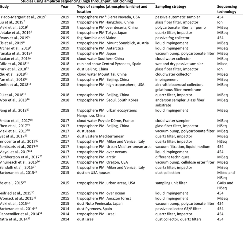
Table S1: List of investigations on the airborne microbial community taxonomic structure using high through put sequencing (not cloning) to date to the best of our knowledge.
Table S2: List of investigations whose datasets have been used to do the multivariate analysis PCoA in the Figure 2.
Chapter 2
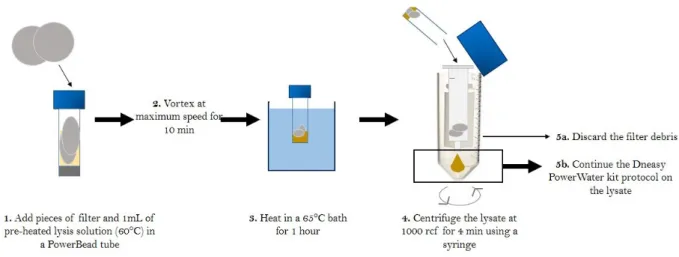
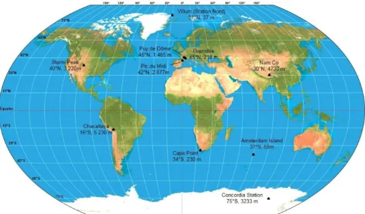
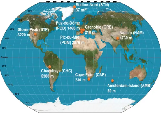
Figure 1: Summary of the modified DNA extraction protocol developed for quartz filters. Figure 2: Global distribution of the sampling sites and their respective elevation above sea level.
Figure 3: Organic concentrations on different types of filters.
Figure 4: OC and 16S rRNA gene concentrations measured at the different sampling sites. Table 1: Summary of sampling sites characteristics.
Table 2: Number of 16S rRNA gene copies per mm2.
Table 3: Organic carbon concentrations expressed in µg per cm2.
Chapter 3
Figure 1: Map showing the geographical location and elevation from sea level of the nine sampling sites.
Figure 2: Heatmaps of the relative abundances (the relative abundances are centered and scaled) of the fifty most abundant bacterial genera and fungal species in the dataset.
Figure 3: Hierarchical cluster analysis (average method) of the Bray-Curtis dissimilarity matrices based on the V3-V4 region of the 16S rRNA gene (genus level) and ITS region (species level).
Figure 4: Distribution of the sites based on the different data sets.
Table 1: Summary of bacterial and fungal abundances and bacterial (genus level) and fungal (species level) Chao1 richness estimations averaged per site and associated to a standard deviation.
Table 2: Temporal variability of the microbial community structure and meteorological conditions at each site.
Figure S1: Hierarchical cluster analysis (average method) of the Bray-Curtis dissimilarity matrices based on the V3-V4 region of the 16S rRNA gene and ITS region.
15
Figure S3: Hierarchical cluster analysis (average method) on the Euclidean distance matrix calculated on the PM10 chemistry data.
Figure S4: Distance-based RDA analyses.
Figure S5: Wind roses covering the sampling time at each site.
Figure S6. Backward trajectories calculated over 3 days at each site using HYSPLIT. Figure S7: Q-Q plots of the multiple linear regressions.
Table S1: Information on samples.
Table S2: Presentation of the different MODIS land covers from Friedl et al., 2002.
Table S3: Estimation of mean bacterial cell concentration per cubic meter of air in near-surface air above the different landscapes reported in Burrows et al., 2009.
Table S4: Total abundance (number of annotated sequences), contribution in each site and relative abundance per site of the first fifty most abundant bacterial genera.
Table S5: Total abundance (number of annotated sequences), contribution in each site and relative abundance per site of the first fifty most abundant fungal species.
Table S6: Bacterial genera and fungal species characterizing the different sites or groups of sites identified using hierarchical cluster analyses based on both bacterial and fungal community structures.
Table S7: Average concentration (and standard deviation) of the chemicals per site in ng/m3
of air.
Table S8: Multiple linear regression results. Chapter 4
Figure 1: Central position of Puy-de-Dôme in France and relative surfaces of the landscapes surrounding the site in a perimeter of 50 km based on the MODIS land surfaces.
Figure 2: Log10 of the bacterial and fungal cell concentration estimated by the number of 16S and 18S rRNA gene copies per cubic meter of air and per sample.
Figure 3: Hierarchical cluster analysis (average method) on the airborne bacterial community structure and airborne fungal community structure based on the Bray-Curtis dissimilarity matrices.
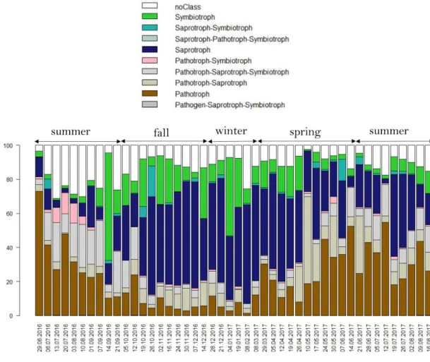
Figure 4: Trophic mode of the fungal species.
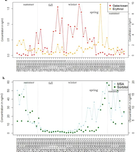
Figure 5: Temporal evolution of the relative abundance of several fungal species and bacterial genera over the year.
Figure 6: Temporal evolution of the concentration of chemical species over the year. Figure S1: Monthly NASA satellite images of Puy-de-Dôme surrounding surfaces.
Figure S2: Rarefaction curves of the number of bacterial genera and fungal species per season. Figure S3: Venn diagrams showing the number of shared and unique bacterial genera and fungal species between the samples of each season after rarefaction.
Figure S4: Temporal evolution of the relative abundance of four fungal phytopathogens over the year.
Figure S5: Heatmap showing the temporal evolution of the relative abundances of the fifty most abundant bacterial genera and fungal species in the dataset.
Figure S6: Hierarchical cluster analysis (average method) on the particulate matter chemical concentrations based on the Euclidean distance matrix.
Figure S7: Constrained Analysis of Principal Coordinates.
Figure S8: Temporal evolution of the temperature and relative humidity over the year. Figure S9: Meteorological conditions per season.
16
Table S1: Information on the samples.
Table S2: Bacterial and fungal concentration and richness averaged per season.
Table S3: Top 25 of the bacterial genera and fungal species observed in Puy-de-Dôme, and average percentages of these bacterial genera and fungal species per season.
Table S4: Weekly variability of the bacterial and fungal composition per season
Table S5: Richness in fungal species annotated as pathotroph, symbiotroph and saprotroph averaged per season and according to FUNGuild.
Table S6: Grouping of the fifty most abundant bacterial genera and fungal species based of the trend of their relative abundance over the year.
Table S7: Chemical concentrations averaged per season. Chapter 5
Figure 1: Map showing the geographical location and elevation from sea level of our nine sampling sites and the geographical position of whose public metagenomes come from. Figure 2: Percentage of fungi-associated sequences and bacteria-associated sequences over the total number of sequences annotated as belonging to fungal and bacterial genomes in the metagenomes.
Figure 3: PCoA analysis of the Bray-Curtis dissimilarity matrix based on the functional potential structure of each site.
Figure 4: Average number of hits of sporulation related functions per 10000 annotated sequences from all sequences, fungi-associated sequences and bacteria-associated sequences per site.
Figure 5: Average number of hits of UV protection related functions per 10000 annotated sequences from all sequences, fungi-associated sequences and bacteria-associated sequences per site.
Figure 6: Average number of hits of oxidative stress cell response related functions per 10000 annotated sequences from all sequences, fungi-associated sequences and bacteria-associated sequences per site.
Figure 7: Average number of hits of cell death related functions per 10000 annotated sequences from all sequences, fungi-associated sequences and bacteria-associated sequences per site.
Figure 8: Average number of hits of desiccation response related functions per 10000 annotated sequences from all sequences, fungi-associated sequences and bacteria-associated sequences per site.
Figure 9: Conceptual model that might explain the higher ratio between fungi and bacteria in air relatively to airborne microbial cell sources such as soil and ocean water.
Table S1: Information on the sites.
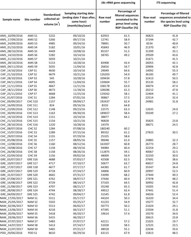
Table S2: Standardized collected air volume and sampling starting date of each air sample that we collected for this study.
Table S3: Functional richness and evenness after rarefaction per site, based on the SEED functional classes.
Table S4: qPCR on the 16s rRNA gene and on the 18S rRNA gene on air and soil samples, and ratio between these qPCRs.
Table S5: Top 50 of the SEED functions observed in the air samples considering all the sequences (i.e. bacteria and fungi-associated sequences).
Table S6: SIMPER analysis results on groups identified on the PCoA analysis based on all sequences.
17
Figure S1: Surrounding landscapes of the sampling sites in a 50 km perimeter based on the land cover MODIS approach.
Figure S2: PCoA analysis of the Bray-Curtis dissimilarity matrix based on the functional potential structure of each air sample.
Figure S3: PCoA analysis of the Bray-Curtis dissimilarity matrix based on the functional potential structure of each site.
Figure S4: Average number of hits of hydrogen peroxide catabolic process related functions per 10000 annotated sequences from all sequences, fungi-associated sequences and bacteria-associated sequences per site.
Figure S5: Functional richness after rarefaction from all sequences, fungi-associated sequences and bacteria-associated sequences per site.
Figure S6: Average number of hits of methane monooxygenase process related functions per 10000 annotated sequences from all sequences per site.
Figure S7: Average number of hits of lipoate synthase and chromosome plasmid partitioning protein ParA per 10000 annotated sequences from all sequences per site.
Chapter 6
Figure 1: Overview of the role of different factors in controlling microbial communities of the planetary boundary layer.
18
List of peer-reviewed publications
*Publications on the thesis work
Dommergue A., Amato P., Tignat-Perrier R., Magand O., Thollot A., Joly M., Bouvier L., Sellegri K., Vogel T.M., Sonke J.E., Jaffrezo JL, Andrade M., Moreno I., Labuschagne C., Martin L., Zhang Q. and Larose C. Methods to investigate the global atmospheric microbiome. Front. Microbiol. 2019. Published in Frontiers in Microbiology.
Tignat-Perrier R., DommergueA., ThollotA., KeuschnigC., MagandO., VogelT.M., LaroseC.
Global airborne microbial communities controlled by surrounding landscapes and wind conditions. Under review in Scientific Reports.
Tignat-Perrier R., Dommergue A., Thollot A., Magand O., Vogel T.M., Larose C. Microbial
functional signature in the planetary atmospheric boundary layer. In preparation for
publication in Environmental Microbiology.
Tignat-Perrier R., Dommergue A., Thollot A., MagandO., Amato P., VogelT.M., Larose C.
Seasonal changes of local landscapes drive airborne microbial community variation. In
preparation for publication in FEMS.
*Side publications
Els N., Larose C., Baumann-Stanzer K., Tignat-Perrier R., Keuschnig C., Vogel T.M., Sattler B. Microbial Composition in Seasonal Timeseries of Free-Tropospheric Air and Precipitation Reveals Community Separation. 2019. Published in Aerobiologia.
Sanchez-Cid C., Tignat-Perrier R., Franqueville L., Delaurière L., Schagat T., Vogel T.M. Sequencing depth rather than DNA extraction determines soil bacterial richness discovery.
20
Chapter 1: Bibliography - Airborne microbial communities of
the troposphere
Introduction
The study of microorganisms in the atmosphere goes back to the 19th century when Pasteur
cultivated microorganisms from the air. At that time, the principal issue was tracing the spread of diseases to the aerial dispersion of bacterial pathogens. Later, in the late 20th century, with
the discovery of a rich airborne microbial world, attention broadened to the origin, transport dynamics, survival, as well as the airborne microorganism’s role in air quality, meteorology and chemical cycles within the atmosphere.
The lowest part of the atmosphere that surrounds the Earth’s surface is the troposphere. It is the densest and most dynamic layer in terms of chemistry and physics of aerosols (i.e. atmospheric particles). It represents a large volume of 6.6 billion cubic kilometers that is five times larger than that of oceans (1.3 billion cubic kilometers). The troposphere harbors complex chemical reactions and meteorological phenomena that are intricately linked and lead to the coexistence of gas, solid (i.e. particulate matter from a size of a few nanometers to millimeters like sand dust) and liquid phases (i.e. clouds, rain, fog).
These phases represent different potential biological niches (Fig. 1) that are all studied in aeromicrobiology (supplementary Table S1). Based on the literature1–3, these niches harbor different microbial communities (Fig. 2) and different airborne microbial concentrations that might be due to significant differences in physico-chemical characteristics that constrain either microbial life within the niches or the destruction of specific members from their source. For example, liquid-phase associated microorganisms (i.e. cloud, rain and fog water) might be particularly different from microorganisms in the dry phase of the atmosphere (i.e. free microorganisms suspended in the gas phase and microorganisms attached to particulate matter such as sand dust) (Fig. 2).
21
Figure 1: The different biological niches existing in the troposphere. Airborne microorganisms have been observed in the liquid phase (i.e. cloud, fog and rain water), gas phase (as free cells) and solid phase (as cells attached to aerosols from a microscopic to macroscopic size such as sand dust). These different niches might exchange microorganisms and represent different physico-chemical conditions for airborne microorganisms.
Figure 2: PCoA analysis of the Bray-Curtis dissimilarity matrix based on the bacterial community structure (amplicon sequencing) of samples coming from cloud water, rain, fog, planetary boundary layer and free troposphere particulate matter. The corresponding studies are listed in supplementary Table S2.
22
In addition to their aqueous nature, clouds are characterized by specific chemistry and physics compared to the dry troposphere. Clouds dissolve chemical species in water and support new and/or faster chemical reactions4 that lead to an enhanced formation of secondary organic
aerosols as well as strong oxidants like H2O2 and radicals5,6. Clouds are at the heart of current
investigations on airborne microbial metabolism. In the laboratory, several culture-based and microcosm studies have shown that cloud-associated microorganisms can degrade the main carboxylic compounds found in cloud water (i.e. formate, acetate, formaldehyde7). Microbial
communities were shown to influence the oxidative capacity of clouds through the reduction of oxidants like H2O26. These chemical specificities are associated with physical processes like
the ongoing presence of successive condensation-evaporation cycles and freeze-thaw cycles of the cloud water8–12. These processes create a highly specific biological niche in clouds that
might constrain microbial life13. Clouds might themselves be formed on microbial cells
originating from the dry troposphere as they offer a surface for the condensation of water vapor and act as cloud condensation nuclei (CCN). The role of microorganisms as CCN and ice nuclei in cloud formation has been intensively investigated14–18.
Cloud-associated microbial communities and more generally liquid-phase associated microorganisms are not the center of interest of this PhD. Rather, we focused on free and particulate-matter associated microorganisms found in the dry phase of the troposphere (i.e. gas and solid phases). Moreover, we were specifically interested in the lower part of the troposphere (i.e. the planetary boundary layer – Fig. 3) representing the atmosphere that directly surrounds us. The way the planetary boundary layer interacts with humans, crops and diverse ecosystems like soils and oceans makes the understanding of its microbial composition of utmost importance. While planetary boundary layer microorganisms (from the dry phase) seem to vary significantly over space and time, the number of investigations remains limited (supplementary Table S1). Moreover, investigations on planetary boundary layer microorganisms are mainly of small spatial-scale (Fig. 4) and use different sampling strategies (supplementary Table S1) making them difficult to compare.
23
Figure 3: Vertical stratification of the first layers of the atmosphere and concentration range of the number of bacterial cells per cubic meter of air based on qPCR data in these layers.
*Free troposphere related studies: Tanaka et al. (2019)19, Zweifeil et al. (2012)20, DeLeon-Rodriguez et al. (2013)21.
Boundary layer related studies: Bertolini et al. (2013)22, Zhen et al. (2017)23, Genitsaris et al. (2017)24, Gandolfi et
al. (2015)25, Cho et al. (2011)26, Park et al. (2018)27.
Figure 4: Grouping of the investigations on the airborne microbial community taxonomic structure using high through put sequencing (not cloning – see supplementary Table S1) depending on the atmospheric niche studied (i.e. liquid versus dry phase, planetary boundary layer versus free troposphere) and the scale of the study (i.e. small or large scale studies; only for the dry phase of the planetary boundary layer related studies). Small scale studies include studies at one or a few sites; large scale studies include studies at several sites (regional or continental scale). The number of studies per group is indicated under the group name.
24
This Chapter will review knowledge and knowledge gaps regarding major questions in microbial ecology of the troposphere such as “Are airborne microorganisms undergoing a
selective process during aerosolization?”, “Is airborne microbial functional composition related to the physical and chemical conditions characterizing the atmosphere?”, “Are airborne microorganisms metabolically active?” and “What factors structure airborne microbial communities at varying temporal and spatial scales?” with a special interest for the dry
troposphere. When necessary, we will present data and hypotheses obtained from investigations of airborne microbial communities from other atmospheric biological niches like clouds. We will discuss their relevance and their potential inference to airborne microbial communities of the dry troposphere.
We will start by describing the microbiological characteristics of the troposphere, especially in the gas and solid phases (i.e. the dry troposphere). We will then present potential interactions between airborne microbial communities and their physico-chemical environment as well as the potential contribution of airborne microorganisms to biogeochemical cycles and the role of the physico-chemical environment on constraining airborne microbial life. Finally, we will identify the main factors potentially responsible for the geographical distribution and temporal dynamics of troposphere microbial communities. The review will be followed by an outline of the objectives, hypotheses and relevance of the thesis work in this context.
Microbiological characteristics of the troposphere Bioaerosols in the troposphere
The atmosphere consists of a layer of gases that is retained around the Earth by gravity. It is vertically stratified from the Earth’s surface to the exosphere, which is the upper layer separating the Earth’s atmosphere from space. The first layer that immediately surrounds the Earth is called the troposphere (Fig. 3). The troposphere is the densest layer in terms of aerosols and, consequently, the most dynamic layer in terms of aerosol physics and chemistry. Aerosols are tiny particles floating in the air and created either by condensation of gases or directly emitted by the Earth’s surface. We will use either the term particulate matter (PM) or aerosols to refer to atmospheric particles, although aerosols consist of both the suspended particles (i.e. particulate matter) and their surrounding gas. The troposphere is also the layer
25
with the largest concentration of bioaerosols. Bioaerosols can represent around 30% of the atmospheric aerosol load (aerosols > 1 µm) in urban and rural air, and up to 80% in pristine rainforest air28–32. Bioaerosols are aerosols of biological origin and include plant debris, pollen,
microorganisms (bacteria, fungal cells and spores, viruses as well as larger eukaryotic microorganisms like protozoans), and biological secretions. In the tropospheric layer, microbial cell concentrations can reach up to 106 cells per cubic meter of air in its lowest and
densest part close to the ground (Fig. 3). The troposphere height extends up to ten to fifteen kilometers above sea level and the stratosphere is above it. The stratosphere is a layer of free atoms in which radical chemical reactions occur and is commonly called the ozone layer4. It is
a layer with low air density and is composed of low particulate matter and ion concentrations and likely very low microbial concentrations33. Microorganisms have been isolated from as
high as 77 km in the stratosphere34, although it might represent the upper altitudinal limit.
These microorganisms found in the stratosphere remained viable35,36, even though the
stratosphere combines challenging conditions, such as cold temperatures down to -70°C, exposure to destructive UV radiations, intense desiccation due to the extremely low relative humidity (down to 0%), high radical concentrations, hypobaric conditions, and low nutrient concentrations etc.
Tropospheric aerosol properties, mainly number and mass concentration, size distribution, chemical composition, over time, space and geography have been thoroughly studied37.
Although investigations on the concentration and diversity of airborne microbial communities are becoming more numerous, their distribution on particulate matter remains unknown. Airborne microbial cells might exist mainly as aggregates or attached to particulate matter, while airborne fungi might exist mainly as single spores38. Particulate matter size range is
broad, starting from less than one nanometer (example of secondary aerosols created by the condensation of gases) up to hundreds of micrometers like sand dust. Microbial cells entering freely in the atmosphere might attach to existing particulate matter or other microbial cells39.
Conversely, particle-attached microbial cells might detach from their support in the air. Based on a compilation of data from more than one hundred investigations, Clauss et al. (2015)38
determined that 15% of cultivable airborne bacterial cells were on particles < 2.1 µm (size) and 25% on particles > 7.2 µm, and that cultivable airborne fungal spores and cells were mainly distributed on particles between 1 and 3.2 µm (median-based values) on average in outdoor air. The distribution size was shown to depend on aerosolization processes occurring and
26
different meteorological conditions at the time of aerosolization such as air relative humidity38. Using a culture-independent concentration estimation approach, Sippula et al.
(2013)40 also observed that 77% of total bacteria were found in the size fraction > 2.4 µm.
Airborne microbial concentrations tend to show a vertical gradient with high concentrations near the Earth’s surface and decreasing concentrations with altitude up through the troposphere and into the stratosphere2,19 (Fig. 3). Simultaneous airplane samples collected at
690, 1000 and 3127 m altitude above sea level showed decreasing numbers of cultivable microorganisms per cubic meter of air with increasing altitude41. While airborne microbial
concentrations have been repeatedly measured in the lower troposphere around our planet using culture-dependent and molecular analyses23–27,42–44, cell concentrations in the upper troposphere and stratosphere remain unknown. To date, only one culture-independent investigation has been carried out in the stratosphere33. Transport of particles and microbial
cells might be restricted between the upper troposphere and the stratosphere. While the upper troposphere supplies the stratosphere in microorganisms and chemical species involved in global ozone depletion, the stratosphere is a source of free radicals for the upper troposphere4. Although unknown, the stratosphere might support a low microbial diversity
formed of microorganisms resistant to the extreme environmental conditions33 (see next
section). Current knowledge about stratospheric microorganisms is scarce and almost exclusively based on culture-based approaches, which limits investigations to a small fraction (< 1%) of the whole community. In-flight collection of stratospheric microorganisms remains expensive and an engineering challenge, which due to the very low concentration of cultivable cells in the stratosphere suffers from aircraft-associated contamination.
Planetary boundary layer versus free troposphere and vertical distribution of airborne microbial communities
The troposphere is itself divided in two layers, the planetary boundary layer and the free troposphere (Fig. 3). The planetary boundary layer interacts with the Earth’s surface and represents the densest layer harboring the largest concentration of particulate matter and bioaerosols4. This PhD will focus specifically on the planetary boundary layer because it has
the highest concentration of bioaerosols and it is the layer that surrounds most of the human population. Air mass dynamics within the planetary boundary layer are subject to mechanical and thermal convective turbulence that is controlled in part by the ground roughness and
27
Earth’s surface heat4. As a consequence, the boundary layer’s height changes according to
location and even time of day throughout the year. In contrast, the free troposphere is driven horizontally by geostrophic wind and is vertically stable.
Aerosolized microbial cells generally enter the lowest layer of the atmosphere, the planetary boundary layer, from which an as yet unknown quantity might be transferred to the free troposphere. Some microbial inputs from the Earth’s surface could directly reach the free troposphere during specific and violent meteorological events like dust storms or volcanic eruptions. The free troposphere can also be in direct contact with the Earth’s surface. For example, surfaces of mountain peaks like Storm-Peak (+ 3220 m, Colorado, USA) and Pic-du-Midi (+ 2876 m, France) can at times be in the free troposphere during the night. Puy-de-Dôme site (+ 1465 m; France) is known to be influenced by free tropospheric air masses during certain meteorological conditions (more often in winter).
Once in the planetary boundary layer or the free troposphere, microbial cells will be transported over short or long distances depending on the meteorological conditions (windy, wet and dry precipitation), and if they travel freely or attached to a bigger particle. In the planetary boundary layer, airborne microorganisms have been shown to have a residence time of a few days before returning to the Earth’s surface due to gravitation or precipitation if they behave like non biological aerosols45. In the free troposphere, their residence time
might be several days during which they might be transported over thousands of kilometers46.
Despite an obvious continuum of the troposphere and because of differences in aerosol dynamics, chemical composition and physical conditions, the planetary boundary layer and the free troposphere might support different microbial communities2,19 (Fig. 2). The free
troposphere is more stable than the planetary boundary layer in terms of physics and aerosol dynamics and might also be more stable in airborne microbial concentration and composition. Studies on the structure of oceanic microbial communities reported a biogeography and a vertical distribution of the microbial diversity47–51. Like the atmosphere, the ocean represents
a continuous fluidic environment that is vertically stratified. Some microbial oceanographic studies showed that microbial community structure and abundance in the surface layers are less stable than those in the mesopelagic zone (the layer under the surface layer) due to abiotic and biotic perturbations that make the marine surface layer environment less stable49,52. Some studies on the vertical distribution of airborne microbial communities
28
suggested that some microbial taxa might be filtered out during vertical transport2. Due to
their size, the largest and densest airborne microbial cells might be less prone to reaching the free troposphere than lighter cells. This hypothesis was supported by the observed increase in the ratio between bacteria and fungi at a remote mountain site in Austria (+ 3106 m)2.
Another explanation could be that microbial cells floating in the free troposphere have more time to undergo selection and adaptation to the abiotic conditions as compared to those in the planetary boundary layer, so that only the microorganisms that are the most resistant to the harsh tropospheric conditions (UV radiation, cold temperature, radicals etc.) survive. Thermophilic strains with high resistance towards extreme conditions, which are often identified in heavy dust events, were shown to be ubiquitous and significantly increased in relative abundance in the free troposphere as compared to the planetary boundary layer at a remote mountain site in Austria (+ 3106 m)2.
Interactions between physico-chemical characteristics and airborne microorganisms in the troposphere
Physico-chemical characteristics of the atmosphere that might constrain microbial life
A variety of chemical substances interact with tropospheric microorganisms and subsequently might have an effect on them. Solid and liquid aerosols can be composed of essential nutrients (carbon, oxygen, nitrogen, phosphorus, sulfur, hydrogen, trace minerals) that could be used as source of energy and matter for microbial metabolism. The troposphere also contains free radical species (the hydroxyl radical OH is the most common and reacts with nearly every chemical species) and compounds at potential toxic concentrations (heavy metals, persistent organic compounds, antibiotics) that may have negative effects on microbial development and survival. Temperature and photochemical processes constantly alter the aerosol properties as well as their chemical composition. The physical and chemical characteristics of the atmosphere that we think constitute the main constraints of microbial life in the dry troposphere are presented below.
UV radiation. UV radiation levels can be extremely high and destructive in the atmosphere.
The highly energetic wavelengths (UV-C ~190-290 nm and UV-B ~290-320 nm) are responsible for direct DNA damage that could be lethal. Longer wavelengths (UV-A ~320-400 nm and visible light ~400-800 nm) contribute to intra-cellular reactive oxygen species (ROS)
29
production that can cause subsequent oxidative damage to DNA, RNA, lipids and proteins, altering microbial metabolism and survival53,54. Some microorganisms have developed a range
of protection mechanisms against UV radiation. Cell aggregation, association with particles and production of carotenoid pigments to scavenge ROS are all mechanisms used by environmental microorganisms to reduce the effects of destructive UV radiation. The stratosphere supports by far the highest levels of UV radiation found on Earth as levels increase by around 11% with every 1000 m in altitude (WHO). Data on the impact of UV radiation on airborne microorganisms comes mainly from investigations using high UV levels such as those found in the upper troposphere or stratosphere layer. Smith et al. (2011)36
showed that UV radiation was the most biocidal factor in the low stratosphere, and could kill up to 99.9% of Bacillus subtilis spores after 96 h. However, the authors pointed out that spore resistance might be dependent on the environment the cells germinated in55–57.
Consequently, UV resistance might have been higher if the spores were directly isolated from the stratosphere and not germinated in culture media like was done in the study. Microbial strains isolated from the upper troposphere and lower stratosphere exhibited a higher resistance to UV radiation as compared to those from the troposphere at ground level58. Some Deinococcus and Streptomyces strains showed an extreme UV resistance and tended to form
aggregates in culture medium. These aggregates were suggested to be a protection mechanism58. With the exception of sporulation and cell aggregation, no other protective
mechanism against UV radiation has been observed in airborne microbial communities. UV radiation levels and consequently the need for UV protection mechanisms might depend on the height of the troposphere (i.e. planetary boundary layer or free troposphere height), geography (for example the tropics harbor higher UV levels) and surface conditioning (i.e. surface reflectance)59.
Temperature shock and freeze-thaw cycles.At the same altitude, atmospheric temperature is highly dependent on the latitude and longitude of the site. It also decreases by 0.6 to 1°C for every 100 m increase of altitude and can reach -70°C in the upper stratosphere. Upward aerial transport of microorganisms with high-speed winds could occur rapidly and airborne microorganisms might suffer large temperature shocks. Airborne microorganisms present in an air parcel transported from the surface to 1 km altitude can undergo a temperature decrease of 5 to 10°C and a substantial increase in relative humidity4. Cold temperature and
30
freeze-thaw cycles generally occur at high latitudes, high altitudes and/or in clouds and so do the resulting impact. They slow down microbial metabolism, decrease membrane fluidity, and influence protein refolding. Freeze-thaw cycles could additionally lead to mechanical stress that might damage the cell membrane60–62. Freeze-thaw cycles were shown to alter the
survival of microbial strains following UV radiation, H202 exposure and osmotic shock when
these factors were tested individually on strains isolated from clouds belonging to
Pseudomonas, Sphingomonas, Arthrobacter and the yeast Dioszegia8. To date, no specific
mechanism of protection against cold temperature and freeze-thaw cycles known to be used by environmental extremophile microorganisms63 has been observed in airborne microbial
communities.
Relative humidity and condensation/evaporation cycles.The troposphere harbors the whole range of relative humidity (RH) values, from values near 0% in the upper troposphere up to 100% above ground level. Investigations on the survival of aerosolized microorganisms under different RH showed different results depending on the species. While the survival of airborne
Flavobacterium was not affected by RH ranging from 25 to 99% at 24°C64, mid-range RH negatively impacted mycoplasma survival but not RH values outside of this range65. In the
environment, desiccation resistance is generally associated to ionizing radiation resistance66– 70. Yet, the mutual nature of the underlying mechanisms remains unknown. In the
environment, the molecular mechanisms underlying desiccation resistance remain poorly defined and seem to involve wax ester biosynthesis71 and DNA reparation mechanisms.
Desiccation, like radiation, tends to induce DNA damage70,72.
Evaporation/condensation cycles of water vapor occur in the troposphere, both in the dry troposphere and in clouds. In a water droplet, evaporation can concentrate metabolites in the near environment of the cells by up to 1000 times8. Evaporation/condensation cycles induce
osmotic changes, leading to water fluxes between the intracellular and extracellular compartment of the cell to maintain osmolarity. These water fluxes can provoke cell damage, increase the concentration of metabolites in cells, and increase the concentration of compounds like radicals and metals around the cell73,74. Alsved et al. (2018)75 showed that
during evaporation, Pseudomonas syringae survival was enhanced when the relative humidity rapidly reached the level where salts become solid. Hence, small and salty liquid droplets were suggested as a more suitable environment when exposed to evaporation than large and
31
slightly salty liquid droplets75. Microbial cells could use compatible solutes that are
osmoprotectants to control water fluxes. However, the effect of deleterious evaporation/condensation cycles on for airborne microbial communities and the mechanisms they use to protect themselves are unknown.
Concentration of radicals. The potential impact of the oxidizing nature of the atmosphere that
is characterized by an enhanced presence of radicals (OH, O2-), nitrate radicals and OH
precursors such as hydrogen peroxide (H2O2)4,11 on airborne microorganisms has been mainly
investigated in cloud water. Joly et al. (2015)8 tested the effect of different concentrations of
hydrogen peroxide on the survival of different microbial strains isolated from cloud water. They showed that the 50% lethal concentration of H202 was different among the strains, and
ten times higher than the typical concentration found in Puy-de-Dôme cloud water. Increases in ROS (reactive oxygen species) could occur during other environmental stresses, like UV radiation, as discussed above. They could be deleterious to DNA, RNA, proteins, lipids in cells and can lead to cell death. Anti-oxidant molecules such as vitamins, glutathione, carotenoid pigments and specific enzymes could help deal with an excess of radicals. Yet, the mechanisms involved in the resistance of airborne strains to high concentration of radicals remain unknown8.
Physical selection versus microbial adaptation of airborne microbial communities
The question as to whether atmospheric chemistry and physics might be controlling factors in leading to the survival and/or development of microbial taxa with specific functions in the atmosphere remains open. On the one hand, the harsh physical and chemical conditions of the troposphere might cause the death of non-resistant cells, a process we consider as physical selection. Resistant cells might survive, and even develop if they are active and growing (discussed in a following section). On the other hand, microbial adaptation (i.e. genetic changes in the genome in response to the physical and chemical conditions) might also occur. This would increase adaptation to the tropospheric environment.
In the troposphere, microorganisms in the gas and solid phase face UV radiation, low temperature, low relative humidity and the presence of radicals which can all affect microbial survival or metabolism as discussed above. These conditions become more and more intense as altitude increases and are considered as extreme at the top of the troposphere36. UV
32
radiation levels can be both extremely high (stratosphere) and relatively low (planetary boundary layer) at different heights. While UV radiation might be a critical factor in shaping airborne microbial communities through the selection of resistant microorganisms over a certain height of the troposphere, this remains unknown for the lower troposphere. Survival studies have mainly been done under simulated cloud and stratospheric conditions, and on isolated cultivable microorganisms of an atmospheric origin. While Smith et al. (2011)36
showed that UV radiation was the most biocidal factor in the low stratosphere, Joly et al. (2015)8 suggested that freeze-thaw cycles and osmotic shock were the most damaging factors
for microorganisms in clouds when these factors were tested individually on isolated strains. Survival mechanisms such as dormancy, sporulation, aggregation between cells or with particulate matter, and microbial resistance to the extreme conditions encountered in the atmosphere are relatively common in the environment8. Fungal spores have evolved to
survive and disseminate through the troposphere. They are known to be particularly resistant to atmospheric conditions, and especially to desiccation, UV radiation and oxidative stress76.
Yet, their resistance might have been selected for on Earth surfaces before being aerosolized. Some might resist the physical selection but might not adapt while suspended in the air. While resistant microbial cells were observed in the air, the question about whether these resistant cells represent the majority of the airborne microbial cells remains. Little is known about the survival mechanisms of both airborne bacterial and fungal cells, and the ratio between resistant and sensitive cells in the air. We expect a higher abundance of resistant cells as conditions are more intense as altitude increases in the troposphere. Yang et al. (2008)58
showed that microbial strains isolated from the upper troposphere exhibited a higher resistance to UV radiation as compared to strains from the atmosphere at ground level. Microbial cells resistant to extreme conditions exist in the major sources of airborne microbial cells, (e.g., in soil and water). Survival of airborne cells might be the result of an innate resistance (like fungal spores) or a resistance acquired while aerially transported. Genetic changes in airborne microbial genomes allowing a better survival and/or metabolism (and even development) in the atmosphere might be expected. However, microbial cells might face constantly changing conditions during aerial transport (i.e. changes in temperature, UV radiation, condensation/evaporation of water etc.), which could prevent their adaptation. In the ocean, a faster evolution of microorganisms than ocean currents can disperse them has been suggested in the Atlantic and Pacific oceans (oceanic surface current speed around 0.05
33
m/s;77,78). However, air currents could be 100 even 1000 times faster than surface oceanic
currents. Inputs of new cells through aerosolization from Earth surfaces are large and continuous in the planetary boundary layer. The free troposphere might receive less cells than the layers close to the ground, and these cells might have initiated a selection process within the planetary boundary layer. We can thus expect to observe the effects of physical selection and microbial adaptation more in the free troposphere as compared to the planetary boundary layer2.
Physical selection and microbial adaptation in the troposphere, if occurring, might lead to a functional differentiation of airborne microbial communities in response to atmospheric conditions as compared to their source environments. The impact of these processes on the functional potential of airborne microbial communities will be addressed in Chapter 5 of this thesis.
Metabolic activity and growth of airborne microbial communities
Activity and growth. Airborne microbial cells might be a mix of dead and living cells. The
atmosphere harbors carbonaceous sources and inorganic components essential for microbial metabolism, but the stressful conditions (i.e. UV radiation, radicals, desiccation, low temperature etc.) might affect the microbial metabolic potential of the living cells. UV radiation in particular has been shown to be a critical factor restraining microbial activity of the oceanic surface bacterioplankton79–84. It has been shown that irradiance affected
bacterioplankton more in spring and summer83 and that the activity could be suppressed up
to 40% in the top five meters of the water column in near shore waters80. Metabolic activity
in the atmosphere could be restricted to specific microbial cells resistant to the atmospheric conditions, as well as cells embedded in particulate matter and protected from the potential selective conditions (UV radiation, radicals, desiccation etc.)85. The first to date and still one
of the rare functional metagenomic studies on airborne microorganisms from the dry troposphere was conducted in New York City and San Diego (USA), and revealed that airborne microorganisms carry a rich panel of putative functional genes86. Planetary boundary layer
and cloud isolated microbial strains have been shown to metabolize the carbonaceous compounds found in the atmosphere5,87,88. rRNA-based studies identified the taxonomy of the
potential active microbial taxa in the dry troposphere and cloud water7,89,90. Epiphytic,
34 Acidiphilium, Pseudomonas, Comamonas) have been suggested as the most active organisms
due to their physiological properties (resistance to temperature and humidity shifts, high levels of UV radiation etc.) compatible with their maintenance in the dry troposphere and clouds89,91. The same was observed for fungi with plant pathogens and saprophytic taxa
(Pleosporales, Magnaporthales, Xylariales, Conioscyphales etc.) potentially showing the highest activities89,91. In clouds, it has been suggested that bacteria might be more active than
fungi based on a transcriptomic study13.
Airborne microbial growth and reproduction have been suggested in cloud water. Half of the tested strains (11 out of 20) originating from Puy-de-Dôme (France) cloud water have been able to grow at 5°C, which is the average temperature of Puy-de-Dôme clouds7. Sattler et al.
(2001)92 suggested that bacterial production in cloud water might range from 3.6 to 19.5 days
(production measurement at 0°C), which was comparable to those of phytoplankton in the ocean, i.e. about a week93. Temperature in the planetary boundary layer might be higher than
0°C; consequently, microbial replication time might be less than 4 days. Residence time in the air might be a critical factor for airborne microorganisms to reproduce, as microbial replication time might be of the same order as residence time.
Role in atmospheric chemistry. Airborne microorganisms might transform atmospheric
chemical compounds and play a role in biogeochemical cycles17,87. In clouds, microorganisms
have been shown to use the main carboxylic compounds (monoacid and diacid compounds: formate, acetate, lactate, succinate, formaldehyde and methanol), organic nitrogen as well as the radical precursor H202 after cultivation5–7,12,88. Using a liquid medium mimicking the
composition of cloud water and a temperature at 5°C (average temperature of low-altitude clouds), biological activity was shown to drive the oxidation of carbonaceous compounds during the night (90 to 99%), while contributing 2 to 37% of the reactivity during the day alongside radical reactions mediated by photochemistry5. Studies on the metabolic activity of
airborne microbial cells in situ present major technical issues. Most of the studies evaluating the metabolic potential of airborne microbial communities are based on cultivable microorganisms, and in this way are studies whose conditions (physics and chemistry) are far from those found in the atmosphere. Given the high taxonomic and functional microbial diversity found in the troposphere, we suppose that airborne microorganisms could have an impact on different biogeochemical cycles. The potentially significant contribution of chemical