HAL Id: inserm-00136076
https://www.hal.inserm.fr/inserm-00136076
Submitted on 13 Mar 2007
HAL is a multi-disciplinary open access archive for the deposit and dissemination of sci-entific research documents, whether they are pub-lished or not. The documents may come from teaching and research institutions in France or abroad, or from public or private research centers.
L’archive ouverte pluridisciplinaire HAL, est destinée au dépôt et à la diffusion de documents scientifiques de niveau recherche, publiés ou non, émanant des établissements d’enseignement et de recherche français ou étrangers, des laboratoires publics ou privés.
(filgrastim) after cyclophosphamide (4 g/m2) enhance
the peripheral blood progenitor cell harvest: results of
two randomized studies including 120 patients.
Philippe Quittet, Patrice Ceballos, Ernesto Lopez, Zhao-Yang Lu, Pascal
Latry, Catherine Becht, Eric Legouffe, Nathalie Fegueux, Carole Exbrayat,
Damien Pouessel, et al.
To cite this version:
Philippe Quittet, Patrice Ceballos, Ernesto Lopez, Zhao-Yang Lu, Pascal Latry, et al.. Low doses of GM-CSF (molgramostim) and G-CSF (filgrastim) after cyclophosphamide (4 g/m2) enhance the pe-ripheral blood progenitor cell harvest: results of two randomized studies including 120 patients.. Bone Marrow Transplantation, Nature Publishing Group, 2006, 38 (4), pp.275-84. �10.1038/sj.bmt.1705441�. �inserm-00136076�
Cyclophosphamide (4g/m²) Enhances the Peripheral Blood Progenitor Cell
Harvest : Results of two Randomized Studies including 120 patients.
Quittet P1, Ceballos P1, Lopez E1, Lu ZY2, Latry P1, Becht C1, Legouffe E1, Fegueux N1, C
Exbrayat1, Pouessel D1, V Rouillé1
, Daures JP3, Klein B2, Rossi JF1*.
1, Service Hématologie et Oncologie médicale, Hopital Lapeyronie, 34295 Montpellier, France.
2, Unité de Thérapie Cellulaire, Hopital St-Eloi, 34295 Montpellier, France. 3, Unité de Biostatistiques, IURC Montpellier.
to whom correspondence should be sent : E-mail address: jf-rossi@chu-montpellier.fr
ABSTRACT
The use of a combination of G-CSF and GM-CSF to G-CSF alone, after cyclophosphamide (4g/m²) was compared in 2 randomized phase III studies, including 120 patients. In study A, 60 patients received 5 x2 µg/kg/day of CSF and GM-CSF compared to 5 µg/kg/day of CSF. In study B, 60 patients received 2.5 x2 µg/kg/day CSF and GM-CSF compared to G-CSF alone (5 µg/kg/day). With the aim to collect at least 5 x 10⁶/kg CD34 cells in a maximum
of 3 large volume leukapherisis (LK), 123 LK were performed in study A, showing significant higher number of patients reaching 10 x 106/kg CD34 cells (21/29 in G+GM-CSF arm vs
11/27 in G-CSF arm, P= .00006). In study B, 109 LK were performed, with similar results (10/27 vs 15/26, P= .003). In both the study, the total harvest of CD34 cells/kg was 2-fold higher in G-CSF plus GM-CSF group (18.3 x 10⁶ in study A and 15.85 x 10⁶ in study B) than
in G-CSF group (9 x 10⁶ in study A and 8.1 x 10⁶ in study B), a difference particularly seen in
multiple myeloma, with no significant difference in terms of mobilized myeloma cells between G-CSF and GM-CSF groups.
INTRODUCTION
Autologous peripheral blood progenitor cells (PBPC) provide a rapid and sustained hematopoïetic recovery after the administration of high dose therapy in patients with haematological malignancies and certain solid tumors (1-5). The mobilization of PBPC is usually achieved with the use of haematopoietic growth factors (HGFs) alone or in combination with chemotherapy, in which case a higher yield of CD34+ cells can be reached and collected (6). It is generally recognized that granulocyte-colony-stimulting factor (G-CSF) as a single agent mobilizes more CD34+ cells than does granulocyte-macrophage-colony stimulating factor (GM-CSF) (7,8 and for review, 9). Other HGFs, such as Flt3 ligand (10), interleukin-3 (11), stem cell factor (12-13) and erythropoietin (14) or antagonists of SDF-1-CXCR4 (15) have been tested generally in combination for mobilizing PBPC. The recommended dose of CSF after chemotherapy is 5µg/kg/d (16). Increasing the dose of G-CSF superior to 5µg/kg/d (17, 18) or changing the administration schedule (19) has not been proven to improve PBPC mobilization. More recently, the use of a single dose of pegfilgrastion was demonstrated as equivalent to a daily administration of filgrastim for mobilizing PBPC (20). It has been shown the therapeutic relevance of high dose of CD34 cells in autologous PBPC transplantation. The infusion of 5x10⁶ CD34/kg minimum results in
rapid engraftment, reduces the transfusion need and may have a clinical influence (21-23). The synergistic effect of co-administration of HGFs like G-CSF and GM-CSF on PBPC mobilization has been suggested in phase I/II study (24), in order to improve the harvest of CD34 cells (25). Several non-randomized or randomized clinical trials have been performed, showing a little or no benefit for sequential administration of standard doses (5-10 µg/kg/d) of GM-CSF and G-CSF (26-37). Neverthelesss, the use of these HGFs was not well explored and particularly the minimal efficient dose for their concomitant administration, following high dose cyclophosphamide (CY). We report here two randomized studies in order to
determine first, whether the concomitant administration of GM-CSF and G-CSF could improve the PBPC mobilization and collection to achieve a target yield of 5 x 10⁶ CD34
MATERIALS AND METHODS
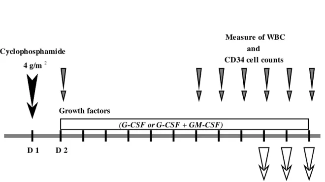
Study design.
As shown on Fig.1, this was a randomized, open-label, unicenter study, including two parts (study A and study B). The primary objective of this study was to achieve 10x106 CD34
cells/kg in a minimal number of leukapheresis. Calculation of the number of subjects was based on the fact that the percentage of the subjects reaching more than 10x106 CD34 cells/kg
in the combination arm will be twice than that observed in the single-cytokine arm. With a power of 90% and a 5% risk, the number of patients is 25 per arm. For that reason, we decide to include 30 patients per arm due to the possibility of unevaluable patients. There was no stratification. The secondary objectives of this study include the tolerance, the factors influencing the mobilization of progenitor cells, the percentage of myeloma cells mobilized in PB, and the difference of mobilization at the level of 5x10⁶ CD34 cells/kg between the 2
arms. In study A, 60 patients were randomized to receive after CY (4 g/m2) the
co-administration of 5 µg/kg/day (d) of rHu-GM-CSF (Molgramostim, Schering-Plough, Kenilworth, NJ, USA) and 5 µg/kg/d of rHu-G-CSF (Filgrastim, Amgen, Thousand Oaks, CA) (G + GM-CSF, total HGF dose = 10 µg/kg/d) compared to 5 µg/kg/d of G-CSF (Filgrastim). In study B, 60 subsequent patients have received the same design of treatment with 2.5 µg/kg/d of GM-CSF and 2.5 µg/kg/d of G-CSF (G + GM-CSF, total dose of HGF = 5 µg/kg/d) compared to 5 µg/kg/d of G-CSF. The 4 g/m2 CY was administred by intravenous
(IV) route to all the patients followed 24 hours later by the HGF administration subcutaneously until the last day of leukapheresis (LK). For patients receiving the 2 HGFs, they were injected in separate sites.
Patient Eligibility
Written informed consent was obtained from all patients enrolled in the study. Patients with histologically confirmed history of cancer and requiring a high dose myeloablative
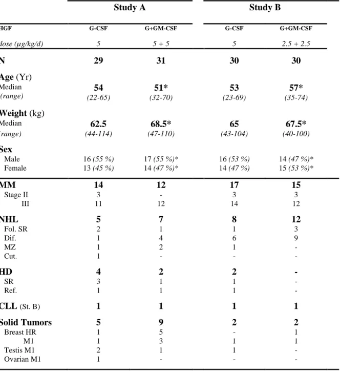
chemotherapy with PBPC rescue were eligible. Disease status and demographic characteristics are detailed in Table I. Inclusion criteria were: age between 18 and 65 years, performance status < 2, half-life expectancy of at least 6 months, normal organ function as defined by serum creatinine, transaminase <2 times normal range, cardiac ejection fraction within the institution's normal range, and normal blood count (ANC > 1.5 x 10⁹/L, platelets > 100 x
109/L) at the day of mobilization chemotherapy. Exclusion criteria were: patients who had
received HGF within 2 weeks before study entry, patients with other malignancies within 5 past years, HIV1 or 2, HTLV 1 or 2, hepatitis C sero-positivity, hepatitis B positive virological status, patients with psychiatric, addictive or any disorder which compromised their ability to give truly informed consent.
Stem cell collection.
White blood count (WBC) were assessed daily after the beginning of chemotherapy. When it reaches 0.5 109/L, the percentage of circulating CD34 cells was monitored daily according
to the ISHAGE protocol, by using a phycoerythrin-conjugated (PE) antibody to CD34 (Q-Bend 10, Immunotech, Marseille, France) (38). For each labelled sample, 30,000 events were recorded on forward-versus-side scatter dot-plot using a FACScan (Becton-Dickinson, San-Jose, CA, USA). The estimated number of CD34 cells/kg that can be collected in one LK was calculated by the central laboratory according to the following formula: (%CD34 x WBC x 8/kg body weight (8 being a coefficient corresponding approximately to 2 blood volumes in L). Then, a LK was started if the estimation of harvestable CD34 number was > 106 CD34/kg.
If the estimation of harvestable CD34 cell number was below 106 CD34/kg the collection was
delayed. A maximum of three LK was attempted to reach a minimum target of 5 x 10⁶
CD34/kg. For MM patients, at this time, we performed a CD34 selection in order to decrease contaminating tumoral cells. For that reason, due to the loss of cells during this procedure, we attempt to collect 10 x 106 CD34/kg. Large volume LK was performed through a dual
peripheral venous puncture using a Cobe Spectra separator version 4 (CS 3000; Baxter Healthcare Corp Lakewood, Co). Median time of processing was 5 hours and the mean volume of processed blood ranged between 12 to 16 L.
Analysis of product of leukapheresis.
The determination of CD34 cell content in the LK was carried out according to the following procedure. Incubation of 106 cells with 30% AB serum (100 µL) for 10 mn, two
washings with PBS, suspension in a final volume of 100 µL, incubation with 20 µL of monoclonal antibody CD34 conjugated with PE, or 20 µL of murine IgG recognizing no human antigen and conjugated with PE as a negative control, or 5 µL anti-CD 45 conjugated with PE as a positive control for 30 mn at 4°C, cell lysis followed by two washings and resuspension in 200 µL PBS. Cells are analysed with a FACSCAN cytometer. At least 30,000 events were acquired for each sample.
In patients with Multiple Myeloma (MM), myeloma cells were enumerated by FACS analysis using the FITC-conjugated MI15 (anti-CD138 mAb (39). 106 cells were incubated
with 1 µg MI15FITC (10 µL) or to 1 µg IgGFITC negative control (20µL) and fluorescence was
analysed with FACSCAN cytofluorometer. No assesment of CD34 cells was performed on the LK product.
Autologous stem cell transplantation (ASCT).
After a resting time of a maximum of 8 weeks, patients received high dose chemotherapy (HDC) followed by autologous PBPC transplant. MM patients were scheduled to receive two subsequent HDC and PBPC transplants. The first HDC was 140 mg/m2 Melphalan. At least
three months later, patients received the second HDC consisting of 200 mg/m2 melphalan or
140mg/m² melphalan and total body irradiation delivering 12 Gy followed by 5 µg/kg/d G-CSF support administrated from day 2 after HDC and until ANC > 1.5 x 10⁹/L on two
consisting on 300 mg/m2 BCNU, Etoposide 200 mg/m2/d for four days, Cytarabine 200
mg/m2/d for four days and Melphalan 140 mg/m2 without post-transplant G-CSF support.
Breast cancer, testis and ovarian cancer patients were scheduled to receive one or two HDC and PBPC transplant. The following parameters of haemotologic recovery were analyzed: day of ANC > 0.5 x 10⁹/L, day of platelet count > 50 x 109/L, cost estimation of the whole
transfusion procedure and duration of hospitalization. Statistical analysis.
An intent-to-treat analysis was done taking into account all patients who received CY and HGFs. A Wilcoxon non-parametric test was carried out to assess the comparability of group at randomization. The comparison of number of LK per group were evaluated by an exact Fischer's test. The median values of CD34 cells harvested in the different groups were compared by a Mann-Withney non-parametric test. The comparison of distribution of patients with 1 to 3 LK to reach the pre-defined threshold was done by the Chi 2 test. If none LK was performed it is considered as mobilization failure and not evaluated. Data were analysed using SAS (Statistical Analysis System) software version 6.08.
RESULTS
Patient population (Table I)
One hundred twenty consecutive patients were enrolled and randomized in the department. All patients had a disease requiring high-dose chemotherapy and PBPC transplantation as salvage therapy. They were: 58 (48%) MM patients (stage II or III), 32 (27%) non-Hodgkin Lymphoma (NHL) (large cell type with IPI≥3 or sensitive relapse), 8 (7%) Hodgkin disease (HD) (sensitive relapse or refractory), and 4 (3%) chronic lymphocytic leukemia (CLL) (Binet stage B and C), and 18 (15%) solid tumors (ST) including 13 metastatic (or high-risk) breast cancer, 4 metastatic testis cancer, 1 metastatic ovarian cancer. All patients were newly diagnosed and received a range of 3 to 16 courses of chemotherapy before PBSC mobilization lasting less than one year.
Sixty patients were included in study A (5 µg/kg/d G-CSF plus 5 µg/kg/d GM-CSF versus 5 µg/kg/d G-CSF): 26 MM, 12 NHL, 6 HD, 2 CLL, 14 ST.
Sixty subsequent patients were included in study B (2.5 µg/kg/d G-CSF plus 2.5 µg/kg/d GM-CSF versus 5 µg/kg/d G-CSF): 32 MM, 20 NHL, 2 HD, 2 CLL, 4 ST.
4 were not evaluable for the following events at time of chemotherapy: infection at time of chemotherapy (1 case), disease progression or relapses (2 cases), death before mobilization (1 case). Patients characteristics were comparable, particularly age, sex ratio, body weight and blood cell count before mobilization.
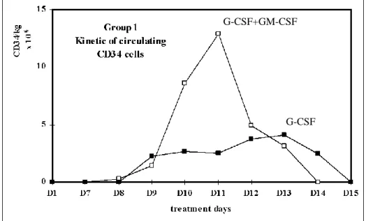
Kinetics of mobilization of circulating CD34 PBPC
In study A, the circulating CD34 cells appear in the peripheral blood (PB) respectively between day 9 to 12 in the G-CSF group and day 9 to 13 in G-CSF plus GM-CSF group. In study B, the circulating CD34 cells appear between day 9 to 11 in the G-CSF group and day 9 to 13 in G-CSF plus GM-CSF group (data not shown).
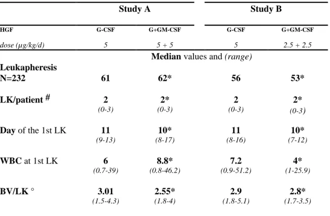
There was a good correlation between the estimated number of CD34 cells and the collected number of CD34 cells measured (Fig. 2). Parameters of LK procedures are shown in Table II. A total of 232 cytaphereses were performed: 123 in study A, and 109 in study B.
One to three large volume LK were performed to harvest the required number of CD34 cells. The following parameters of LK were analysed: number of LK processed per patient (LK/patient), the day of first LK after CY, WBC at the first LK, and the median number of blood volumes treated per LK (BV/LK). None of these parameters were statistically different between the randomized groups.
Number of patients achieving a harvest of 5 x 10⁶ and10 x 106 CD34 cells/kg
In study A, 123 LK were performed (61 in the G-CSF group, 62 in the G + GM-CSF group). The distribution of patients undergoing one, two or three LK were identical in both groups. Among the patients who have undergone LK, the proportion of patients reaching a minimum target of 5 x 10⁶ CD34 cells/kg was not statistically different between the G-CSF
arm (21/27, 77.8%) and the G-CSF plus GM-CSF arm (25/29, 86.2%). When the objective of CD34 collection was 10 x 106 cells/kg, the proportion of patients reaching this target was
significantly higher in the G-CSF plus GM-CSF arm (21/29, 72.4%) compared to the G-CSF arm (11/27, 40.7%: P= .00006).
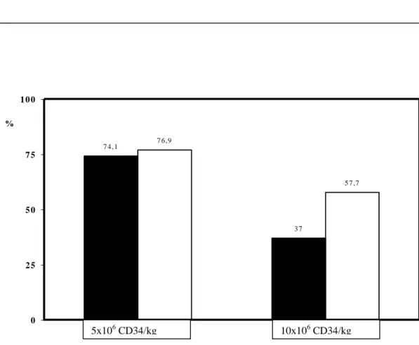
In study B, a total of 109 LK were performed (56 in G-CSF arm and 53 in G-CSF plus GM-CSF arm). The distribution of patient undergoing one, two or three LK are identical in both groups. Among the patients who have undergone LK, the proportion of patients reaching a minimum target of 5 x 10⁶ CD34 cells/kg was not statistically different between the G-CSF
arm (20/27, 74.1%) and the G-CSF plus GM-CSF arm (20/26, 76.9%). When the objective of CD34 collection was 10 x 106 cells/kg, the proportion of patients reaching this target was
significantly higher in the G-CSF plus GM-CSF (arm 15/26, 57.7%) compared to the G-CSF arm (10/27, 37%: P= .003) (Fig 3).
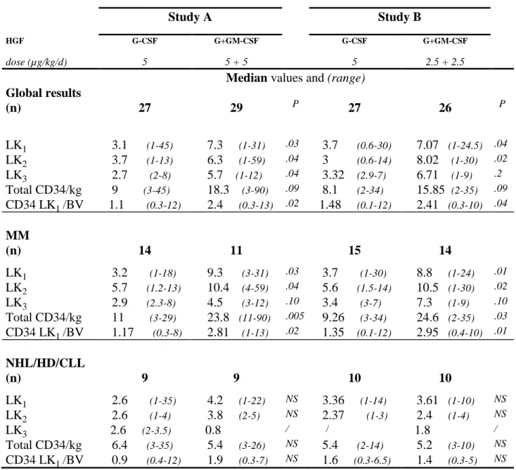
CD34 cell harvest (Table III).
In study A, the median number of CD34 cellsx 106 /kg collected in LK1, LK2 or LK3 in
the G-CSF and G + GM-CSF groups is respectively 3.1 (range 0.9-44.7), 3.7 (1.2-13.1), 2.7 (1.6-7.6) versus 7.3 (1-30.9), 6.28 (0.8-59), 5.72 (0.8-12.5). The total number of CD34 cells x106 /kg harvested is 2 fold higher in the G + GM-CSF group 18.3 90) compared to 9
(3-45) in the G-CSF group (P= .09).
In study B, the median number of CD34 cells x 106 /kg collected at LK1, LK2, LK3 in
the G-CSF and G + GM-CSF groups is respectively 3.7 (range 0.6-30.1), 3.0 (0.6-14.3), 3.3 (2.9-7) versus 7.07 (0.9-24.5), 8.02 (0.7-29.8), 6.71 (0.6-9.3). The total number of CD34 x 106
/kg harvested is about 2 fold higher in the G + GM-CSF group 15.85 (2.5-34.9) compared to 8.1 (1.6-33.9) in the G-CSF group (P= .09).
To avoid a bias due to the variable number of LK, and due to the variable blood volume treated during one LK, we have evaluated the efficacy of CD34 PBPC collection by calculating the number of CD34 cells x 106 /kg collected during the first LK according to the
number of blood volume processed (CD34 LK1/BV). In study A, this parameter was again increased about 2 fold in the G-CSF plus GM-CSF group 2.4 13) compared to 1.1 (0.3-12) in the G-CSF group (P= .02). In study B, the same observation could be done in the G + GM-CSF group: 2.52 (0.3-10.2) versus 1.37 (0.2-11.6) in the G-CSF group (P= 0.04).
Patients with MM
In the study A : 14 MM patients received G-CSF and 12 received G-CSF plus GM-CSF (11 are evaluable). The total CD34 cells x 106 /kg harvested is 2.2 fold higher in the G-CSF plus
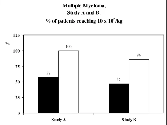
GM-CSF group compared to the G-CSF group respectively 23.8 (11.1-89.9) versus 11.02 (3.2-29.2) (P= .005). The CD34 LK1/BV is 2.4 fold higher in the G-CSF plus GM-CSF group 2.81 (0.9-13.3) versus 1.17 (0.3-8.1) in the G-CSF group (p=0.02). In the MM patients, we recommended to collect a minimum target of 10 x 106 CD34 cells/kg before the CD34
positive selection. In these conditions, 11/11 (100%) of patients receiving the G-CSF plus GM-CSF combination reach this target compared to only 8/14 (57%) in the G-CSF group (P = .02) (Fig 4). The same observation were done in patients of study B, 17 MM patients received G-CSF (15 are evaluable) and 15 received G-CSF plus GM-CSF (14 are evaluable). The total CD34 cells x 106 /kg harvested is about 2.5 fold higher in the G-CSF plus GM-CSF group
(24.6 , range: 2.5-34.9) compared to the G-CSF group (9.26, range: 2.8-33.9) (P= .03). The CD34 LK1/BV is 2.2 fold higher in the G-CSF plus GM-CSF group 2.95 x 10⁶/kg (0.4-10.2)
compared to 1.35 x 10⁶/kg (0.1-11.6) (P= .014). 12/14 (86%) of patients receiving the G-CSF
plus GM-CSF combination reach the defined target of 10 x 106 CD34+ cells/kg compared to
only 7/15 (44%) in the G-CSF group (P= .03) (Fig. 4).
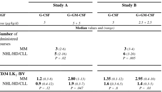
Patients with other lymphoid malignancies (NHL, HD and CLL)
Mobilization by CY and G-CSF plus GM-CSF did not significantly improve the amount of collected CD34 cells compared to the CY and G-CSF mobilization especially the CD34 LK1/BV was not significantly increased (Table III). The comparison between myeloma and lymphoma patients is detailed in Table IV. MM patients received less chemotherapy courses (median 3, range: 2-6) while lymphoma patients received a median of 6 courses (median 2-16) chemotherapy courses (P = .02). As shown on Fig 5, no differences were seen between the two arms (G-CSF and G+GM-CSF) in the group of patients with more than one year of chemotherapy before mobilization.
In study A, among patients receiving G-CSF, the CD34 LK1/BV is not significantly higher in MM 1.2 x 10⁶ (0.3-8) than in Lymphoma 0.9 x 10⁶ (0.4-12) (P= .12) while among
patients receiving G-CSF plus GM-CSF, the CD34 LK1/BV is 1.5 higher in MM (2.8 x 10⁶ ,
range :1-13) than in Lymphoma (1.9 x 10⁶ , range: 0.3-7) (P= .047). In study B, same
observations was made, the CD34 LK1/BV is not significantly higher in MM than in lymphoma (P= .8) while among patients receiving G-CSF plus GM-CSF, the CD34 LK1/BV
is 2.1 higher in MM (2.95 x 10⁶ , range: 0.4-10) than in Lymphoma (1.4 x 10⁶ , range: 0.3-5)
(P= .01).
Patients with solid tumors
Due to of low number of patients with solids tumors and their heterogenicity, none comparison was made between the two groups of treatment.
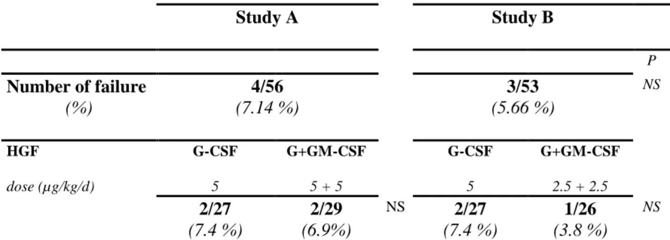
Mobilization failure
The percentage of patient that failed to mobilize sufficient PBSC was similar in study A (4/56, 7%) and in study B (3/53, 6%). None difference has been observed according to the HGF treatment group (Table V).
Adverse events
No serious adverse events were associated with the administration of cyclophosphamide at
4 g/m2. Grade 1 and 2 nausea and vomitis were easily controlled by systematic administration
of andosetron. In study A, three patients with full dose of G + GM-CSF experienced grade 2 symptoms (chill, bone and muscle pains, tachycardia) within the first two days. These patients stopped themself the GM-CSF. No patient experienced grade 1 and 2 toxicities during HGF treatment in the study B.
Effects of HGF on residual plasma cells (PC) mobilization in MM patients
In study A, an estimation of residual PC in product of LK was possible in 19 patients. The number of PC was 4.6 x 10⁶/kg in the G-CSF group and 5.39 x 10⁶/kg in the G-CSF plus
GM-CSF group (P = NS).
In study B, the numeration of residual PC in LK was done in 21 patients with MM. The number of PC was 2.58 x 10⁶/kg in the G-CSF group and 6.19 x 10⁶/kg in the G + GM-CSF
group (P = NS).
When comparing the total PC collected to the total CD34 cells collected in each patient, the PC/CD34 ratio is in study A 1 for 2.4 in G-CSF group and 1 for 4.42 in the G-CSF plus
GM-CSF group (P = NS). In study B, the PC/CD34 ratio is 1 for 3.6 in G-CSF group and 1 for 3.97 in the G-CSF plus GM-CSF group (P = NS) showing no enhancement of PC mobilization by G-CSF plus GM-CSF regimen.
Hematologic recovery after myeloablative treatment
97 patients were transplanted: 49 patients in Study A, 48 in study B. All MM patients were scheduled to receive a transplantation with a graft purged by CD34 positive selection. In study A, the CD34 positive selection was done in only 9/14 (64.3%) MM patients receiving G-CSF and 11/11 (100%) in patients receiving G-CSF plus GM-CSF (P= .03). In study B, this regimen was done in only 8/15 (53.3%) patients receiving G-CSF and 13/14 (92.8%) patients receiving G-CSF plus GM-CSF (P= .02).
Because of purification and double HDC in MM, the median number of CD34 cells infused was similar in all group of treatment and ranged between 3.78 to 4.32 x 10⁶/kg. As
expected, all patients had an engraftment, the median times to reach an ANC of > 0.5 x 10⁹/L
and platelet > 50 x 109/L were respectively 11 days (range 9 to 17) and 15 days (8-34). The
transfusion cost is estimated at 1,619 $ (265-8,752). The median duration of hospitalisation was 24 days (16-94).
DISCUSSION
These two randomized studies demonstrate two major points:
-the combination of GM-CSF plus G-CSF following CY results in a 2 fold enhancement of the number of PBPC, in a safe and efficient manner for an homogenous group of newly diagnosed patients;
-the concomitant administration of these two HGFs, had additive effects, as observed by the superiority of the combination at equivalent doses (5µg/kg vs 2.5+2.5µg/kg).
Most of the patients included in these studies, achieved a minimal target of 5 x 10⁶
CD34/kg, but more patients receiving G-CSF plus GM-CSF achieved a target of 10 x 106
CD34/kg. This result was obtained with a median of 2 LK. No adverse effects related to LK procedures have been reported. Although CD34 cell dose is currently considered to be the prefered indicator of satisfactory engraftment, the optimal CD34 cell dose for autologous transplantation remains to be defined. A collection of a threshold value of at least 3 to 5 x 10⁶
CD34/kg is recommended to insure rapid and successful engraftment and long term hematological recovery. A dose-response relationship is evident between the number of CD34 cells per kilogram infused and neutrophil and platelet engraftment kinetics, decrease hospital stay and cost (40-42). Infusion of very high dose superior to 15 x 106 CD34/kg was shown to
be associated with a shorter time for platelet recovery compared to that observed for patients receiving between 2.5 and 15 x 106 CD34/kg (43). However, Dercksen et al found no
difference for neutrophil recovery when the number of CD34+ cells were superior to 6 x 10⁶
cells/kg (44). Recently, different groups observed a linkage between the number of CD34 harvested and reinfused, and survival or duration of the response in different diseases including breast cancer and multiple myeloma (45, 46). The mecanism of such effect remains uncertain, but may be linked to a high number of immunocompetent cells collected in addition to the stem cells, as suggested by Porrata et col. who observed a correlation between
progression-free survival and the number of lymphocytes readministered (46). In addition, GM-CSF had several immune activities, including differentiation of dendritic cells from monocytes and it has been associated to different strategies of immune therapy including anti-tumoral vaccination (46, 47). These observations may represent a rationale for re-discovering the role of GM-CSF for mobilizing both haematopoietic stem cells and immune cell effectors (48,49). The response we made through this study appears important to define the minimal efficient dose for a combination of G-CSF and GM-CSF, prior to add other compounds for mobilization such as selective CXCR4 antagonists (15).
We observed that the efficacy of dual HGFs was more evident in patients having MM, where 2.5 fold higher PBSC are collected with the combination of HGFs. However, no difference have been observed in the group of lymphoma’s patients. The impressive results observed with MM compared to the lymphoma group, are probably due to prior chemotherapy compared to the lymphoma group. Some investigations have highlighted the importance of prior exposure to the specific stem cell toxicity of chemotherapy containing alkylating agent and/or anthracycline and their impact on mobilization efficacy (50). Haas R and col. suggest the same findings showing a loss of 0.2 x 10⁶ CD34+ cells/kg per chemotherapy course
including alkylating agent (51). In that way, it seems that 6 courses of chemotherapy appears as a significant critical threshold. This feature is probably not corrected by the HGF treatment, including the co-administration of G-CSF and GM-CSF. The dexamethasone administred preferentielly to MM patients should also stimulate the hematopoïesis in synergy to HGFs. The role of glucocorticoids, espacially dexamethasone, on proliferation of haemotopoïetic progenitors has been suggested in vitro (52, 53). This particular benefit observed in MM patients and the fact that we use a concomitant administration, may represent the differences observed between the literature and our results.
The timing of harvest is one of the major parameter of successfull yield of CD34+ PBSC. The criteria used to define the optimal harvesting day was the peripheral WBC or mononuclear cells. But, these criterias are associated with a weak correlation to the CD34 cells amount in the product of leukapheresis (54). At the present time, the absolute number of circulating CD34 cells is correlated closely with the CD34 cell yield of the corresponding leukapheresis product. The usual critical threshold of circulating CD34 cells as indicator of onset of cytapheresis is 40 CD34 cells per µL (55). But, this method doesn't provide an estimation of CD34 cells collection. We use an estimation of the CD34 PBSC yield determined just before each cytapheresis to give directly an estimation of the final collect of CD34+ PBSCs.
In addition to PBSC mobilization, the mobilization of tumoral cells remains likely. The role of these residual tumoral cells in the graft for the relapse after transplantation is still controversial (56, 57). In this study, we evaluate the tumors cells co-mobilization in MM patients. We have shown no trend to mobilize more tumoral cells with G-CSF plus GM-CSF regimen than that observed with G-CSF alone. All patients recovered after myeloablative treatment.
As expected, no clear benefit appears in terms of engraftment, transfusion cost according to HGF regimen. The median number of CD34 cells infused to the patients in all treatment group is comparable (3.8 to 4.32 x 10⁶ CD34+/kg). The majority of the patients having MM or
breast cancer were scheduled to receive a double intensive chemotherapy followed by a CD34 immunoselected graft rescue. This schedule was possible more frequently in the G-CSF plus GM-CSF mobilization regimen (93 to 100% of patients) than in G-CSF group (53 to 64% of patients).
Future insight of graft manipulation as high level tumor cell purging, genetic manipulation, monocytes mobilization in view of anti-tumoral vaccination by dendritic cells, ex-vivo
expansion will invariably result in loss of a substantial fraction of hematopoietic progenitor cells harvested, a situation that needs to collect higher number of hematopoietic stem cells. The combination of cytokines, including GM-CSF may represent a particular interesting combination, in addition to the effects of GM-CSF on the immune system, as demonstrated in vaccination programs.
REFERENCES
1. Attal M , Harousseau JL, Stoppa AM et al. A prospective, randomized trial of autologous bone marrow transplantation and chemotherapy in multiple myeloma. N Engl J Med 1996; 335: 91-97.
2. Harousseau JL.Stem cell transplantation in multiple myeloma (0, 1, or 2).Curr Opin Oncol 2005 ; 17: 93-98.
3. Gorin NC. Autologous stem cell transplantation in hematological malignancies. Springer Semin Immunopathol 2004 ; 26: 3-30.
4. Philip T, Guglielmi C, Hagenbeek A et al. Autologous bone marrow transplantation as compared with salvage chemotherapy in relapses of chemotherapy-sensitive non-Hodgkin's lymphoma. N Engl J Med. 1995; 333: 1540-1545.
5. Gratwohl A, Schmid O, Baldomero H, et al. Accreditation Committee of the European Group for Blood and Marrow Transplantation. Haematopoietic stem cell transplantation (HSCT) in Europe 2002. Changes in indication and impact of team density. A report of the EBMT activity survey. Bone Marrow Transplant 34: 855-875, 2004.
6. Demirer T, Buckner CD, Bensinger WI. Optimization of stem cell mobilization. Stem Cells 1996; 14 : 106-116.
7. Lane TA, Law P, Maruyama et al. Harvesting and enrichment of HPC mobilized into the peripheral blood of normal donors by GM-CSF or G-CSF: potential role in allogenic transplantation. Blood 1995; 85: 275-282.
8. Peters WP, Rosner G, Ross M et al. Comparative effect of GM-CSF and G-CSF on priming of peripheral blood progenitor cells for use with autologous bone marrow after high dose chemotherapy. Blood 1993, 81: 1709-1719.
9. Gazitt Y. Comparison between granulocyte colony-stimulating factor and granulocyte-macrophage colony-stimulating factor in the mobilization of peripheral blood stem cells. Curr Opin Hematol 2002, 9 : 190-198.
10. Lyman SD. Biologic effects and potential clinical applications of Flt3 ligand. Curr Opin Hematol 1998; 5: 192-196.
11. Haas R, Ehrhardt R, Witt B et al. Autografting with peripheral blood stem cells mobilized by sequential interleukin-3/granulocyte-macrophage colony-stimulating factor following high-dose chemotherapy in non-Hodgkin's lymphoma. Bone Marrow Transplant 1993; 12: 643-9.
12. Stiff P, Gingrich R, Luger S,A et al. Randomized phase 2 study of PBPC mobilization by stem cell factor and filgrastim in heavily pretreated patients with Hodgkin's disease or non-Hodgkin's lymphoma. Bone Marrow Transplant 2000; 26: 471-481.
13. Prosper F, Sola C, Hornedo J et al. Mobilization of peripheral blood progenitor cells with a combination of cyclophosphamide, r-metHuSCF and filgrastim in patients with breast cancer previously treated with chemotherapy. Leukemia 2003; 17: 437-441.
14. Waller CF, von Lintig F, Daskalakis A, et al. Mobilization of peripheral blood progenitor cells in patients with breast cancer: a prospective randomized trial comparing rhG-CSF with the combination of rhG-CSF plus rhEpo after VIP-E chemotherapy. Bone Marrow Transplant. 1999; 24: 19-24.
15. Liles WC, Rodger E, Broxmeyer HE, et al. Augmented mobilization and collection of CD34+ hematopoietic cells from normal human volunteers stimulated with granulocyte-colony-stimulating factor by single-dose administration of AMD3100, a CXCR4 antagonist. Transfusion. 2005; 45: 295-300.
16. Ozer H, Armitage JO, Bennett CL, et al. 2000 update of recommendations for the use of hematopoietic colony-stimulating factors: evidence-based, clinical practice guidelines. J Clin Oncol 2000; 18: 3558-3585.
17. Andre M, Baudoux E, Bron D, et al. Phase III randomized study comparing 5 or 10 microg per kg per day of filgrastim for mobilization of peripheral blood progenitor cells with chemotherapy, followed by intensification and autologous transplantation in patients with nonmyeloid malignancies. Transfusion. 2003; 43: 50-57.
18. Jones HM, Jones SA, Watts, MJ, et al. The effects of variation of the dose and schedule of G-CSF administration after low dose cyclophosphamide (1.5 g/m2) on
both neutrophil and peripheral blood haemopoietic progenitor cell levels. Blood 1993; 82 (suppl 1) 233a.
19. Carrion R, Serrano D, Gomez-Pineda A, Diez-Martin JL.A randomised study of 10 microg/kg/day (single dose) vs 2 x 5 microg/kg/day (split dose) G-CSF as stem cell mobilisation regimen in high-risk breast cancer patients. Bone Marrow Transplant 2003; 32: 563-567.
20. Green MD, Koelbl H, Baselga J, et al. A randomized double-blind multicenter phase III study of fixed-dose single-administration pegfilgrastim versus daily filgrastim in patients receiving myelosuppressive chemotherapy. Ann Oncol. 2003; 14: 29-35. 21. Siena S, Schiavo R, Pedrazzoli P, Carlo-Stella C: Therapeutic relevance of CD34+
cell dose in blood cell transplantation for cancer therapy. J Clin Oncol 2000: 18:1360-1377.
22. Weaver CH, Hazelton B, Birch R, et al. An analysis of engraftment kinetics as a function of the CD34 content of peripheral blood progenitor cell collections in 692 patients after the administration of myeloablative chemotherapy. Blood 1995; 86: 3961-3969.
23. Schulman KA, Birch R, Zhen B, et al. Effect of CD34+ cell dose on resource utilization in patients after high-dose chemotherapy with peripheral-blood stem-cell support. J Clin Oncol 1999; 17: 1227.
24. Winter JN, Lazarus HM, Rademaker A, et al. Phase I/II study of combined granulocyte stimulating factor and granulocyte-macrophage colony-stimulating factor administration for the mobilization of hematopoietic progenitor cells. J Clin Oncol 1996, 14: 277-286.
25. Charrier S, Chollet P, Bay JO, et al. Hematological recovery and peripheral blood progenitor cell mobilization after induction chemotherapy and GM-CSF plus G-CSF in breast cancer. Bone Marrow Transplantation 2000; 252: 705-710.
26. Bolwell B, Goormastic M, Yanssens T, et al. Comparison of G-CSF with GM-CSF for mobilizing peripheral blood progenitor cells and for enhancing marrow recovery after autologous bone marrow transplant. Bone Marrow Transplant 1994; 14: 913-918.
27. Spitzer G, Adkins D, Mathews M, et al. Randomized comparison of G-CSF + GM-CSF vs G-GM-CSF alone for mobilization of peripheral blood stem cells: effects on hematopoietic recovery after high-dose chemotherapy. Bone Marrow Transplant 1997; 20: 921-930.
28. Weisdorf D, Miller J, Verfaillie C, et al. Cytokine-primed bone marrow stem cells vs peripheral blood stem cells for autologous transplantation : a randomized comparison of GM-CSF vs G-CSF. Biol Blood Marrow Transplant 1997, 3: 217-223.
29. Hohaus S, Martin H, Wassmann B, et al. Recombinant human granulocyte and granulocyte-macrophage colony-stimulating factor (G-CSF and GM-CSF) administered following cytotoxic chemotherapy have a similar ability to mobilize peripheral blood stem cells.Bone Marrow Transplant 1998; 22: 625-630.
30. Comenzo RL, Sanchorawala V, Fisher C, et al. Intermediate-dose intravenous melphalan and blood stem cells mobilized with sequential GM+G-CSF or G-CSF alone to treat AL (amyloid light chain) amyloidosis. Br J Haematol 1999; 104 : 553-539.
31. Siena S, Bregni M, Gianni AM. Mobilization of peripheral blood progenitor cells for autografting: chemotherapy and G-CSF or GM-CSF. Baillieres Best Pract Res Clin Haematol 1999; 12: 27-39.
32. Madero L, Gonzalez-Vicent M, Molina J, et al. Use of concurrent G-CSF + GM-CSF vs G-CSF alone for mobilization of peripheral blood stem cells in children with malignant disease. Bone Marrow Transplant 2000; 26: 365-369.
33. Weaver CH, Schulman KA, Wilson-Relyea B, et al. Randomized trial of filgrastim, sargramostim, or sequential sargramostim and filgrastim after myelosuppressive
chemotherapy for the harvesting of peripheral-blood stem cells. J Clin Oncol 2000; 18: 43-53.
34. Koc ON, Gerson SL, Cooper BW, et al. Randomized cross-over trial of progenitor-cell mobilization: high-dose cyclophosphamide plus granulocyte colony-stimulating factor (CSF) versus granulocyte-macrophage colony-stimulating factor plus G-CSF. J Clin Oncol 2000; 18: 1824-1830.
35. Gazitt Y, Callander N, Freytes CO, et al. Peripheral blood stem cell mobilization with cyclophosphamide in combination with G-CSF, CSF, or sequential GM-CSF/G-CSF in non-Hodgkin's lymphoma patients: a randomized prospective study. J Hematother Stem Cell Res 2000; 9: 737-748.
36. Gazitt Y, Shaughnessy P, Liu Q. Differential mobilization of CD34+ cells and lymphoma cells in non-Hodgkin's lymphoma patients mobilized with different growth factors. J Hematother Stem Cell Res 2001; 10: 167-176.
37. Armitage JO. Emerging applications of recombinant human granulocyte-macrophage colony-stimulating factor. Blood 1998; 92: 4491-4508.
38. Sutherland DR, Anderson L, Keeney M, et al. The ISHAGE guidelines for CD34+ cell determination by flow cytometry. International Society of Hematotherapy and Graft Engineering. J Hematother 1996; 5: 213-226.
39. Costes V, Magen V, Legouffe E, et al. The Mi15 monoclonal antibody (anti-syndecan-1) is a reliable marker for quantifying plasma cells in paraffin-embedded bone marrow biopsy specimens. Hum Pathol 1999; 30: 1405-1411.
40. Duggan PR, Guo D, Luider J, et al. Predictive factors for long-term engraftment of autologous blood stem cells. Bone Marrow Transplant. 2000; 26: 1299-1304.
41. Uyl-de Groot CA, Huijgens PC, Rutten FF: Colony-stimulating factors and peripheral blood progenitor cell transplantation, benefits and costs. Pharmaco Economics 1996; 10: 23-35.
42. Bensinger W, Appelbaum F, Rowley S, et al. Factors that influence collection and engraftment of autologous peripheral-blood stem cells. J Clin Oncol 1995; 13: 2547-2555.
43. Ketterer N, Salles G, Raba M, et al. High CD34 (+) cell counts decrease hematologic toxicity of autologous peripheral blood progenitor cell transplantation. Blood 1998; 91: 3148-3155.
44. Dercksen MW, Rodenhuis S, Dirkson MK, Schaasberg WP, Baars JW, van der Waal E, Slaper-Cortenbach IC, Pinedo HM, Von dem Borne AE, van der Schoot CE, Gerritsen WR: Subsets of CD34+ cells and rapid hematopoietic recovery after peripheral-blood stem-cell transplantation. J Clin Oncol 1995; 13: 1922-1932.
45. Wahlin A, Eriksson M, Hultdin M. Relation between harvest success and outcome after autologous peripheral blood stem cell transplantation in multiple myeloma. Eur J Haematol. 2004; 73: 263-268.
46. Porrata LF, Gertz MA, Geyer SM, et al. The dose of infused lymphocytes in the autograft directly correlates with clinical outcome after autologous peripheral blood hematopoietic stem cell transplantation in multiple myeloma. Leukemia. 2004; 18: 1085-1092.
47. O’Neil DW, Adams S, Bhardwaj N. Manipulating dendritic cell biology for the active immunotherapy cancer. Blood 2004; 104: 2235-2246.
48. Dranoff G. GM-CSF-based cancer vaccines. Immunol Rev. 2002; 188: 147-154. 49. Gazitt Y. Immunologic profiles of effector cells and peripheral blood stem cells
mobilized with different hematopoietic growth factors. Stem Cells. 2000; 18: 390-398.
50. Koumakis G, Vassilomanolakis M, Hatzichristou H, et al. Predictive factors affecting mobilization and peripheral blood stem cell (PBSC) collection using single apheresis (SA) for rescuing patients after high-dose chemotherapy (HD.CHE) in various malignancies. Bone Marrow Transplant 1996; 18: 1065-1072.
51. Haas R, Möhle R, Frühauf S, Goldschmidt H, et al. Patient characteristics associated with successful mobilizing and autografting of peripheral blood progenitor cells in malignant lymphoma. Blood 1994; 83: 3787-3794.
52. von Lindern M, Zauner W, Mellitzer G, et al. The glucocorticoid receptor cooperates with the erythropoietinreceptor and c-Kit to enhance and sustain proliferation of erythroid progenitors in vitro. Blood 1999; 94: 550-559.
53. Dror Y, Ward AC, Touw IP, Freedman MH. Combined corticosteroid/granulocyte colony-stimulating factor (G-CSF) therapy in the treatment of severe congenital neutropenia unresponsive to G-CSF: Activated glucocorticoid receptors synergize with G-CSF signals. Exp Hematol. 2000; 28: 1381-1389.
54. Armitage S, Hargreaves R, SamsonD, et al. CD34 counts to predict the adequate collection of peripheral blood progenitor cells. Bone Marrow Transplant 1997; 20: 587-591.
55. Möhle R, Murea S, Pförsich M, et al. Estimation of the progenitor cell yield in a leukapheresis product by previous measurement of CD34+ cells in the peripheral blood. Vox Sang 1996; 71: 90-96.
56. Rill DR, Moen RC, Buschle M, et al. An approach for the analysis of relapse and marrow reconstitution after autologous marrow transplantation using retrovirus-mediated gene transfer. Blood 1992; 79: 2694-2700.
57. Vora AJ, Toh CH, Peel J, Greaves M. Use of granulocyte colony-stimulating factor (G-CSF) for mobilizing peripheral blood stem cells:risk of mobilizing clonal
myeloma cells in patients with bone marrow infiltration. Br J Haematol 1994; 86: 180-182.
Figure and Table legends.
Table I. Demographic and disease status of patients at diagnosis. Table II. Parameters of leukapheresis (LK) procedures
Table III. Study A and B: Collection of CD34+ PBSC (x 106/kg) for harvested patients (n) in each group.
Table IV: Differences between MM and Lymphoma
Table V. Mobilization failure in patients receiving the hematopoietic growth factor
Figure 1. Design of the study: mobilization and collection of CD34 cells.
Figure 2. Correlation between the estimated number of CD34 cells (%CD34 x WBC x 8)/kg body weight (8 being a coefficient corresponding approximately to 2 blood volumes in L) and the number of collected CD34 cells.
Figure 3. Percentage of patients reaching 5 or 10x 106 CD34 cells/kg in study B.
Figure 4. Percentage of patients with multiple myeloma and reaching 10x 106 CD34 cells/kg.
Figure 5. Kinetic of circulating CD34 cells in patients with less (A) and more (B) than one year of prior chemotherapy.
Table I. Study A Study B HGF dose (µg/kg/d) G-CSF 5 G+GM-CSF 5 + 5 G-CSF 5 G+GM-CSF 2.5 + 2.5 N 29 31 30 30 Age (Yr) Median (range) (22-65)54 (32-70)51* (23-69)53 (35-74)57* Weight (kg) Median 62.5 68.5* 65 67.5* (range) (44-114) (47-110) (43-104) (40-100) Sex Male 16 (55 %) 17 (55 %)* 16 (53 %) 14 (47 %)* Female 13 (45 %) 14 (47 %)* 14 (47 %) 15 (53 %)* MM 14 12 17 15 Stage II 3 - 3 3 III 11 12 14 12 NHL 5 7 8 12 Fol. SR 2 1 1 3 Dif. 1 4 6 9 MZ 1 2 1 - Cut. 1 - - - HD 4 2 2 - SR 3 1 1 - Ref. 1 1 1 - CLL (St. B) 1 1 1 1 Solid Tumors 5 9 2 2 Breast HR 1 5 - 1 M1 1 3 1 1 Testis M1 2 1 1 - Ovarian M1 1 - - -
MM= Multiple Myeloma, stage according to Salmon-Durie classification. NHL= Non-Hodgkin's Lymphoma, Fol.= follicular, Dif.= diffuse, MZ= mantle zone, Cut.= cutaneous, SR= sensitive relapse, Ref.= refractory. HDK = Hodgkin disease, (all NHL and HDK are stage IV according to Ann Harbor classification). CLL= Chronic Lymphocytic Leukemia, stage according to Binet Classification. Breast, HR= high risk according to Scarff-Bloom/Richardson score > 6 and/or more than 6 loco-regional metastatic nodes, M1 presence of long-distance metastase according to UICC-TNM classification. Ovarian= Ovarian cancer.
Table II. Study A Study B HGF dose (µg/kg/d) G-CSF 5 G+GM-CSF 5 + 5 G-CSF 5 G+GM-CSF 2.5 + 2.5
Median values and (range) Leukapheresis N=232 61 62* 56 53* LK/patient # 2 (0-3) 2* (0-3) 2 (0-3) 2* (0-3) Day of the 1st LK 11 (9-13) 10* (8-17) 11 (8-16) 10* (7-12) WBC at 1st LK 6 (0.7-39) 8.8* (0.8-46.2) 7.2 (0.9-51.2) 4* (1-25.9) BV/LK ° 3.01 (1.5-4.3) 2.55* (1.8-4) 2.9 (1.8-5.1) 2.8* (1.7-3.5)
LK/patient# = Leukapheresis undergone by patient. WBC= White Blood Count x 109/L. BV/LK ° = number of Blood Volume processed during a leukapheresis.
Table III. Study A Study B HGF dose (µg/kg/d) G-CSF 5 G+GM-CSF 5 + 5 G-CSF 5 G+GM-CSF 2.5 + 2.5
Median values and (range) Global results (n) 27 29 P 27 26 P LK1 3.1 (1-45) 7.3 (1-31) .03 3.7 (0.6-30) 7.07 (1-24.5) .04 LK2 3.7 (1-13) 6.3 (1-59) .04 3 (0.6-14) 8.02 (1-30) .02 LK3 2.7 (2-8) 5.7 (1-12) .04 3.32 (2.9-7) 6.71 (1-9) .2 Total CD34/kg 9 (3-45) 18.3 (3-90) .09 8.1 (2-34) 15.85 (2-35) .09 CD34 LK1 /BV 1.1 (0.3-12) 2.4 (0.3-13) .02 1.48 (0.1-12) 2.41 (0.3-10) .04 MM (n) 14 11 15 14 LK1 3.2 (1-18) 9.3 (3-31) .03 3.7 (1-30) 8.8 (1-24) .01 LK2 5.7 (1.2-13) 10.4 (4-59) .04 5.6 (1.5-14) 10.5 (1-30) .02 LK3 2.9 (2.3-8) 4.5 (3-12) .10 3.4 (3-7) 7.3 (1-9) .10 Total CD34/kg 11 (3-29) 23.8 (11-90) .005 9.26 (3-34) 24.6 (2-35) .03 CD34 LK1 /BV 1.17 (0.3-8) 2.81 (1-13) .02 1.35 (0.1-12) 2.95 (0.4-10) .01 NHL/HD/CLL (n) 9 9 10 10 LK1 2.6 (1-35) 4.2 (1-22) NS 3.36 (1-14) 3.61 (1-10) NS LK2 2.6 (1-4) 3.8 (2-5) NS 2.37 (1-3) 2.4 (1-4) NS LK3 2.6 (2-3.5) 0.8 / / 1.8 / Total CD34/kg 6.4 (3-35) 5.4 (3-26) NS 5.4 (2-14) 5.2 (3-10) NS CD34 LK1 /BV 0.9 (0.4-12) 1.9 (0.3-7) NS 1.6 (0.3-6.5) 1.4 (0.3-5) NS
CD34+ PBSC are harvested during one or two or three maximum large volume leukapheresis (LK). LK1 = 1 st Leukapheresis, LK2 = 2 nd Leukapheresis, LK3 = 3 rd Leukapheresis. CD34 LK1/BV= CD34+ cells harvest in the 1 st LK in function of number of blood volume. P values were determined with a Wilcoxon test.
Table IV. Study A Study B HGF dose (µg/kg/d) G-CSF 5 G+GM-CSF 5 + 5 G-CSF 5 G+GM-CSF 2.5 + 2.5
Median values and (range)
Number of administred courses MM NHL/HD/CLL 3 (2-6) 5 (2-16) P = .02 3 (3-4) 6 (3-20) P = .005 CD34 LK1 /BV MM NHL/HD/CLL 1.2 (0.3-8) 0.9 (0.4-12) P = .12 2.80 (1-13) 1.9 (0.3-7) P = .047 1.35 (0.1-12) 1.6 (0.3-6.5) P = .8 2.95 (0.4-10) 1.4 (0.3-5) P = .01
Table V. Study A Study B P Number of failure (%) 4/56 (7.14 %) 3/53 (5.66 %) NS HGF dose (µg/kg/d) G-CSF 5 G+GM-CSF 5 + 5 G-CSF 5 G+GM-CSF 2.5 + 2.5 2/27 (7.4 %) 2/29 (6.9%) NS 2/27 (7.4 %) 1/26 (3.8 %) NS
Number and percentage of patients who received the HGF and impaired the mobilization p = Fisher's exact test
Figure 1. D 2 Measure of WBC CD34 cell counts D 1 Cyclophosphamide 4 g/m Growth factors (G-CSF or G-CSF + GM-CSF) Determination of CD34 cell and in each leukapheresis 2
Figure 3. 7 4 ,1 3 7 7 6 ,9 5 7 ,7 0 2 5 5 0 7 5 1 0 0 % 5x10⁶ CD34/kg 10x106 CD34/kg
Figure 4. Multiple Myeloma, Study A and B, % of patients reaching 10 x 106/kg 57 47 100 86 0 25 50 75 100 125 Study A Study B %
Figure 5. A B G-CSF+GM-CSF G-CSF G-CSF G-CSF + GM-CSF