Development and Mechanistic Analysis of in vivo Liposomal Nanoparticle Delivery of siRNA and mRNA
By
J. Robert Dorkin B.A. Biological Chemistry Swarthmore College, 2006
SUBMITTED TO THE DEPARTMENT OF BIOLOGY IN PARTIAL FULFILLMENT OF THE REQUIREMENTS FOR THE DEGREE OF
DOCTOR OF PHILOSOPHY AT THE
MASSACHUSETTS INSTITUTE OF TECHNOLOGY FEBRUARY 2016
2016 Massachusetts Institute of Technology. All Rights reserved.
Signature of Author: -Certified by: Accepted by:
Signature redacted
,j9
/
J. Robert Dorkin September 21st, 2015Signature redacted
0
Daniel G. AndersonAssociate Professor of Chemical Engineering Thesis Advisor
Signature redacted
OT HNOLOGY
Michael Hemann Associate Professor of Biology Co-Chair, Biology Graduate Committee
Development and Mechanistic Analysis of in vivo Liposomal Nanoparticle Delivery of siRNA and mRNA
By
J.
Robert DorkinSubmitted to the Department of Biology on September 21st, 2015
in Partial Fulfillment of the Requirements for the Degree of Doctor of Philosophy Abstract
A limited number of siRNA and mRNA based therapeutics have been developed largely due to the difficulties of efficaciously delivering RNA to cells in vivo. Liposomal nanoparticles (LNPs) have shown some success, but their design is limited by both a lack of information concerning the optimal lipid composition and insufficient data regarding the LNPs' interaction with their environment. Further elucidation of the physical properties of LNPs as well as their mechanisms of action will improve future development of RNA therapeutics.
Using a combinatorial lipid library, we identified four design criteria that are required for efficacious LNP delivery of siRNA: the presence of a tertiary amine, having lipid tails that are 13 carbons long, having three or more lipid tails, and having an LNP pKa of 5.4 or more. LNPs meeting all four of these conditions demonstrated 100% probability of efficaciously delivering siRNA to hepatocytes in vivo.
While numerous LNPs have been developed for siRNA delivery, few have been developed for mRNA delivery. Utilizing LNPs optimized for siRNA to deliver mRNA instead could rapidly reduce development time and cost for mRNA therapeutics. Here, we
demonstrate that the relative efficacy of 48 different ionizable lipids were comparable for both siRNA and mRNA delivery, but that several formulation parameters must be modified for optimal mRNA delivery. These include a lower molar percent of the ionizable lipid, having a higher total lipid to RNA weight ratio, and containing conical phospholipids
instead of cylindrical phospholipids.
Using LNPs to deliver RNA to cells other than hepatocytes has proven difficult. By incorporating a positive surface charge on the LNPs we redirected liver targeted liposomes to lung endothelial cells. Examination of the plasma proteins bound to the LNPs revealed apolipoprotein (Apo) B and ApoE attached to the hepatocellular targeted LNPs, with serum albumin and fibrinogen were bound to the lung targeted LNPs, and ApoAl found on both types of LNPs. Subsequent in vitro experiments demonstrated that VLDL and HDL are important for hepatocellular and lung endothelial cell delivery, respectively. Plasma proteins function by improving the cellular uptake of the LNPs, as we demonstrated that ApoE is essential for hepatocellular uptake via macropinocytosis.
Acknowledgements
While I am unabashedly arrogant in my own scientific capabilities, even I must concede that my success is largely due to the multitude of brilliant, caring, and, er, unconventional people who have continually supported me both in my scientific career, and in the other aspects of my life. While thanking everyone individually would require another several chapters, there are some people who are directly responsible for my completing graduate school whom I would like to mention; the rest will have to settle for knowing they are in my heart.
To begin with, I want to thank Dr. Anderson for very generously letting me join his lab. His lab has been the perfect incubator for scientific discovery; I have had enough independence to explore whatever mad experiments entered my brain, while receiving enough guidance to make sure that my research remained feasible, relevant, and at least partially coherent. He has also afforded me with the opportunity to work with some
amazing collaborators both in academia and in industry, exposing me to more exciting and innovative projects than a graduate student could ever hope for.
Another advantage of working in Dr. Anderson's lab has been the amazing post-docs from whom I have had the privilege of learning. Of the many intelligent and helpful
researchers in his lab, three in particular really guided me in my research: Dr. Katie
Whitehead, Dr. Yizhou Dong, and Dr. Arturo Vegas. All three were invaluable in teaching me new skill sets, sharing their projects with me, and helping me design my own projects. They have always been available and willing to help me, while still treating me with the respect
of a peer. They all have found prestigious faculty positions, and their future graduate students will be very lucky to have them.
My work would have also been impossible without the help from Jung Yang, Faryal Mir, and Philip Chang. Without their technical skills I would still be in the animal facility, quietly cursing at the mice. More than just helping me with physical labor, they have offered suggestions and advice, helping me shift through the mountains of data to find the answers buried within.
I would like to thank the plethora of collaborators that I've had in the lab: Kevin Kauffman, Owen Fenton, Matthias Oberli, Siddharth Jhunjhunwala, Luke Ceo, Hao Yin, Piotr Kowalski, Ben Tang, Gaurav Sahay, Andreas Reichmuth, Lavanya Thapa, Karsten Olejnik, Omar Kahn, Amy Lin, Alan Chiu, and many more. I greatly appreciate them putting up with me, and indulging my insane ideas. Working with them, and the Anderson lab as a whole, has been a fun and insightful experience. I hope to continue collaborating with them in the future.
I greatly appreciate the guidance of my thesis committee members, Dr. Phillip Sharp and Dr. Michael Hemann, as well as my external examiner, Dr. Angela Koehler. My
experiments benefited greatly from their suggestions and guidance. They helped to keep me on track, and made sure that I graduated in six years, and not sixteen.
I am deeply grateful and apologetic to Betsey Walsh, Tara Fawaz, Tiffany Greaves, and Connie Beal. I am constantly amazed by how organized you all are, and I can only image you are constantly amazed by how organized I'm not. Thank you for the numerous messages, reminders, and re-reminders. And thank you for not killing me when I still forgot.
Before even coming to MIT I was set on the RNA formulation path by two fantastic people: Dr. Vicki Sato, and Dr. Akin Akinc. Dr. Sato's guidance directed me to Alnylam after college, where I spent two and a half extremely productive and educational years as a research associate working with Dr. Akinc. I have no idea why he let me do half the things I did, but I will be forever grateful for the opportunities and independence he gave an overly-confident under-prepared recent graduate like myself. The two of them started me on my career path, and I am extremely lucky to know them both.
During my time at BB&N, Swarthmore, Alnylam, and MIT, I have been surrounded by incredible friends. Brilliant, humorous, and utterly deranged individuals, they have not kept me sane, but rather have joined me in my madness. I could not be more fortunate to have them all in my life; they are family, loyal and steadfast. They have seen me through hard times, and if it comes to it, they will see me through worse. Thank you.
And last, but certainly not least, I have to thank my family. Thank you Freya, for the wonderful support you've shown me this summer; whether it was in Newton or in Maine, you kept me sane while I was trying to work on my thesis. I know I sometimes try your patience, but thank you for putting up with me. Thank you Molly for being such a good sister to me; raising our parents isn't easy, and I don't think I could do it without you. You may have fled the country, but you are always in my thoughts, and a mere ocean won't save you from my nudging.
Thank you Mom and Dad. You are tremendously supportive and always have been. I can do no wrong in your eyes, and you have fought tooth and nail to defend me. In my darkest times, when I didn't like myself, your faith in me kept me going. Thank you Dad for the many scientific discussions. You have been an inspiration, and I hope, through my research, I can help people the way you have. Thank you Mom for encouraging my weirdness. You make me proud to be myself, and I appreciate that to no end.
And, because I am a narcissist, I'm going to thank myself. Good going Robert - you're pretty damn awesome.
Table of Contents
Title Page 1
Abstract 3
Acknowledgements 4
Table of Contents 6
List of Figures and Tables 8
Abbreviations 9
Chapter 1: Introduction 10
Overview 11
Therapeutic Platforms for Disease Treatment 11
Barriers to Systemic RNA Delivery 13
Delivery Vehicles for Systemic RNA Administration 17
Liposomal Concepts Addressed in this Thesis 21
Chapter 2: Determining the structural motifs required for efficacious ionizable lipids via examination of a degradable, acrylate-based
compound library 24
Structual Motifs Important to Efficacious Ionizable Lipids 25 Combinatorial Library Synthesis and in vitro Screening 26 In vivo Lipid Testing and Formulation Optimization 28
LNP Biodistribution 31
Ionizable Lipid Structural Analysis 32
Second Generation Library Synthesis and Screening 34
Methods and Materials 36
Chapter 3: Examining the role of apolipoproteins on efficacy and cellular
uptake using novel lipopeptide based LNPs 40
Overview 41
Lipopeptide Iterative Screening 41
Lipopeptide Biodistribution 47
The Effect of Apolipoproteins on LNP Cellular Uptake and siRNA Expression 49
LNP Delivery in ApoE and LDLR Knockout Mice 51
Chapter 4: Optimizing LNPs for mRNA delivery to the liver 59
Formulation Optimization Parameters 60
LNP Parameter Screening in vivo 65
Formulation Efficacy Across RNA Sequence and Mouse Strain 69
Methods and Materials 74
Chapter 5: Examining the efficacy of ionizable lipids for
both siRNA and mRNA delivery 76
Ionizable Lipid Library Design 77
Ionizable Lipid Screen with siRNA and mRNA 79
Methods and Materials 83
Chapter 6: Determining the lipoprotein involvement for LNP delivery
to the liver versus to the lungs 85
Liposomal Biodistribution Overview 86
Biodistribution of Charge Variant LNPs 88
Liposomal Plasma Protein Binding Profile 94
Discussion 102
Methods and Materials 104
References 110
Appendix A: Nucleic acid sequences 117
Appendix B: Alkyl amine structures 119
Appendix C: Lipopeptide siRNA entrapment 124
Appendix D: Liposomal characteristics for the DOE 127
Appendix E: Mass spectrometry analysis 129
List of Figures and Tables
Figure 1.1: Barriers to RNA delivery in vivo. 16
Figure 1.2: Liposomal mediated RNA delivery in vivo. 20 Figure 2.1: Combinatorial library of degradable lipids. 27
Figure 2.2: In vitro LNP efficacy. 28
Figure 2.3: In vivo LNP efficacy. 30
Figure 2.4: LNP biodistribution and pharmacokinetics. 32 Figure 2.5: Characterization correlating with efficacy. 34 Figure 2.6: The structure and silencing of the rationally designed library. 36 Figure 3.1: Structure and synthesis of lipopeptides. 43
Figure 3.2: Lipopeptide silencing in vivo. 44
Figure 3.3: Dipeptide and poly-peptide based lipids. 46
Figure 3.4: Dose response curve for cKK-E12. 47
Figure 3.5: siRNA biodistribution 48
Figure 3.6: Tissue and cellular LNP specificity. 49
Figure 3.7: Lipoprotein effect on LNP silencing. 50
Figure 3.8: LNP cellular uptake in vitro. 54
Figure 3.9: Silencing in wild type (WT), ApoE knockout and LDLR knockout mice. 55 Figure 4.1: Phospholipid structure and membrane formation. 62
Figure 4.2: Phospholipid Structure. 63
Table 4.1: Composition parameters for each of the formulation libraries tests 66 Figure 4.3: EPO expression across LNP parameters. 68 Figure 4.4: Luciferase expression and tissue distribution. 70 Figure 4.5: Tissue luminescence across mouse strains. 71
Figure 4.6: FVII silencing in vivo. 72
Figure 5.1: Poly(glycoamidoamine)-lipid brush composition. 79 Table 5.1: Formulation composition for siRNA and mRNA LNPs. 80
Figure 5.2: PGAALB in vivo efficacy. 82
Figure 6.1: Amphipathic lipids DODAP and DOTAP. 89
Table 6.1: Liposomal composition and surface charge. 89 Figure 6.2: Biodistribution of LNP formulated mRNA. 92 Figure 6.3: Luciferase expression by cell type in the lungs. 93
Figure 6.4: Ionizable lipids. 94
Figure 6.5: Biodistribution and expression across ionizable lipid types. 95
Figure 6.6: Protein binding profile for LNPs. 97
Figure 6.7: Relative protein binding levels. 98
Figure 6.8: Turbidity of LNPs incubated with or without plasma. 99 Figure 6.9: In vitro luminescence of LNP transfected cells. 101
Abbreviations Apo -Apolipoprotein Cy5.5 - Cyanine 5.5 DODAP -1,2-dioleoyl-3-dimethylammonium-propane DOPC -1,2-dioleoyl-sn-glycero-3-phosphocholine DOPE -1,2-dioleoyl-sn-glycero-3-phosphoethanolamine DOTAP -1,2-dioleoyl-3-trimethylammonium-propane DMG-mPEG2000 - 1,2-dimyristoyl-sn-glycero-3-phosphoethanolamine-N-[methoxy(polyethylene glycol)-2000] DSG-mPEG2000 - 1,2-distearoyl-sn-glycero-3-phosphoethanolamine-N-[methoxy(polyethylene glycol)-2000] DSPE -1,2-distearoyl-sn-glycero-3-phosphoethanolamine DSPC -1,2-distearoyl-sn-glycero-3-phosphocholine ECso - half maximal effective concentration
EIPA - 5-N-ethyl-N-isoproamiloride
EPO - Erythropoietin
FVII - Factor VII serum clotting protein
Gal - galactarate
GAPDH -Glyceraldehyde 3-phosphate dehydrogenase
Glu - glutamic acid
Hii - inverted hexagonal
HDL - high density lipoprotein
ID L- intermediate density lipoprotein
LDL - low density lipoprotein
LDLR - low density lipoprotein receptor
LNP - liposomal nanoparticle
mRNA - messenger RNA
PEI - Polyetheleneimine
PNP - Polymeric nanoparticle
Pten - phosphate and tensin homolog
RISC - RNA Induced Silencing Complex
RNAi - RNA interference
siRNA - small interfering RNA
Tar - tartrate
TNS - 2-(p-toluidinyl)naphthalene-6-sulphonic acid
Chapter 1 Introduction
Overview
Virtually all diseases, including cancer, heart disease, diabetes, and the common cold, involve an abnormal expression of proteins. Whether it is the undesirable
upregulation of an oncogene, the absence of a tumor suppressor, or the presence of an exogenous viral protein, protein expression often determines the phenotypes observed in individuals suffering from diseases. To that end, many therapeutics have been developed with the intent of eliminating, modifying, or increasing specific proteins in the body. Developing the ability to regulate individual proteins allows for control of the disease symptoms, and in many cases can result in the elimination of the disease itself (Herzog, Cao, & Srivastava, 2010).
Therapeutic Platforms for Disease Treatment
Numerous types of therapeutics have been examined and developed to regulate protein expression in humans. The use of small molecule drugs are the oldest example of protein modification, with many natural compounds being used for medicinal purposes prior to the development of modern medicine (Koehn & Carter, 2005). Small molecules function in many different ways, including protein degradation, inhibition, modification, activation, and upregulation. While many of these small molecule compounds are effective and selective, many more bind to off target proteins or bind to proteins in untargeted cells, resulting in a plethora of side effects (Bender et al., 2007). Furthermore, not all proteins are easily targetable, and small molecules cannot be used to introduce an exogenous protein
An alternative method for protein regulation involves the direct delivery of a therapeutic protein. Insulin and factor IX are two common therapeutic proteins which are directly injected into patients, and are used to treat diabetes and hemophilia respectively
(Dewitt, Hirsch, Care, & Pro, 2014; Orthner, Anderson, & Kosow, 2009). However, the application of protein based therapeutics is limited. For cytoplasmic, nuclear, or
transmembrane proteins, obtaining the correct cellular distribution is extremely difficult. In addition, because many proteins are non-catalytic in nature or have a short half-life, either large dosages or continuous dosing of the therapeutic protein is required. Direct protein delivery is also not an effective method for eliminating problematic endogenous proteins.
The development of gene therapy provided an alternative method to regulate protein production in cells. Experiments performed in vitro demonstrated that by
delivering DNA into the nucleus, a cell could be modified to produce a specific protein, with the appropriate post-translational modifications and cellular localization, for the entire lifetime of the cell (Herzog et al., 2010). Translating the work in vitro to in vivo, however, has proven extremely difficult. Delivery of DNA to cells in vivo faced numerous obstacles, including serum nucleases, tissue biodistribution, cellular uptake, endosomal escape, and finally entry into the nucleus (Escriou, Ciolina, Helbling Leclerc, Wils, & Scherman, 1998; Jiao et al., 1992; Tamkovich et al., 2006). Ultimately these barriers have so far proven insurmountable for many therapeutics, and while research on gene therapy continues, relatively few DNA based drugs have demonstrated success in the clinic.
As an alternative to gene therapy, recent research has focused on the development of RNA based therapeutics. Two major types of RNA are currently being developed for
therapeutic use: messenger RNA (mRNA) and small interfering RNA (siRNA). Just as with DNA, mRNA encodes for a specific protein, allowing for selective introduction or up regulation of a target protein, with the necessary post-translational modifications and cellular localization. mRNA also faces many of the same obstacles as DNA for in vivo delivery - save one. While DNA requires entry into the nucleus in order to be transcribed,
mRNA needs only to enter the cytoplasm for successful translation. The absence of this barrier has proven significant, as preliminary data have shown positive mRNA expression
in vivo (Kormann et al., 2011; Zangi et al., 2013). One difference in using mRNA rather than
DNA is the duration of action; while mRNA is capable of producing a significant amount of protein, mRNA is degraded much more rapidly than DNA. For diseases that require
constitutive expression of a protein, this can be a disadvantage, though for some therapeutic applications, a transient protein expression is optimal (Zangi et al., 2013), making an mRNA based therapeutic preferable in those instances.
The discovery of RNA interference (RNAi) has resulted in the generation of a new class of RNA, siRNA (Fire et al., 1998). By synthetically generating double-stranded RNA that is complementary to a segment of an mRNA transcript, scientists can selectively target mRNA for degradation via the RNA Induced Silencing Complex (RISC). Through careful
design of the siRNA sequence, an individual protein can be targeted for silencing in vivo. Although varying in activity and structure, siRNA delivery faces many of the same obstacles
as mRNA delivery (K. a Whitehead, Langer, & Anderson, 2009). Despite these barriers, siRNA based therapeutics have been developed, several of which have begun clinical
Barriers to Systemic RNA Delivery
Between the use of siRNA and mRNA, researchers can selectively silence or express virtually any endogenous or exogenous protein; this ability allows scientists to treat a myriad of diseases and disorders with potentially far fewer side effects than by treating them with many of the small molecule drugs that currently dominate the market. Despite their therapeutic potential, however, relatively few mRNA and siRNA based drugs are currently under development. The primary obstacle preventing RNA therapeutics from entering the clinic is not the efficacy of the RNA itself, which has repeatedly demonstrated success in vitro, but rather the difficulty of delivering nucleic acid based drugs to the cytoplasm of the diseased cells in vivo. There are numerous barriers to the delivery of siRNA and mRNA drugs systemically.
The first obstacle encountered for systemically injected RNA, is plasma stability. Unmodified RNA is rapidly degraded by numerous circulating exonucleases and
endonucleases (Figure 1.1a) (Sorrentino, 1998). Certain chemical modifications, such as 2'-fluoro groups and phosphorothioates, have been developed to help stabilize and protect siRNA from nuclease activity (Chiu & Rana, 2003). However, while these modifications can be included in the synthetically constructed siRNA, they are not readily included in mRNA, which is synthesized enzymatically via in vitro transcription. Furthermore, while
chemically modified siRNA displays a high degree of efficacy, chemically modified mRNA has demonstrated a decrease in efficacy as compared to the unmodified form (Thess et al., 2015). Alternative methods are required to protect mRNA from degradation during circulation.
If the RNA is not degraded during circulation, the next obstacle faced is biodistribution; intravenously injected siRNA is rapidly cleared by the kidneys (K. A. Whitehead et al., 2014), while we have found the majority of unformulated mRNA locates to the liver (Figure 1.1b). While these two tissue types are implicated in numerous
diseases, modifying the biodistribution of the RNA to target other tissue types dramatically increases the number of clinical applications available.
Provided that the siRNA or mRNA has localized to the appropriate tissue type, the next obstacle is cellular uptake. Both siRNA and mRNA are heavily charged molecules, and cannot freely diffuse across the hydrophobic region of the membrane (Figure 1.1c). There are numerous methods by which cells take up material from their surrounding
environment (macropinocytosis, caveloi mediated endocytosis, clatherin mediated endocytosis, etc.) (Khalil, Kogure, Akita, & Harashima, 2006); however, RNA is not readily taken up into most cell types (Figure 1.1d). Cellular uptake of RNA can be dramatically improved by delivering with a carrier that directly interacts with the cell membrane, or binds to a surface receptor.
Finally, once the RNA is endocytosed, it must exit the endosome and enter the cytoplasm, where siRNA and mRNA bind to RISC or the ribosomal complex respectively (Figure 1.1e). Again, due to the charged nature of the RNA, simple diffusion across the endosomal membrane is virtually impossible; some disruption must occur in order for the RNA to escape. This process if currently not well understood. Failure to exit the endosomal pathway before entering the lysosome results in the exposure to numerous exonucleases
4C
RNase Degradation -J- -_- _ ____-J Plasma Membranea
b
Kidneys Endothelial Liningd
C WO,7b&0P
Lysosome.0
f
Ribosomal Complexe
R RISC EndosomeFigure 1.1: Barriers to RNA delivery in vivo. (a) Systemically injected RNA face
degradation by circulating RNases. (b) Unformulated siRNA localized to the kidneys, while unformulated mRNA distributes to the liver. (c) Due to the high charge density of the RNA, it does not readily diffuse across the plasma membrane. Instead, the RNA must be taken up by an alternative method (d) such as macropinocytosis or endocytocis. (e) mRNA and siRNA must escape the endosome in order to bind to either the ribosomal complex or RISC
respectively; diffusion across the endosomal membrane is again impeded by the high charge density of the RNA. (f) Failure to escape the endosome results in transitioning to the lysosome, where the RNA is exposed to RNases, and degraded.
Delivery Vehicles for Systemic RNA Administration
In order to overcome these barriers to RNA delivery in vivo, several types of delivery vehicles have been developed. Among these different platforms are viral particles, ligand
conjugates, and liposomal nanoparticles. No single delivery method is perfect, but some methods have demonstrated greater practicality than others.
Due to their natural capability to deliver RNA to specific cell types in vivo, viruses have been examined as potential delivery vehicles for siRNA and mRNA. While viruses are capable of successfully delivering these compounds, several difficulties reduce their potential for clinical application. Immunostimulation often results in the clearance of viral particles before they have a chance to infect the target cell, as well as generating the risk of creating a potentially lethal cytokine cascade. From a practical standpoint the use of viral therapeutics faces other obstacles, as large scale production of virions can be difficult, especially in replication incompetent strains (Verma & Somia, 1997).
Another method for delivering RNA systemically involves the use of ligand conjugates. Covalent attachment of cellular receptor ligands has been demonstrated to successfully deliver siRNA to specific cell types in vivo. (Akinc et al., 2010) The difficulty with using conjugates is that while the ligand may be successful at targeting a specific cell type for cellular uptake, it conveys no mechanism for endosomal escape. As a result, ligand-conjugated siRNA that are endocytosed must survive exposure to nucleases in the
lysosome in order to enter the cytoplasm. While extensive chemical modification has
chemical modifications into mRNA backbone. As a result, the mRNA is still vulnerable to degradation by circulating RNases, reducing the potential for the use of direct ligand conjugation for mRNA delivery.
Liposomal nanoparticles (LNPs) have demonstrated tremendous potential for nucleic acid delivery in vivo. Structurally, most LNPs consist of a single lamellar or a multilamellar shell around an RNA core (Figure 1.2a). These nanoparticles can be
extremely stable in an aqueous environment, even in the presence of negatively charged plasma proteins. This physical barrier serves to protect the RNA from degradation by sterically occluding the nucleases circulating in the blood, allowing for intravenous delivery without requiring any chemical modifications on the RNA. The surface of the liposome can also be modified to evade the immune system, allowing for repeat injections without a loss in efficacy (Akinc et al., 2009).
The size, composition, and charge of the LNP have a notable impact on the biodistribution of the entrapped RNA (Ishiwatari et al., 2012; Sato et al., 2008). While siRNA has a length of only 7.5nm (Schroeder, Levins, Cortez, Langer, & Anderson, 2010), liposomes are much larger, typically 50-200nm in diameter; this increase in size prevents clearance from the kidneys, where the vascular fenestration size is only between 20-30nm (Gaumet, Vargas, Gurny, & Delie, 2008). The fenestration of the endothelial lining for most tissues is even smaller, less than 10nm in size. In contrast the liver (Audouy, Leij, Hoekstra, & Molema, 2002), the spleen, and most tumors generally have larger fenestrations in their vascular system, allowing liposomes to easily penetrate and transfect epithelial cells (Figure 1.2b). The endothelial cells themselves can be a viable tissue target, with several LNPs having been shown to deliver RNA to lung endothelial cells.
In addition to modifying tissue biodistribution, LNPs can also facilitate cellular uptake. Some liposomes are modified with ligands that are capable of binding to proteins on the cell's surface, which in turn allow for either passive or active uptake. Other
liposomes bind plasma proteins during circulation, and when the plasma protein binds to a cell surface receptor and is endocytosed, the LNP is taken up as well (Figure 1.2c).
Alternatively, it has been theorized that the positive surface charge of the LNP drives cellular uptake by binding to the negatively charged surface proteins and lipids. Regardless of the specific method employed, the presence of the LNP dramatically increases the
cellular uptake of the entrapped RNA.
Although there are many potential pathways for the LNP to be taken up, such as macropinocytosis, clatherin dependent endocytosis, and caveolae mediated endocytosis (Khalil et al., 2006), most uptake methods result in the liposome and the entrapped RNA entering the endosomal pathway (Figure 1.2d). There are several theories as to how LNPs aid in RNA escaping the endosome and entering the cytoplasm (Allen & Cullis, 2013) (Figure 1.1e). One theory suggests that the ionizable lipids act as a proton sponge; as the lipids become protonated, additional counter ions are pumped into the endosome causing it to swell and rupture. Another theory suggests that the lipids in the LNP insert into the endosomal membrane, which destabilizes the membrane and allows the RNA to exit into the cytoplasm. Depending on the structure and composition of the LNP, both theories may be applicable to various degrees.
a
Plasma Proteins Endothelial Lining C Plasma Membraned
Ribosomal Complex RISC Endosomee
Figure 1.2: Liposomal mediated RNA delivery in vivo. (a) The LNP generally consists of a single or multi-lamellar lipid membrane around an RNA core, sterically obstructing
access from the circulating RNases. (b) The size of the LNP modulates the biodistribution of the RNA, as few organs have endothelial fenestrations large enough to allow the LNP to pass through. (c) The LNP binds to the surface of the target cell through either electrostatic interactions or through protein mediated interactions. (d) The LNP may be taken up into the cell through multiple pathways, including macropinocytosis, clatherin mediated endocytosis, and caveolae mediated endocytosis. (e) LNPs enhance endosomal escape of
RNA by disrupting the endosomal membrane stability.
From a development standpoint, liposomes have many additional benefits. Some liposomes are self-assembling, with the RNA automatically becoming entrapped due to electrostatic interactions with the cationic or ionizable lipids. Physical characteristics, such as particle size and surface charge, are heavily influenced by composition, allowing for LNP formation in a highly reproducible and scalable manner. Since the biodistribution is largely independent of the RNA sequence, liposomes are also very modular, meaning that once an LNP has been developed for a specific cell type, numerous different RNAs can be delivered with the same formulation, ultimately allowing for rapid development of numerous
therapeutics for a specific cell type. Liposomes can also be highly stable, retaining their size and entrapment even after multiples months of storage at 37'C (Akinc et al., 2009). This stability allows for easier transportation and storage of the therapeutics, increasing both their shelf life and their ease of access.
Liposomal Concepts Addressed in this Thesis
Due to all of the advantages in using LNPs as a delivery platform for RNA, it is no surprise that they are being actively researched and developed; many siRNA formulated liposomes have already reached clinical trials (M. E. Davis et al., 2010), while many more have demonstrated great success in a pre-clinical setting. But despite the extensive
research already done on liposomal delivery of RNA, many obstacles and questions remain. Toxicity has always been a potential obstacle for LNPs; ionizable and cationic lipids have induced weight loss, cytokine stimulation, and even tissue necrosis at high doses.
kg' for anti-FVII siRNA, with mice manifesting toxicity at a dose of 10 mg kg1 (Akinc et al.,
2009). Subsequent generations of lipids have demonstrated a greater increase in efficacy, but the tolerated dose level has remained relatively unchanged. The majority of these second generation lipids have few, if any, labile functional groups, making them difficult for the cells to degrade. This has raised concerns for repeat dosing of LNPs; even if the
ionizable lipid is non-immunogenic, and well tolerated at a single dose, evidence has suggested that the lack of degradability may result in accumulated toxicity from repeat
dosing. One of the focuses of my research has been on the development of labile or biocompatible lipids, to improve the tolerability of not just a single dose, but of chronic treatment.
Despite the successes in developing LNPs for siRNA delivery in vivo, relatively little is known about the structure-function relationship between ionizable lipids and RNA delivery. While many efficacious lipids have been developed, they have been discovered primarily as a part of large library screens (Akinc et al., 2008, 2009; Love et al., 2010; Mahon et al., 2010); no lipids have been successfully designed ab initio. Aside from the presence of an amine core and hydrophobic poly-carbon tails, very little is known about what structural motifs are important for RNA delivery. This lack of knowledge significantly
reduces the pace at which new and potentially more efficacious compounds can be
discovered. Through examination of efficacious and non-efficacious lipids, my research has focused on better understanding the lipid structural motifs important for effective RNA delivery. This knowledge will ultimately allow for more rationally designed lipids in the future, reducing both the time and cost required to develop new delivery agents.
The use of liposomes for RNA delivery has been accomplished primarily with siRNA, thus far. As more mRNA therapeutics are under development, the question arises as to whether LNPs designed for siRNA delivery in vivo are also viable for mRNA delivery. While both therapeutics consist of RNA, the difference in molecular size, hybridization, and charge density can potentially alter both the size and structure of the LNP as well as
endosomal escape, and overall efficacy. In order to determine whether siRNA optimized LNPs can be used for mRNA delivery as well, we examined the effect of two properties on RNA delivery: the structure of the ionizable lipid, and the composition of the LNP.
Finally, while numerous LNPs have been developed to deliver RNA to hepatocytes
in vivo, relatively few LNPs exist for delivery to other tissue types. Historically it has been
demonstrated that more positively charged nanoparticles have increased biodistribution and efficacy in the lungs. To that end, my research includes efforts to redirect liver-targeted liposomes to the lung through the modification of LNP surface charge. In addition, I
examine the role of plasma proteins, profiling the proteins bound to the different LNPs, as well as determining their effect on liposomal efficacy for different cell types.
Chapter 2
Determining the structural motifs required for efficacious ionizable lipids via examination of a degradable, acrylate-based compound library
This chapter contains previously published data:
Whitehead, K. A., Dorkin,
J.
R., Vegas, A.J.,
Chang, P. H., et al. (2014). Degradable lipid nanoparticles with predictable in vivo siRNA delivery activity. Nature Communications, 5.Structual Motifs Important to Efficacious Ionizable Lipids
While many efficacious lipids have been developed for siRNA delivery in vivo, discovery of these compounds has primarily been achieved through screening large lipid libraries (Akinc et al., 2008, 2009; Love et al., 2010; Mahon et al., 2010). Generating and screening these libraries requires a tremendous amount of time, effort, and resources; furthermore, there is no guarantee that such libraries will contain a compound with
improved efficacy or tolerability over the current generation of lipids. The scope and cost of these screens could be dramatically reduced, however, though the implementation of rational design. Establishing which structural motifs aid or hinder in liposomal delivery would allow for the development of smaller lipid libraries while improving the probability of developing a compound with increased efficacy.
Unfortunately, relatively little is known about the structure-function relationship between ionizable lipids and RNA delivery in vivo. In order to better examine this
relationship, a combinatorial library was designed by Dr. Kathryn Whitehead. This library consisted of 1400 unique compounds, comprised of 280 alkyl-amines (Appendix B), and 5 different tails. The amine cores and lipid tails were combined via Michael addition (Figure 2.1). The relative ease of this synthesis allowed for the generation of such a large library, which ultimately provides more analytical power. In addition, the resulting lipids contain
one or more ester groups. Inclusion of an ester groups provides a labile functional group, which can then be hydrolyzed or potentially degraded by the numerous esterases in the
Combinatorial Library Synthesis and in vitro Screening
The 1400 lipids from the combinatorial library were formulated using a previously published composition (Love et al., 2010); the ionizable lipid was combined with 1,2-distearoyl-sn-glycero-3-phosphocholine (DSPC), cholesterol, and 1,2-dimyristoyl-sn-glycero-3-phosphoethanolamine-N-[methoxy(polyethylene glycol)-2000] (DMG-mPEG2000) at a molar ratio of 50:10:38.5:1.5 and with an ionizable lipid:siRNA weight ratio of 5:1. For
in vitro testing, the LNPs were formulated with firefly luciferase siRNA, and applied to HeLa
cells expressing both firefly luciferase and Renilla luciferase. After treatment, the relative ratio of firefly to Renilla luminescence was examined, and 82 compounds demonstrated greater than 50% silencing in vitro (Figure 2.2a). Examination of these 82 compounds revealed several structural motifs that were enriched for compared to the initial library. The relative hit rate for a structural motif was determined as the (percentage of
compounds with that motif that demonstrated >50% silencing in vitro) - (the percentage of compounds with that motif in the original library).
Among the 82 most efficacious compounds from the in vitro screen, several structural motifs were over or under represented as compared to the original library. Compounds containing lipid tails that were 12 or 13 carbons long had a relative hit rate increase of 5%, while the C10 tail showed the greatest decrease in hit rate, at -8% (Figure
2.2b). The number of tails in the lipid also demonstrated a correlation with efficacy, with compounds having three or more tails demonstrating a higher hit rate than compounds with only two tails (Figure 2.2c). Various functional groups within the amine core also influenced efficacy, with secondary amines, tertiary amines, alcohols, piperazines, and a
brached core appearing to improve delivery, while ethers, five member rings, and six member rings demonstrated decreased efficacy.
Amine Acrylate Tail
+ 0
cx--N_'
o-cx
o N 0 C f NCx 0 H2N OH 113 H2N N NH2 H ON H OH 120 H2N ^ O..H - NH, H2N s- -- ^--"OH 122 H2N, N N NH H H '- N NH2 N)N H lop 123 H2N N H H HNotk NH2 H2N 134 144 NA NH2 H2N N 154 H2N 2 H H H H . O H 156 H2N N H N, N H,-NH H HO,,) r-<'OH 158 HNNH2 H2N N - OH 161 H2N O O NH2 H2N N 164 H2N , H H2 H2N- NH2 NH 191 NX I OH 195 OH HA '-_NIA O H NHH2 H NH 193 H 196 OH H2 NH2 NH2 OH 200 H2N HN NNH 205 2 N H 27H2N N NH2 217 2 218 H H 219 N HH 235 H2N 302 H2N N 303 304 H 305 H2N NH2 306 014 013 012 Oi 011 0 010 313 H2N N.., ) 347 H2N NH2 H2N 371Figure 2.1: Combinatorial library of degradable lipids. A library of ionizable lipids was generated by combining primary and secondary amines with fully saturated poly-carbon acrylate tails via Michael Addition. A portion of the amines used in the library are included, as well as the 5 acrylate tails. (X = 10-14).
Lipidoid 25 32 36 64 68 77 80 81 86 87 94 99 109 110
e
1.0
a
cU QD (0 01 500 Experiment no. 0 0 00: 0% 0 ire0 V*o IV C 1 -1 -30 U 1,000 (U (D-
Lipidoid tail 010 011 O 12 013 014 5M 0 3 5 4 5 6 7 8 Subsitution number 30 15iii1 A EU -30 VU C (Ui '5 0Figure 2.2: In vitro LNP efficacy. (a) HeLa cells expressing both firefly and Renilla luciferase were treated with LNPs containing anti-firefly luciferase siRNA. 24 hours post administration the relative firefly:Renilla luminescence ratio was determined. Compounds demonstrating less than 50% firefly luminescence relative to Renilla luminesce are
highlighted in red. The relative hit rate of compounds was determined for various
structural motifs: (b) the length of their lipid tail, (c) the number of tails in the lipid, and (d) the structure of the amine core.
In vivo Lipid Testing and Formulation Optimization
In order to further evaluate the efficacy of the ionizable lipids, the 82 most efficacious compounds, as well 14 less efficacious compounds, were examined for their capabilities to target hepatocytes for silencing in vivo. The LNPs were formulated containing an siRNA sequence that targets protein clotting factor VII (FVII). FVII is
produced exclusively by hepatocytes, and therefore any decrease in the observed protein level is a direct indication of siRNA efficacy in hepatocytes; any potential transfection of
00
~t:v
5W* ' U ~0 0 0 00~ * 0* 00. 0 0 * *0 0 0.5-0.0 0 5a 0 . -5.-10-M
I
0) (U V (U 'C 0 C CD E 0 UU E C iFrErc Ialternative tissues is not registered in this assay. In addition, FVII is a secreted protein, which allows for the protein levels to be determined easily, and with minimal invasion, by examining blood samples.
The formulations were injected intravenously at a total siRNA dose of 5 mg kg-1. Of
the 96 compounds examined, 15 of them demonstrated >95% FVII silencing two days later (Figure 2.3a). These 15 compounds were subsequently injected at lower doses to
determine their ECSOs, which determined to range from 0.05 mg kg-1 to 2 mg kg-1 (Figure 2.3b).
The liposomal formulation was then optimized to determine the maximal level of efficacy. The optimization was performed using the ionizable lipid 304013, which had demonstrated an EC5O of 0.1 mg kg-1. Slight variations in the mole percent of DMG-mPEG20oo resulted in significant changes in liposomal efficacy, with a 0.25% variation resulting in as much as a three-fold change in EC5O. Ultimately the maximal efficacy was observed when using a DMG-mPEG2000 mole percent between 0.5% and 1% (Figure 2.3c); the lipid molar ratio of 50: 10.75:38.5:0.75 for the ionizable lipid:DSPC:cholesterol:DMG-mPEG2000 was determined to be the considered the optimal formulation for 304013, resulting in an EC5O of 0.02 mg kg-1for these LNPs.
b
0
Of E 0 U, 0 20 E 00 # 80 100 Experiment# -0- 0.1 mg/kg -a- 0.03 mg/kg 0.15 0.5 0.75 1 PEG %d
.5 Q. Ir 1.25 1.5 5 11 0.1 b K Kb K Kb K 5 1.0- 0.5-0 0.01 0.03 0.1 mg/kg 6 12 Days Post-Injection 18Figure 2.3: In vivo LNP efficacy. (a) The 96 most efficacious compounds, as determined from the in vitro screen were formulated with anti-FVII siRNA and injected intravenously
into mice at a total siRNA dose of 5 mg kg-1. The FVII serum protein levels were
determined 48 hours later, and compared relative to a PBS treated group. (b) The 15 top performing ionizable lipids were injected in vivo at lower doses to determine their relative EC50s for FVII silencing. (c) The ionizable lipid 304013 was formulated with varying mole percents of DMG-mPEG20oo and injected intravenously at either 0.1 mg kg-1 or 0.03 mg kg-1. (d) The duration of action for the optimized 304013 LNP was determined for three dose levels: 0.1 mg kg-1, 0.03 mg kg-1, and 0.01 mg kg-1. (Data points represent group mean + standard deviation, n=3)
a
.5 1.0-0.5- I N uimE~
0. C 1.0-.5 (D Wu 0.5-A' 0 I- --The duration of action was examined for the optimized 304013 LNP at three doses: 0.1 mg kg-1, 0.03 mg kg-1, and 0.01 mg kg-1. A single injection of anti-FVII siRNA at a dose of 0.1 mg kg-1 resulted in greater than 95% silencing, with the FVII serum protein levels returning to baseline after 18 days (Figure 2.3d). Regardless of the initial dose, the rate of recovery was comparable.
LNP Biodistribution
In order to determine whether any tissues other than the liver were potentially being transfected by the LNPs, we examined the relative biodistribution of the siRNA. Both
formulated and unformulated fluorescently labeled siRNA were injected intravenously. One hour post injection the liver, spleen, kidneys, heart, lungs, pancreas, uterus, ovaries,
thymus, muscle, and fat were examined ex vivo. The three organs demonstrating the high accumulation of siRNA were the liver, spleen, and kidneys (Figure 2.4a). Unformulated siRNA localized predominantly to the kidneys; 14% of the fluorescence signal was seen in the liver, with 1% in the spleen, and 71% in the kidneys. Formulated siRNA showed
dramatically higher levels in the liver and spleen, with 42% of the fluorescent signal seen in the liver, 24% in the spleen, and 18% in the kidneys. Cross sections of the organs were examined to determine the relative localization of the siRNA within the different tissues
(Figure 2.4b). A fairly homogenous distribution is observed for the formulated siRNA in the liver, while in the spleen the siRNA appears to be primarily localized to the red pulp. In the kidneys the siRNA seems to permeate the renal cortex; a lack of signal in the rest of the
Concurrent to examining the biodistribution, we examined the clearance rate of the formulated siRNA, to ensure that the majority of the nanoparticles had been taken up at a one hour time point, and were not in circulation. Fluorescently labeled siRNA was
formulated and injected intravenously. Blood was drawn at various time points, and fluorescence in the serum was determined (Figure 2.4c). The earliest observed time point was at 20 seconds, and by 6 minutes only half of the signal remained, demonstrating a rapid clearance of nanoparticles. At one hour post-injection, less than 10% of the initial siRNA remained in circulation, suggesting that the biodistribution observed at a one hour timepoint was indicative of the final nanoparticle distribution.
a liver spleen kidneys b liver spleen kidneys C
E
naked 2
siRNA
ft
304013
LNP0 LNP0 io o io io 160
Time Post-injection (min)
Figure 2.4: LNP biodistribution and pharmacokinetics. (a) Unformulated siRNA (top row) localized primarily in the kidneys (71%), while the LNP formulated siRNA (bottom row) localized primarily to the liver (42%), with some observed in the spleen (24%), and significantly less found in the kidneys (18%). (b) The cross-sections of the tissues reveal disperse distribution of the siRNA in the liver, with splenic siRNA localized primarily in the
red pulp, and the siRNA in the kidneys primarily in the renal cortex. (c) (Data points represent group mean + standard deviation, n=3)
Ionizable Lipid Structural Analysis
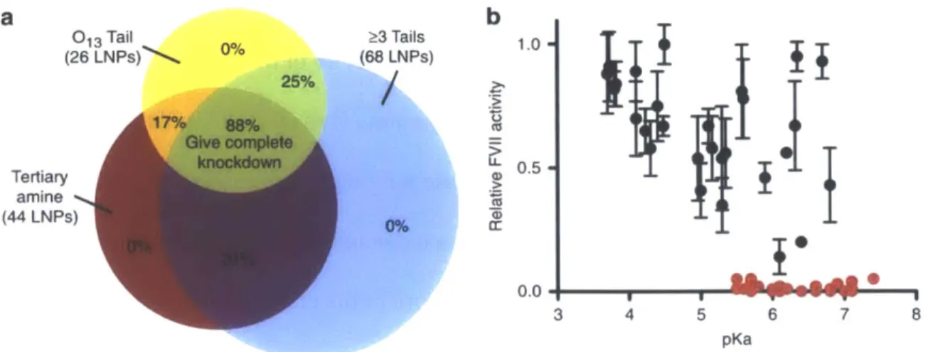
Having successfully optimized the liposomal formulation, and verified the relative tissue targeting of the LNPs, we sought to further investigate which structural motifs of the 96 compounds tested in vivo, correlated with a high degree of efficacy (Figure 2.5a). Of
these lipids, 26 (yellow circle) were found to contain 013 tails, while 68 of them have 3 or more tails (blue circle), and with respect to the amine core, 44 of them were synthesized from an alkyl-amine containing at least one tertiary amine (red circle). Lipids containing all three properties demonstrated an 88% success rate for a high degree of silencing FVII protein production in vivo, while elimination of even one of these criteria resulted in a decrease to only a 17-31% success rate. If two or more of the criteria were not met, FVII silencing was not observed at the tested dose of 5 mg kg-1.
In addition to examining the structural motifs of the individual lipids, the physical characteristics of the formulated LNPs were examined as well. One parameter that
demonstrated a strong correlation with efficacy was the pKa of the nanoparticle. The pKa of the LNP has been shown to have a significant effect on the efficacy of the nanoparticle
(Jayaraman et al., 2012; Zhang, Fan, Levorse, & Crocker, 2011). Of the LNPs examined, only nanoparticles with a pKa of 5.4 or greater demonstrated a high degree of silencing in vivo
(Figure 2.5b). When this additional parameter is considered in addition with the three structural motifs previously mentioned, the predictive power becomes even greater; 100% of the compounds that meet all four criteria demonstrated a high level of efficacy in vivo.
b 013 Tail 03 Tails 1.0 (26 LNPs) 25% (68 LNPs) 050 Tertiary 0.5 amine (44 LNPs) 0.0 3 4 5 6 7 8 pKa
Figure 2.5: Characterization correlating with efficacy. (a) The probability that
containing a structural motif will result in >95% silencing in vivo at a dose of 5 mg kg-1. (b) The relative in vivo FVII silencing of LNPs with respect to their pKa. (Data points represent group mean + standard deviation, n=3)
Second Generation Library Synthesis and Screening
In order to determine the validity of our hypothesis, a second lipid library was generated. Amine cores containing tertiary amines, and capable of undergoing Michael addition with three or more acrylates, were combined with 013 tails to generate a new library of lipids (Figure 2.6a). The lipids were then formulated with anti-FVII siRNA and the pKa was determined. All of the LNPs showed pKa values > 5.4, except for liposomes
containing either 509013 or 510013. We predicted that all of the compounds from the second library, except for 509013 and 510013, would demonstrate a high degree of
silencing in vivo. This prediction proved correct; when tested in vivo all of the compounds except 509013 and 510013 demonstrated greater than 95% silencing at a dose of 5 mg kg-1 (Figure 2.6b).
As we have demonstrated, we can dramatically improve the probability of synthesizing an efficacious lipid of this type by making sure to include three structural
parameters: the inclusion of a tertiary amine in the amine core, the presence of three or more lipid tails, and using lipid tails that are 13 carbons long. When the initial library of
1400 compounds was generated with no structural parameters, only 15 lipids, roughly 1%, demonstrated a high level of silencing in vivo; however, when the second library was
designed around these three motifs, greater than 80% of the compounds showed efficient silencing of hepatocytes in vivo. When the fourth parameter, the pKa of the LNP, was included, the rate of prediction is raised to 100%. Together, these parameters provide an invaluable tool for the design and screening, markedly reducing the time, cost, and number of animals required to develop new LNPs for therapeutic delivery.
a b 1.0 -500 H2N -N - NH2 501 1 501 H2N -,N--N --- NH2 N -NH2 502 H2N 503 H2N NH2 504 H2N- N -NH2 505 H2N N N NH2 506 NH2 508 H2N N N 509 H2N N, N NH2 510 510 H2 N-,N ? -- NH2 514 H2N '^'NH2 \yeNNH2 515 H2N, N -N,--^N -'/NH2 I I 0.5-0.0 >95% siencing @ 5 mg kg-1 5 2 0.5 siRNA dose (mg kg-')
Figure 2.6: The structure and silencing of the rationally designed library. (a) The amine cores combined with O13 tails to generate the second library. (b) The dose response curves for the LNPs that fit all four parameters established for high levels of efficacy in vivo.
Methods and Materials
Amine-lipid Synthesis. The alkyl-amines for the initial library screen were purchased from Sigma Aldrich, Alfa Aesar, Acros Organics, and CHESS Organics. The acrylates were
obtained from Scientific Polymer Products and Hampford Research, Inc. For the 500 series, the alkyl amines were synthesized by reacting secondary amines with sodium cyanide to form nitriles. These nitriles were then reduced to primary amines by using lithium
aluminum hydride (Gamage et al., 2001). The amine-lipids were synthesized by combining
cc
5M13 502013 508013 505013 013 514013 503013 0.1 2the alkyl-amines with the acrylates and stirring for 3 days at 90'C. In vitro experiments were conducted using the crude material, while in vivo experiments were performed using either crude material, or material that had been purified using a Teledyne Isco
Chromatography system.
LNP Formulation. The LNPs were formulated using an ionizable lipid, cholesterol (Sigma Aldrich), DSPC (Avanti Polar Lipids), and DMG-mPEG2000 (Alnylam Pharmaceuticals); the lipids were combined at a molar ratio of 50:38.5:(11.5-X):X respectively in 90% ethanol and 10% 10mM sodium citrate buffer, pH 3, comprising the organic phase. Unless
otherwise stated in the text, X = 1.5. The aqueous phase consisted of the siRNA dissolved in a solution of 10mM sodium citrate buffer, pH 3. Equal volumes of the organic and aqueous phases were mixed using a microfluidics device (Chen et al., 2012), and immediately diluted with a two-fold volume of 1X PBS, pH7.4. The lipidoid:siRNA weight ratio was 5:1, unless otherwise stated in the text. After formulation the particles were dialyzed against 1X PBS, pH 7.4, for at least 90 minutes.
In vitro transfection. HeLa cells stably modified to express both firefly and Renilla luciferase (American Type Culture Collection, Manassas, VA) were seeded in a white 96-well plate at a density of 15,000 cells per 96-well. Cells were transfected at a concentration of 40nM of formulated anti-firefly luciferase siRNA (Dharmacon). Firefly and Renilla
In vivo silencing. All animal experiments were conducted with the approval of the Institutional Animal Care and Use Committee (IACUC). Female C57BL/6 mice at least 6 weeks of age (Charles River Laboratories) were injected intravenously via the tail vein with either PBS, naked siRNA, or siRNA formulated LNPs. The siRNA sequence for FVII provided by Alnylam Pharmaceuticals may be found in Appendix A. Two days post injection blood was obtained from the mouse, and the FVII levels in the serum were determined using a Biophen FVII kit (Aniara Corporation).
Biodistribution. LNPs were formulated with siRNA that had been labeled with Cy5.5 on the 5'-end of the sense strand (provided by Alnylam Pharmaceuticals). Female C57BL/6 mice were injected with the LNPs at a dose of 1 mg kg-1 of total siRNA; 1 hour post
injection, the mice were euthanized and the organs were removed and imaged using an IVIS system (Calipur Life Sciences).
Blood clearance. LNPs formulated with Cy5.5-labelled siRNA was injected at a dose of 0.5 mg kg-1 into female C57BL/6 mice via the tail vein. Blood samples were collected at the various time points via the retroorbital vein, except for the final time point which was collected via a cardiac puncture. The serum was isolated from the samples, and was imaged and quantified using an Odyssey CLx imaging system (LI-COR Biosciences).
Nanoparticle characterization. The total concentration of siRNA and the percent of entrapped siRNA were determined using the Quant-iT RiboGreen RNA assay (Invitrogen).
Particle sizes were determined using a ZETAPals analyzer (Brookhaven Instruments). Zeta potential measurements are acquired on a Zetasizer Nano ZS (Malvern).
pKa measurements. The pKa of the LNPs were determined as previously described (Heyes, Palmer, Bremner, & MacLachlan, 2005). Briefly, solutions of 20mM sodium phosphate, 25mM citrate, 20mM ammonium acetate, and 150mM NaCl were titrated to a series of pHs ranging from 2 to 12. These solutions were aliquoted into a black 96-well flat bottom plate in quadruplicate. The LNPs and TNS were added to each of the wells such the final concentrations were 20tM and 6tM respectively. The fluorescence of each well was
determined using a Tecan plate reader and using an excitation wavelength of 322nm and an emission wavelength of 431nm.
Chapter 3
Examining the role of apolipoproteins on efficacy and cellular uptake using novel lipopeptide based LNPs
This chapter contains data previously published:
Dong, Y., Love, K. T., Dorkin,
J.
R., Sirirungruang, S., et al. (2014). Lipopeptide nanoparticles for potent and selective siRNA delivery in rodents and nonhuman primates (vol 111, pg 3955, 2014). PNAS, 111(15), 5753.Overview
While the analysis of the degradable lipid library in Chapter 2 elucidates some of the structural motifs and characteristics important for LNP delivery, it provides limited
information as to the mechanism of action. Although historically it has been postulated that hepatocellular delivery of LNPs is primarily size dependent, it has been demonstrated that localization to the liver by nanoparticles <100nm in size is insufficient for the successful transfection of hepatocytes in vivo (Santel et al., 2006; Semple et al., 2010). Recent publications have provided evidence that apolipoprotein E is essential for hepatocellular delivery in vivo (Akinc et al., 2010; Bisgaier, Siebenkas, & Williams, 1989; Yan et al., 2005), but only a small subset of apolipoproteins and isotypes have been examined thus far. In this chapter we examine a larger subset of apolipoproteins, determining both their effect on hepatocellular delivery, and determine their role in cellular uptake.
The investigation into the apolipoprotein modulated delivery was performed using a novel lipid library developed by Dr. Yizhou Dong. This library was designed around the use of amino acids and dipeptides as the ionizable core of the lipopeptide. By structuring the compound around a biocompatible core, the idea was to create a compound that would be degradable by the cell, thus reducing the toxicity and improving the therapeutic
window.
Lipopeptide Iterative Screening
each of the 20 amino acids with three different lipid tails (Figure 3.1); each lipid tail
consisted of a fully saturated twelve carbon long tail with either a terminal aldehyde (A12), acrylate (012), or epoxide group (E12) to allow for the addition to the amine groups on the lipopeptide core. For the liposomal formulation, the lipopeptides were combined with
cholesterol, DSPC, and DMG-mPEG2000 using a lipid molar ratio of 50:38.5:10:1.5, and with a lipid to siRNA weight ratio of 5:1. As with the acrylate-based library, siRNA targeting the serum protein FVII was used for in vivo formulation in order to select for liposomes
that successfully targeted hepatocytes. Of the initial 60 lipopetides that were examined, only thirteen LNPs demonstrated any siRNA entrapment (Appendix C); the rest of the LNPs were discarded, and were not tested in any further capacity. The thirteen formulations with entrapped siRNA were then injected in vivo at a dose of 1 mg kg1. Of the 13 LNPs
injected, only one of the lipopeptides tested, K-E12, demonstrated greater than 50% silencing. (Figure 3.2 series 1)
a
Amino Acids
0 HNI OH \:=N NH2 H 0 HOOC OH NH2 E Et 0 Me OH NH7 I 0 0 0 0H2N OH i-Pr OH MeS OH H2NOC OH
NH2 NH2 NH2 NH2 K L M N o 0 NH 0 -r1OH H2NOC OH H2N N OH NH NH2 NH2 P Q R 0 H ClH23 A12 i O' 0,C12H25 0 012 L>-C1OH21 E12 0 HO OH NH2 S OH 0 Me 'OH NH2 T
b
R H2N OH + 0 Amino Acids i-Pr-yJL OH NH2 V 0 HN2 OH NH2 W W O NaHB(OAc)3,L
H C11H 23 THF, rt A12 0 OH HO ~ NH Y R C11H-23 ,N yOH OH C11H23 0 TEA, IPA 0 R O'C2H5 C1H2'O N OH O 90 OC H 012 0 C EtOH CH2 1 R0 E>-12 W 1 5 0 H NC C10H E12 H 21,50C 0 0 HS -- , OH HOOC OH NH2 NH 2 C D 0 Me OH NH2 A 0 PhV-f' OH NH2 FTails
0 (1 OH NH2 G I-2.0 Screening at a dose of 1 mg/kg Screening at a dose of 0.1 mg/kg
Series 1 Seriea 2
0)
J hit rate: 1.7% (1 out 60) hit rate: 23% (10 out 43)
c 1.5 12-14 carbon tail
Z) lengths were favorable
*-0
2
0.5F)0.0- 3.
ipopeptidNNe
leirng n
vivo.Li
4
LoiiitidJJ
Lat n-F
IULL~
wr
ineceditrveosli iTeFII serum level s wer xaine 4i horlte n
0WY 000 a a
concentrations; Data points represent group mean +standard deviation , n=3)
Due to the efficacy of the lysine based lipopeptide, a second library was generated using dipeptide or polypeptide cores containing lysine (Figure 3.3a). For a subset of the dipeptides, a cyclization was performed to produce the diketopiperizine (Figure 3.3b). In total, 43 compounds were generated for the second library, 20 of which were capable of forming stable particles with entrapped siRNA (Appendix C). These LNPs were then
injected intravenously at a dose of 1 mg kg-1; 10 of the LNPs injected displayed greater than 50% FVII silencing, with 8 compounds showing more than 80% silencing (Figure 3.2 series 2).
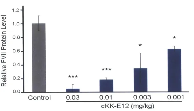
In order to further differentiate between the more efficacious compounds, LNPs demonstrating greater than 80% silencing were re-injected at a dose of 0.1 mg kg-1 (Figure 3.2). cKK-E12 demonstrated the greatest degree of silencing, with more than 95% protein