HAL Id: tel-01598875
https://tel.archives-ouvertes.fr/tel-01598875
Submitted on 30 Sep 2017HAL is a multi-disciplinary open access archive for the deposit and dissemination of sci-entific research documents, whether they are pub-lished or not. The documents may come from teaching and research institutions in France or abroad, or from public or private research centers.
L’archive ouverte pluridisciplinaire HAL, est destinée au dépôt et à la diffusion de documents scientifiques de niveau recherche, publiés ou non, émanant des établissements d’enseignement et de recherche français ou étrangers, des laboratoires publics ou privés.
Towards genome-wide, single-molecule analysis of
eukaryotic DNA replication
Francesco de Carli
To cite this version:
Francesco de Carli. Towards genome-wide, single-molecule analysis of eukaryotic DNA replication. Genetics. Université Pierre et Marie Curie - Paris VI, 2016. English. �NNT : 2016PA066600�. �tel-01598875�
Université Pierre et Marie Curie
École doctorale Complexité du Vivant
Institut de Biologie de l'École Normale Supérieure (IBENS) Eukaryotic Chromosome Replication team
Towards genome-wide, single-molecule analysis of
eukaryotic DNA replication.
Dissertation by
Francesco De Carli
For the Degree of Doctor of Philosophy
Directed by Dr. Olivier Hyrien
Presented and publically debated on September 28th, 2016 before a jury composed of: Pr. Gilles Fischer President of the Jury UPMC (FR)
Dr. Aloys Schepers Referee Helmholtz Zentrum (DE)
Dr. Bénédicte Michel Referee I2BC (FR)
Dr. Torsten Krude Examiner Univ. of Cambridge (UK)
3
Acknowledgements
I would like to thank the members of the jury: Bénédicte Michel and Aloys Schepers, to accept to revise my thesis in the middle of August. Thanks to Gilles Fischer, to chair the committee, and to Torsten Krude, to fight the Brexit and to come from Cambridge. Thanks to Arach Goldar, to be always present all along my PhD, and to amaze me with a mixture of biophysics and magic (yes, I’m still trying to understand why, theoretically, the YOYO-1 signal shouldn’t be “double”). Thanks to the others that followed me during the PhD: to Gaël Millot, I would have never been able to include the nanochannel results in this thesis without your “scoop” on the existence of this technology, and to Jean-François Allemand to give me the access to the white-room at the physic department and to be always available to discuss.
Many thanks to the “captain of the ship”, Olivier Hyrien, to accept me at the IBENS, and to give me the full responsibility of this project («You are responsible for all your successes
and all your failures»). Thank you to always force me to think with my head (every time I enter
in your office with a question, I exit with a reply, and with two or three novel questions!), and to never limit my “brilliant” ideas (which were immediately summarised with: «You are
probably the most expensive M2 student of all the times». Ok, it was true, but check the price
of the AlexaFluor-dUTP!). Thank you for the good wine (oups…time) spent together, and to always remember me why we do research. Thanks to keep doing science for science (and not for the glory from the scientific community). Many thanks to Benoit: you brought a breath of fresh air in the lab, and a lot of good science. I would have never finished the manuscript without your help and support. I’m looking forward to start new experiments with your criticisms and your competences. Thanks to “wonder-woman” Mag: biologist, bioinformatic and mother (super Pablo!): how can you do that? I wish to be half as efficient as you are. Thank you, Xia, to share your thesis preoccupations with me, and to work so hard for three years: we have never given up for one second even in the most difficult periods, downstairs. Thank you, Nikita, to be “brave” and accept to start the huge bioinformatics effort necessary to analyse my data. I hope your thesis will be full of joy (I also hope you give me some results tomorrow. No, really. Where are my results?). Thanks to the old lab members. To Nat: I wish you were here with your family. I’ll never forget your positive energy that is still supporting me. Thank you Malik! To stay with me to distribute 12 mL of Xenopus egg extracts in 5 µL aliquots at 2 am, and to teach me how to seduce a frog. Our frozen extracts still partially works 2 years after…that’s not magic: it’s the touch of the sun from the south. Thanks to the last arrived: Florence. I would have liked an engineer before the end of my thesis (I’d have slept a bit more…), but I’m very happy to wait if this engineer is you!
Thanks to the doctoral school “Complexité du Vivant”, for the PhD grant. And to some other French institutions: thank you to consider me “Italian” on French-reserved grants, and to consider me “French” on foreign-reserved grants. That’s kind.
Thanks to the collaborators. To the ENS in Lyon (Alain Arneodo, Benjamin Audit, Françoise Argoul and Stéphane Roux) to make me waking up at 4 am, but also to enchant my soul with wavelet transforms and some other mathematical things that I promise…I would if I could (understand). Thanks to Auguste Genovesio (IBENS): your platform is the most important tool in this building for us, poor “wet-lab” researchers! Thank you, Elton Rexhepaj, cause this **** computer doesn’t want to understand my language (but, apparently, your is ok). Thanks to Valérie Barbe (Genoscope): in a scientific world where “time is money” you believed in our ideas, and you still continue to do it. I am grateful for your excitement in this nanochannel
4
project and for your patience. Thank you, Wahiba, to wonderfully finalise my samples and to run the Irys beast.
Thanks to the functional genomic (S2) section. I’m sorry for shouting in the corridor, for the angry emails about the microscope, for the chairs flying in the radioactive room, for my daily, “non-diplomatic” (euphemism) way to resolve problems. Thank you Hervé, to be the one understanding that I simply like when “things work” and to respect my constant nervousness. Thank to your team. To Isa, you’ve been a great colleague and a dear friend since the beginning. You still are. Emélie (yes, you’re still here for me): we miss you, but the world needs even more passionate biology teachers. Thanks to Joanne, Virginia, Rémi, Rahul, Raphael, Jerome for the time spent together. Thanks to OB to bring tons of “useful” material on my bench, to Lydia to constantly animate the section, and to Floriane, Anne-Catherine and Xiao. Thank you, Alice, to be always available for me and to have brought to the section such a positive energy and your amazing lab. Thank you Laure, to make me smile every single time I meet you, even without talk (and to not make me pay 20 cents for every bad word I’ve said). I’m actually depressed for your departure. Thank you Vinko. You were also here with me quite all the Saturdays and the Sundays until 10 pm. I would have never been able to finish this year, without your friendship. Thanks for the 5,000 Italian coffees, for the pizzas/sushi, and for the drinks, despite the unacceptably expensive price of beers in France. Thanks to Caro: I’m keeping all your post-it on my desk wall, and in my heart. I bring you with me, always. Thank you, Chloé: I’ve no idea how you do, but every time I talk to you, you say something that I don’t expect and I laugh till I can’t breathe…do not stop doing that, please! Thanks to Benoit, to remember me that your flat is less expensive and bigger (but outside Paris, gne gne). Thanks to the EM team, especially to Guillaume, to remember me that I should stay calm, and to Vero, to incite my daily rebellions and to always say what she thinks. Thanks to the bioinformaticians of the section, especially to Yves, to be able to link all of us together, and to Alexandra, to always smile (yeah, you’re miss sunshine of the section). Thanks to the past members of the S2, especially the Darzacq lab, to introduce me to the fluorescent microscopy.
Thanks to Brigitte. You would deserve a page, all for you. I don’t have words to say how much we need you. Thank you to be my “mother” at IBENS. Thank you, Abdoul, to make her nervous. You are a cool couple. Many thanks to Martine (laverie) and to all the informatics service to make our job here possible, and to be so incredibly efficient.
Thanks to the S2 microscopy room. To “steal” me 4 months of my thesis, and a ridiculous amount of time per week. Thanks to Xavier (and Astou!), to teach me everything about the Nikon Ti. Thanks to help me in putting the furniture together, and to mount/test/maintain that “monster”. Thank you to injure yourself (twice) for our section. That black room wouldn’t exist without you (my images, as well). Thank you Benjamin, to help me whenever you could, and to Nikon, to make a software as expensive as its number of bugs.
Thanks to all the friends from the M2 at the Magistère Européen de Génétique. Fra, Lara, Ale, Madda, Giulia, Teo (Deb, Cri, …): every time we meet we have great fun. It’s like the time never pass and I always feel you by my side.
Thanks to my family here: Jack, Frarro, Andre. You have made me feel at home, every day, for 3 years. Thanks for the inniorantia time, for the Appa Alti Livelli, for the food and the good wine, for the water leakages every 2 weeks, for the door blocked and the nights outside the flat, for the hole in the gas pipe to hang a lantern, for the 1500 euros-extortion of the boiler
5
and the weeks without light in the toilet. Don’t ask me HOW, but all this **** was a fantastic experience, with you. Thanks to support me despite my many limits.
Thanks to Marco (God only knows how much I miss you), Stefania (to never let me go, to keep forgiving me for my absence) and Marta (I will never forget that 14th July, before your
departure). You, famiglia cuore, you taught me the value of friendship. Thanks to Nat and our group (especially to Turo and Chiara), to ask me, constantly, what we are doing here. Thanks to the Italian friends in Paris, especially Robi, I miss the time of our disputes.
Thanks to Paolo, Frenchy, David, Tati, Nic, Beppe, Margio, Claude. Some friends are always close in your heart, it doesn’t matter the distance. You are as the good ageing wine.
Dulcis in fundo, thanks to my family. To my mother, to show me that believe in
Something is the most rational way to reply to our desire for happiness (yes, science can’t). Thanks for your constant support and for the sacrifices done for our family. To my father, to show me that miracles do exist and that in life nothing is lost, when you want to restart. To my sisters Marta and Miriam, ‘cause sometimes you need to leave to realise what you’ve left. I miss you, I miss big-Stefano on one side, and Ale, Japo, Michele and Niccoló on the other side. Thanks for your help, to keep me company during all these years, and to make my very shorts Italian trips gorgeous. Thanks to my grandma Rosetta, example of extraordinary faith and of untiring joy of life, and to Elio, Giannina and Gelmino, to keep an eye on me from up there.
Thank you, Laurianne. Thank you for believing in me. Thank you, to constantly show me that science is not sufficient be happy. Thank you to accept my limits and to love our differences. Thank you, to make me always smile with your simplicity and to comfort me. Thank you, to stick with me despite my periodical nervousness and the too many moments of absence along this year together. This thesis and my stay here in Paris during difficult times wouldn’t have been possible, without you. Thanks to Sylvie, Michel (Funny), Matthieu, Marie-Laure et les mamies! For the short time spent together, and to make me feel part of your family.
For the people that, arrived at this line, did not found (unjustly) their name: I’m sorry. Thanks to have contributed, in a way or another, to the realisation of this manuscript. Please to forgive my omission and to understand my emotion at the time of writing.
7
« L'unica gioia al mondo è cominciare. E' bello vivere perché vivere è cominciare, sempre ad ogni istante. »
« The only joy in the world is to begin. It is good to be alive because living is beginning, always, at every moment. »
Cesare Pavese
« One day the machines will be able to solve all the problems, but none of them can deliver us one. »
8
Abstract
In eukaryotes, DNA replication starts at multiple origins that are activated following a specific program. Population methods allow genome-wide analysis of DNA replication. However, single-molecule methods are required to monitor cell-to-cell variability, detect rare events and measure individual replication fork speeds. With the existing techniques, newly-synthesized DNA is labelled with thymidine analogs and revealed with fluorescent antibodies. Fibres containing a locus of interest can be identified by fluorescent in situ hybridization. These steps are complex and the throughput is low. This work proposes novel, antibody-free tools to detect replication tracts and identify the locus of origin of all DNA molecules at much higher throughput. DNA replicated in the presence of a fluorescent dUTP was purified and specifically barcoded by using a nicking endonuclease, followed by limited nick-translation with another fluorescent dUTP. This allowed alignment to a reference genome map. DNA was then stained with the fluorescent DNA intercalator YOYO-1. Direct epifluorescence revealed the DNA molecules, their replication tracts and their barcodes in three distinct colours. Replicated segments showed a stronger YOYO-1 fluorescence, demonstrating that replication bubbles can be directly detected without metabolic labelling. Finally, these tools were coupled to a nanofluidic device: DNA was driven into 13,000 parallel nanochannels and automatically imaged, massively increasing the throughput. Altogether, these results provide a starting point for genome-wide, single-molecule mapping of DNA replication in eukaryotic organisms. Keywords: DNA replication; molecular combing; DNA barcoding; single-molecule; nanochannel; epifluorescence.
9
Résumé
Vers l’analyse en molécule unique de la réplication de l’ADN eucaryote à l’échelle du génome entier.
Chez les eucaryotes, la réplication de l’ADN démarre au niveau de multiples origines activées suivant un programme précis, qui peut être analysé à l'échelle du génome sur des populations cellulaires. Cependant, l'étude de la variabilité intercellulaire, la détection d'évènements rares et la mesure de la vitesse des fourches de réplication nécessitent des analyses en molécule unique. Avec les techniques actuelles, l’ADN néosynthétisé est marqué avec des analogues de la thymidine et révélé par des anticorps fluorescents. Les molécules d’intérêt sont identifiées par hybridation fluorescente in situ. Ces étapes sont complexes et le débit est faible. Cette thèse développe de nouvelles méthodes de détection et d'identification des molécules d'ADN réplicatives sans anticorps et à haut débit. L’ADN est répliqué en présence d’un dUTP fluorescent, purifié puis marqué en code-barre spécifique permettant l’alignement sur le génome de référence par coupure avec une endonucléase simple brin et incorporation d’un autre dUTP fluorescent. L’ADN est ensuite coloré avec un intercalant fluorescent, le YOYO-1. Les molécules d’ADN, leurs segments néorépliqués et leurs code-barres sont observés en trois couleurs différentes par épifluorescence directe. Les segments répliqués ont une fluorescence YOYO-1 plus intense, ce qui permet de détecter les bulles de réplication sans marquage métabolique. Ces outils ont été couplés à un dispositif nanofluidique dans lequel l’ADN est conduit dans des milliers de nanocanaux et imagé automatiquement, ce qui augmente massivement le débit. L'ensemble de ces résultats ouvre la voie à la cartographie pangénomique de la réplication de l’ADN en molécule unique.
Mots clés : réplication de l’ADN; peignage moléculaire; code-barre fluorescent; molécule unique; nanocanaux; épifluorescence.
10
Table of contents
Acknowledgements ... 3 Abstract ... 8 Résumé ... 9 Table of contents ... 10 List of abbreviations ... 13 I. Introduction ... 17I.1 Prerequisites on DNA replication ... 17
I.2 The DNA replication background: early studies ... 19
I.2.1 The first visualisation of replication “bubbles”: a fibre autoradiography story ... 19
I.2.2 The discovery of eukaryotic replicator: ARS in plasmids as a replication assay 21 I.2.3 Origins have different efficiencies: the advent of 2D agarose gel electrophoresis 22 I.2.4 Origins are redundant: novel insights from electron microscopy studies ... 24
I.2.5 The eukaryotic initiator (ORC) and the pre-replicative complex (pre-RC) ... 25
I.3 Insights from early replication studies ... 26
I.4 Pre-“omics” studies ... 27
I.4.1 Random vs. site-specific initiation in metazoans ... 27
I.4.2 The DHFR locus: a closer look at replication initiation reveals controversies .... 28
I.4.3 Narrow and efficient origins vs broad, initiation zones ... 29
I.4.4 Temporal control of origin activation: early studies ... 30
I.5 The advent of genome-wide analysis of DNA replication ... 31
I.5.1 Technologies for rapid pangenomic mapping of the ORIs ... 31
I.5.2 Genome-wide analysis of DNA replication: what did we learn? ... 34
I.5.2.1 Replication timing: contributions from genome-scale methods (CTRs, TTRs) 34 I.5.2.2 Spatiotemporal replication program ... 35
I.5.2.3 Spatiotemporal program of DNA replication: missing elements? ... 35
I.5.2.4 Sequence-specific metazoan origins? “G-rich” evidences from λ-SNS studies 36 I.5.2.5 Whole-genome nucleotide compositional skew & replication fork directionality analysis ... 37
I.5.2.6 The advent of OK-seq ... 40
I.5.2.7 General features of mammalian genome replication ... 41
I.5.3 Limits of the current genome-wide techniques ... 41
I.5.3.1 Whole-genome ORI mapping: a matter of (missing) concordance ... 41
I.5.3.2 SNS, the λ-exo concern: about the reliability of current genome-wide methods 43 I.6 Interplays between transcription and replication ... 44
I.7 Towards single-molecule, genome-wide ORI mapping ... 46
I.7.1 Advantages of single-molecule techniques ... 46
I.7.2 DNA combing: a powerful tool for single-molecule analysis of DNA replication 46 I.7.3 Limits of the current single-molecule techniques ... 49
I.7.4 Possible achievement of genome-wide, single-molecule techniques ... 51
I.7.5 Inspiring tools for developing novel, high-throughput, single-molecule techniques ... 52
11
I.7.7 Strategies ... 55
II. Methods ... 57
II.1 Preparation of polystyrene coated coverslips ... 58
II.1.1 Glass coverslips cleaning ... 58
II.1.2 Polystyrene coating ... 58
II.2 Alkaline and neutral agarose gel electrophoresis to monitor fluorescent dUTP incorporation ... 59
II.2.1 Neutral agarose gel ... 59
II.2.2 Alkaline agarose gel ... 59
II.2.3 Signal acquisition ... 59
II.3 Fluorescent imaging of replicating nuclei in Xenopus egg extracts ... 60
II.4 Construction of a lariat DNA structure ... 61
II.4.1 Design of the experiment ... 61
II.4.2 Protocol ... 63
II.5 Studying DNA replication using the nanochannel device ... 64
II.5.1 Isolation of genomic DNA from Xenopus egg extracts ... 64
II.5.2 Fluorescent barcoding, DNA staining and sample run ... 64
III. Results ... 65
III.1 Premise ... 65
III.2 Molecular DNA combing ... 66
III.2.1 Surface testing, definition of optimum DNA combing conditions ... 66
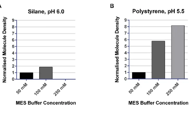
III.2.1.1 Density of adsorbed DNA molecules ... 67
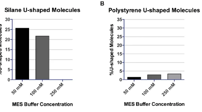
III.2.1.2 Percentage of exploitable DNA fibres ... 71
III.2.1.3 Stretching of DNA fibres (i.e. evaluation of the stretching factor) ... 74
III.2.1.4 Alignment of DNA fibres ... 80
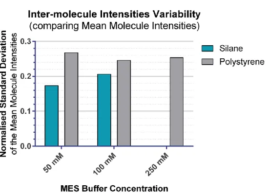
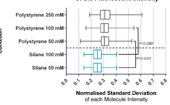
III.2.1.5 Uniformity of the fibres’ fluorescent signal ... 83
III.2.2 Fluorescent labelling of specific nicking endonucleases sites ... 90
III.2.2.1 Early essays ... 90
III.2.2.2 The problem of the adjacent nicks (i.e. “fragility hotspots”) ... 95
III.2.2.3 Definition of a working protocol (Nt.BspQI + Taq pol + PreCR repair)105 III.2.3 Fluorescent replication signal detection ... 111
III.2.3.1 Fluorescent dUTP incorporation: nuclei reconstitution experiments ... 111
III.2.3.2 Discontinuity of fluorescent dUMP incorporation ... 113
III.2.3.3 Observation of doubling of DNA fluorescent intensity along a combed Y-shaped DNA substrate ... 125
III.2.4 Fluorescent replication signal detection on barcoded DNA molecules ... 127
III.2.4.1 Early essays ... 127
III.2.4.2 Definition of a working protocol ... 128
III.2.5 The first fluorescent replication map of the bacteriophage lambda genome (De Carli et al 2016) ... 133
III.3 High throughput single-molecule analysis of DNA replication using nanochannel arrays 142 III.3.1 Description of the nanochannel system ... 142
III.3.2 Test the continuity of the fluorescent replication signal ... 145
III.3.3 Evaluating the YOYO-1-based bubble detection ... 147
III.3.4 The bacteriophage lambda fluorescent replication map: a comparison with the DNA combing technique ... 149
IV. Discussion ... 151
IV.1 Challenges encountered in developing novel single-molecule tools ... 152
12
IV.1.2 Challenges in developing a fluorescent DNA barcoding ... 152
IV.1.3 Challenges in developing a direct fluorescent bubble detection ... 154
IV.1.4 Challenges in combining fluorescent bubble detection with fluorescent barcoding156 IV.2 Challenges encountered in adapting the tools for nanofluidic analysis ... 157
IV.3 Antibody-free DNA combing vs. nanofluidics: current potential and limitation .. 159
IV.4 The bacteriophage lambda story: DNA combing vs nanochannel ... 162
IV.5 Towards genome-wide, single-molecule analysis of eukaryotic DNA replication 164 IV.5.1 Capacity of the technologies at present ... 164
IV.5.2 Potential of the technologies ... 165
IV.6 Future of the single-molecule techniques. ... 166
IV.6.1 Is the DNA combing era at the end? ... 166
IV.6.2 Meeting the dynamic studies of DNA replication ... 166
Résumé détaillé de la thèse ... 169
Bibliography ... 175
Table of illustrations ... 183
13
List of abbreviations
2D/3D: 2 Dimensions / 3 Dimensions λ-DNA: Bacteriophage lambda DNA λ-exo: Lambda exonuclease
A: Adenine
ACS: ARS Consensus Sequence AF: AlexaFluor
AF555-dUTP: Alexa Fluor555-aha-dUTP AF647-dUTP: Alexa Fluor647-aha-dUTP ARS: Autonomously Replicating Sequence BAC: Bacterial Artificial Chromosome bp: base-pair(s)
BrdU: 5-Bromo-2'-deoxyuridine C: Cytosine
Cdc: Cell division cycle CDK: cyclin dependent kinase CGI: CpG Island
ChIP: Chromatin ImmunoPreciptiation CHO: Chinese Hamster Ovary
CldU: 5-Chloro-2'-deoxyuridine CMG: Cdc45, MCM, Gins complex CNV: Copy Number Variant
CpG: 5'-C-phosphate-G-3' CTR: Constant Timing Region Cy: Cyanine
dACG: dATP+dCTP+dGTP
dATP: 2'-Deoxyadenosine 5'-triphosphate Dbf4: Dumb bell former 4
dCTP: 2'-Deoxycytidine 5'-triphosphate DDK: Dbf4 Dependent Kinase
dGTP: 2'-Deoxyguanosine 5'-triphosphate DHFR: Dihydrofolate reductase
14 Dig: Digoxigenin
DNA: Deoxyribonucleic acid DNAse: Deoxyribonuclease
dNTP: Deoxyribonucleotide triphosphate dsDNA: double-stranded DNA
dTTP: 2'-Deoxythymidine 5'-triphosphate dUTP: 2´-Deoxyuridine, 5´-Triphosphate EdU: 5-ethynyl-2'-deoxyuridine
EM: Electron Microscopy
FISH: Fluorescent In Situ Hybridization
FRET: Fluorescence-Resonance Energy Transfer G: Guanine
G1/G2-phase: Gap 1 / Gap 2 phase G4: G-quadruplex
h: hour(s)
Hi-C: Chromosome Conformation Capture coupled to high-throughput sequencing HR: Homologous Recombination
IdU: 5-Iodo-2′-deoxyuridine IOD: Inter Origin Distance IP: Immunoprecipitation
kb: kilobase-pairs, 103 base-pairs (bp) Mb: Megabase-pairs, 106 base-pairs (bp) MBT: Midblastula Transition
MCM: Mini Chromosome Maintenance NE: Nicking Endonuclease
MES: 2-(N-morpholino)ethanesulfonic acid M-phase: Mitosis
min: minutes
NGM: Next Generation Mapping NGS: Next Generation Sequencing NHEJ: Non-Homologous End-Joining NS: Nascent Strand(s)
nt: nucleotide(s)
15 ORC: Origin Recognition Complex ORI: Origin of replication
PCR: Polymerase Chain Reaction Pol: Polymerase
pre-RC: pre-Replication Complex px: pixel(s)
rDNA: Ribosomal DNA
RFB: Replication Fork Barriers RFD: Replication Fork Directionality RIP: Replication Initiation Point RNA: Ribonucleic acid
rRNA: ribosomal RNA seq: sequencing
SM: Single-Molecule SNS: Short Nascent Strands S-phase: Synthesis
ssDNA: single-stranded DNA T: Thymidine
TAD: Topologically Associated Domains TCA: Trichloroacetic Acid
TDP: Timing Decision Point
TIRFM: Total Internal Reflection Fluorescent Microscopy TTR: Transition Timing Region
17
I. Introduction
I.1
Prerequisites on DNA replication
The “genetic research” era that led to the DNA replication studies, opened with the identification of the molecule responsible for the genetic inheritance. Thomas H. Morgan’s fruit flies experiments demonstrated that genes, the physical and functional units of heredity, are located on chromosomes. This notion, termed “Chromosomal Theory of Heredity” (Morgan 1910), however, could not identify the element responsible for the genetic inheritance. Both deoxyribonucleic acid (DNA) and proteins were plausible candidates. It took almost two decades to discover that the genetic information is transferrable (Griffith 1928), and other sixteen years to prove that the “transforming principle” is the DNA (Avery et al 1944). Residual sceptical scientists, arguing that genes of bacteria must have very different compositions compared to those of complex organisms, finally accepted DNA as the genetic material after the bacteriophage T2 infection experiments conducted by Alfred D. Hershey and Martha C. Chase (Hershey & Chase 1952). Meanwhile Erwin Chargaff had shown that the pyrimidine and purine bases are present in equal proportion in the DNA, and that the composition of the DNA varies between organisms (Vischer & Chargaff 1948). The rigorous validation of the Chargaff’s rules was finally provided by James D. Watson and Francis H. C. Crick with the elucidation of the DNA structure (Watson & Crick 1953). This paper, a cornerstone of the modern research, not just confirmed the Chargaff’s rules and that the DNA is the chemical substance defining the genes, but inspired an entire era with its famous citation:
"It has not escaped our notice that the specific pairing we have postulated immediately suggests a possible copying mechanism for the genetic material."1
Eventually, this intuition of the semi-conservative replication model was demonstrated only four years later (Meselson & Stahl 1958, Taylor et al 1957). Since then, many discoveries were done, setting the stage for DNA replication studies. Today, we know that despite the huge variability in known eukaryotic genome sizes (i.e. spanning from the 2.25 million base pairs of the parasite Encephalitozoon intestinalis to the stunning 133 billion base pairs of the marbled
1 Watson JD, Crick FH. 1953. Molecular structure of nucleic acids; a structure for deoxyribose nucleic acid. Nature
18
lungfish Protopterus aethiopicus), the DNA is highly organised and its duplication is tightly regulated within a dedicated conserved time-slot, the S-phase.
It is in this frame that the DNA replication takes places: this very delicate process of life responsible for the genetic inheritance is meticulously orchestrated, together with all the other necessary operations, at any moment, within the “concert” of the cell survival.
19
I.2
The DNA replication background: early studies
I.2.1 The first visualisation of replication “bubbles”: a fibre autoradiography story In 1963, Cairns coupled 3H-thymidine metabolic labelling of replicating DNA in
Escherichia coli cells, with fibre spreading and autoradiography. For the very first time DNA
replication could be physically visualised. Replication appeared to start at a single site, and to be bidirectional (Cairns 1963). At the meantime genetic studies conducted on the same organism by Jacob, led to the formulation of the “Replicon Theory”: a cell contains one or several autonomous units of DNA replication, termed "replicons", that each require a cis-acting element, named “replicator”, and a trans-acting element, the “initiator”, for their replication (Jacob & Brenner 1963)(Figure I-1 A). The interaction between these two elements results in an autonomous replication unit called “replicon”. It took almost ten years to isolate the replicator, oriC (Yasuda & Hirota 1977), and another five to determine the initiator, DnaA (Chakraborty et al 1982), but finally the theory was proved by in vitro reconstitution of the initiation process (Bramhill & Kornberg 1988). It is the initiator itself that is in charge of binding and unwinding the replicator in order to recruit the helicase (DnaB) directly on single-stranded DNA. Finally, the recruitment of additional factors culminate with the assembly of two divergent replication forks (reviewed in Bell and Kaguni (2013)). Almost a decade after Cairns, his discoveries were confirmed by Prescott and Kuempel (1972), but the position of the replication start site was still unknown. Then, in 1968, Huberman and Riggs successfully adapted the DNA fibre autoradiography analysis to the study of mammalian chromosomes. Many key points could be outlined. DNA replication is bidirectional also in eukaryotic cells, but starts at multiple sites (Huberman & Riggs 1968)(Figure I-1 B). These sites were therefore called “replication origins” (ORIs), and appeared to be coordinated in clusters. Clusters of five to ten replicons2 complete replication synchronously in about one hour, implying sequential
activation of different clusters along the S-phase (8 to 10 hours, typically). The DNA fibre autoradiography also allowed measurements of fork velocities (2-3 kb/min)3 and quantification of the distances between replicons (20-400 kb)(Huberman & Riggs 1968). This was doable because the structure of the DNA was already known (Watson & Crick 1953): each residue in the double helix occupies 3.4 Å, therefore, it is possible to establish a correspondence between the µm distance (measured by autoradiography) and its number of bases. Later studies revealed
2 In eukaryotes, the word “replicon” refers to the DNA replicated from a single origin. 3 kb = kilobase-pairs, 103 base-pairs (bp).
20
no agreement comparing the number of replication units within different clusters (R. Hand observed very few replication units within the cluster (Hand 1975), while Willard and Latt reported several dozens of replicons (Willard & Latt 1976)). Moreover, Yurov and Liapunova (1977) proved that mammalian replicons could be much longer than previously thought, up to 2 Mb4. It was clear from all these experiments that, although the fork velocities were almost constant among different cell types and studies, the intervals between ORIs and the synchrony of their activation were highly variable. The reasons accounting for this “flexibility” in ORI usage were unknown, but scientists reasoned that it could be a way to fit specific needs of the cell. The cell needs change the replication program according to its developmental and differentiation stage (reviewed in Berezney et al (2000)). The concept of flexibility of ORIs usage was further stimulated when Taylor showed that holding the cells at the beginning of the S-phase increases the replicon number by reducing ORIs spacing (Taylor 1977). Therefore, replication stress can modify the “normal” DNA replication pattern, which is not anymore to be considered immutable.
4 Mb = Megabase-pairs, 106 base-pairs (bp).
21 Figure I-1: Basic mechanisms of DNA replication.
A| The replicon model as proposed by Jacob and Brenner (1963): the replicon is a circular structure harbouring two genetic determinants. A trans-acting element, the initiator protein, coded by a “structural gene” (SG1, blue), recognises a specific cis-acting element, the replicator (orange). This interaction allows the beginning of DNA replication. B| DNA fiber autoradiography of mammalian chromosomes (Chinese hamster cells, CH). CH cells were pulse labelled with 3H-thymidine for 30 min, and chased by
non-radioactive thymidine for another 45 min. Cells were collected and lysed. The DNA trapped on dialysis filters was subjected to autoradiography. Replication occurs at several starting sites (i.e. replication origins, indicated by orange arrows) and proceed in a bidirectional fashion from each of these. Adapted from Huberman and Riggs (1968).
I.2.2 The discovery of eukaryotic replicator: ARS in plasmids as a replication assay Despite the fast progression of knowledge, it was still unclear whether replication ORIs correspond or not to specific genomic sequences in eukaryotes. The replicon model inspired novel essays to study DNA replication: selectable markers coded in specific plasmids could be maintained independently of chromosomal replication only if the plasmid contained a replication ORI (Figure I-2). Thanks to this idea, eukaryotic replicators were first isolated from the budding yeast Saccharomyces cerevisiae (Stinchcomb et al 1979). Specific sequences smaller than 100-150 bp conferred to other DNA molecules the ability to autonomously replicate when transfected into host cells. Such sequences were therefore called “autonomously replicating sequences” (ARSs). Yeast ARSs share little sequence homology (<10%) with each other. However, an 11-bp degenerated T-rich consensus sequence, called “ARS consensus sequence” (ACS), is necessary – although not sufficient – to establish an ARS. Later studies showed that a second non-consensus element, located at the 3’ of the ACS, is also required (Newlon & Theis 1993). Please notice that, a priori, the genetic replicator may not necessarily coincide with the replication origin (i.e. the site where replication actually begins). Replicators isolated in bacteria and viral genomes did act as replication origins, strongly supporting this idea, but the definitive proof in yeast required the invention of a more powerful DNA replication assay: the 2D agarose gel electrophoretic analysis.
22
Figure I-2: autonomously replicating sequence (ARS) assay.
ARS assay allows to identify sequences that behave as replicators (ORIs). A| The genome is fragmented in random pieces that are cloned in a plasmid vector that contains a specific selectable marker. B| The library is transfected to the host cells, which contain all the replication machinery (initiator and additional markers). C| If the cell is able to grow in a selecting media it means that it incorporated a plasmid with the ability to autonomously replicate within the cell. The colony can be isolated and expanded for further ARS characterisation. Alternatively, the cell can gain the ability to grow by integrating the selectable marker into one of its chromosomes by integrative recombination, but this is a rare event.
I.2.3 Origins have different efficiencies: the advent of 2D agarose gel electrophoresis In 1987, Brewer and Fangman and Huberman et al independently invented 2D agarose gel electrophoretic techniques to map replication origins (Brewer & Fangman 1987, Huberman et al 1987). The technique used by Brewer and Fangman was in fact first developed by Bell and Byers to identify X-shaped recombination intermediates (Bell & Byers 1983).
In the Huberman et al "neutral/alkaline" 2D gel technique, total genomic DNA was digested with a restriction enzyme and subjected to a first, neutral gel electrophoresis that separated restriction fragments according to mass. The replication intermediates were separated from the corresponding unreplicated fragments because of their increased mass and lower electrophoretic mobility. The second migration (i.e. second dimension, 2D) was performed at a 90° angle, under alkaline conditions that denatured the DNA and separated the parental and nascent strands. After blotting and revealing DNA with specific probes covering different regions of the target fragment, it was possible to detect DNA ORIs based on the electrophoretic
23
pattern of the separated nascent strands (Huberman et al 1987). This method, however, was very sensitive to the nicks introduced in the parental DNA during isolation and manipulation.
In the "neutral/neutral" 2D gel technique of Brewer and Fangman, the first migration is similar to the neutral/alkaline technique but the second migration is performed in neutral conditions, at both high voltage and high agarose concentration (Brewer & Fangman 1987)(Figure I-3). These conditions permit to separate restriction fragments based on the branch topology: branched structures have lower electrophoretic mobility compared to un-branched molecules of the same mass and fragments of similar mass that carry a single fork or two convergent or divergent forks migrate differently (for a review about 2D gels, I suggest the original one written by Fangman and Brewer (1991)). Later on, their neutral/neutral 2D electrophoresis analysis was widely employed, resulting in an explosion of new concepts. We initially learnt that, on ARS plasmids, replication starts always and only at ARS sequences (Brewer & Fangman 1987). 2D gels also showed that in their chromosomal context, the ARS sequences were activated (i.e. ORIs “fired”) in very different fractions of the cell cycles (from 0 to 100%), introducing the concept of ORI “efficiency”. This efficiency turned out to correlate with the time at which the ORI was activated: efficient ORIs tend to fire earlier in the S-phase than inefficient ones. Moreover, 2D gels discriminate whether a given locus is replicated from an internal initiation or passively replicated by forks emanating from outside origins.
24
Figure I-3: Analysis of replication intermediates by neutral/neutral 2D gels.
After digesting the total DNA with a restriction enzyme to isolate the region of interest, the restriction digest is submitted to a first electrophoretic migration (1D, orange arrows) that separate the fragments based on their mass. The unreplicated restriction fragment of interest has a mass of 1n. If the fragment was completely replicated, just before dividing the 2 daughter molecules would have a double mass of 2n. A second electrophoretic migration (2D, blue arrows) is performed at high voltage and agarose percentage in order to separate fragments of equivalent mass but different shapes. The 2D migration is performed in a direction orthogonal to the 1D. Branched molecules have a retarded migration compared to linear molecules, but are quicker than bubbles. Finally, the gel is blotted and probed with specific probes allowing to resolve fragments containing a simple fork (simple Y-shaped molecules), 2 diverging forks (bubbles, replication ORIs) or 2 converging forks (double-Ys, termination). Adapted from Hyrien (2015).
I.2.4 Origins are redundant: novel insights from electron microscopy studies
Cloning experiments on the yeast genome allowed an estimation of the density of ARS elements. At the beginning of the 90s, moieties of the chromosomes 3, 4 and 5 had been systematically tested for the presence of ARSs, revealing about one ARS element every 20 kb. Meanwhile, electron microscopy (EM) of replicating DNA had confirmed the replicon sizes observed in yeast by fibre autoradiography (Petes et al 1974). With EM it was no longer necessary to expose the fibres to the photographic emulsion for several months in order to reveal the radiolabeled tracts. However, ARS elements seemed to be in excess over the real ORIs number: while cloning experiments estimated a density of one ARS every ~20 kb, EM observations detected one active ORI every ~40 kb. In a 2D gel electrophoresis study of the replication intermediates performed by Linskens & Huberman in 1988, it was shown for the first time that only a small proportion of the ARSs are actually fired within a given genomic region (Linskens & Huberman 1988). This is consistent with the observation described above that ORI efficiencies are broadly distributed (from 0% to 100%). Thus, chromosomes contain more ARSs than the number of activated ORIs in a single S-phase. Experiments elegantly narrated in Fangman and Brewer (1991) suggested that ARS could be silenced, e.g. by the
25
proximity of the telomere. ARSs that do not fire under normal circumstances, could be activated by perturbing chromatin structure, inactivating checkpoints, or delaying their passive replication by perturbing fork progression, and were called “dormant ORIs”.
I.2.5 The eukaryotic initiator (ORC) and the pre-replicative complex (pre-RC)
The eukaryotic initiator was first isolated in yeast, as a heterohexameric complex called the origin recognition complex (ORC) that binds the yeast replicator in vitro (Bell & Stillman 1992). Mutations of the ORC genes, indeed, resulted in defects in ORI firing. It was shown that ORC also interacts with ARSs in vivo throughout the cell cycle (Diffley & Cocker 1992, Diffley et al 1994). However, ORC binding is not sufficient to start DNA replication. Additional proteins are recruited by ORC in G1-phase, to form a pre-replicative complex (pre-RC, Diffley and Cocker (1992)). ORC, together with replication factors Cdt1 and Cdc6, load the minichromosome maintenance proteins 2-7 (MCM2-7), which form the core motor of the replicative helicase, in an inactive form around double-stranded DNA. This event is commonly described as origin “licensing”. Please note that, in contrast to the bacterial initiator, ORC is unable to unwind the replicator alone. During S-phase, S-phase protein kinases Dbf4-dependent (DDK) or Cyclin-dependent (CDK) and many other factors switch the inactive MCM2-7 into an active conformation leading to the assembly of two diverging replisomes (for a detailed review about the proteins involved in the ORIs licensing and firing, see Fragkos et al (2015)). Although a larger number of MCM proteins than ORC can be loaded on the DNA during G1-phase, only a small fraction actually fire in the subsequent S-phase. In higher eukaryotes, unfired MCMs proved to be a responsive backup system for both helping to complete the normal S-phase replication (Lucas et al 2000), as well as rescuing artificially stalled forks (Woodward et al 2006).
26
I.3
Insights from early replication studies
From the replication studies presented above (spanning from the early 60s to the early 90s), it was clear that the replication process was much more complex and dynamic than initially thought. In 30 years of DNA replication studies, we came across unexpected concepts like the “flexibility” in ORI usage (both in space and time), the “efficiency” of a given ORI and finally the notion of ORIs “redundancy”.
For these reasons, the search for DNA replication ORIs became the major topic since the beginning, immediately after the Cairns’ experiments in 1963 (Hyrien 2015, Urban et al 2015). Understanding the nature of the DNA synthesis initiation sites is clearly the keystone in understanding eukaryotic cell proliferation.
Hereafter I highlight the key, unsolved questions, which I believe animated the replication debate then as now:
1. What determines where the potential replication ORIs are located? 2. What decides which, among all the potential ORIs, will be activated? 3. What defines when in the S-phase, a given ORI will be activated?
27
I.4
Pre-“omics” studies
I.4.1 Random vs. site-specific initiation in metazoans
Eukaryotic DNA contains many DNA replication start sites. By regulating when and where DNA replication begins, the cell ensures that the entire genome is duplicated within a reproducible time. If ORIs selection was completely random, segments of the genome may fail to replicate at each cell cycle, which would be catastrophic for genome stability. This is because random ORIs positioning would imply a geometric distribution of inter origin distances (IODs), resulting in a large number of unreplicated segments at the end of the S-phase (i.e. a problem known as the “random completion problem”, described by Laskey et al (1985) and revised by Hyrien et al (2003)). Strikingly, exogenous DNA from any source could be replicated when microinjected in Xenopus laevis eggs or incubated into a Xenopus egg extract, a system able to recapitulate the steps occurring during eukaryotic DNA replication (Harland & Laskey 1980, Mechali & Kearsey 1984). For plasmids, the replication efficiency was independent of their sequence, but dependent of their size (i.e. the bigger the plasmid, the more efficient the replication). A similar experiment performed in human cells gave comparable results (Krysan et al 1989). Interestingly, autonomous replication assays generally failed to isolate metazoan DNA replication ORIs, but useful information could be gathered by 2D gel electrophoresis: in
Xenopus (either in vitro and ex vivo) as well as in human cells, 2D gels of replicating plasmids
were consistent with random initiation events (Hyrien & Mechali 1992, Krysan & Calos 1991). More importantly, non-transcribed chromosomal ribosomal DNA5 (rDNA) and tandem repeats of histone genes were also found to replicate randomly in early (pre mid-blastula transition, pre-MBT6) embryos of Xenopus and Drosophila, respectively (Hyrien & Mechali 1993,
Shinomiya & Ina 1991). These experiments showed that random initiation is not a peculiar feature of exogenous DNA, but is compatible with the physiological nuclear replication. It was later demonstrated that spatially random initiation is compatible with a reproducible replication ending time because 1) potential origins are redundant and 2) the rate of origin activation increases as S-phase progresses, which speeds up replication of the occasional large genomic
5 rRNA genes in eukaryotes are organised in ~300-400 tandem repeats to form clusters (on different
chromosomes). These clusters are commonly called "ribosomal DNA" (rDNA), while coding for rRNAs.
6 The mid-blastula transition (MBT) is a specific moment during the blastula stage of embryonic development in
28
segments that failed to activate origins in early S-phase (Herrick et al 2000, Hyrien et al 2003, Lucas et al 2000).
In contrast to these results, many EM and 2D gel studies in Xenopus, Drosophila and sea urchins showed that transcriptionally active rDNA is preferentially replicated starting from the intergenic spacer between the rDNA’s repeats (reviewed in Hernandez et al (1993)). The obvious question is: when is the transition between random and site-specific initiation taking place? The transition appeared at the MBT stage both in Xenopus and in Drosophila (Hyrien et al 1995, Sasaki et al 1999). Thus, the establishment of the transcription activity interestingly coincides with the spatial specification of replication initiation, suggesting an epigenetic control of DNA replication. It is interesting to note that the absence of “classic” replicators in higher eukaryotes does not mean that the position of ORIs cannot be specified by other means than DNA sequence. Several potential epigenetic mechanisms have indeed been proposed for replication origin specification in metazoans (see Hyrien (2015)). This is in stark contrast with the DNA-sequence based origin specification observed in budding yeast.
I.4.2 The DHFR locus: a closer look at replication initiation reveals controversies Many insights about mammalian DNA replication came from the study of the Chinese hamster dihydrofolate reductase (DHFR) gene. This locus was initially studied in a methotrexate-resistant Chinese hamster ovary (CHO) cell line (i.e. CHOC400), harbouring ~1000 copies of a 240 kb region containing the DHFR gene. By labelling synchronised cells with 3H-thymidine at the onset of the S-phase and visualizing the earliest labelled amplified restriction fragments, it was shown that replication initiates at specific sites within the repeated units (Heintz & Hamlin 1982). All these early-labelled fragments mapped within a 55 kb “spacer” located between the DHFR and the 2BE2121 genes (Looney & Hamlin 1987). Interestingly, the two genes were transcriptionally active, reminiscent of the Xenopus and
Drosophila discovery presented above (see paragraph I.4.1), and transcribed in a convergent
direction. These results suggested that it might be possible to precisely map the replicators within the spacer and gave the general hope that DNA replication initiation in the mammalian chromosomes could be sequence specific. Early studies revealed the presence of at least two preferential initiation regions (ori-β and ori-γ) within the intergenic spacer. However, 2D gels studies brought evidence that initiation could happen basically everywhere inside the 55 kb-long spacer (reviewed in Hamlin et al (2010)). Thus, these authors proposed the term of “initiation zone”. The dispersive and inefficient nature of initiation events within the DHFR
29
initiation zone was later confirmed by mapping the location of in vivo-labelled short nascent strands (SNS) or the strandedness of in vivo-labelled Okazaki fragments using hybridization to cloned DNA probes (Hamlin et al 2010) and at a single-molecule level by DNA combing (Lubelsky et al 2011). Altogether these studies highlighted somewhat favoured initiation sites (ori-β, ori-β’ and ori-γ), but these only accounted for a low fraction of initiation events through the zone.
I.4.3 Narrow and efficient origins vs broad, initiation zones
Before the “omics7” era, and especially before the advent of the “next generation
sequencing8” (NGS) techniques, ORIs had to be mapped locus-by-locus. Because of the difficulty in isolating the rare and fragile initiation events, very few ORIs were mapped in the early 2000s (Hyrien 2015, Urban et al 2015). In terms of ORI location, the most studied loci were the MYC and the β-globin genes (together with the DHFR gene above described, see paragraph I.4.2). For MYC, both strand-polarity and nascent strand abundance assays (Figure I-4 A-D) identified a localised ORI 5’ to the MYC gene (McWhinney & Leffak 1990, Vassilev & Johnson 1990). Experiments detected ORI activity after ectopic relocation (Malott & Leffak 1999). All these experiments suggested that MYC gene replication is under the control of a narrow and efficient initiation zone located just upstream of the gene. Analogous results were obtained for the β-globin gene: a thin initiation zone was mapped between the δ-globin and the
β-globin genes and this ORI was suppressed by deleting a 5 kb DNA fragment straddling it
(Kitsberg et al 1993). Moreover, ectopic relocation experiments gave similar results to the ones obtained at the MYC locus (Aladjem et al 1998). Thus, as for MYC, a narrow and efficient ORI seemed the most appropriate description of the mechanism ensuring replication of the β-globin locus. However, more recent nascent strands experiments revealed dispersed initiation events both at the MYC (Waltz et al 1996) as well as the β-globin (Kamath & Leffak 2001) genes. Single-molecule DNA combing experiments (see paragraph I.7.2 for an explanation of the technique) at fragile site FRA6E detected broad and scattered initiations over ~800 kb covering the PARK2 gene with a slight preference for its upstream region (~200 kb, Palumbo et al (2010)). Thus, despite the early mapping studies targeting specific loci suggested highly efficient and narrow initiation sites, later analysis converged towards the definition of broad
7 « Omics » refers to the fields in biology research ending with the “-omic” suffix. Omics experiments are all
characterised by studying pools of biological molecules, ideally the totality of these molecules, thanks to their high-throughputs.
8 « Next-generation sequencing » refers to non-Sanger-based high-throughput DNA sequencing technologies. In a
30
initiation zones. In such zones, initiation appears to be widely dispersed. Many other studies supported the idea of broad initiation zones by single-molecule observations: a region covering 1.5 Mb on the chromosome 14q11.2 showed large initiation zones that preferentially mapped in intergenic regions (Lebofsky et al 2006), but also the mouse Igh, the human POU5F1,
NANOG and FRM1 loci (Gerhardt et al 2014, Schultz et al 2010), as well as the human
telomeres (Drosopoulos et al 2012), showed broad initiation zones. To conclude, from the early 2D gels analysis in metazoans (where dispersed initiations where proposed to account for the inability to detect efficient ORIs), to the more recent discoveries by single-molecule techniques, broad initiation zones appears to be a general feature of mammalian cells.
I.4.4 Temporal control of origin activation: early studies
In eukaryotic cells, different genomic regions replicate at different times during the phase. This may be explained if unequally spaced ORIs were all activated at the start of S-phase, so that the most distant regions from an origin would be the latest to replicate. However, late replicating regions are large enough to contain multiple ORIs (Drouin et al 1990). Furthermore, the rate of eukaryotic chromosome replication is rather constant during the S-phase, whereas it should continuously decrease if no new ORIs were activated after the start of S-phase (Fangman & Brewer 1991). In addition, early autoradiography experiments had shown that ORIs are organised in clusters that are activated at different times in S-phase. Analysis of the right telomere of yeast chromosome V (Ferguson et al 1991) showed that the telomere is replicated late in S-phase from the closely located ARS 501, whose activity is only detected late in S-phase by 2D gels. Ectopic relocation experiments shuffling ARS501 (late) within the ARS1 (early) chromosomal context (and vice versa) proved that the activation time of these ARS elements depended on their position rather than on the relocated DNA sequences (Ferguson & Fangman 1992). This highlighted a novel level of complexity in the control of eukaryotic DNA replication initiation: the determinants for ORI function are separable from the determinants of ORI firing time. In the case of ARS 501 it is the telomere that creates a signal for late ORI activation (Wellinger & Zakian 1989). The mechanisms were unclear. Replication timing needed to be studied genome-wide in order to infer the replication program.
31
I.5
The advent of genome-wide analysis of DNA replication
I.5.1 Technologies for rapid pangenomic mapping of the ORIs
Before the “omics” era, the 2D gels analysis (explained in paragraph I.2.3), as well as the purification of replication bubbles9, short nascent strands10 (SNS) and Okazaki fragments11 were only applied to small portions of the genome (see DePamphilis (1997) for a review). The same consideration is true for DNA combing12 (further described in paragraph I.7.2). The advent of DNA microarrays and of the NGS techniques resulted in explosion of the population-based, genome-wide replication studies (Hyrien 2015, Rhind & Gilbert 2013, Schepers & Papior 2010, Urban et al 2015). Early approaches to map replication ORIs (see section I.2) were successfully coupled to these novel technologies massively increasing their throughput and our knowledge (Figure I-4). Coupling chromatin immunoprecipitation (ChIP) on ORC binding sequences with DNA microarrays (ChIP-chip) or deep sequencing (ChIP-seq) allowed to study ORC binding sites in yeast, fruit flies and human (Dellino et al 2013, MacAlpine et al 2010, Miotto et al 2016, Xu et al 2006). After enrichment of SNS in sucrose gradient, immunoprecipitation (IP) of 5-bromo-2’-deoxyuridine (BrdU)-labelled SNS was also coupled with microarrays chip)(Lucas et al 2007) or sequencing (BrIP-SNS-seq)(Mukhopadhyay et al 2014), permitting whole-genome ORI mapping. A variant of the technique consists in sorting the BrdU-labelled DNA at increasing time-points during the S-phase (Repli-seq). The relative amount of SNS at each point in the different fractions allows to compute the replication timing profile, genome-wide (Chen et al 2010, Hansen et al 2010). Alternatively to nascent DNA, ORIs can be defined at the midpoint of small, growing replication bubbles. Bubbles are trapped in the agarose gel, because of the occasional polymerisation of agarose fibres through their circular moiety. They are then recovered from the gel and libraries are submitted either to DNA microarray hybridisation (Bubble-chip)(Mesner et al 2011) or to deep sequencing (Bubble-seq)(Mesner et al 2013). The most
9 Replication “bubbles” or “eyes” refer to the open, unwound region where DNA replication occurs. Replication
bubbles are isolated in agarose: after cell lysis and proteins digestion they can be trapped in the agarose matrix thanks to their shape.
10 Short nascent strands (SNS) purification consists in isolating the small fragments of single-stranded DNA
(ssDNA) of 1-1.5 kb that are synthetized at the replication forks immediately after the ORI firing. They are usually purified on a sucrose gradient.
11 Okazaki fragments are short, ssDNA fragments that are synthetized at the lagging strand during DNA
replication. They are ~1000-2000 nucleotides (nt) long in prokaryotes but only ~100-200 nt in eukaryotes. Okazaki fragments are also purified on sucrose gradient.
12 DNA (or molecular) combing is a technique to produce an array of stretched DNA that allows the investigation
32
used technique to map DNA replication ORIs at a genome-wide level takes advantage of an enzyme called lambda exonuclease (λ-exo) to enrich for SNS (λ-SNS). The idea is inherited from a late version of replication initiation point13 (RIP) mapping and is to digest the parental DNA from the 5’ ends: the nascent DNA molecules that contains a 5’ RNA primer are protected, and therefore not digested. λ-SNS has been coupled both to microarrays (λ-SNS-chip) as well as to NGS (λ-SNS-seq) and successfully used to study Drosophila, mouse and human genome replication (Cadoret et al 2008, Cayrou et al 2012, Cayrou et al 2011, Karnani et al 2010, Sequeira-Mendes et al 2009, Valenzuela et al 2011). The latest existing methods detect ORIs based on the transition between leading and lagging strands. The idea of mapping the leading/lagging transition was initially used at the DHFR locus (Handeli et al 1989). Today the concept of mapping the transition between continuous and discontinuous synthesis has been joined to the sequencing readout. Okazaki fragments were purified following DNA ligase I repression in a degron-tagged yeast construct (McGuffee et al 2013, Smith & Whitehouse 2012), which allowed accumulation of unligated Okazaki fragments, or following pulse-labelling with 5-ethynyl-2'-deoxyuridine (EdU), size selection on a sucrose gradient, covalent joining of biotin to EdU by “click” chemistry, and capture on streptavidin-coated magnetic beads in human cells (Petryk et al 2016). Purified Okazaki fragments were sequenced preserving the strand information. The relative abundance of Okazaki fragments from the two complementary DNA strands can be used to map replication fork directionality and identify replication initiation and termination sites genome-wide (see paragraph I.5.2.6). Summarising, DNA ORI maps and replication timing profiles could be traced for the entirety of the genome of Drosophila, mouse, and human. In general, the replication maps obtained by different methods were highly resolutive but not always in agreement, while the timing profiles were highly reproducible but not enough resolutive to map single ORIs (Hyrien 2015, Rhind & Gilbert 2013, Urban et al 2015).
13 RIP mapping consists in mapping the transition from continuous to discontinuous replication by phophorylating
the 5’ end of all DNA fragments, followed by primer removal in order to expose the 5’-hydroxyl group (previously end labelled with 32P).
33
Figure I-4: Population-based replication mapping techniques.
Schematic drawing of A| a growing replication bubble, B| nascent strands (NS), C| Okazaki fragments (OK) and D| leading strands. E-G| Representation of the existing techniques to analyse DNA replication by nascent strands abundance or polarity. E| In order to isolate short nascent strands (SNS) without contaminant short single-strands (SSS) due to sheared and nicked DNA, various methods were used. 1) Cells can be lysed directly inside the agarose well to reduce further manipulation. 2) SNS can be purified by recovering BrdU-positive SSS using anti-BrdU antibodies after size selection excluding Okazaki fragments on alkaline gel, neutral sucrose gradient, or isopycnic centrifugation. 3) Finally, since NS are synthetized elongating an RNA primer, SNS can be enriched thanks by a λ-exonuclease digestion removing all the SSS that are not protected by an RNA. F| Okazaki fragments can also be isolated using different approaches. 1) Cells are radio-labelled and selected on alkaline gel prior to elution of 50-300 nt-long SSS. 2) Alternatively, they were isolated by size selection on ligase-deficient yeast cells. 3) Recently, OK were isolated by size-selection on denatured, EdU-labelled DNA. Biotin was coupled to EdU thanks to “click”-chemistry and EdU-positive SSS were captured on streptavidine beads. G| Leading strand bias can also be used to find replication ORIs by isolating BrdU-NS with an isopycnic separation on cells treated with emetine to block lagging strand synthesis. See text for references. Modified from Hyrien (2015).
34
I.5.2 Genome-wide analysis of DNA replication: what did we learn?
I.5.2.1 Replication timing: contributions from genome-scale methods (CTRs, TTRs) The advent of genome-scale studies strongly boosted our knowledge of the replication timing. The Repli-chip and Repli-seq techniques consist in sorting asynchronous, BrdU-pulse-labelled cells according to their total DNA content, into 2-6 successive stages of the S-phase and then hybridising the immunoprecipitated Br-DNA to microarray (Repli-chip) or submit it to deep-sequencing (Repli-seq) to draw a comprehensive picture of the DNA replication temporal organisation (see Rhind and Gilbert (2013) for a review). Repli-chip profiles obtained after sorting cells in two (early and late) compartments of S-phase showed megabase-sized, early or late constant timing regions (CTRs) separated by 100-600 kb timing transition regions (TTRs)(Figure I-5 B). Because of their large size, CTRs were presumed to contain multiple synchronous replicons, whereas TTRs were presumed to replicate by unidirectional progression of a single fork travelling from an earlier- to a later-replicating domain (Farkash-Amar et al 2008, Norio et al 2005, Ryba et al 2010). Repli-seq profiles obtained from 4-6 compartments of S-phase questioned the CTR/TTR dichotomy because a broad distribution of replication timing gradients was observed genome-wide, suggesting that TTRs can also replicate by sequential firing of multiple ORIs, known as the cascade model (Frum et al 2009, Guilbaud et al 2011, Hyrien et al 2013)(see paragraph I.5.2.5 for further details).
The size of the replication timing domains is variable among eukaryotes depending on genome complexity. The typical scale ranges from [75-250 kb] in Drosophila to [hundreds of kb-several Mb] in mammals. Despite these different sizes, replication profiles from different organisms share the same features and are qualitatively similar (Rhind & Gilbert 2013). In vertebrates, early-replicating regions are usually rich in active genes, with a solid positive correlation between transcription and early replication timing detected in higher eukaryotes (in metazoans, at least 3/4 of the coding genome is replicated in the first half of the S-phase) (Hiratani et al (2008), for a review see Rhind and Gilbert (2013)). However, these features are not sufficient to specify the replication timing (Rivera-Mulia & Gilbert 2016). Later studies have proposed that chromatin accessibility is the most reliable predictor for replication timing (Gindin et al 2014). Replication timing is also clearly related to the development of an organism, under the control of epigenetic factors. In mammals, more than 50% of the genome is subject to extensive changes in replication timing during development (Hiratani et al 2008, Ryba et al 2010).
35 I.5.2.2 Spatiotemporal replication program
A strong connection has long been observed between replication timing and spatial nuclear organisation. In mammals, early-replication occurs within the inner part of the nucleus while late-replication happens at the periphery (see Rhind and Gilbert (2013) for a review). Recent genome-wide analyses by chromatin conformation capture (Hi-C14) have revealed a strong correlation between replication timing and the 3D chromatin organisation. In fact, this correlation is one of the strongest ever observed in genomics (Rhind & Gilbert 2013). Chromatin appears to be organised in two compartments that are spatially separated and that correlate with early- and late-replicating domains (Lieberman-Aiden et al 2009, Ryba et al 2010). Later Hi-C studies with an increased resolution revealed that chromatin is organised in self-interacting domains called topologically associating domains15 (TADs). Very interestingly, TADs often coincides with the replication timing domains (Pope et al 2014).
I.5.2.3 Spatiotemporal program of DNA replication: missing elements?
DNA replication timing is a 2-step mechanism: the location and the firing time of ORIs is specified in G1-phase, but this program is only executed later in S-phase. One interesting discovery is that some DNA replication factors are limiting for ORI firing. In yeasts, initiation factors such as Cdc45, Sld2/3 and DDK were shown to be less abundant than the number of available ORIs and therefore to limit the rate of ORI firing (Mantiero et al 2011, Patel et al 2008, Wu & Nurse 2009). It is proposed that limiting factors are first captured by the most avid ORIs then can be recycled to lower efficiency ORIs only after firing of the most avid ones. What defines origin avidity for the limiting factor(s) is unclear, although it has been shown that the chromatin context established in G1, affects the probability of a given ORI to compete for limiting factors in S-phase (Kim et al 2003). It is also possible that the diffusion rate of the activators differs between early- and late-replicating nuclear domains (Gauthier & Bechhoefer 2009).
It is still debated whether establishing a specific spatiotemporal DNA replication program improves the fitness, or if replication timing simply reflects the chromosomal
14 Hi-C is an extension the chromosome conformation capture (3C) technique coupled to high-throughput
sequencing. Hi-C is capable of identifying long-range interactions, genome-wide.
15 Topologically associating domains (TADs) can be thought as "chromosome neighborhoods". They can range