HAL Id: tel-01599030
https://tel.archives-ouvertes.fr/tel-01599030
Submitted on 1 Oct 2017
HAL is a multi-disciplinary open access archive for the deposit and dissemination of sci-entific research documents, whether they are pub-lished or not. The documents may come from teaching and research institutions in France or abroad, or from public or private research centers.
L’archive ouverte pluridisciplinaire HAL, est destinée au dépôt et à la diffusion de documents scientifiques de niveau recherche, publiés ou non, émanant des établissements d’enseignement et de recherche français ou étrangers, des laboratoires publics ou privés.
application for biomaterials
Lydie Séon
To cite this version:
Lydie Séon. Polymer multilayers : fundamental aspects and application for biomaterials. Other [cond-mat.other]. Université de Strasbourg, 2014. English. �NNT : 2014STRAE014�. �tel-01599030�
THESE
Pour obtenir le grade deDocteur de l’Université de Strasbourg
Discipline : Chimie-physique
Présentée et soutenue le 30 septembre 2014 par
Lydie Séon
Polymer multilayers: fundamental aspects
and application for biomaterials
Membres du jury
Directeurs : Fouzia BOULMEDAIS Chargée de recherche, HDR, CNRS, Strasbourg Jean-Claude VOEGEL Directeur de recherche, INSERM, Strasbourg
Rapporteurs : Bernard MARTEL Professeur, Université Lille 1, Lille Catherine AMIEL Professeur, Université Paris 12, Paris
Examinateurs : Marie-Hélène METZ-BOUTIGUE Directeur de recherche, INSERM, Strasbourg Alexander ZELIKIN Professeur, Université d’Aarhus, Danemark
Comme un travail de thèse ne se réalise pas tout seul, je souhaiterais remercier toutes les personnes qui y ont participé et qui m’ont apporté leur aide et leur soutien durant mes trois années de thèse.
Ce travail de thèse a été réalisé dans les laboratoires de l'Institut Charles Sadron, UPR 22-CNRS, au sein de l'équipe Ingénierie Macromoléculaire aux Interfaces ainsi qu’à l'Unité INSERM UMR 1121, Biomatériaux et Bioingénierie, à Strasbourg. Je tiens donc à remercier les directeurs de ces laboratoires : Jean-François Legrand puis Jean-Michel Guenet pour la direction de l’ICS et Jean-Claude Voegel puis Pierre Schaaf pour la direction du laboratoire INSERM. Je remercie également Marie-Hélène Metz-Boutigue pour m’avoir accueillie dans son équipe pour la partie antimicrobienne de mon sujet.
Je tiens à remercier Fouzia Boulmedais et Jean-Claude Voegel, mes directeurs de thèse, pour m’avoir confié ce sujet de thèse multidisciplinaire. Grâce à votre confiance, j’ai pu mener différents projets à bien, rencontrer de nombreuses personnes et apprendre beaucoup aussi bien scientifiquement que humainement. En particulier, je remercie chaleureusement Fouzia pour sa grande disponibilité, pour ses conseils, pour nos nombreux échanges et aussi pour m’avoir donné la possibilité de m’exprimer lors de conférences nationales et internationales. Mon petit MRSA me suit désormais partout où je vais !
Je remercie les membres du jury, Catherine Amiel, Marie-Hélène Metz-Boutigue, Alexander Zelikin et Bernard Martel, pour l’intérêt porté à mon manuscrit ainsi qu’aux riches échanges lors de la soutenance.
Ce travail de thèse a nécessité l’aide de différentes personnes au sein de l’équipe IMI mais aussi au sein de l’ICS et je les en remercie, en particulier Gwenaëlle Cado, Gaulthier Rydzeck, César Rios Neyra et Audrey Parat pour m’avoir permis de commencer ma thèse dans de bonnes conditions, Loïc Jierry et Tony Garnier pour leur aide très précieuse en synthèse, André Schroeder pour m’avoir donné accès à l’ITC et m’avoir appris à traiter les données, Thanh Chau Dalencon pour les analyses XPS, l’équipe de Gero Decher qui a toujours été là pour m’ouvrir les portes du labo de QCM et pour prendre le temps de discuter
Je remercie également toutes les personnes de l’INSERM qui ont participé à ce travail de thèse et qui m’ont permis de mieux comprendre les biologistes! Merci à Armelle Chassepot (culture cellulaire), Rizwan Aslam (tests antimicrobiens), Sophie Hellé (tests de Kirby), Cosette Betscha (HPLC), Céline Marban (tests inflammatoires), Roxane Fabre (microscopie confocale) pour leur formation. Merci à Bernard Senger, Dominique Vautier et Philippe Lavalle pour les conversations enrichissantes. Merci à tous les doctorants et les post-docs qui m’ont de multiples fois aidé : Christophe, Julien, Morgane, Roxane, Engin et Florian. Enfin, un très grand merci à Aurélie Schwarzentrüber qui a été une stagiaire fabuleuse et qui grâce à son efficacité et son indépendance m’a permis de gagner beaucoup de temps à la fin de ma thèse.
Je tiens également à remercier Francis Schneider pour ses informations sur les besoins de l’hôpital concernant les cathéters ce qui m’a permis de m’accrocher à un but concret, Halima Kerdjouj qui a pris plus d’une fois le temps de m’écouter et de répondre à mes questions sur la biologie, Grégory Francius pour ses expériences en AFM force et Jean-Marc Strub pour ses analyses Maldi-Tof.
Pour faire face aux embûches parfois semées sur le chemin de la thèse, j’ai eu la chance d’être entourée au quotidien de doctorants, post-doc et stagiaires qui ont apporté une ambiance joviale et un soutien précieux pendant ces trois ans de thèse. Alex et Johan, quelle joie d’avoir mené cette aventure ensemble du début à la fin ! Clément, Cécile, Elodie, Fabien, Tony, Pauline, Cyril : tout simplement merci pour tous les moments que nous avons partagé ! Merci aussi à l’extension de la Schaaf’s team : Eric et Véronique !
Je terminerai en remerciant ma famille pour son appui sans condition et également Andreas qui a été mon soutien de tous les jours pendant ces trois années.
List of abbreviations iv
Introduction 1
Chapter 1: Bibliographic overview
5
1.1 Coating strategies ... 71.1.1 Langmuir-Blodgett films ... 7
1.1.2 Self-assembled monolayers ... 8
1.1.3 Polymer adsorption and grafting ... 8
1.2 Polyelectrolyte multilayer films ... 9
1.2.1 Layer-by-layer assembly ... 9
1.2.2 Deposition methods ... 10
1.2.3 Growth regimes ... 12
1.2.4 Tunable properties ... 14
1.3 Multilayer films based on host-guest interactions ... 18
1.3.1 From electrostatic to other kinds of interactions ... 18
1.3.2 Inclusion complexes ... 19
1.3.3 Polymer multilayers based on host-guest interactions ... 23
1.4 Multilayer films as antimicrobial coating ... 25
1.4.1 Medical context: hospital acquired infections ... 25
1.4.2 Antimicrobial peptides ... 27
1.4.3 Polyelectrolyte multilayers as antimicrobial surfaces ... 31
Chapter 1 references ... 63
Chapter 2: Materials and methods
81
2.1 Materials ... 832.1.1 Polyelectrolytes ... 83
2.1.2 Neutral polymers ... 86
2.1.3 Host and guest molecules ... 87
2.1.4 Antimicrobial peptides ... 87
2.1.5 Bis-maleimide linkers ... 88
2.2.3 Atomic force microscopy ... 94
2.2.4 Confocal laser scanning microscopy ... 97
2.2.5 Isothermal titration calorimetry ... 99
2.2.6 High-performance liquid chromatography ... 101
2.3 Biological characterizations ... 103
2.3.1 Buildup in cell culture plates ... 103
2.3.2 Cytocompability tests ... 103
2.3.3 Antibacterial and antifungal tests ... 104
2.3.4 Hemolysis assays ... 106
2.3.5 Inflammatory assays ... 106
Chapter 2 references ... 109
Chapter 3: Influence of the interaction strength between supramolecular
complexes on the topography of neutral polymer multilayers
111
3.1 Introduction ... 1133.2 Association constant of the ββββ-CD/guest complexes ... 114
3.3 Neutral polymer multilayer buildup based on β-CD/guest interactions ... 117
3.4 Influence of the host-guest affinity on neutral polymer multilayer buildup ... 124
3.5 Conclusion ... 130
Chapter 3 references ... 131
Chapter 4: Antibacterial and antifungal self-defensive polysaccharides
multilayer films
135
4.1 Introduction ... 1374.2 Physical-chemical characterization of CHI/HA-CTL-C films ... 138
4.3 Antibacterial and antifungal assays of CHI/HA-CTL-C films ... 142
4.4 Mechanism of pathogen growth inhibition of CHI/HA-CTL-C films ... 144
4.5 Biocompatibility tests of CHI/HA-CTL-C films ... 146
4.6 Conclusion ... 148
5.1 Introduction ... 155
5.2 Syntheses of CTL-C based dimers and dendrimer ... 155
5.2.1 Choice of the linkers ... 156
5.2.2 Syntheses of CTL-C dimers and dendrimer monitored by HPLC ... 157
5.3 Determination of the minimal inhibitory concentration... 161
5.3.1 CTL-C dimers and dendrimer G1 ... 162
5.3.2 “D”-CTL-C ... 163
5.4 Evaluation of the toxicity ... 164
5.5 Evaluation of the inflammatory response ... 165
5.6 Conclusion ... 167
Chapter 5 references ... 168
Chapter 6: Toward the design of a robust antimicrobial coating for medical
catheter
169
6.1 Introduction ... 1716.2 Chemical crosslinking of PEM films and mechanical characterization ... 172
6.3 PU surface modification and covalent grafting of a PEM film ... 177
6.3.1 Development of flat PU surfaces ... 178
6.3.2 Functionalization of PU surfaces ... 181
6.3.3 Buildup up of CHI-SH/HA-P-CTL-C films on PU surfaces ... 183
6.4 Buildup of antimicrobial step-by-step crosslinked PEM films on PU surfaces ... 184
6.5 Conclusion ... 185
Chapter 6 references ... 187
Conclusion and outlooks 189
Annexe A: Syntheses of PAH-CD, PHPMA-CD and PHPMA-X (X = Ad, Fc and Py) 194
A
Ad Adamantane
Ad-COOH Adamantane-1-carboxylic acid AFM Atomic force microscopy
ALG Alginate
AMP Antimicrobial peptide
B
BSA Bovine serum albumin
C
C. albicans Candida albicans
CD Cyclodextrin
β-CD β-cyclodextrin
CFU Colony forming unit
CgA Chromogranin A
CHI Chitosan
CSLM Confocal laser scanning microscopy
CTL Cateslytin
CTL-C Cateslytin with a cysteine residue at the C-terminal end
CX Chlorhexidine
D
DMEM Dulbecco’s Modified Eagle Medium
E
E. coli Escherichia coli
EDTA Ethylenediaminetetraacetic acid
F
Fc Ferrocene
Fc(MeOH)2 1’-ferrocenedimethanol
FITC Fluorescein isothiocyanate
H
HA Hyaluronic acid
HEP Heparin
HEPES 4-(2-hydroxyethyl)-1-piperazineethanesulfonic acid HGF Human gingival fibroblast
HPLC High performance liquid chromatography
I
M
M. luteus Micrococcus luteus
Mal Maleimide
MALDI-TOF Matrix-Assisted Laser Desorption/Ionisation-Time Of Flight
MB Methylene blue
MHB Mueller Hinton Broth
MIC Minimal inhibitory concentration
MRSA Methicillin-resistant Staphylococcus aureus
N
NMR Nuclear magnetic resonance
NP Nanoparticle
O
OD Optical density
P
P Pyridyl disulfide
PAA Poly(acrylic acid)
PAAm Polyacrylamide
PAH Poly(allylamine hydrochloride) PBS Phosphate buffered saline
PDA Polydopamine
PDADMAC Poly(diallyldimethylammonium chloride) PDMS Polydimethylsiloxane
PEG Poly(ethylene glycol)
PEI Polyethylenimine
PEM Polyelectrolyte multilayer
PFA Paraformaldehyde
PGA Poly(L-glutamic acid)
PHPMA Poly(N-hydroxypropylmethacrylamide)
PLL Poly(L-lysine)
PMAA Poly(methacrylic acid) PSS Poly(stryrene sulfonate)
PU Polyurethane
PVAm Polyvinylamine
Py Pyrene
Q
QAC Quaternary ammonium compounds
QCM Quartz crystal microbalance
S. epidermis Staphylococcus epidermis
SAM Self-assembled monolayer
SH Thiol
SPR Surface plasmon resonance SWNT Single-wall carbon nanotube
T
THF Tetrahydrofuran
TRIS 2-amino-2-hydroxymethyl-propane-1,3-diol
X
Introduction
The surface of a material is the privileged location where the interactions between the material and its environment take place. In the field of biomaterials, the challenge is to control these interactions to enhance their biocompatibility. This can be achieved by tailoring the surface properties of the biomaterial, especially through the application of a coating.
A very versatile and convenient coating technique is the layer-by-layer (LbL) assembly of polyelectrolytes, which consists in the alternated deposition of polyanions and polycations. The cohesion of the polyelectrolyte multilayer (PEM) films is ensured by electrostatic interactions. However, it was extended to other driving forces such as hydrogen bonding or metal-ion coordination. Only few studies described the use of host-guest interactions to build multilayer films and none of them focused on the topography of the formed films.
The advantage of the LbL technique mainly comes from the fact that any substrate, whatever its nature or geometry, can be coated and also from the width of possibilities for further functionalization. Since more than 10 years, INSERM UMR 1121 and ICS focused on the development and the application of PEM films in the biomedical field. Several biomolecules have been embedded in PEM films such as proteins, enzymes, peptides and hydrophobic drugs to give different properties to the biomaterial, among them catalytic activities, anti-inflammatory properties and antimicrobial properties.
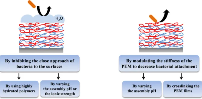
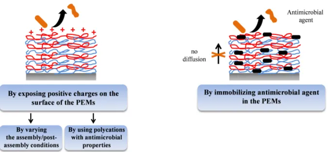
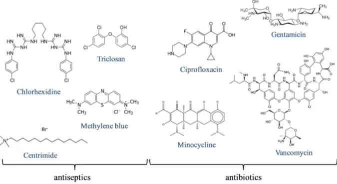
The prevention of pathogen colonization of implantable medical devices constitutes a major medical and financial issue. Indeed, the formation of microbial biofilms at the surface of biomaterials leads if untreated to chronic microbial infection, inflammation, tissue necrosis and eventually to death. Microbial biofilms are extremely difficult to remove because of their high resistance to antibiotic treatment. The only way to treat them is to remove the device from the patient. The key to stop infections lies in killing bacteria that come near the device before they form a biofilm. PEM films were developed for the functionalization of implants in order to inhibit the adhesion and the proliferation of microbes such as bacteria, fungi and yeasts. Three main kinds of PEM coatings have been developed to reduce microbial colonization of biomaterial surfaces: adhesion-resistant coatings (based on pegylated polyelectrolytes or stiff PEM films), contact-killing coatings (based on chitosan, quaternary ammonium polymers or carbon nanotubes) and antimicrobial agents leaching coatings (based on metal/metal ions, oxygen reactive species, antibiotics or natural antimicrobial peptides).
However, none of the developed PEM films exhibited both antibacterial and antifungal properties. So, it would be interesting to develop PEM films that can be multi-defensive, i.e. active against bacteria, fungi and yeasts.
My thesis work deals with the physical chemical study of polymer multilayer films based on supramolecular interactions and with antimicrobial peptides based molecules and multilayer films. In the first part of the manuscript, we were interested in the buildup of neutral polymer multilayer films based on host-guest interactions and in particular on the influence of the strength of the inclusion complex on their topography. In the second part, in collaboration with Pr. Francis Schneider (Centre Hospitalier Universitaire d’Hautepierre, Strasbourg) and Dr. Marie-Hélène Metz-Boutigue (INSERM U1121), cateslytin, a natural antimicrobial peptide derived from the natural processing of chromogranin A, was immobilized in polysaccharide multilayer films to develop self-defensive and multi-defensive coatings against bacteria and yeasts. Antimicrobial and anti-inflammatory properties of cateslytin functionalized dimers and dendrimers were also studied in order to improve the bioactivity of the original peptide. Finally, the functionalization of polyurethane, a polymer used for catheters, was achieved by polydopamine chemistry and the step-by-step cross-linking of polysaccharide films embedding cateslytin.
The manuscript of the thesis is divided in six chapters. In chapter 1, different surface coating methods allowing the functionalization of materials are described and more details concerning the layer-by-layer deposition technique are given. In particular, studies on multilayer films based on host-guest interactions are reported. Then, after a brief description of natural antimicrobial peptides and their properties, a complete state of the art of antimicrobial polyelectrolyte multilayer films is given. Chapter 2 presents the materials and methods (physico-chemical and biological) used during this thesis.
In chapter 3, we investigated the buildup and the topography of neutral polymer multilayer films. The step-by-step deposition of poly(N-hydroxypropylmethacrylamide) bearing β-cyclodextrin hosts and different hydrophobic guests (adamantane, ferrocene, pyrene) induces the formation of a continuous or a droplet-like film depending on the strength of the inclusion complex. This strength can be modulated either by changing the hydrophobic guest involved in the buildup or by the type and the concentration of the salt present during the film buildup. This study is a first attempt to rationalize the evolution of the coating structure as a function of the strength of the interactions between the polymers constituting the multilayer film.
Chapter 4 describes the development of a new self-defensive antibacterial and antifungal coating based on a polysaccharide multilayer film with embedded antimicrobial peptides. Cateslytin including a cysteine residue at the C-terminal position was grafted on hyaluronic acid and assembled with chitosan to form a PEM film. The optimal number of layers required for a complete inhibition of Micrococcus luteus, Staphylococcus aureus and
Candida albicans was determined. The antimicrobial mechanism was also studied thanks to
confocal microscopy.
In chapter 5, cateslytin functionalized dimers and dendrimers were synthesized to improve the bioactivity of the original peptide. Their antibacterial and antifungal properties, their toxicity and for some, their inflammatory properties were assessed and compared to cateslytin.
Chapter 6 reports the development of a robust self-defensive antimicrobial coating for medical catheter using the polydopamine chemistry and the step-by-step cross-linking of polysaccharide multilayer films. While keeping the antimicrobial properties of the PEM film, an increase of its mechanical properties as well as its covalent attachment onto a catheter was pursued.
Finally, a general conclusion is drawn and some outlooks, in particular for the development of antimicrobial coatings for biomaterial application, are given.
Chapter 1:
Chapter 1:
Bibliographic overview
Summary
1.1 Coating strategies ... 7 1.1.1 Langmuir-Blodgett films ... 7 1.1.2 Self-assembled monolayers ... 81.1.3 Polymer adsorption and grafting ... 8
1.2 Polyelectrolyte multilayer films ... 9
1.2.1 Layer-by-layer assembly ... 9
1.2.2 Deposition methods ... 10
1.2.3 Growth regimes ... 12
1.2.4 Tunable properties ... 14
1.3 Multilayer films based on host-guest interactions ... 18
1.3.1 From electrostatic to other kinds of interactions ... 18
1.3.2 Inclusion complexes ... 19
1.3.3 Polymer multilayers based on host-guest interactions ... 23
1.4 Multilayer films as antimicrobial coating ... 25
1.4.1 Medical context: hospital acquired infections ... 25
1.4.2 Antimicrobial peptides ... 27
Cateslytin ... 31
1.4.3 Polyelectrolyte multilayers as antimicrobial surfaces ... 31
This chapter briefly describes the different surface coating methods allowing the functionalization of materials. Among them, the layer-by-layer technique will be further discussed. First based on electrostatic interactions, this technique has been extended to other types of interactions such as the host-guest interactions for example. The interest of layer-by-layer films in the biomaterials field will also be presented. We will in particular focus on the development of antimicrobial coatings since their development was one of the goals of my PhD.
1.1 Coating strategies
Control of the interactions between material surfaces and their environment can be achieved by tailoring their surface properties, especially through the application of coatings. Different techniques of surface functionalization forming a thin film on a surface have been developed usually depending on the nature of both the material and the deposited molecules or macromolecules.
1.1.1 Langmuir-Blodgett films
One of the first surface functionalization technique was proposed by Langmuir and Blodgett in the 1930s [1]. The Langmuir-Blodgett technique allows the deposition of one or several monolayers of amphiphilic molecules on a solid surface with high control of the spatial arrangement. A layer of amphiphilic molecules is first formed at a liquid-air interface and then transferred to a substrate (Figure 1.1). This process can be repeated several times which allows obtaining several monolayers. However, this technique is restricted to amphiphilic molecules and to planar surfaces. Furthermore, due to the weakness of the interactions involved, the obtained coatings have only a limited stability. Nowadays, the Langmuir-Blodgett technique is mainly used in the study of lipid bilayers mimicking cell membranes.
Figure 1.1: Schematic illustration of Langmuir-Blodgett deposition of amphiphilic molecules by withdrawing of
1.1.2 Self-assembled monolayers
An alternative technique to obtain organic thin films on the surface of a material is the self-assembled monolayer (SAM) technique. SAMs are ordered molecular assemblies spontaneously formed by the adsorption of surfactants on a solid surface [3] (Figure 1.2). For example, thiols and disulfides functionalized organic molecules can form highly ordered SAMs on gold surfaces. This technique provides very dense, stable and homogeneous films. Functionalities can be brought to the surface by using functional alkanethiols bearing for example carboxylic acid, alcohol or amine groups as tail groups [4]. The SAMs technique is restricted to surfaces that have appropriate interactions with thiols, thus essentially noble metal surfaces and silanes.
Figure 1.2: Schematic illustration of SAM deposition. The thiol head groups bind to the metal substrate; the
alkane chains form a well-organized interface and act as spacers. The tail groups are located at the interface with the medium and define the surface properties. [5]
1.1.3 Polymer adsorption and grafting
Polymers can be brought to the surface by simple adsorption from solution, by spin casting or by solvent evaporation. Adhesion is in this case mainly obtained by the sum of van der Waals attractions between the individual polymer segments and the surface. For example, poly(ethylene oxide)/poly(propylene oxide)/poly(ethylene oxide) (PEO/PPO/PEO) triblock copolymers were used to anchor PEO chains on hydrophobic surfaces. While PPO segments interact with the hydrophobic surface, PEO chains are extended in the bulk solution [6].
Covalent grafting allows to anchor the polymers more firmly on the surface, either by introduction of reactive groups at chain ends and/or side chains on polymers that can be grafted to the surface (“grafting to”) or by using surface-initiated polymerization (“grafting from”) [7]. As example of a “grafting to” approach, Mansky et al. synthesized by living radical polymerization a series of hydroxyl-terminated random copolymers of styrene and
methyl methacrylate at different ratios. The copolymers reacted then with silanol groups on a silicon wafer surface to form tethered random polymer brushes [8].
1.2 Polyelectrolyte multilayer films
The layer-by-layer (LbL) method was developed by Prof Gero Decher at the beginning of the 1990s [9]. It consists in the alternated deposition of polyanions and polycations through electrostatic interactions. The LbL technique presents a lot of advantages among them the possibility of being used on all kinds of surfaces. Also, it does not require the use of any organic solvent.
1.2.1 Layer-by-layer assembly
Polyelectrolyte multilayer (PEM) films are obtained by the layer-by-layer deposition method through the alternated deposition of negatively charged polymer chains (polyanions) and positively charged polymer chains (polycations) on a substrate (Figure 1.3). The substrate, usually negatively charged, is brought into contact with a polycation solution allowing the adsorption of positively charged chains on the surface through electrostatic interactions. A rinsing step is usually employed after each polyelectrolyte deposition to eliminate weakly adhering polymer chains. The surface becomes positively charged (charge overcompensation) and a layer of polyanions can thus be adsorbed by the same process. Again charge overcompensation takes place and at the end of the polyanion deposition the surface is negatively charged allowing the deposition process to be continued again with a polycation. This process can be repeated until the desired film thickness is obtained.
Figure 1.3: Schematic representation of a polyelectrolyte multilayer film buildup by successive adsorptions of
1.2.2 Deposition methods
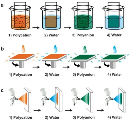
Different processes have been developed for the buildup of PEM films: the dip-coating, the spin-coating and the spray-coating method.
Dip-coating method
The dip-coating method (Figure 1.4a) has been introduced in the 1990s by Decher et al. to form for example poly(allylamine hydrochloride)/poly(styrene sulfonate) (PAH/PSS) films [10]. The substrate was alternately dipped in polycation and polyanion solutions, each dipping being followed by a rinsing step. This simple technique can be used on various substrates, whatever their shape. The time needed for the adsorption of the polyelectrolytes on the substrate ranges typically from 5 to 20 minutes. Dip-coating is thus a slow deposition method. However, one of the advantages of this method is the low consumption of products since the same polyelectrolyte solution can be used for several deposition steps. The thickness and the roughness of the obtained films depend mainly on the immersion time, the film growth regime, the ionic strength, the pH and the temperature (see 1.2.3 and 1.2.4).
Figure 1.4: Schematic representation of the processes used to build PEM films: (a) dipping LbL assembly, (b)
spin-assisted LbL assembly and (c) spray-assisted LbL assembly. The growth of the films is based on the repetition of the cycle (steps 1 to 4). [11]
Spin-coating method
Used over several decades for the application of thin organic films on planar surfaces, the spin-coating method has been used in the 2000s to decrease the buildup time of PEM films [12]. In this process, the polyelectrolyte solution is first deposited on the substrate which is then accelerated rapidly to the desired rotation speed. Due to the action of the centrifugal force, the liquid solution flows radially and its excess is ejected off the substrate. This allows a uniform covering of the substrate by a polyelectrolyte monolayer (Figure 1.4b). No rinsing step is required in this technique so the oppositely charged polyelectrolyte solution can be immediately deposited to form the second layer. The deposition time of a monolayer is about 1 minute; spin-coating is thus a very fast deposition method. The thickness and the roughness of the PEM films depend mainly on the rotation speed and the viscosity of the polyelectrolyte solutions. For example, a low rotation speed or a high concentration of polyelectrolytes in solution leads to thicker films [12]. A planar surface and a reasonable size of the substrate are required to use the spin-coating process.
Spray-coating method
The spray-coating method has been reported for the first time in the literature by Schlenoff et al. who sprayed alternately PSS and poly(diallyldimethylammonium chloride) (PDADMAC) solutions on a vertical substrate, allowing the drainage of the excess solution [13] (Figure 1.4c). Each deposition step was followed by a rinsing step which was performed by spraying pure water. Highly uniform films were obtained. The major advantages of the spray-coating method compared to the dip-coating one are that spraying is fast and allows the coating of large surface areas. However, the spray-coating method consumes a high amount of polyelectrolytes solution. Most of the sprayed solution is indeed lost by drainage and not reusable afterwards. Spraying time, spraying distance, pH, ionic strength and concentrations of the polyelectrolyte solutions have an influence on the thickness and the roughness of the films. Izquierdo et al. showed that the rinsing step can be omitted without impairing the film buildup [14]. This led Porcel et al. to propose, in 2005, to spray simultaneously the polyanion and the polycation solutions on a substrate held vertically [15]. This process, now denoted as the Simultaneous Spray Coating of Interacting Species (SSCIS), leads to organic films whose thickness increases linearly with the spraying time. The method was recently shown to be quite more general, applying (i) to almost any kind of polycation/polyanion systems, (ii) to polyelectrolyte/small multi-charged molecule systems, (iii) to polyelectrolyte/nanoparticles
systems and even (iv) to the simultaneous spraying of two inorganic solutions [16]. This simultaneous spraying method allows reducing the buildup time of polyelectrolyte films since the spraying time can be divided by two but the films are generally rougher and no longer structured [17].
1.2.3 Growth regimes
Two types of growth regimes are observed for PEM films, generally named linear and exponential growth, which are represented figure 1.5.
Figure 1.5: Evolution of the thickness of a () PEI-(PSS/PAH)i system and a ( ) PEI-(PGA/PLL)i system depending on the number of deposited bilayers i. The first system is linear whereas the second one is exponential. [18] (PGA: poly(glutamic acid); PLL: poly-L-(lysine))
Linear growth
In the case of linearly growing films, the thickness or the mass increment of the adsorbed pair of layers (one layer of polyanion and one layer of polycation, also called bilayer) is constant whatever the number of adsorption cycles. The polyelectrolytes from the solution interact only with the top outer layer of the film forming stratified PEM films as it was shown by neutron reflectometry measurements [19]. PAH/PSS film is one of the most prominent example of linearly growing films [9, 19].
Exponential growth
Until 1999, it was assumed that the thickness of PEM films always increased linearly or super-linearly with the number of deposition steps. The super-linearly growth, which is a slightly faster growth than the linear one, was attributed to an increase of the film roughness
along the whole buildup process [20, 21]. In 1999, Hubbell and coworkers found that the LbL deposition of poly(L-lysine) (PLL) and alginate (ALG) led to the formation of gel-like films where the thickness is exponentially increasing with the number of deposition steps instead of the usually observed linear thickness increase [22]. Shortly later, Picart and coworkers [23-25] found that it was not an isolated example: exponentially growing films constitute a new class of polyelectrolyte multilayers. Exponentially growing films are characterized by an exponential thickness and mass increase with the number of adsorption cycles. The exponential growth can be explained by the diffusion of at least one of the polyelectrolytes into the entire film during each deposition step. When the film is brought into contact with the polyelectrolyte of opposite charge, the polyelectrolyte that has already diffused into the film diffuses again partly out. Once it reaches the film-solution interface, it is complexed by the oppositely charged polyelectrolyte from the solution. These complexes remain bound to the film and form an additional layer on the top of the film. The mass and the thickness of this layer being proportional to the amount of polyelectrolytes that diffused in and out of the film, the film growth becomes exponential. The obtained films are highly hydrated and thus resemble to hydrogels. Most polypeptides and polysaccharides films have an exponential growth. That is for example the case of the poly(L-lysine)/hyaluronic acid (PLL/HA) [26] or PLL/poly(L-glutamic acid) (PLL/PGA) systems [27].
Some exponentially growing films enter into a linear growth phase after a certain number of deposition steps. This has been first reported by Hübsch et al. [28] for the buildup of a (PGA0.5-PSS0.5/PAH)n multilayer film (Figure 1.6) where the growth goes from
exponential to linear after deposition of 26 layers.
Figure 1.6: Thickness of a (PGA0.5-PSS0.5/PAH)n multilayer film. There is an exponential growth until the 26th layer and then a linear regime. [28]
Such a transition was also described later in the case of PLL/HA [29] and seems to be valid for all exponentially growing PEMs. The reason for the exponential to linear transition is still not fully understood. It could be related to the fact that, when the polyelectrolytes diffuse out of the film, only the first chains reaching the film-solution interface can form polycation/polyanion complexes that remain bound to the film. The interactions between the surface and the complexes are not sufficient anymore to be maintained on the surface and they diffuse in the solution.
The growth regime of a PEM film was related to the interaction strength between the two polyelectrolytes. Linearly growing films, like PAH/PSS, are based on polyelectrolyte pairs that have a negative complexation enthalpy and hence an enthalpic contribution to the free energy of complexation. In this case the buildup process is both enthalpically and entropically driven. On the other hand exponentially growing films, like PLL/PGA, are based on polyelectrolytes that have a positive complexation enthalpy and so the complexation is entirely entropically driven [30].
Moreover, depending on the buildup parameters, some PEM films based on the same two polyelectrolytes can be exponential or linear. The influence of the main parameters on the PEM buildup is described in the next paragraph.
1.2.4 Tunable properties
Many parameters have an influence on the buildup of PEM films, especially on their growth mode, thickness, roughness and density.
Ionic strength
The ionic strength of the polyelectrolyte solutions used during the buildup of the PEM films has a strong influence on their thickness and on their growth mode. An increase of salt concentration usually leads to thicker films [31-33]. For example, PAH/PSS multilayer films were reported to possess an average bilayer thickness increasing from 10.9 to 22.6 Å for NaCl concentrations increasing from 0 to 2.0 mol.L-1 [32]. At low ionic strength, the polyelectrolytes adopt a “flat” or rigid-rod like conformation since the charges on the polyelectrolyte repel each other, whereas they adopt a “loopy” one at high ionic strength
(Figure 1.7). A higher thickness of PEM films is thus obtained at high ionic strengths due to the adsorption of polyelectrolytes in “loopy” conformation [32].
Figure 1.7: Evolution of a polycation chain conformation with the ionic strength.
Richert et al. [34] showed the influence of the ionic strength on the growth regime of a chitosan/hyaluronic acid (CHI/HA) PEM film. When the polyelectrolytes are dissolved in 10-4 M NaCl, the CHI/HA film possesses a linear growth but tends to an exponential growth when built in the presence of 0.5 M NaCl (Figure 1.8).
Figure 1.8: Effect of the ionic strength on the buildup of a (CHI/HA) multilayer film followed by quartz crystal
microbalance. The system goes from linear to exponential with a NaCl concentration increase. [34]
Nature of the counterions
The nature of the counterions has also a major influence on the PEM buildup. Salomäki et al. showed for example that the thickness of a PDADMAC/PSS multilayer film is depending on the anion present in solution, keeping sodium as cation [35]. Ions can be ranked by their degree of hydration, especially anions because they usually have a higher effect on water than cations. The smaller the hydration sphere of the anion is, the higher is the interaction between the anions and the positive charges of the polyelectrolytes. In the case of
+ + + + + + + + + + + + ionic strength
weakly hydrated ions, the anions interact more strongly with the positive charges of the polycations which leads to a reduction of the polyanion/polycation interactions resulting in an increase of the film thickness as well as its roughness. This effect is similar to the one obtain by increasing the ionic strength. Thus, the use of cosmotropic anions, i.e. highly hydrated anions, leads to thinner films, whereas the use of chaotropic ones, i.e weakly hydrated anions, allows the buildup of thicker films and eventually even exponentially growing ones [35] (Figure 1.9). A comparable study was performed on a PAH/PSS film. This PEM film was reported to be two times thicker for chaotropic counterions than for cosmotropic ones, but linear growth was kept in both cases [36].
Figure 1.9: Effect of the counterion on the buildup of a (PDADMAC/PSS) multilayer film in a 0.1 M sodium salt
solution of the corresponding anion. The system goes from linear to exponential when using anions going from cosmotropes to chaotropes. [35]
Temperature
The influence of the temperature on the buildup of PEMs has been mainly studied on linearly growing films. Salomäki et al. [37] showed that a PDADMAC/PSS multilayer film grows linearly at 15°C and 25°C whereas the buildup becomes exponential for higher temperatures, 45°C and 55°C (Figure 1.10). This growth mode change is due to a faster diffusion of the polyelectrolytes at higher temperatures. Relying on a model, Salomäki et al. proposed that every LbL buildup is inherently exponential and turns linear when the diffusion rate is not fast enough to transport the polyelectrolyte within the entire thickness of the formed layer.
Figure 1.10: Effect of the temperature on the buildup of a (PDADMAC/PSS) multilayer film in a 0.1 M NaBr
solution followed by the acoustic impedance evolution of a gold-covered quartz substrate. The system goes from linear to exponential with a temperature increase. [37]
Other parameters can influence the PEM buildup such as the pH in the case of weak polyelectrolytes [38, 39], the molecular weight of the polyelectrolytes [40, 41] or their concentrations [42].
The influence of pH has been studied for PEM films involving weak polyelectrolytes because their ionization degree depends on pH. A variation in pH induces a change on the number of charges on the polyelectrolytes which leads therefore to a change of the polymer chain conformation in solution. For example, when the polymer chains are highly ionized, they adopt a “flat” conformation leading to thin films. This effect is similar to the one described previously for the ionic strength. Rubner and coworkers showed that a variation of pH has a large effect on a PAH/Poly(acrylic acid) (PAA) system. By varying the pH between 2.5 and 8.5, they could modulate the thickness of the adsorbed PAA layer over the range of 5 to 80 Å. It was also reported that the control of the ionization degree by pH allowed the control of the bulk and surface composition of the PEM film [39].
The molecular weight of the polyelectrolytes plays a role in the diffusion process. Kujawa et al. showed that the thickness of an exponentially growing CHI/HA film depends on the molecular weight of both polyelectrolytes: the diffusing one (CHI) and the non-diffusing one (HA) [41]. Short polymer chains can diffuse easily in contrary to long polymer chains, which diffusion can be blocked
1.3 Multilayer films based on host-guest interactions
The cohesion of PEM films is primarily based on electrostatic interactions but it was extended over the years to other driving forces such as hydrogen bonding [43, 44], metal-ion coordination [45] or host-guest interactions. After a brief description of hydrogen bonding and metal-ion coordination based PEM, a closer interest will be taken on host-guest interactions especially the one involving β-cyclodextrin.
1.3.1 From electrostatic to other kinds of interactions
The buildup of PEM films based on other driving forces than the electrostatic interactions allowed a new control of the cohesion of the assemblies. Some elements will be given in this paragraph about hydrogen bonding and metal-ion coordination.
Hydrogen-bonding based PEM
The hydrogen bond [46], whose energy is between 5 and 30 kJ.mol-1, is stronger than a van der Waals interaction but weaker than an ionic bond. Rubner’s team proposed in 1997 to use this type of interaction to build multilayer films [44]. Films made of polyaniline and a hydrogen-bonding polymer such as poly(ethylene oxide), poly(acrylamide) or poly(vinyl alcohol) could be assembled in a layer-by-layer manner. PAA/poly(4-vinylpyridine) (PVP) films were also assembled at low pH, PAA being fully protonated [47]. By modifying the pH, it was possible to break the hydrogen bonds leading to the film dissolution above pH 6.9 [48]. This can be explained by an increasing ionization of PAA carboxylic acid groups when the pH is increased. This induces an electrostatic repulsion between the layers. Hydrogen-bonding films or capsules with tunable dissolution have been extensively studied for biomedical application [49].
Metal-ion coordination based PEM
In 1998, Xiong et al. introduced the coordination chemistry in the LbL assembly field by building up poly(copper styrene 4-sulfonate)/PVP films. By infrared spectroscopy, they showed that the buildup is possible thanks to the formation of a complex involving copper ions, pyridine groups of PVP and sulfonate groups of poly(copper styrene 4-sulfonate). Using a bifunctional polyelectrolyte (BiPE) bearing bipyridine metal ion receptors and positively charged ammonium groups, Krass et al. [45] built a multilayer film with either electrostatic
cohesion when BiPE was assembled with a negatively charged polymer such as PSS or by metal coordination cohesion with BiPE only (Figure 1.11). In the first case, the layers can reversibly accept and release transition metal ions. In the second case, the layers are disassembled with a complexing agent such as EDTA.
Figure 1.11: Structure of the bifunctional polyelectrolyte (BiPE) bearing bipyridine metal ion receptors and
positively charged ammonium groups. BiPE can be assembled through electrostatic interactions with negatively charged polymers such as PSS (top) or it can be assembled through metal ion coordination (bottom) [45].
1.3.2 Inclusion complexes
The “host-guest interaction” expression is used to designate an inclusion complex between a molecule, i.e. the guest, that is trapped within the cavity of a broader one, i.e. the host. Guest molecules can be encapsulated by more than one host or a host can interact with multiple guests. Figure 1.12 represents a 1:1 inclusion complex between a cyclodextrin (CD) and a guest compound.
A common host molecule: β-cyclodextrin
In the pharmaceutical and biomedical field, cyclodextrins (CD) [50] are the most common used host molecules, especially β-cyclodextrin. CDs are cyclic oligosaccharides composed of D-glucose subunits that are joined by α-1,4 glucosidic linkages. These natural molecules, obtained by enzymatic degradation of starch, possess a truncated cone structure. The CD’s hydroxyl groups are located at the outer surface of the molecule which makes CD water-soluble and let a hydrophobic cavity within it (Figure 1.13).
Figure 1.13: (a) Chemical structure of β-cyclodextrin and (b) its schematic 3D representation.[50]
Because of this hydrophobic interior cavity, CD has the ability to form inclusion complexes with various non-polar molecules. The main driving forces of the inclusion complexes are hydrophobic and van der Waals interactions. An important factor for the formation of an inclusion complex is the size of the guest. If the guest is too small, the distance between the guest and the interior of the CD is too high and the intermolecular forces will not exist. If the guest is too large, complex formation cannot happen due to steric hindrance. Among the numerous guest molecules, adamantane (Ad) and ferrocene (Fc) are the ones that have the highest binding affinity with β-CD. Ad is a cycloalkane that consists of three rings fused to each other in a chair conformation (Figure 1.14a). Fc is an organometallic compound that is composed of two planar cyclopentadienyl rings bound to a central iron atom from opposite side like a sandwich (Figure 1.14b).
Figure 1.14: Guest molecules of β-cyclodextrin: (a) adamantane and (b) ferrocene.
a b
Association constant
The stability of a host-guest complex is described by its association constant K. In the case of a 1:1 inclusion complex with CD, K is defined as following for a guest G:
+ ⇌ − K = [ ]
[ ][ ] (1.1)
Where [G] is the concentration in guest molecule; [CD] is the concentration in cyclodextrin molecules and [ − ] the concentration in host-guest complexes. The association constant
provides a qualitative measurement of the binding affinity. The higher K is, the higher is the binding affinity. For inclusion complexes between CD and a guest molecule, association constants are typically in the range of 103-105 M-1 [51]. For example, at 25°C, log K = 4.26 for CD/ferrocene carboxylate in pure water at pH 8.5 [52] and log K = 3.33 for β-CD/ferrocene carboxylate in 50 mM phosphate buffer at pH 8.6 with 0.1 M NaCl [53]. Thermodynamic parameters of the complexation of cyclodextrins with hydrophobic guests, including association constants, have been mainly determined with microcalorimetry techniques, especially isothermal titration calorimetry since it is the most sensitive method available at the moment. But a wide variety of other experimental methods are found in the literature: electronic absorption, circular dichroism, fluorescence, nuclear magnetic resonance, electron spin resonnance spectroscopy, gas- and liquid-phase chromatography, capillary electrophoresis, pH potentiometry, the use of ion selective electrodes, kinetic experiments and solubility determination [53]. Whereas calorimetric methods allow the direct and simultaneous determination of the association constant and the reaction enthalpy, other methods have to be used carefully since they allow access to the thermodynamic parameters from the van’t Hoff equation based on some assumptions which can impair the accuracy of the results.
Influence of the environment on the association constant
Since the pioneering experiments of Hofmeister about specific ion effects [54], numerous studies about salts affecting inclusion complexes equilibrium involving β-CD have been reported in the literature [55-57]. Depending on the type of salt present in solution, the binding affinity is stronger or weaker. Some key elements to understand the role of the salts on the inclusion complex will be presented.
One first has to remind that all molecules in an aqueous solution are surrounded by surface-hydrating water molecules. Thus, the specific association of host and guest in solution
is regulated by their desolvation. Thermodynamic studies have shown that β-CD/Ad complexation in aqueous solution involves the release of water from the interacting surfaces (Figure 1.15). It was estimated by Harries et al. that 15 to 25 water molecules are released during the formation of this complex [51].
Figure 1.15: Schematic of the complexation reaction between adamantane carboxylate and β-CD. The scheme
shows that the release of surface-hydrating water molecules from both interacting molecules is required in order to make it possible for the adamantane carboxylate to fit in the β-CD cavity. [51]
The Hofmeister series constitutes a classification of ions depending on their ability to stabilize the structure of proteins. Except some relative positions in the series, they are basically ranked by their degree of hydration (Figure 1.16). The weakly hydrated anions (exhibiting weaker interactions with water) are called chaotropes whereas the strongly hydrated ones (exhibiting strong interactions with water molecules) are called cosmotropes.
Figure 1.16: Hofmeister series: ranking of anions by their hydration degree. The hydration degree is decreasing
from the left to the right of the scheme.
Several studies pointed out that the stability and the association constant of β-CD/Ad [58], γ-CD/Ad [59], β-CD/nabumetone [60], β-CD/1-butanol [61], β-CD/phenylbenzothiazole derivatives [62], CD/nitroxide radical probe [63] complexes can be modulated by co-solutes such as salts. Chaotropic anions have the tendency to destabilize host-guest complex formation in contrary to cosmotropic anions that promote it. From the literature, it is known that chaotropic anions, like ClO4- [64-66] and SCN- [65, 67], can both form 1:1 complexes
with β-CD with a stability constant log K = 1.42 [65] and 0.99 [65], respectively. In contrast, cosmotropic ones, like SO42- and F- anions, do not form 1:1 inclusion complexes with β-CD,
since their stability constant are negative [64, 66]. It has been proposed that the inside of β -CD cavity is composed of positively polarized carbon atoms leading to the inclusion of
HPO
42-SO
42-
C
2H
3O
2-F
-Cl
-Br
-I
-ClO
4-SCN
hydrophobic anionic guest [68]. In the case of ClO4- and SCN- anions, hydrophobic
interactions are presumed to contribute to their inclusion within the β-CD cavity. Since SO4
2-and F- anions are strongly hydrated, they do not bind to β-CD. The decrease of affinity constant when a chaotropic anion is involved can be explained by the competition with the guest for the binding site. The increase of the association constant when a cosmotropic anion is involved is correlated with the water molecules released from both the β-CD cavity and the guest upon the formation of the complex. The presence of a cosmotropic anion such as SO4
2-or F- disturbs indeed the internal structure of water around the β-CD and its guest, rendering the releasing step of water easier and thus displacing the equilibrium toward the complex formation. Both phenomena are exacerbated when the salt concentration is higher.
1.3.3 Polymer multilayers based on host-guest interactions
β-CD is readily available and is also an effective host for a large range of hydrophobic
guests including drug molecules. So, β-CD was often embedded in multilayer films based on polyelectrolyte/β-CD [69], polyelectrolyte/β-CD-modified polyelectrolyte [70], polyelectrolyte/poly(CD) [71] or even more recently poly(CD)/poly(CD) systems [72].
But only few examples of multilayer films based on host-guest interactions between β-CD and a guest molecule are reported in the literature. In 2002, Suzuki et al. reported the first assembly based on host-guest interactions [73]. They showed that electrostatic repulsions between PAH chains can be overcome through interactions between ferrocene moieties grafted on the PAH chains and a β-CD homobifunctionalized molecule. Three deposition steps of PAH-Fc alternating with the β-CD homobifunctionalized molecule were successfully performed on a gold substrate (Figure1.17).
Figure 1.17: Assembly of positively charged-ferrocene-modified poly(allylamine) multilayer on a gold surface
Huskens and coworkers developed then a layer-by-layer assembly of adamantane-functionalized dendrimers and β-CD-adamantane-functionalized gold nanoparticles on a β-CD SAM, obtaining well-defined thin multilayer films (Figure 1.18) [74]. Using nano-imprint lithography or transfer printing, they created 3D supramolecular assemblies with controlled size and geometry.
Figure 1.18: Assembly of adamantane-functionalized dendrimers and β-CD-modified gold nanoparticles. [74]
Later, in 2006, van der Heyden et al. reported the first assembly only based on host- and guest-modified polymers [75]. They showed that chitosan chains onto which were grafted respectively β-CD and Ad moieties can be alternately deposited on a gold surface covered with Ad-functionalized SAMs. PEM films constituted of only 2 to 4 layers were built depending on the density of Ad moieties exhibited by the surface (Figure 1.19). By using Fc- and β-CD-modified poly(allylamine), Wang et al. succeeded in depositing at least 8 bilayers on CaCO3 particles [76]. The alternated incubation of the substrate in PAH-CD aqueous and
PAH-Fc methanol solution allowed decreasing both the charge repulsion and the polymer dissolution during the film formation.
Figure 1.19: Shematic representation of the influence of the SAMs adamantly densities on the self-assembly
buildup: (A) high, (B) medium and (C) low adamantly ratio exhibited by the SAM. [75]
Since the use of polyelectrolytes limits the number of effective deposition cycles, Dubacheva et al. developed a new approach by using neutral polymers. They succeeded in an unlimited host-guest multilayer film buildup in aqueous solution by using alternately neutral poly(N-hydroxypropylmethacrylamide) chains bearing respectively β-CD and Fc moieties.
Yet, none of these studies focused on the topography of the formed film and this will be one of the goals of my PhD.
1.4 Multilayer films as antimicrobial coating
PEM films have been developed for a large range of applications in fields as different as photodiodes [77, 78], optical devices [79], filtration membranes [80, 81], fuel cell membranes [82], biologically active membranes [83], drug release [84, 85] or biologically active coatings [86-88]. Here, we are interested in their use as antimicrobial coatings since it is in the context of my thesis. After a short presentation of the hospital acquired infections, a description of the antimicrobial peptides will be given. Finally, the different studies about PEM films used as antibacterial and antifungal coatings will be reported.
1.4.1 Medical context: hospital acquired infections
Implantable medical devices are widely used in surgery not only to replace altered or lost tissues but also in critical care for fluid or gas administration using catheters or tracheal tubes, respectively. These devices constitute an open gate for pathogen invasion [89]. Prevention of pathogen colonization of medical implants constitutes a major medical and financial issue since nosocomial infections represent one of the most serious complications after surgery or critical care. Indeed each year in Europe, 5% of patients admitted to hospitals suffer from hospital-acquired infections leading to a mortality of 10% [90]. The most encountered pathogens in nosocomial infections are Staphylococcus aureus (S. aureus) and
Candida albicans (C. albicans). S. aureus [91] is a gram-positive bacterium and is one of the
most virulent bacteria leading to high rates of device-related systemic infections and mortality. C. albicans is the most common human yeast pathogen and possesses the ability of forming biofilms that are sources of local and systemic infections. Biofilms are communities of pathogens embedded in a polysaccharide matrix adhering to a surface. These communities can form within a few hours [92]. First, free-floating bacteria encounter a submerged surface (such as a catheter), attach to it within a few minutes and start then to produce extracellular polymeric substances which allow the development of a complex three-dimensional structure: the biofilm (Figure 1.20).
Figure 1.20: The biofilm life cycle: (1) attachment of free-floating microorganisms to a surface and production
of extracellular polymer substances (EPS) that lead to irreversible attachment, (2) maturation of the biofilm and (3) detachment of clumps or individual cells and propagation of the biofilm. [93]
Reaching a critical size, the biofilm can propagate through detachment of individual cells or groups of cells that attach to another surface. Bacteria are 10 to 1000 times more resistant to antibiotic under a biofilm form than under a planktonic one. Biofilms are responsible for 2/3 of hospital-acquired infections developed in the blood. In particular, C.
albicans biofilms allow the formation of S. aureus microcolonies on their surface and by
symbiotic interactions even enhance S. aureus resistance to antibiotics increasing the frequency or the severity of diseases [94].
The capacity of bacteria to more or less strongly adhere on a surface and to build a biofilm is depending on the kind of bacteria. Bacteria can be classified by their shape. For example, Staphylococcus epidermis (S. epidermis) is a spherical cocci and Escherichia coli (E. coli) is a cylindrical bacilli. But they are also commonly classified as Gram positive or Gram negative (Figure 1.21). Gram positive bacteria such as S. epidermis exhibit an outermost multilayered peptidoglycan cell wall, embedded with teichoic acid polymers at the top of the inner cell membrane that can include ion channels and protein receptors. In contrast, Gram negative bacteria such as E. coli exhibit a single peptidoglycan layer between a lipopolysaccharide-rich outer layer and phospholipid-rich inner layer [95].
Figure 1.21: Classification of bacteria by shape and then by their outermost cell envelope composition [95].
Prevention of pathogen colonization of implantable medical devices constitutes a major medical and financial issue. For these reasons, there is a growing interest in the biomaterial field to provide new coatings and materials to prevent bacterial adhesion and biofilm formation.
1.4.2 Antimicrobial peptides
Antimicrobial peptides (AMPs) [96-99] are endogenous polypeptides that are part of the innate immune system of multicellular organisms including animals, plants, bacteria, fungi, viruses and humans. More than 800 different AMPs have been isolated in the last decades. Used in the macromolecular range, they are known to display a broad spectrum of antimicrobial activities against various microorganisms such as viruses, fungi, bacteria and parasites.
Classification of AMPs
AMPs form a chemically and structurally heterogeneous family, which renders it hard to define them, but most of them still share some common features. They generally are constituted of less than 50 amino acids and possess a cationic amphiphilic character. They have indeed the ability to adopt an amphipathic shape in which clusters of hydrophobic and hydrophilic amino acids segregate. Several classifications of AMPs exist in the literature but they are often classified into five categories based on their amino acid composition and structure: anionic, linear amphipathic α-helical, cationic peptides enriched in specific amino
acids, peptide fragments and peptides with cysteines that form intramolecular bonding (see Table 1.1).
Table 1.1: Classification of antimicrobial peptides by their amino acid composition and structure. Inspired from
Brogden et al. [100].
Class Characteristics Examples
Anionic peptides Contain glutamic or aspartic acids
Maximin H5 from amphibians [101], dermcidin from humans [102] Linear cationic α-helical
peptides Lack in cystein
Cecropin P1 from nematodes [103], magainin 2 from amphibians [104] Cationic peptides enriched
for specific amino acids
Rich in proline, arginine, phenylanaline, glycine,
tryptophan
Abaecin from bees [105], indolicidin from bovines [106]
Peptide fragments Cationic, β-turn Lactoferrin from lactoferrin [107], cathelicidins from mammals [108] Charged peptides with
cysteine Contain 1 to 3 disulfide bonds
Defensins A from insects [109], protegrin from porcines [110]
The first group is composed of anionic peptides. These peptides are rich in aspartic acid or glutamic acid, which provide negative charges. The second group contains linear cationic α-helical peptides that are disordered in solution but that adopt an α-helical secondary structure once in contact with membranes. The third group is composed of linear peptides enriched in specific amino acids such as proline, tryptophan, glycine, histidine and arginine. AMPs of this group lack of cysteine residue, making them very flexible and fluid in solution. The forth group is formed by charged peptides that are fragments of large proteins. The fifth group is composed of AMPs rich in cysteine residues. They are usually cyclic peptides with
β-sheets that are stabilized by disulfide bonds.
Mechanism of action
Despite their vast diversity, most AMPs act by disrupting the integrity of the microbial cell membrane. Diamond et al. [98] proposed a mechanism of action in three steps: attraction, attachment and insertion of the peptides in the membrane. Attraction is presumed to occur via electrostatic interactions between the cationic peptides and the negatively charged moieties on the microbial cell membrane. Beside electrostatic interactions, hydrophobic and polar residues
play a crucial role in the attachment step. AMPs can indeed adopt a secondary structure, which is energetically favorable, once close to phospholipids membranes. The last step is the insertion of the peptides in the membrane. Several models of penetration have been found that depend on the class of the AMPs and also on the microorganisms [100, 111]. The three main models are the carpet model, the barrel-stave and the toroidal pore model (Figure 1.22). In the case of the carpet model, there is an accumulation of AMPs on the bilayer surface until a saturation point is reached where the membrane disintegrates and forms micelles. In the case of the barrel-stave model, AMPs form a bundle in the membrane with a pore in the center. The hydrophobic regions of the AMPs interact with the phospholipids of the membrane while the hydrophilic ones are oriented inward into the water-filled pore. The toroidal pore model is rather similar to the barrel-stave model. They differ by the fact that the AMPs are always associated with the lipid head groups even when perpendicularly inserted into the lipid bilayers.
Figure 1.22: Schematic representation of some action mechanisms of AMPs disrupting the microbial cell
membrane. (A) Barrel-stave model: AMPs insert themselves into the membrane perpendicularly. (B) Carpet model: Small areas of the membrane are coated with AMPs with hydrophobic sides facing inward leaving pores behind in the membrane. (C) Toroidal pore model: This model resembles the barrel-stave model but in this case AMPs are always in contact with phospholipid head groups of the membrane. The blue color represents the hydrophobic parts of AMPs whereas the red color represents their hydrophilic part.[111]
Besides the membrane disruption, AMPs have other ways to kill a pathogen. Once they have penetrated inside the cell, AMPs act on intracellular targets of cytoplasmic
components crucial to proper cellular physiology. They can for example inhibit protein or cell-wall synthesis and also interact with DNA or RNA [98].
Chromogranin A and its derived antimicrobial peptides
Chromogranin A (CgA) is a protein composed of 439 amino acid residues for humans and 431 for bovines and belongs to the granin family. This protein is located in secretory vesicles of endocrine, immune and neuron cells [112]. Chromofungin, vasostatin, catestatin and its active form cateslytin are four antimicrobial peptides derived from CgA (Figure 1.23) [113, 114]. We took an interest in particular for cateslytin, corresponding to CgA344-358,that is
a positively charged (5+) arginine-rich AMP and that possesses antimicrobial activities against both bacteria and yeasts. In the intragranular matrix, CgA fragments result from the processing of CgA by numerous enzymes such as prohormone convertases (PC1 and 2), aminopeptidases and carboxypeptidases. In the extracellular medium, larger forms may be processed by kallikrein located at the plasmic membrane level and by circulatory proteolytic enzymes such as plasmin and thrombin [115]. In addition, some virulent bacteria produce proteolytic enzymes, for example Glu-C protease for S. aureus, and might continue the natural processing of CgA to generate the chromofungin and catestatin fragments during infections [115, 116].
Cateslytin
As antimicrobial peptide of interest for my thesis, the antimicrobial mechanism of action of cateslytin will be briefly described here. It was demonstrated that cateslytin interacts with fungal membrane by adopting an aggregated antiparallel beta-sheet structure at the negatively charged surface of the bacterial membranes [118] and generates pores of 1 nm diameter [119] that destabilize then the membranes leading to the bacteria death. The five arginine residues that CTL possesses are essential for the binding to the negatively charged lipids of the bacterial membrane.
1.4.3 Polyelectrolyte multilayers as antimicrobial surfaces
Prevention of microbial adhesion and proliferation on implant surfaces is a topic of major medical and financial importance. Three main kinds of coatings have been developed to reduce microbial colonization of material surfaces: adhesion-resistant, contact-killing and antimicrobial agent leaching coatings (Figure 1.24). PEM films were developed for the functionalization of implants in order to inhibit the adhesion and the proliferation of microbes such as bacteria, fungi and yeasts. After a brief description of the methods used to evaluate anti-adhesive and antimicrobial properties of PEM films, the state of the art will be described in details for PEM films.