Core Circadian Clock Genes Regulate
Leukemia Stem Cells in AML
The MIT Faculty has made this article openly available.
Please share
how this access benefits you. Your story matters.
Citation
Puram, Rishi V. et al. “Core Circadian Clock Genes Regulate
Leukemia Stem Cells in AML.” Cell 165, 2 (April 2016): 303–316 ©
2016 Elsevier Inc
As Published
http://dx.doi.org/10.1016/J.CELL.2016.03.015
Publisher
Elsevier
Version
Author's final manuscript
Citable link
http://hdl.handle.net/1721.1/116753
Terms of Use
Creative Commons Attribution-Noncommercial-Share Alike
Core circadian clock genes regulate leukemia stem cells in AML
Rishi V. Puram1,2,3, Monika S. Kowalczyk3, Carl G. de Boer3, Rebekka K. Schneider1, Peter G. Miller1,4, Marie McConkey1, Zuzana Tothova1,3,4, Héctor Tejero5, Dirk Heckl1,6, MarcusJärås1,7, Michelle C. Chen1, Hubo Li2,4, Alfred Tamayo8, Glenn S. Cowley3, Orit Rozenblatt-Rosen3, Fatima Al-Shahrour1,5, Aviv Regev3,9,10, and Benjamin L. Ebert1,3,4,*
1Division of Hematology, Department of Medicine, Brigham and Women’s Hospital, Harvard Medical School, Boston, MA, 02115
2Program in Biological and Biomedical Sciences, Harvard Medical School, Boston, MA, 02115 3Broad Institute of Harvard University and MIT, Cambridge, MA, 02142
4Dana-Farber Cancer Institute, Harvard Medical School, Boston, MA, 02215
5Translational Bioinformatics Unit, Spanish National Cancer Research Centre (CNIO), Madrid, Spain, 28029
6Department of Pediatric Hematology and Oncology, Hanover Medical School, Hanover, Germany, 30625
7Lund University, Department of Clinical Genetics, Lund, Sweden, 22184 8Harvard Medical School, Department of Neurobiology, Boston, MA, 02115
9Department of Biology, Massachusetts Institute of Technology, Cambridge, MA, 02139 10Howard Hughes Medical Institute, Chevy Chase, Maryland, 20815
SUMMARY
Leukemia stem cells (LSCs) have the capacity to self-renew and propagate disease upon serial transplantation in animal models, and elimination of this cell population is required for curative therapies. Here, we describe a series of pooled, in vivo RNA interference (RNAi) screens to identify essential transcription factors (TFs) in a murine model of acute myeloid leukemia (AML) with genetically- and phenotypically-defined LSCs. These screens reveal the heterodimeric, circadian rhythm TFs Clock and Bmal1 as genes required for the growth of AML cells in vitro and in vivo. Disruption of canonical circadian pathway components produces anti-leukemic effects, including impaired proliferation, enhanced myeloid differentiation, and depletion of LSCs. We
*Correspondence: Benjamin L. Ebert, CONTACT INFORMATION: [email protected], Address: 1 Blackfan Circle, Karp Building
(CHRB 05.125), Boston, MA, 02115, Phone: 617-355-9060.
AUTHOR CONTRIBUTIONS
R.V.P., O.R.R., F.A., A.R., and B.L.E. designed and supervised the research. R.V.P., M.S.K., C.G.D., R.K.S., P.G.M., M.M., Z.T., H.T., D.H., M.J., M.C.C., and H.L. performed the research. A.T. provided experimental assistance for the continuous bioluminescence studies. R.V.P., M.S.K., C.G.D., R.K.S., M.M., G.S.C., F.A., and B.L.E. analyzed the data. R.V.P. and B.L.E. wrote the manuscript.
Publisher's Disclaimer: This is a PDF file of an unedited manuscript that has been accepted for publication. As a service to our
HHS Public Access
Author manuscript
Cell
. Author manuscript; available in PMC 2017 April 07.Published in final edited form as:
Cell. 2016 April 7; 165(2): 303–316. doi:10.1016/j.cell.2016.03.015.
A
uthor Man
uscr
ipt
A
uthor Man
uscr
ipt
A
uthor Man
uscr
ipt
A
uthor Man
uscr
ipt
find that both normal and malignant hematopoietic cells harbor an intact clock with robust circadian oscillations, and genetic knockout models reveal a leukemia-specific dependence on the pathway. Our findings establish a role for the core circadian clock genes in AML.
INTRODUCTION
Acute myeloid leukemia (AML) is a clonal disorder of hematopoietic stem and progenitor cells (HSPCs) characterized by enhanced proliferation and impaired differentiation. Recurrent mutations in transcription factors (TFs) and epigenetic regulators identified in AML (Dohner et al., 2015; Ley TJ, 2013) suggest that aberrant transcriptional circuits are a common feature of leukemogenesis. Collectively, these circuits drive oncogenic gene expression programs that inhibit differentiation and activate self-renewal, generating
leukemia stem cells (LSCs) responsible for the initiation and propagation of disease (Chao et al., 2008; Reya et al., 2001; Somervaille and Cleary, 2006).
In leukemias with rearrangements of the Mixed Lineage Leukemia (MLL) gene, activation of a self-renewal circuit involving the Hox gene cluster is a key aspect in the transformation of committed myeloid progenitor cells (Krivtsov et al., 2009). Studies have identified multiple transcription factor dependencies within this circuitry, including HoxA9, Meis1, β-Catenin, Myc, and Myb, demonstrating that re-wired transcriptional circuits in LSCs may be targeted by blocking the activity of single TFs (Faber et al., 2009; Wang et al., 2010; Wong et al., 2007; Zuber et al., 2011a). Although both LSCs and normal hematopoietic stem cells (HSCs) share the hallmark feature of self-renewal, differences in their transcriptional wiring and the degree of functional redundancy within their stemness circuits may offer selective therapeutic opportunities (Huntly and Gilliland, 2005; Novershtern et al., 2011).
Recent studies demonstrate that leukemia-specific targets may be identified using short-hairpin RNA (shRNA) screens to interrogate multiple genes in primary AML cells in vivo (Jaras et al., 2014; Miller et al., 2013; Zuber et al., 2011b). These screens employ disease models that reflect the phenotypic and functional cellular heterogeneity observed in human AML (Lapidot et al., 1994) and aim to study leukemia cells in the context of the niche (Lane et al., 2009; Schepers et al., 2015). In vivo screening using primary cell models enables the identification of both cell autonomous and cell non-autonomous mechanisms relevant to disease biology. We therefore used pooled, in vivo shRNA screening to identify transcription factor dependencies in leukemia stem cells.
RESULTS
Pooled In Vivo RNA Interference Screening in LSCs
To identify essential TFs in leukemia and to highlight transcriptional mechanisms of self-renewal in LSCs, we performed an in vivo RNA interference (RNAi) screen using a serial transplantation model of MLL-AF9-driven, myelomonocytic AML derived from dsRed+ mice (Figure S1A). LSCs in this model are aggressive, immunophenotypically-defined, and may be enriched by prospectively sorting dsRed-labeled cells with high expression of the c-Kit cell surface marker (Hartwell et al., 2013) (Figure S1B). The short-latency, high
A
uthor Man
uscr
ipt
A
uthor Man
uscr
ipt
A
uthor Man
uscr
ipt
A
uthor Man
uscr
ipt
penetrance quaternary transplant leukemias are highly enriched for LSCs and are suitable for in vivo screening (Miller et al., 2013).
Given the limited number of genes that can be effectively examined in such an in vivo screen, we first identified candidate TFs on the basis of gene expression (Bullinger et al., 2004; Chen et al., 2008; Krivtsov et al., 2009; Metzeler et al., 2008; Novershtern et al., 2011; Somervaille et al., 2009; Valk et al., 2004; Wang et al., 2010). We selected DNA-binding proteins that have higher expression in stem cells (LSCs or HSCs) compared to myeloid progenitor cells and are co-expressed with the canonical LSC regulators HoxA9 and Meis1. The top 152 genes, based on this analysis, were selected for the screen (Table S1A). To test the functional importance of these candidate genes, we generated a custom pool of 770 lentiviral shRNAs (approximately 5 shRNAs per gene) and 7 control shRNAs that lack complementarity with any known murine genes. Freshly sorted quaternary transplant leukemia cells were transduced with 6 subpools of lentiviral shRNAs. An aliquot of these cells was banked 24 hours later, and the remainder was transplanted into sublethally irradiated mice (5 replicate mice per subpool). Two weeks after transplantation, dsRed+ leukemia cells were banked from the bone marrow of moribund recipient mice (Figure S1C). Genomic DNA was harvested from both banks of cells and shRNAs were PCR amplified and sequenced on the Illumina platform (Moffat et al., 2006).
We compared the quantitative representation of shRNAs in the input and leukemic bone marrow samples, and observed strong concordance between biological replicates (Figures 1A and S1D). We identified 35 transcriptional regulators targeted by at least 3 shRNAs depleted greater than 20-fold (Table S1B), indicating that these genes are essential for AML cells in vivo. Top hits from the screen are established regulators of LSC function, including HoxA9, Meis1, β-Catenin, Pbx1, and Myc (Figure 1B). Other genes that scored are implicated in myeloid specification (PU.1), Myc activity (Mlxip), PML bodies (Pml, Mzf1, Trim27, Rere), Mef2c signaling (Mef2c, Foxj3), and the circadian rhythm (Clock and Bmal1). For key hits, we validated that top scoring shRNAs effectively decrease expression of their target gene (Figure S1E).
Clock and Bmal1 are Required for Leukemia Cell Growth
Two hits from our in vivo screen were Clock and Bmal1, encoding bHLH domain-containing TFs that regulate transcription as a heterodimer (Huang et al., 2012). Multiple shRNAs targeting Clock or Bmal1 were depleted greater than 20-fold in vivo, with several shRNAs depleted up to 1000-fold (Figure 1B). Clock and Bmal1 are core components of an autoregulatory loop that drives robust oscillations in gene expression to regulate circadian physiology (Partch et al., 2014; Wager-Smith and Kay, 2000). In the hematopoietic system, the circadian clock regulates HSC egress from the bone marrow microenvironment
(Mendez-Ferrer et al., 2008), hematopoietic engraftment (D’Hondt et al., 2004; Rolls et al., 2015), and bone marrow mitotic activity (Clark and Korst, 1969; Smaaland et al., 1991). Cell autonomous and cell non-autonomous functions of the circadian rhythm in leukemia have not been rigorously established.
A
uthor Man
uscr
ipt
A
uthor Man
uscr
ipt
A
uthor Man
uscr
ipt
A
uthor Man
uscr
ipt
We validated a role for Clock and Bmal1 in leukemia in vivo with shRNAs co-expressed with GFP from single lentiviral vectors. We transduced murine MLL-AF9 leukemia cells with individual shRNAs, transplanted the cells into sublethally irradiated recipient mice, and compared the percentage of GFP+ leukemia cells before transplant and after 2 weeks in vivo. GFP+ cells expressing Clock or Bmal1 shRNAs were strongly depleted in vivo, while cells expressing the luciferase (Luc) control shRNA were stable over time (Figure 1C). To evaluate whether this effect is independent of the bone marrow microenvironment or cell engraftment, we performed an equivalent experiment in vitro with cytokine supplementation (IL3, IL6, and SCF). Similar to the phenotype in vivo, GFP+ cells were depleted over 12 days in culture following transduction with Clock or Bmal1 shRNAs but not with the Luc control (Figure 1D).
To account for possible off-target effects of the shRNAs, we performed a cDNA rescue experiment. We generated a retroviral vector that co-expresses GFP and a mutant Bmal1 cDNA with silent point mutations in the shRNA binding sites. We sequentially transduced murine leukemia cells with the rescue vector and Bmal1 shRNAs, followed by puromycin selection of shRNA-expressing cells. In this experiment, an increase in the percentage of GFP+ cells reflects selection for the shRNA-resistant Bmal1 cDNA and confirms on-target effects. Indeed, we observed a significant increase in the GFP+ fraction in vitro for two Bmal1 shRNAs (A1 and A6) normalized to the Luc control (Figure 1E).
As an added confirmation of specificity, we used CRISPR-Cas9-based genome editing to introduce loss-of-function mutations in the Bmal1 gene in primary murine leukemia cells. We transduced quaternary transplant MLL-AF9 cells with lentiviral SFFV-driven Cas9 followed by puromycin selection of cells with stable Cas9 expression (Heckl et al., 2014). Leukemia cells were then transduced with a separate lentiviral vector that co-expresses RFP657 and a single guide RNA (sgRNA) targeting luciferase, Bmal1, or HoxA9 as a positive control. Consistent with the shRNA results, a sgRNA targeting Bmal1 was progressively depleted (Figures 1F–G and S1F).
These data demonstrate that Bmal1 and its heterodimeric partner Clock are functionally required for LSCs. Accordingly, we found that Bmal1 expression is induced by MLL-AF9 (Figure S1G) and is correlated with c-Kit, a marker of LSCs in our serial transplantation model of AML (Figures 2A and S2A–B; Table S2).
Circadian Disruption Induces Myeloid Differentiation and Impaired Cell Cycle Progression
We next examined the consequences of acute disruption of the circadian rhythm in AML using a small molecule. SR9011 is a potent and specific agonist of the nuclear hormone receptors RevErb α and Rev-Erb β, which inhibit Bmal1 transcription via a feedback loop (Burris et al., 2013; Solt et al., 2012). Treatment of quaternary transplant MLL-AF9 cells with SR9011 for 3 days in vitro resulted in a dose-dependent reduction in cell viability (EC50 = 1.8 μM) (Figure 2B). In agreement with known roles of Rev-Erb α and Rev-Erb β
within the circadian pathway, short duration treatment of leukemia cells with the drug resulted in decreased expression of Bmal1 and its target gene Per2 (Figure 2C). In addition, we observed dose-dependent suppression of key regulators LSC self-renewal (HoxA9,
A
uthor Man
uscr
ipt
A
uthor Man
uscr
ipt
A
uthor Man
uscr
ipt
A
uthor Man
uscr
ipt
Meis1, Ctnnb1), indicating that the circadian rhythm circuitry influences functions related to stemness (Figure 2D).
Consistent with these observations, we found that Clock or Bmal1 shRNAs in leukemia cells induce morphologic changes of monocytic differentiation, including decreased nuclear to cytoplasmic ratio and enhanced granularity (Figure 2E). Using flow cytometry, we observed reduced cell surface expression of stem cell marker, c-Kit, and increased expression of myeloid differentiation markers, CD11b and Gr1 (Figures 2F–G and S2C). Additionally, leukemia differentiation following circadian gene knockdown is associated with a reduction in the percentage of actively dividing cells (S/G2/M) and an increase in the fraction of cells in G0/G1 (Figures 2H and S2D).
Murine Leukemia Cells Harbor a Functional Circadian Clock
Our finding that AML cells are dependent on Clock and Bmal1 expression implies that leukemia cells, and perhaps their normal hematopoietic counterparts, harbor an intact circadian clock that regulates rhythmic gene expression. To test this possibility in primary murine leukemia cells, we used reporter mice with an in-frame knock-in of luciferase at the 3′ end of the Per2 gene (Yoo et al., 2004). We generated primary MLL-AF9 leukemias in the Per2::Luc genetic background, transplanted these leukemias into wild type (WT) recipients entrained to a 12 hour light - 12 hour dark (LD12:12) schedule, and followed the mice for 4 to 6 weeks for the development of disease (Figures S2E–F). Continuous bioluminescence recordings of leukemic spleen explants from moribund mice revealed oscillations with a period of approximately 24 hours (Figure 3A). Thus, murine leukemia cells have the capacity for circadian-dependent gene expression independent of the suprachiasmatic nucleus (SCN), the dominant circadian pacemaker in the central nervous system.
Additionally, continuous bioluminescence measurements of purified Per2::Luc MLL-AF9 bone marrow cells suspended in liquid culture with cytokines confirmed that these rhythms are not dependent on an intact stromal microenvironment (Figure S2G).
Canonical Circadian Machinery Operates in Human AML Cells
We next examined whether the circadian circuitry is intact in human AML, as it is in murine leukemia. We found that MLL-WT (SKM-1) and MLL-rearranged (THP-1) leukemia cell lines are highly sensitive to CLOCK and BMAL1 knockdown using multiple shRNAs (Figures 3B and S2H–I). In THP-1 cells, reciprocal co-immunoprecipitation assays demonstrated a physical association between CLOCK and BMAL1, as would be expected for cells with an intact circadian mechanism (Figure 3C).
To define a set of CLOCK/BMAL1 targets in human AML, we performed chromatin immunoprecipitation sequencing (ChIP-Seq) for both TFs. We called peaks individually for CLOCK (269 peaks) and BMAL1 (9584 peaks) in THP-1 cells and defined a
high-confidence set of 140 regions associated with 229 genes bound by both factors (Figure S2J; Table S3A). Pathway analysis revealed that CLOCK/BMAL1 targets are significantly enriched for genes involved in the circadian rhythm (“Circadian Rhythm – Mammal”; MSigDB; Hyper FDR 2.09E-8), including BHLHE40/41, CRY1/2, NR1D1, PER1/2, and DBP (Figure 3D; Table S3B). Gene ontology (GO) analysis demonstrated that
high-A
uthor Man
uscr
ipt
A
uthor Man
uscr
ipt
A
uthor Man
uscr
ipt
A
uthor Man
uscr
ipt
confidence loci are involved in diverse cellular processes, including metabolism,
differentiation, immune function, cell cycle regulation, and proliferation, as reported in other tissue types (Miller et al., 2007; Richards and Gumz, 2012) (Figure 3E). In addition, we found CLOCK/BMAL1 binding to Wnt/β-Catenin pathway genes (WNT11, FZD3, WISP1, CSNK2A3) and several genes associated with cancer (HRAS, CEBPA, ZEB1, EGLN2). E-Box sites are among the most enriched motifs in the high-confidence binding regions, consistent with the known specificity of these TFs (Weirauch et al., 2014) (Figure 3F; Table S3C).
Identification of Additional Circadian Regulators in Leukemia Cells
Given evidence for binding of CLOCK/BMAL1 to multiple conserved circadian genes in human AML cells, we hypothesized that additional components of the pathway are required for LSC function. We evaluated this possibility by performing an in vivo minipool screen in murine MLL-AF9 cells with shRNAs targeting 22 genes in the canonical circadian pathway, including Clock and Bmal1 and several key regulators. This screen confirmed a role for Clock/Bmal1 and highlighted additional pathway genes (Per1/3, Cry1, and Csnk1e/2a1/2b), raising the possibility that both positive and negative elements of the circadian
transcriptional-translational feedback loop impact LSC function (Figure 3G; Table S4). Interestingly, a subset of the hits from this screen (Per1, Cry1) are directly bound by the Clock/Bmal1 complex and may represent essential mediators of Clock and Bmal1 activity in AML (Figure 3G).
Role of the Circadian Rhythm Pathway in Normal Hematopoiesis
Having established a functional requirement for Clock and Bmal1 in leukemia, we next examined their roles in normal hematopoiesis. Continuous bioluminescence studies with spleen explants from healthy Per2::Luc transgenic mice entrained to LD12:12 revealed persistent oscillations with a period of approximately 24 hours (Figure 4A). The circadian activity observed in these mice could reflect either rhythms in hematopoietic cells or oscillations in extra-hematopoietic cells within the spleen. We thus performed bone marrow transplantation experiments to study the rhythm specifically in HSPCs and their progeny. We harvested bone marrow from healthy Per2::Luc mice and transplanted c-Kit enriched cells to lethally irradiated WT mice. Continuous bioluminescence recordings of spleen explants from these mice revealed robust rhythms with a period of approximately 24 hours (Figure 4B).
The presence of an intact clock in normal hematopoietic cells, in conjunction with evidence that Clock and Bmal1 are more highly expressed in HSPCs compared to differentiated cells (Figures 4C–D), raises the possibility that the circadian rhythm impacts HSC function. We sought to determine the effect of circadian gene disruption on normal hematopoiesis using germline Bmal1 knockout mice that lack a functional circadian pacemaker in all tissues (Bunger et al., 2000). These mice display premature death, infertility, organ shrinkage, age-related pathologies such as cataracts, and increased reactive oxygen species, indicating that Bmal1 may be required for normal tissue stem cell homeostasis (Alvarez et al., 2008; Kondratov et al., 2006; Kondratov et al., 2009).
A
uthor Man
uscr
ipt
A
uthor Man
uscr
ipt
A
uthor Man
uscr
ipt
A
uthor Man
uscr
ipt
We used multi-parameter flow cytometry (Figures S3A–D) to assess Bmal1−/− mice (Figure S4A) for defects in hematopoietic function and/or differentiation. We first gated lineage−, Scal+, c-Kit+ (LSK) cells, with further separation based on the SLAM markers CD48 and CD150, to examine the stem cell compartment in these mice (Kiel et al., 2005). We identified a significant reduction in the percentage of both LSK cells and long-term HSCs (LT-HSCs) in Bmal1−/− animals compared to age-matched Bmal1+/− controls (Figures 4E– G; Table S5). However, we did not observe a corresponding reduction in the frequency of mature myeloid or lymphoid cells in the bone marrow or spleens of Bmal1−/− mice, possibly due to compensatory mechanisms (Figure 4H and S4B). Similarly, an evaluation of myeloid progenitor cells did not reveal significant abnormalities in common myeloid progenitor (CMP), granulocyte monocyte progenitor (GMP), or megakaryocyte erythroid progenitor (MEP) populations (Figure 4I; Table S5).
Because germline Bmal1−/− mice have disrupted central pacemaker function, globally altered molecular and behavioral rhythms, and diverse tissue phenotypes, the stem cell finding in these mice may be related to either cell autonomous or cell non-autonomous effects. Consequently, we sought to highlight cell intrinsic effects by inactivating Bmal1 in adult hematopoiesis with preservation of the circadian rhythm in the SCN and non-hematopoietic peripheral tissues.
We examined the role of Bmal1 in hematopoietic cells in competitive repopulation assays using conditional knockout mice in which LoxP sites flank the critical bHLH domain of Bmal1 (Storch et al., 2007). We mixed WT cells expressing CD45.1 with equal numbers of either Bmal1f/f; MxCre+ cells or Bmal1+/+; MxCre+ cells marked by CD45.2, and
transplanted the mix to lethally irradiated CD45.1+ recipient mice. Following engraftment, we treated the full cohort of mice with polyinosinic-polycytidylic acid (poly(I:C)), inducing Cre expression and Bmal1 inactivation in Bmal1f/f; MxCre+ hematopoietic cells. We serially measured the peripheral blood CD45.2 chimerism by flow cytometry over 24 weeks in vivo, and did not identify significant abnormalities in repopulating ability or multi-lineage differentiation of Bmal1 knockout HSPCs compared to control HSPCs (Figures 4J and S4C– F). Importantly, we did not detect differences in the frequencies of CD45.2-derived LSK cells or LT-HSCs between the 2 groups, and no significant hematopoietic phenotypes were observed with secondary transplantation (Figures 4K–L and S4G–H).
Consistent with these data, we found that normal murine LSK cells are resistant to Bmal1 knockdown with multiple shRNAs (Figure S4I). In addition, analyses of non-competitive Bmalf/f; MxCre+ mice and germline Per2 mutant (Per2m/m) mice (Zheng et al., 1999) did not reveal gross hematopoietic deficits (Figures S5A–I). Collectively, these studies demonstrate that loss of Bmal1 (or Per2) in adult hematopoiesis is well tolerated.
Genetic Knockout Confirms a Leukemia-Specific Dependence on Bmal1
We used the Bmal1 conditional knockout model to evaluate the phenotypic consequences of complete loss of Bmal1 activity on leukemia maintenance. We generated murine MLL-AF9 leukemias in the Bmal1f/f background and retrovirally transduced the cells with PIG-Cre, resulting in efficient excision (Figure 5A). Consistent with our shRNA and CRISPR-Cas9
A
uthor Man
uscr
ipt
A
uthor Man
uscr
ipt
A
uthor Man
uscr
ipt
A
uthor Man
uscr
ipt
results, homozygous Bmal1 loss resulted in an in vitro leukemia growth defect compared to the Bmal1+/+ control (Figure 5A).
We next evaluated the effect of Bmal1 inactivation on the initiation and propagation of MLL-AF9 leukemia. We treated Bmal1f/f; MxCre+ or Bmal1+/+; MxCre+ animals with poly(I:C), harvested bone marrow from these mice, and confirmed excision (Figure S5J). C-Kit enriched cells from both groups of mice were retrovirally transduced with MIG-MLL-AF9 and transplanted into lethally irradiated WT recipients. Although we did not observe differences in leukemia latency or overall survival between the primary transplant groups, secondary recipient mice receiving Bmal1−/− MLL-AF9 cells lived significantly longer than mice receiving Bmal1+/+ MLL-AF9 cells (Figure 5B). Thus, Bmal1 is required for disease propagation.
Given these findings, in conjunction with the HSC knockout studies, we reasoned that AML cells may be differentially sensitive to circadian disruption with the Rev-Erb α/β agonist SR9011. To test this hypothesis, we measured the viability of quaternary transplant MLL-AF9 cells and normal murine LSK cells following treatment with SR9011 for 3 days in vitro. We found that murine leukemia cells are more sensitive than HSPCs to various concentrations of drug (Figure 5C). Similarly, SR9011 had greater activity in human AML cell lines (NOMO-1 and MOLM-13) compared to CD34+ cord blood cells purified from healthy donors (Figure 5D). These data indicate that myeloid leukemia cells, in contrast to normal hematopoietic cells, are highly sensitive to perturbation of the core circadian rhythm genes.
Finally, we interrogated genomic data from The Cancer Genome Atlas (TCGA) to determine if central circadian components (Clock, Bmal1, and Per2) are somatically disrupted in human AML (Ley TJ, 2013). In the 191 cases in the TCGA AML dataset, we did not identify mutations or homozygous deletions in Clock, Bmal1, or Per2 (Figure S5K), indicating that key components of the circadian circuitry are intact in AML.
DISCUSSION
Cell non-autonomous roles for the circadian rhythm in hematopoiesis are well established (Casanova-Acebes et al., 2013; Lucas et al., 2008; Ferrer et al., 2010; Mendez-Ferrer et al., 2008; Nguyen et al., 2013). Our findings demonstrate a previously
unrecognized role for the core circadian clock genes in AML. We found that disruption of the circadian circuitry, mediated by shRNA knockdown, targeted mutation, or genetic deletion of key TFs, impairs the growth of murine leukemia cells in vivo and human cell lines in vitro.
We evaluated the cellular mechanisms of leukemia depletion and found that circadian perturbation induces myeloid differentiation, including a reduction in cell surface expression of c-Kit, a marker of LSCs. Using the Rev-Erb α/β small molecule agonist SR9011, we identified a dose-dependent transcriptional down-regulation of HoxA9, Meis1, and Ctnnb1; together, these self-renewal TFs cooperate to induce myeloid leukemia in mice and are broadly required in multiple AML models (Heidel et al., 2012; Wang et al., 2010). Similarly,
A
uthor Man
uscr
ipt
A
uthor Man
uscr
ipt
A
uthor Man
uscr
ipt
A
uthor Man
uscr
ipt
in the skin, the circadian clock regulates the expression of stem cell self-renewal genes, including those in the Wnt/β-catenin pathway, and loss of Bmal1 results in stem cell arrhythmia in squamous cell tumors (Janich et al., 2011). Thus, links between the circadian rhythm and self-renewal pathways may be conserved in multiple tissue contexts.
We also found that Clock and Bmal1 regulate cellular proliferation and cell cycle
progression in AML cells, consistent with phenotypes reported in other systems (Grechez-Cassiau et al., 2008; Matsuo et al., 2003; Miller et al., 2007). Although some studies indicate that specific clock genes may act as tumor suppressors (Fu et al., 2002), other studies suggest a more complex relationship between the circadian rhythm and cancer (Antoch et al., 2013). Our finding that AML cells require Clock and Bmal1 expression reinforces recent work demonstrating tumor-maintaining roles for the core circadian rhythm genes in a variety of cancers (Antoch et al., 2008; Cui et al., 2015; Elshazley et al., 2012; Janich et al., 2011; Li et al., 2013; Ozturk et al., 2009; Xiao et al., 2014).
Furthermore, targets and binding partners of CLOCK and BMAL1 are relevant to the biology and survival of AML cells. A previous study demonstrated that CLOCK and BMAL1 form a complex with wild type MLL1, a histone methyltransferase required for MLL-AF9 leukemia (Katada and Sassone-Corsi, 2010; Thiel et al., 2010). This association results in cyclical H3K4 trimethylation, establishing a permissive chromatin state for genome-scale circadian transcription (Koike et al., 2012). If the critical interaction domain of MLL1 is preserved in MLL fusions, CLOCK and BMAL1 may physically bind the MLL-AF9 complex, which includes the H3K79 methyltransferase DOT1L, and target it to genomic loci required for leukemia survival (Bernt et al., 2011).
A critical feature of the circadian rhythm is oscillating gene expression in both the SCN as well as peripheral tissues. Peripheral circadian rhythms are cell-autonomous and occur in the absence of external input (Ko and Takahashi, 2006). Using Per2::Luc knock-in mice, we confirmed that MLL-AF9 leukemia cells have the capacity to cycle. Continuous
bioluminescence recordings of leukemic tissue explants cultured in isolation demonstrated persistent oscillations with a period of approximately 24 hours, indicating the presence of an intact clock in primary AML cells. Rhythms were maintained with serial transplantation, and remarkably, were present in moribund mice with advanced hematologic disease. Consistent with cyclical gene expression patterns, canonical circadian regulatory mechanisms are preserved in leukemia. ChIP-Seq studies demonstrated that the CLOCK/ BMAL1 complex binds to multiple known circadian pathway genes, and binding occurred with specificity to E-box regulatory elements (Yu et al., 2006). The MYC/MAX complex also has specificity for E-box motifs, indicating that MYC targets may display circadian-dependent regulation and contribute to cancer phenotypes. In addition, a subset of Clock and Bmal1 targets, including Per1 and Cry1, were highlighted in a circadian pathway minipool screen as genes required for MLL-AF9 leukemia. Given diverse outputs of the peripheral clock in most tissues, it is likely that a set of targets, rather than a single gene, modulates LSC function.
A
uthor Man
uscr
ipt
A
uthor Man
uscr
ipt
A
uthor Man
uscr
ipt
A
uthor Man
uscr
ipt
Although rhythmic, circadian-dependent gene expression was present in both normal cells and in leukemia, murine genetic knockout studies indicate that Bmal1 is not absolutely required for normal hematopoietic function. Interestingly, in the setting of germline loss of Bmal1, we observed a reduction in the frequency of LSK cells and LT-HSCs. Despite altered stem cell numbers, mature myeloid and lymphoid cells in both the bone marrow and spleen were grossly normal and recent work indicates that HSCs from these mice have preserved function (Ieyasu et al., 2014). Additionally, we found that selective loss of Bmal1 in adult hematopoiesis does not significantly alter HSPC differentiation or multilineage repopulating activity in competitive transplantation assays.
In contrast to normal cells, leukemia cells in the MLL-AF9 model depend on Bmal1 expression. Genetic deletion of Bmal1 in established leukemias results in a competitive disadvantage. Leukemia initiation experiments demonstrated that primary leukemias may be derived from Bmal1-deficient HSPCs transformed with MLL-AF9. However, serial
transplantation revealed that loss of Bmal1 increases leukemia latency and impairs disease propagation. Consistent with the knockout studies, we found that AML cells, as compared to normal HSPCs, are differentially sensitive to circadian perturbation with SR9011.
It remains to be determined if the rhythms of HSCs and LSCs are similar in vivo.
Differences in these rhythms could be exploited for chronotherapy, in which chemoradiation is dosed at a time of day when leukemia cells are selectively sensitive due to cell cycle status and/or expression of specific clock output genes. Additionally, drugs that alter the circadian rhythm may sensitize leukemia cells to traditional anti-cancer therapies and facilitate targeting of cancer stem cells to eliminate disease more effectively.
EXPERIMENTAL PROCEDURES
Mouse Maintenance, Transplants, and Genotyping
All mouse experiments were performed with an IUCAC-approved animal protocol at our facility. Specific strains used in this study include C57BL/6J (Jackson Labs), B6.SJL (Taconic), Actin-dsRed (006051, Jackson Labs), B6.129S4(Cg)-Arntltm1Weit (007668, Jackson Labs), B6.129-Arntltm1Bra (009100, Jackson Labs), B6.129S6-Per2tm1Jt (006852, Jackson Labs), and B6.Cg-Per2tm1Brd; Tyrc−Brd/J (003819, Jackson Labs). CD45.1+ cells isolated from B6.SJL mice (Taconic) were used for the competitive transplantation studies. For all other mouse experiments, C57BL/6J mice (Jackson Labs) were used as controls. Prior to tail vein or retro-orbital transplantation, mice were sublethally (1 × 6 Gy [600 rads]) or lethally (1 × 9.5 Gy [950 rads]) irradiated as specified. A multiplexed PCR reaction was used to genotype Bmal1f/f mice and to assess the efficiency of excision, as described previously (Storch et al., 2007). Per2::Luc knock-in mice were genotyped using primers oIMR6588, oIMR6589, and oIMR6590 according to Jackson Labs protocols.
All mice used in this study were maintained on a standard LD12:12 schedule. Bone marrow and spleen harvests for murine knockout and leukemia studies were generally performed between 1 and 5 hours after the initiation of light (Zeitgeber time, ZT1 to ZT5). Control and experimental cohorts were always harvested at the same time of day.
A
uthor Man
uscr
ipt
A
uthor Man
uscr
ipt
A
uthor Man
uscr
ipt
A
uthor Man
uscr
ipt
Quaternary Transplant MLL-AF9 Leukemia Model
GMPs were isolated from the bone marrow of Actin-dsRed mice, as described previously (Krivtsov et al., 2006). GMPs were spinfected twice (2500 rpm, 90 min, 37° C) with MSCV-MLL-AF9-Neo retrovirus in SFEM (Stem Cell Technologies) supplemented with murine IL3 (mIL3, 10 ng/ml, Peprotech), murine IL6 (mIL6, 10 ng/ml, Peprotech), and murine SCF (mSCF, 10 ng/ml, Peprotech), followed by transplantation to lethally irradiated WT
recipients. After disease onset, leukemic spleen cells were harvested and subsequently transplanted to sublethally irradiated secondary recipients. Two additional rounds of transplantation resulted in the generation quaternary transplant leukemias with high penetrance and short disease latency (Hartwell et al., 2013). Flow cytometry was performed using antibodies for the lineage markers CD3, CD4, CD8, B220, CD19, IL7R, and Ter119 (Caltag), c-Kit (17-1172-83, eBioscience), Sca-1 (11-5981-82, eBioscience), CD34 (11-0341-85, eBioscience), and CD16/32 (25-0161-82, eBioscience).
Isolation of Leukemia Stem Cells
The long bones and hips of moribund quaternary transplant leukemic mice were harvested, cleaned, crushed, and sequentially passed through 100 μM and 70 μM filters (Falcon). Following red blood cells lysis (Qiagen), leukemic bone marrow cells were stained with APC-conjugated anti-c-Kit (clone 2B8; eBioscience) and Hoechst 33258 (H21491, Invitrogen) in PBS with 2% FBS (Omega). LSC-enriched populations were isolated by sorting Hoechst−, dsRed+, c-Kithigh (top 10–20 percent) cells (FACS Aria II, Becton Dickinson). In the quaternary transplant MLL-AF9 model, the c-Kithigh fraction is sufficiently enriched for leukemia-initiating cells, as described previously (Hartwell et al., 2013; Miller et al., 2013). Quaternary transplant leukemia cells were used for experiments presented in Figures 1A–G, 2B–H, and 5C.
ChIP-Seq
ChIP-Seq was performed as previously described (Kowalczyk et al., 2012) with minor modifications. Specifically, THP-1 cells were fixed in 2 mM EGS (Thermo Scientific) for 30 or 60 minutes followed by 1% formaldehyde for 30 or 15 minutes at RT, respectively. Chromatin was sonicated to a size less than 500 bp and then incubated overnight with either anti-Clock IgG (ab3517, Abcam) or anti-Bmal1 IgG (ab3350, Abcam). ChIP-Seq libraries were prepared through the process of DNA end-repair (Epicentre), A-base addition, and adaptor ligation using indexed Illumina adaptors followed by enrichment PCR. All
enzymatic steps were carried out using enzymes from New England Biolabs. Final libraries were pooled, size selected, and sequenced on HiSeq2500 with 25 paired-end reads.
ChIP-Seq Data Analysis
Paired-end reads for both ChIP and input samples were mapped to the hg19 version of the human genome using Bowtie 2 (Langmead and Salzberg, 2012), only keeping read pairs that map uniquely to the genome. Peaks were called using findPeaks from the Homer suite (v4.7.2) (Heinz et al., 2010) with “-style factor -fdr 0.01 -LP 0.01 -P 0.1” parameters and using input data as background. The intersection of peaks for CLOCK and BMAL1 was used to define a high-confidence set of binding sites. Identification of target genes associated
A
uthor Man
uscr
ipt
A
uthor Man
uscr
ipt
A
uthor Man
uscr
ipt
A
uthor Man
uscr
ipt
with these binding sites and functional enrichment analysis were completed using GREAT (v2.0.2) (McLean et al., 2010). H3K4me3 peaks (SRA accession ERS148524) were set as background to control for activity/inactivity of genes. We identified 269 peaks associated with 406 genes for CLOCK and 9584 peaks associated with 8027 genes for BMAL1. Focusing on the intersection of CLOCK and BMAL1 peaks, we identified 140 binding sites associated with 229 genes. Motif analysis of high-confidence sites was performed using RSAT peak-motifs (Thomas-Chollier et al., 2012). Gene ontology analysis (Ashburner et al., 2000) was completed using the GO Slim web portal (http://go.princeton.edu). ChIP-Seq data presented in this study may be accessed on Gene Expression Omnibus (http://
www.ncbi.nlm.nih.gov/geo) with GEO ID GSE70686.
Statistics
Kaplan Meyer analysis (Mantel-Cox test) and EC50 calculations were completed using
GraphPad Prism 6. Statistical analysis was completed using R (www.r-project.org) or Excel (Microsoft) software. Mean values are shown (unless otherwise specified) and error bars represent standard error of the means. Flow cytometry gating and MFI analysis were completed using FlowJo 7.6.1 software. Statistical significance was determined using a two-sided Student’s t-test (*p<0.05, **p<0.01, ***p<0.001).
Detailed descriptions of additional experimental and analytical methods are included in the Supplemental Experimental Procedures.
Supplementary Material
Refer to Web version on PubMed Central for supplementary material.
Acknowledgments
The authors would like to thank Charles Weitz, Scott Armstrong, George Daley, Itay Tirosh, R. Grant Rowe, and Samantha Morris for helpful scientific discussions. The germline Per2 mutant and Bmal1 knockout mice were generous gifts from Tyler Jacks and Thales Papagiannakopoulos. This work was funded by the Harvard Stem Cell Institute, the National Institutes of Health (P01 CA066996 and R01 HL082945), the Klarman Family Foundation, and the Koch Institute Core Grant from the National Cancer Institute (P30-CA14051). D.H. was supported by a Mildred-Scheel fellowship from the German Cancer Foundation. R.V.P. was funded by the Medical Scientist Training Program grant T32GM007753 from the National Institute of General Medical Sciences.
References
Alvarez JD, Hansen A, Ord T, Bebas P, Chappell PE, Giebultowicz JM, Williams C, Moss S, Sehgal A. The circadian clock protein BMAL1 is necessary for fertility and proper testosterone production in mice. J Biol Rhythms. 2008; 23:26–36. [PubMed: 18258755]
Antoch MP, Gorbacheva VY, Vykhovanets O, Toshkov IA, Kondratov RV, Kondratova AA, Lee C, Nikitin AY. Disruption of the circadian clock due to the Clock mutation has discrete effects on aging and carcinogenesis. Cell Cycle. 2008; 7:1197–1204. [PubMed: 18418054]
Antoch MP, Toshkov I, Kuropatwinski KK, Jackson M. Deficiency in PER proteins has no effect on the rate of spontaneous and radiation-induced carcinogenesis. Cell Cycle. 2013; 12:3673–3680. [PubMed: 24091726]
Ashburner M, Ball CA, Blake JA, Botstein D, Butler H, Cherry JM, Davis AP, Dolinski K, Dwight SS, Eppig JT, et al. Gene ontology: tool for the unification of biology. The Gene Ontology Consortium. Nat Genet. 2000; 25:25–29. [PubMed: 10802651]
A
uthor Man
uscr
ipt
A
uthor Man
uscr
ipt
A
uthor Man
uscr
ipt
A
uthor Man
uscr
ipt
Bernt KM, Zhu N, Sinha AU, Vempati S, Faber J, Krivtsov AV, Feng Z, Punt N, Daigle A, Bullinger L, et al. MLL-rearranged leukemia is dependent on aberrant H3K79 methylation by DOT1L. Cancer Cell. 2011; 20:66–78. [PubMed: 21741597]
Bullinger L, Dohner K, Bair E, Frohling S, Schlenk RF, Tibshirani R, Dohner H, Pollack JR. Use of gene-expression profiling to identify prognostic subclasses in adult acute myeloid leukemia. N Engl J Med. 2004; 350:1605–1616. [PubMed: 15084693]
Bunger MK, Wilsbacher LD, Moran SM, Clendenin C, Radcliffe LA, Hogenesch JB, Simon MC, Takahashi JS, Bradfield CA. Mop3 is an essential component of the master circadian pacemaker in mammals. Cell. 2000; 103:1009–1017. [PubMed: 11163178]
Burris TP, Solt LA, Wang Y, Crumbley C, Banerjee S, Griffett K, Lundasen T, Hughes T, Kojetin DJ. Nuclear receptors and their selective pharmacologic modulators. Pharmacol Rev. 2013; 65:710–778. [PubMed: 23457206]
Casanova-Acebes M, Pitaval C, Weiss LA, Nombela-Arrieta C, Chevre R, NAG, Kunisaki Y, Zhang D, van Rooijen N, Silberstein LE, et al. Rhythmic modulation of the hematopoietic niche through neutrophil clearance. Cell. 2013; 153:1025–1035. [PubMed: 23706740]
Chao MP, Seita J, Weissman IL. Establishment of a normal hematopoietic and leukemia stem cell hierarchy. Cold Spring Harb Symp Quant Biol. 2008; 73:439–449. [PubMed: 19022770] Chen W, Kumar AR, Hudson WA, Li Q, Wu B, Staggs RA, Lund EA, Sam TN, Kersey JH. Malignant
transformation initiated by Mll-AF9: gene dosage and critical target cells. Cancer Cell. 2008; 13:432–440. [PubMed: 18455126]
Clark RH, Korst DR. Circadian periodicity of bone marrow mitotic activity and reticulocyte counts in rats and mice. Science. 1969; 166:236–237. [PubMed: 5809594]
Cui M, Zheng M, Sun B, Wang Y, Ye L, Zhang X. A long noncoding RNA perturbs the circadian rhythm of hepatoma cells to facilitate hepatocarcinogenesis. Neoplasia. 2015; 17:79–88. [PubMed: 25622901]
D’Hondt L, McAuliffe C, Damon J, Reilly J, Carlson J, Dooner M, Colvin G, Lambert JF, Hsieh CC, Habibian H, et al. Circadian variations of bone marrow engraftability. J Cell Physiol. 2004; 200:63–70. [PubMed: 15137058]
Dohner H, Weisdorf DJ, Bloomfield CD. Acute Myeloid Leukemia. N Engl J Med. 2015; 373:1136– 1152. [PubMed: 26376137]
Elshazley M, Sato M, Hase T, Yamashita R, Yoshida K, Toyokuni S, Ishiguro F, Osada H, Sekido Y, Yokoi K, et al. The circadian clock gene BMAL1 is a novel therapeutic target for malignant pleural mesothelioma. Int J Cancer. 2012; 131:2820–2831. [PubMed: 22510946]
Faber J, Krivtsov AV, Stubbs MC, Wright R, Davis TN, van den Heuvel-Eibrink M, Zwaan CM, Kung AL, Armstrong SA. HOXA9 is required for survival in human MLL-rearranged acute leukemias. Blood. 2009; 113:2375–2385. [PubMed: 19056693]
Fu L, Pelicano H, Liu J, Huang P, Lee C. The circadian gene Period2 plays an important role in tumor suppression and DNA damage response in vivo. Cell. 2002; 111:41–50. [PubMed: 12372299] Grechez-Cassiau A, Rayet B, Guillaumond F, Teboul M, Delaunay F. The circadian clock component
BMAL1 is a critical regulator of p21WAF1/CIP1 expression and hepatocyte proliferation. J Biol Chem. 2008; 283:4535–4542. [PubMed: 18086663]
Hartwell KA, Miller PG, Mukherjee S, Kahn AR, Stewart AL, Logan DJ, Negri JM, Duvet M, Jaras M, Puram R, et al. Niche-based screening identifies small-molecule inhibitors of leukemia stem cells. Nat Chem Biol. 2013; 9:840–848. [PubMed: 24161946]
Heckl D, Kowalczyk MS, Yudovich D, Belizaire R, Puram RV, McConkey ME, Thielke A, Aster JC, Regev A, Ebert BL. Generation of mouse models of myeloid malignancy with combinatorial genetic lesions using CRISPR-Cas9 genome editing. Nat Biotechnol. 2014
Heidel FH, Bullinger L, Feng Z, Wang Z, Neff TA, Stein L, Kalaitzidis D, Lane SW, Armstrong SA. Genetic and pharmacologic inhibition of beta-catenin targets imatinib-resistant leukemia stem cells in CML. Cell Stem Cell. 2012; 10:412–424. [PubMed: 22482506]
Heinz S, Benner C, Spann N, Bertolino E, Lin YC, Laslo P, Cheng JX, Murre C, Singh H, Glass CK. Simple combinations of lineage-determining transcription factors prime cis-regulatory elements required for macrophage and B cell identities. Mol Cell. 2010; 38:576–589. [PubMed: 20513432]
A
uthor Man
uscr
ipt
A
uthor Man
uscr
ipt
A
uthor Man
uscr
ipt
A
uthor Man
uscr
ipt
Huang N, Chelliah Y, Shan Y, Taylor CA, Yoo SH, Partch C, Green CB, Zhang H, Takahashi JS. Crystal structure of the heterodimeric CLOCK:BMAL1 transcriptional activator complex. Science. 2012; 337:189–194. [PubMed: 22653727]
Huntly BJ, Gilliland DG. Leukaemia stem cells and the evolution of cancer-stem-cell research. Nat Rev Cancer. 2005; 5:311–321. [PubMed: 15803157]
Ieyasu A, Tajima Y, Shimba S, Nakauchi H, Yamazaki S. Clock gene Bmal1 is dispensable for intrinsic properties of murine hematopoietic stem cells. J Negat Results Biomed. 2014; 13:4. [PubMed: 24606809]
Janich P, Pascual G, Merlos-Suarez A, Batlle E, Ripperger J, Albrecht U, Cheng HY, Obrietan K, Di Croce L, Benitah SA. The circadian molecular clock creates epidermal stem cell heterogeneity. Nature. 2011; 480:209–214. [PubMed: 22080954]
Jaras M, Miller PG, Chu LP, Puram RV, Fink EC, Schneider RK, Al-Shahrour F, Pena P, Breyfogle LJ, Hartwell KA, et al. Csnk1a1 inhibition has p53-dependent therapeutic efficacy in acute myeloid leukemia. J Exp Med. 2014; 211:605–612. [PubMed: 24616378]
Katada S, Sassone-Corsi P. The histone methyltransferase MLL1 permits the oscillation of circadian gene expression. Nat Struct Mol Biol. 2010; 17:1414–1421. [PubMed: 21113167]
Kiel MJ, Yilmaz OH, Iwashita T, Terhorst C, Morrison SJ. SLAM family receptors distinguish hematopoietic stem and progenitor cells and reveal endothelial niches for stem cells. Cell. 2005; 121:1109–1121. [PubMed: 15989959]
Ko CH, Takahashi JS. Molecular components of the mammalian circadian clock. Hum Mol Genet. 2006; 15(2):R271–277. [PubMed: 16987893]
Koike N, Yoo SH, Huang HC, Kumar V, Lee C, Kim TK, Takahashi JS. Transcriptional architecture and chromatin landscape of the core circadian clock in mammals. Science. 2012; 338:349–354. [PubMed: 22936566]
Kondratov RV, Kondratova AA, Gorbacheva VY, Vykhovanets OV, Antoch MP. Early aging and age-related pathologies in mice deficient in BMAL1, the core componentof the circadian clock. Genes Dev. 2006; 20:1868–1873. [PubMed: 16847346]
Kondratov RV, Vykhovanets O, Kondratova AA, Antoch MP. Antioxidant N-acetyl-L-cysteine ameliorates symptoms of premature aging associated with the deficiency of the circadian protein BMAL1. Aging (Albany NY). 2009; 1:979–987. [PubMed: 20157581]
Kowalczyk MS, Hughes JR, Garrick D, Lynch MD, Sharpe JA, Sloane-Stanley JA, McGowan SJ, De Gobbi M, Hosseini M, Vernimmen D, et al. Intragenic Enhancers Act as Alternative Promoters. Molecular Cell. 2012; 45:447–458. [PubMed: 22264824]
Krivtsov AV, Feng Z, Armstrong SA. Transformation from committed progenitor to leukemia stem cells. Ann N Y Acad Sci. 2009; 1176:144–149. [PubMed: 19796242]
Krivtsov AV, Twomey D, Feng Z, Stubbs MC, Wang Y, Faber J, Levine JE, Wang J, Hahn WC, Gilliland DG, et al. Transformation from committed progenitor to leukaemia stem cell initiated by MLL-AF9. Nature. 2006; 442:818–822. [PubMed: 16862118]
Lane SW, Scadden DT, Gilliland DG. The leukemic stem cell niche: current concepts and therapeutic opportunities. Blood. 2009; 114:1150–1157. [PubMed: 19401558]
Langmead B, Salzberg SL. Fast gapped-read alignment with Bowtie 2. Nat Methods. 2012; 9:357–359. [PubMed: 22388286]
Lapidot T, Sirard C, Vormoor J, Murdoch B, Hoang T, Caceres-Cortes J, Minden M, Paterson B, Caligiuri MA, Dick JE. A cell initiating human acute myeloid leukaemia after transplantation into SCID mice. Nature. 1994; 367:645–648. [PubMed: 7509044]
Ley TJ, et al. Genomic and epigenomic landscapes of adult de novo acute myeloid leukemia. N Engl J Med. 2013; 368:2059–2074. [PubMed: 23634996]
Li A, Lin X, Tan X, Yin B, Han W, Zhao J, Yuan J, Qiang B, Peng X. Circadian gene Clock contributes to cell proliferation and migration of glioma and is directly regulated by tumor-suppressive miR-124. FEBS Lett. 2013; 587:2455–2460. [PubMed: 23792158]
Lucas D, Battista M, Shi PA, Isola L, Frenette PS. Mobilized hematopoietic stem cell yield depends on species-specific circadian timing. Cell Stem Cell. 2008; 3:364–366. [PubMed: 18940728]
A
uthor Man
uscr
ipt
A
uthor Man
uscr
ipt
A
uthor Man
uscr
ipt
A
uthor Man
uscr
ipt
Matsuo T, Yamaguchi S, Mitsui S, Emi A, Shimoda F, Okamura H. Control mechanism of the circadian clock for timing of cell division in vivo. Science. 2003; 302:255–259. [PubMed: 12934012]
McLean CY, Bristor D, Hiller M, Clarke SL, Schaar BT, Lowe CB, Wenger AM, Bejerano G. GREAT improves functional interpretation of cis-regulatory regions. Nat Biotechnol. 2010; 28:495–501. [PubMed: 20436461]
Mendez-Ferrer S, Battista M, Frenette PS. Cooperation of beta(2)- and beta(3)-adrenergic receptors in hematopoietic progenitor cell mobilization. Ann N Y Acad Sci. 2010; 1192:139–144. [PubMed: 20392229]
Mendez-Ferrer S, Lucas D, Battista M, Frenette PS. Haematopoietic stem cell release is regulated by circadian oscillations. Nature. 2008; 452:442–447. [PubMed: 18256599]
Metzeler KH, Hummel M, Bloomfield CD, Spiekermann K, Braess J, Sauerland MC, Heinecke A, Radmacher M, Marcucci G, Whitman SP, et al. An 86-probe-set gene-expression signature predicts survival in cytogenetically normal acute myeloid leukemia. Blood. 2008; 112:4193–4201. [PubMed: 18716133]
Miller BH, McDearmon EL, Panda S, Hayes KR, Zhang J, Andrews JL, Antoch MP, Walker JR, Esser KA, Hogenesch JB, et al. Circadian and CLOCK-controlled regulation of the mouse transcriptome and cell proliferation. Proc Natl Acad Sci U S A. 2007; 104:3342–3347. [PubMed: 17360649] Miller PG, Al-Shahrour F, Hartwell KA, Chu LP, Jaras M, Puram RV, Puissant A, Callahan KP, Ashton
J, McConkey ME, et al. In Vivo RNAi screening identifies a leukemia-specific dependence on integrin beta 3 signaling. Cancer Cell. 2013; 24:45–58. [PubMed: 23770013]
Moffat J, Grueneberg DA, Yang X, Kim SY, Kloepfer AM, Hinkle G, Piqani B, Eisenhaure TM, Luo B, Grenier JK, et al. A lentiviral RNAi library for human and mouse genes applied to an arrayed viral high-content screen. Cell. 2006; 124:1283–1298. [PubMed: 16564017]
Nguyen KD, Fentress SJ, Qiu Y, Yun K, Cox JS, Chawla A. Circadian gene Bmal1 regulates diurnal oscillations of Ly6C(hi) inflammatory monocytes. Science. 2013; 341:1483–1488. [PubMed: 23970558]
Novershtern N, Subramanian A, Lawton LN, Mak RH, Haining WN, McConkey ME, Habib N, Yosef N, Chang CY, Shay T, et al. Densely interconnected transcriptional circuits control cell states in human hematopoiesis. Cell. 2011; 144:296–309. [PubMed: 21241896]
Ozturk N, Lee JH, Gaddameedhi S, Sancar A. Loss of cryptochrome reduces cancer risk in p53 mutant mice. Proc Natl Acad Sci U S A. 2009; 106:2841–2846. [PubMed: 19188586]
Partch CL, Green CB, Takahashi JS. Molecular architecture of the mammalian circadian clock. Trends Cell Biol. 2014; 24:90–99. [PubMed: 23916625]
Reya T, Morrison SJ, Clarke MF, Weissman IL. Stem cells, cancer, and cancer stem cells. Nature. 2001; 414:105–111. [PubMed: 11689955]
Richards J, Gumz ML. Advances in understanding the peripheral circadian clocks. FASEB J. 2012; 26:3602–3613. [PubMed: 22661008]
Rolls A, Pang WW, Ibarra I, Colas D, Bonnavion P, Korin B, Heller HC, Weissman IL, de Lecea L. Sleep disruption impairs haematopoietic stem cell transplantation in mice. Nat Commun. 2015; 6:8516. [PubMed: 26465715]
Schepers K, Campbell TB, Passegue E. Normal and leukemic stem cell niches: insights and therapeutic opportunities. Cell Stem Cell. 2015; 16:254–267. [PubMed: 25748932]
Smaaland R, Laerum OD, Lote K, Sletvold O, Sothern RB, Bjerknes R. DNA synthesis in human bone marrow is circadian stage dependent. Blood. 1991; 77:2603–2611. [PubMed: 2043764]
Solt LA, Wang Y, Banerjee S, Hughes T, Kojetin DJ, Lundasen T, Shin Y, Liu J, Cameron MD, Noel R, et al. Regulation of circadian behaviour and metabolism by synthetic REV-ERB agonists. Nature. 2012; 485:62–68. [PubMed: 22460951]
Somervaille TC, Cleary ML. Identification and characterization of leukemia stem cells in murine MLL-AF9 acute myeloid leukemia. Cancer Cell. 2006; 10:257–268. [PubMed: 17045204] Somervaille TC, Matheny CJ, Spencer GJ, Iwasaki M, Rinn JL, Witten DM, Chang HY, Shurtleff SA,
Downing JR, Cleary ML. Hierarchical maintenance of MLL myeloid leukemia stem cells employs a transcriptional program shared with embryonic rather than adult stem cells. Cell Stem Cell. 2009; 4:129–140. [PubMed: 19200802]
A
uthor Man
uscr
ipt
A
uthor Man
uscr
ipt
A
uthor Man
uscr
ipt
A
uthor Man
uscr
ipt
Storch KF, Paz C, Signorovitch J, Raviola E, Pawlyk B, Li T, Weitz CJ. Intrinsic circadian clock of the mammalian retina: importance for retinal processing of visual information. Cell. 2007; 130:730– 741. [PubMed: 17719549]
Thiel AT, Blessington P, Zou T, Feather D, Wu X, Yan J, Zhang H, Liu Z, Ernst P, Koretzky GA, et al. MLL-AF9-induced leukemogenesis requires coexpression of the wild-type Mll allele. Cancer Cell. 2010; 17:148–159. [PubMed: 20159607]
Thomas-Chollier M, Darbo E, Herrmann C, Defrance M, Thieffry D, van Helden J. A complete workflow for the analysis of full-size ChIP-seq (and similar) data sets using peak-motifs. Nat Protoc. 2012; 7:1551–1568. [PubMed: 22836136]
Valk PJ, Verhaak RG, Beijen MA, Erpelinck CA, Barjesteh van Waalwijk van Doorn-Khosrovani S, Boer JM, Beverloo HB, Moorhouse MJ, van der Spek PJ, Lowenberg B, et al. Prognostically useful gene-expression profiles in acute myeloid leukemia. N Engl J Med. 2004; 350:1617–1628. [PubMed: 15084694]
Wager-Smith K, Kay SA. Circadian rhythm genetics: from flies to mice to humans. Nat Genet. 2000; 26:23–27. [PubMed: 10973243]
Wang Y, Krivtsov AV, Sinha AU, North TE, Goessling W, Feng Z, Zon LI, Armstrong SA. The Wnt/ beta-catenin pathway is required for the development of leukemia stem cells in AML. Science. 2010; 327:1650–1653. [PubMed: 20339075]
Weirauch MT, Yang A, Albu M, Cote AG, Montenegro-Montero A, Drewe P, Najafabadi HS, Lambert SA, Mann I, Cook K, et al. Determination and inference of eukaryotic transcription factor sequence specificity. Cell. 2014; 158:1431–1443. [PubMed: 25215497]
Wong P, Iwasaki M, Somervaille TC, So CW, Cleary ML. Meis1 is an essential and rate-limiting regulator of MLL leukemia stem cell potential. Genes Dev. 2007; 21:2762–2774. [PubMed: 17942707]
Xiao L, Chang AK, Zang MX, Bi H, Li S, Wang M, Xing X, Wu H. Induction of the CLOCK gene by E2-ERalpha signaling promotes the proliferation of breast cancer cells. PLoS One. 2014; 9:e95878. [PubMed: 24789043]
Yoo SH, Yamazaki S, Lowrey PL, Shimomura K, Ko CH, Buhr ED, Siepka SM, Hong HK, Oh WJ, Yoo OJ, et al. PERIOD2::LUCIFERASE real-time reporting of circadian dynamics reveals persistent circadian oscillations in mouse peripheral tissues. Proc Natl Acad Sci U S A. 2004; 101:5339–5346. [PubMed: 14963227]
Yu W, Zheng H, Houl JH, Dauwalder B, Hardin PE. PER-dependent rhythms in CLK phosphorylation and E-box binding regulate circadian transcription. Genes Dev. 2006; 20:723–733. [PubMed: 16543224]
Zheng B, Larkin DW, Albrecht U, Sun ZS, Sage M, Eichele G, Lee CC, Bradley A. The mPer2 gene encodes a functional component of the mammalian circadian clock. Nature. 1999; 400:169–173. [PubMed: 10408444]
Zuber J, Rappaport AR, Luo W, Wang E, Chen C, Vaseva AV, Shi J, Weissmueller S, Fellmann C, Taylor MJ, et al. An integrated approach to dissecting oncogene addiction implicates a Myb-coordinated self-renewal program as essential for leukemia maintenance. Genes Dev. 2011a; 25:1628–1640. [PubMed: 21828272]
Zuber J, Shi J, Wang E, Rappaport AR, Herrmann H, Sison EA, Magoon D, Qi J, Blatt K, Wunderlich M, et al. RNAi screen identifies Brd4 as a therapeutic target in acute myeloid leukaemia. Nature. 2011b
A
uthor Man
uscr
ipt
A
uthor Man
uscr
ipt
A
uthor Man
uscr
ipt
A
uthor Man
uscr
ipt
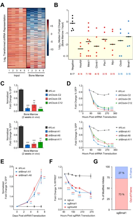
Figure 1. Clock and Bmal1 are required for leukemia cell growth
(A) Pooled RNAi screen identified shRNAs depleting murine MLL-AF9 leukemia cells in vivo. Heat map depicting the log2 transformed representation of individual shRNAs (rows) in the input replicates (n=5, left columns) and leukemic bone marrow replicates (n=5, right columns). Depleted shRNAs are shown in blue. (B) Performance of shRNAs targeting negative control genes (black), positive control genes (red), and circadian rhythm genes (blue). Plots depict the log10 median fold change for each shRNA. Fraction of shRNAs with
fold depletion > 20 is shown. (C–D) Fold change in the percentage of GFP+ murine
MLL-AF9 leukemia cells transduced with single Clock (C2, C6, C12) (n=3), Bmal1 (A1, A6,
A
uthor Man
uscr
ipt
A
uthor Man
uscr
ipt
A
uthor Man
uscr
ipt
A
uthor Man
uscr
ipt
A11) (n=3), or Luc (n=3) shRNAs co-expressed with GFP (C) in vivo or (D) in vitro. (E) Phenotypic rescue of Bmal1 knockdown with an shRNA-resistant Bmal1 cDNA in murine MLL-AF9 leukemia cells. (F) In vitro tracking of Cas9-expressing murine MLL-AF9 leukemia cells transduced with sgRNAs targeting Bmal1 (sgBmal) (n=3), HoxA9 (sgHoxA9) (n=3), or Luc (sgLuc) (n=3) co-expressed with RFP657. (G) Detection of in-frame and out-of-in-frame mutations at the Bmal1 locus in cells expressing Cas9 and sgBmal1 by Sanger sequencing of individual clones. *p<0.05, **p<0.01, ***p<0.001, determined by a two-sided Student’s t-test. Mean values are shown unless otherwise specified, and error bars represent +/− S.E.M.
A
uthor Man
uscr
ipt
A
uthor Man
uscr
ipt
A
uthor Man
uscr
ipt
A
uthor Man
uscr
ipt
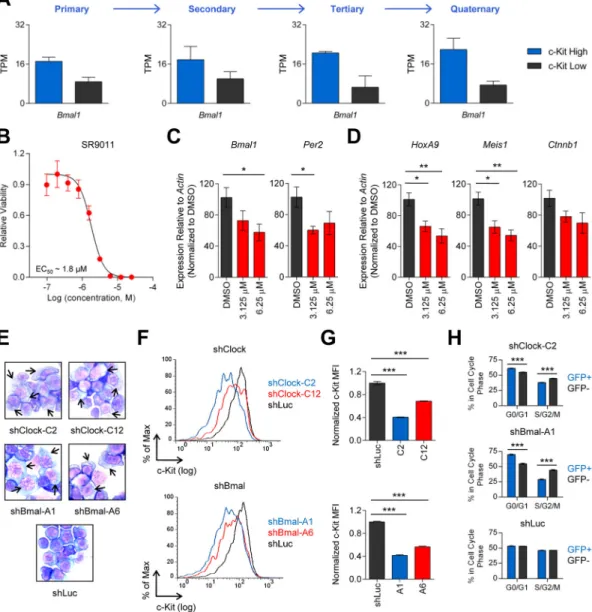
Figure 2. Disruption of the circadian rhythm machinery induces leukemia differentiation
(A) RNA-Seq analysis of Bmal1 expression in c-Kithigh (top 10 percent, n=3) and c-Kitlow
(bottom 10 percent, n=3) leukemic bone marrow cells from primary, secondary, tertiary, and quaternary transplant MLL-AF9 mice. TPM, transcripts per million. (B) Dose response curve showing relative viability (y-axis) of murine MLL-AF9 leukemia cells treated for 3 days with varying concentrations of SR9011 (x-axis, log scale) (n=3). (C–D) Expression of
(C) circadian rhythm genes (Bmal1, Per2) or (D) regulators of LSC self-renewal (HoxA9, Meis1, Ctnnb1), measured by quantitative RT-PCR following treatment of murine MLL-AF9 leukemia cells with 3.125 μM SR9011 (n=4), 6.25 μM SR9011 (n=4), or DMSO (n=4) for 24 hours. (E) Wright-Giemsa staining of murine MLL-AF9 leukemia cells transduced with single Clock (C2, C12) or Bmal1 (A1, A6) shRNAs revealed monocytic differentiation (black arrows). (F) Representative flow cytometry histograms of c-Kit cell surface expression (x-axis, log scale) in GFP+ murine MLL-AF9 leukemia cells expressing Clock (C2, C12), Bmal1 (A1, A6), or Luc shRNAs. (G) Quantification of c-Kit cell surface
A
uthor Man
uscr
ipt
A
uthor Man
uscr
ipt
A
uthor Man
uscr
ipt
A
uthor Man
uscr
ipt
expression by median fluorescence intensity (MFI) analysis. (H) Hoechst33342 cell cycle analysis of murine MLL-AF9 leukemia cells transduced with Clock (C2, n=3), Bmal1 (A1, n=3), or Luc (n=3) shRNAs. Data for both transduced (GFP+) and untransduced (GFP−)
cells within the same replicate are shown. *p<0.05, **p<0.01, ***p<0.001, determined by a two-sided Student’s t-test. Mean values are shown unless otherwise specified, and error bars represent +/− S.E.M.
A
uthor Man
uscr
ipt
A
uthor Man
uscr
ipt
A
uthor Man
uscr
ipt
A
uthor Man
uscr
ipt
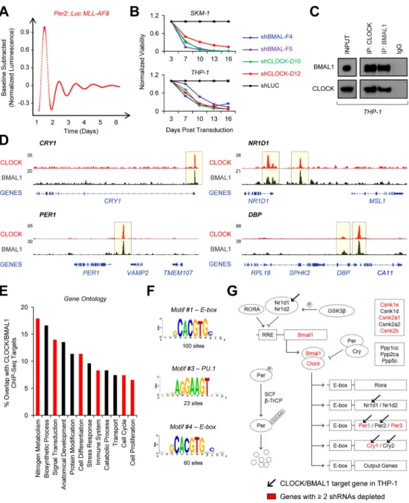
Figure 3. Canonical circadian regulatory mechanisms are intact in primary murine leukemia and human AML
(A) Continuous bioluminescence recording of a leukemic spleen explant from a WT
secondary recipient mouse transplanted with Per2::Luc MLL-AF9 leukemia cells. (B) Cell titer glo assay showing viability of SKM-1 or THP-1 cells following transduction with shRNAs targeting CLOCK (D10, D12) (n=5), BMAL1 (F4, F5) (n=5), or LUC (n=5). Equal numbers of puromycin-selected cells were plated 3 days after transduction and followed for 2 weeks in vitro. Data is normalized to the shLUC control at each timepoint. (C) CLOCK and BMAL1 reciprocal co-immunoprecipitation in THP-1 cells. (D) UCSC genome browser tracks of CLOCK (red) and BMAL1 (black) occupancy at the CRY1, NR1D1, PER1, and
A
uthor Man
uscr
ipt
A
uthor Man
uscr
ipt
A
uthor Man
uscr
ipt
A
uthor Man
uscr
ipt
DBP loci in THP-1 cells based on normalized ChIP-Seq read coverage. Track heights are indicated. (E) Gene ontology analysis of pathways represented by high-confidence CLOCK/ BMAL1 targets. Pathways of interest are highlighted in red. (F) RSEM motif enrichment analysis of high-confidence regions identified in CLOCK and BMAL1 ChIP-seq. (G) Minipool shRNA screen of 22 canonical circadian regulators in murine MLL-AF9 leukemia cells. Hits from the screen are shown in red and high-confidence Clock/Bmal1 ChIP-Seq targets are marked with a black arrow. Mean values are shown unless otherwise specified, and error bars represent +/− S.E.M.
A
uthor Man
uscr
ipt
A
uthor Man
uscr
ipt
A
uthor Man
uscr
ipt
A
uthor Man
uscr
ipt
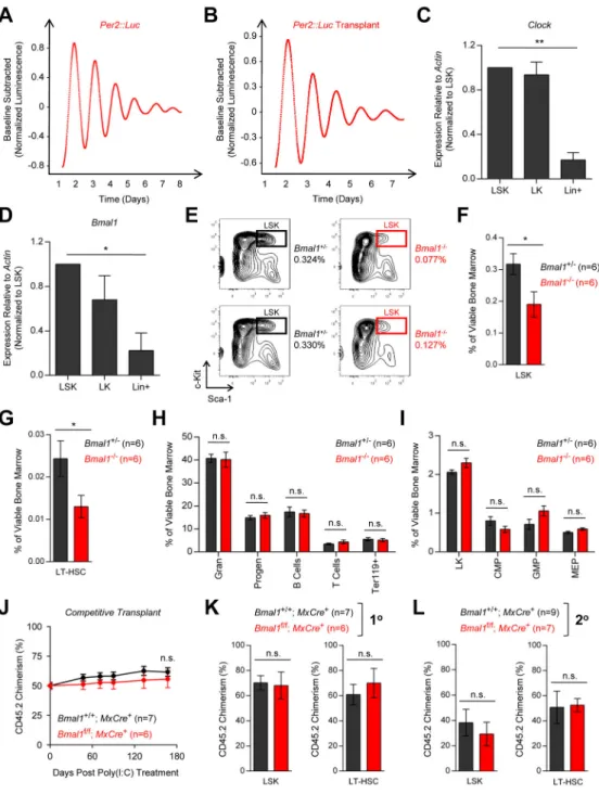
Figure 4. Bmal1 is dispensable for normal HSC function
(A–B) Continuous bioluminescence recording of spleen explant from (A) homozygous
Per2::Luc transgenic mouse or (B) lethally irradiated WT mouse transplanted with c-Kit enriched Per2::Luc bone marrow. (C–D) Expression of (C) Clock or (D) Bmal1 in murine LSK cells (n=3), LK cells (n=3), and lineage+ cells (n=3) purified from wild type mice, as measured by qPCR. (E) Representative flow cytometry contour plots of LSK cells in the bone marrow of germline Bmal1−/− and Bmal1+/− mice. LSK frequency is presented as a
percentage of viable whole bone marrow. (F–I) Quantification of (F) LSK cells, (G) LT-HSCs, (H) lineage+ cells, and (I) myeloid progenitor cells in the bone marrow of germline
A
uthor Man
uscr
ipt
A
uthor Man
uscr
ipt
A
uthor Man
uscr
ipt
A
uthor Man
uscr
ipt
Bmal1−/− (n=6) and Bmal1+/− (n=6) mice. Data is presented as a percentage of viable whole bone marrow. (J) Peripheral blood CD45.2 chimerism in Bmal1+/+; MxCre+ (n=7) and
Bmal1f/f; MxCre+ (n=6) competitive transplant mice following poly(I:C) treatment. (K–L)
CD45.2 chimerism in the bone marrow LSK and LT-HSC compartments of Bmal1+/+;
MxCre+ and Bmal1f/f; MxCre+ (K) primary or (L) secondary competitive transplant mice. *p<0.05, **p<0.01, ***p<0.001, determined by a two-sided Student’s t-test. Mean values are shown unless otherwise specified, and error bars represent +/− S.E.M.
A
uthor Man
uscr
ipt
A
uthor Man
uscr
ipt
A
uthor Man
uscr
ipt
A
uthor Man
uscr
ipt
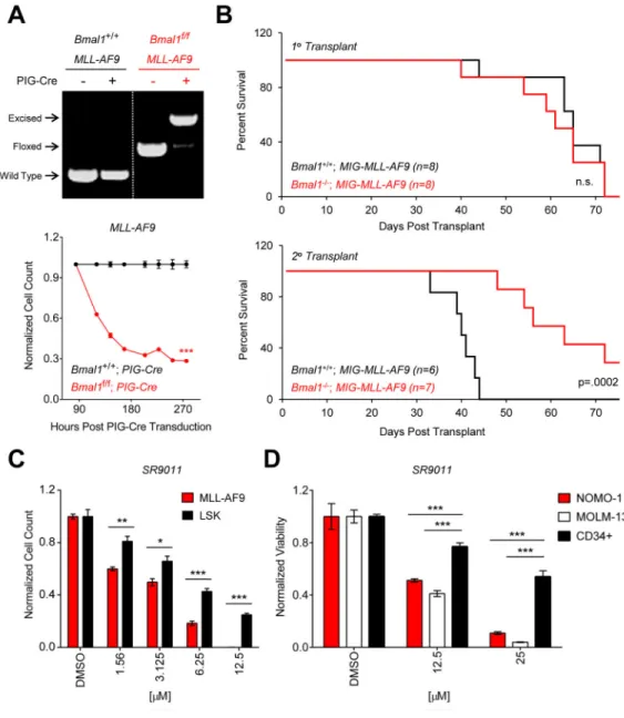
Figure 5. Genetic knockout models reveal a leukemia-specific dependence on Bmal1
(A) Top panel shows genotyping of Bmal1f/f and Bmal1+/+ MLL-AF9 leukemias before and
after transduction with PIG-Cre. Dotted white line indicates spliced gel. Bottom panel shows normalized cell counts of Bmal1f/f (n=4) and Bmal1+/+ (n=4) MLL-AF9 leukemias
transduced with PIG-Cre in vitro. Puromycin-selected cells were plated 84 hours after transduction and followed over time. (B) Bmal1f/f; MxCre+ mice and Bmal1+/+; MxCre+
controls were treated with poly(I:C) to induce hematopoietic-specific Cre expression. Bone marrow cells harvested from these mice were c-Kit enriched, transduced with MIG-MLL-AF9, and transplanted to lethally irradiated WT recipient mice. The resulting Bmal1−/− or
Bmal1+/+ MLL-AF9 leukemias were transplanted to sublethally irradiated secondary
recipients. Kaplan-Meier survival curves for the primary and secondary transplants are shown. (C) Normalized cell counts of quaternary transplant MLL-AF9 cells (n=3) and murine LSK cells (n=3) following treatment with DMSO or SR9011 for 3 days in vitro. (D)
A
uthor Man
uscr
ipt
A
uthor Man
uscr
ipt
A
uthor Man
uscr
ipt
A
uthor Man
uscr
ipt
Cell titer glo assay showing normalized viability of NOMO-1 (n=3), MOLM-13 (n=3), and CD34+ cord blood cells (n=6) following treatment with DMSO or SR9011 for 4 days in vitro. *p<0.05, **p<0.01, ***p<0.001, determined by a two-sided Student’s t-test. Mean values are shown unless otherwise specified, and error bars represent +/− S.E.M.