HAL Id: hal-02335638
https://hal.archives-ouvertes.fr/hal-02335638
Submitted on 28 Oct 2019
HAL is a multi-disciplinary open access
archive for the deposit and dissemination of
sci-entific research documents, whether they are
pub-lished or not. The documents may come from
teaching and research institutions in France or
abroad, or from public or private research centers.
L’archive ouverte pluridisciplinaire HAL, est
destinée au dépôt et à la diffusion de documents
scientifiques de niveau recherche, publiés ou non,
émanant des établissements d’enseignement et de
recherche français ou étrangers, des laboratoires
publics ou privés.
bio-functionalized substrates
Verena Ruprecht, Pascale Monzo, Andrea Ravasio, Zhang Yue, Ekta Makhija,
Pierre Strale, Nils Gauthier, G Shivashankar, Vincent Studer, Corinne
Albigès-Rizo, et al.
To cite this version:
Verena Ruprecht, Pascale Monzo, Andrea Ravasio, Zhang Yue, Ekta Makhija, et al.. How cells respond
to environmental cues -insights from bio-functionalized substrates. Journal of Cell Science, Company
of Biologists, 2017, �10.1242/jcs.196162�. �hal-02335638�
COMMENTARY
SPECIAL ISSUE: 3D CELL BIOLOGY
How cells respond to environmental cues
– insights from
bio-functionalized substrates
Verena Ruprecht
1,2, Pascale Monzo
3, Andrea Ravasio
4, Zhang Yue
4, Ekta Makhija
4, Pierre Olivier Strale
5,
Nils Gauthier
3, G. V. Shivashankar
3,4, Vincent Studer
5, Corinne Albiges-Rizo
6and Virgile Viasnoff
4,7,*
ABSTRACT
Biomimetic materials have long been the (he)art of bioengineering.
They usually aim at mimicking in vivo conditions to allow in vitro
culture, differentiation and expansion of cells. The past decade has
witnessed a considerable amount of progress in soft lithography,
bio-inspired micro-fabrication and biochemistry, allowing the design
of sophisticated and physiologically relevant micro- and
nano-environments. These systems now provide an exquisite toolbox
with which we can control a large set of physicochemical
environmental parameters that determine cell behavior.
Bio-functionalized surfaces have evolved from simple protein-coated
solid surfaces or cellular extracts into nano-textured 3D surfaces
with controlled rheological and topographical properties. The
mechanobiological molecular processes by which cells interact and
sense their environment can now be unambiguously understood
down to the single-molecule level. This Commentary highlights recent
successful examples where bio-functionalized substrates have
contributed in raising and answering new questions in the area of
extracellular matrix sensing by cells, cell
–cell adhesion and cell
migration. The use, the availability, the impact and the challenges of
such approaches in the field of biology are discussed.
KEY WORDS: Biomimetic interface, Environmental sensing,In vitro culture, Mechanobiology, Mechanosensing, Microniches
Introduction
Environment sensing and signaling is increasingly recognized as a
set of fundamental pathways that influence cell behavior, cell fate
and pathologies. These mechanobiological principles contextualize
the gene-expression-centric views that are more traditionally
observed in cell biology. Soluble factors, xenobiotic factors,
nutrients, oxygen and the chemical nature of the extracellular
matrix (ECM) have long been recognized as essential signaling
components of the local cellular microniches. However, cells are
equally sensitive to some biophysical aspects of the environment,
such as the density and mechanical properties of the ECM, physical
confinement and mechanical tension, all of which can elicit, inhibit
or synchronize cell responses.
In vivo, all these parameters are
largely intertwined. Cell
–cell interactions, cell–matrix interactions
and
paracrine
signaling
epitomize
a triad
of intertwined
environmental cues that, at the same time, elicit a cellular
response and are modified by the cells. In the past decade,
combining molecular biology tools with
in vitro reductionist
approaches
involving
bio-functionalized
micro-fabricated
substrates has enabled scientists to identify and understand a
growing number of cellular processes that are governed by
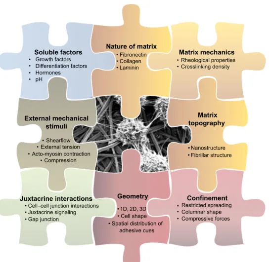
mechanobiological cues (Fig. 1).
This Commentary reviews recent examples of important
processes in the fields of matrix sensing, cell
–cell interactions and
cell migration that have been elucidated using bio-functionalized
surfaces. Many fundamental studies have been carried out in these
fields using exclusively molecular and cell biology tools, such as
gene editing and gene sequencing. We do not intend to minimize the
importance of these approaches. However, this review focuses on
the additional insights that were obtained when
in vitro controls of
the cellular micro-environment are applied. For an exhaustive
view of each field the reader can refer to more specific reviews
(e.g. Bonnans et al., 2014; Cavey and Lecuit, 2009; Kramer et al.,
2013).
Brief overview over bio-functionalized substrates
Bio-functionalized
surfaces
are
typically
substrates
with
controllable
biophysical
properties that
can
elicit
specific
interactions with cells in a close-to-physiological way. The
canonical examples are Petri dishes coated with ECM that has
been simply adsorbed on their surface by incubation from a
solution. Covalent binding can also be achieved by specific surface
chemistry, which ensures proper mechanical coupling with the
underlying substrate and reduces any matrix restructuring. The next
level of complexity consists of patterning in two-dimensional (2D)
adhesive areas. Many techniques can be used to lay down a pattern,
with most being micron-scale approaches usually used in the textile
industry. These include micro-serigraphy or micro-stenciling, which
uses removable membrane with holes of different shapes to mask
the exposed area of the surface (Masters et al., 2012; Ostuni et al.,
2000), and micro-stamping, which uses soft textured material to
‘ink’ the region of contact with proteins (Piel and Thery, 2014b).
Another approach is dip-pen lithography, which uses deposition
of protein with a sharp tip (Salaita et al., 2007). Alternatively, deep
UV patterning uses polymer degradation under intense light to
reveal surfaces with adhesive properties (Azioune et al., 2009). A
resolution down to a few hundreds of nanometers can be achieved
(Fig. 2A).
Multi-protein printing (Fig. 2A) is enabled by repeating these
processes in sequence (Strale et al., 2016). These protein deposition
techniques can be used on substrates of various rigidities (Fig. 2B)
that range from soft hydrogel (1
–10 kPa), elastomers (100 kPa–
1 MPa) to glassy materials (GPa) (Piel and Thery, 2014b). However,
a caveat should be drawn here. The rheological properties felt by the
1Centre for Genomic Regulation (CRG), The Barcelona Institute of Science and
Technology, Dr. Aiguader 88, Barcelona 08003, Spain.2Universitat Pompeu Fabra
(UPF), Barcelona, Spain.3IFOM, Via Adamello, 16, Milano 20139, Italy. 4Mechanobiology Institute, National University of Singapore, 5A Engineering Drive
1, 117411, Singapore.5CNRS, Interdisciplinary Institute for Neuroscience, UMR
5297, Bordeaux F-33000, France.6INSERM, U1209, CNRS UMR 5309, Institute for
Advanced Biosciences, Institute Albert Bonniot, University Grenoble Alpes, La Tronche F-38700, France.7CNRS UMI 3639, 5A Engineering Drive 1, 117411
Singapore.
*Author for correspondence (virgile.viasnoff@espci.fr)
V.V., 0000-0003-3949-2244
Journal
of
Cell
cells might differ from that of the substrate owing to the adsorbtion
and deformability of the ECM proteins (Smith et al., 2007). The
rheological properties of the substrates can also be controlled by
adsorbing patterned or unpatterned functionalized supported lipid
bilayers with various fluidity (Fig. 2B).
Furthermore, another layer of complexity can be added by
imprinting soft or rigid topographic features (e.g. pillars, groves or
pits) onto the substrates (Fig. 2C). Most of these substrates are 2D
or quasi 2D, although three-dimensional (3D) matrix surrogate
hydrogels with controllable properties can also be devised. Inert
biocompatible polymeric backbones can be functionalized with
proteinaceous residues of a given nature and density. The density
of crosslinking sites can also be tailored using different types of
molecular reaction (e.g. click chemistry, acrylate based or
hydrogen bonds). 3D microstructured rigid substrates can also be
fabricated and coated with proteins (Klein et al., 2011) to create
fibrillar environments (Fig. 2B). Moreover, microwells can be used
to structure the 3D spatial arrangement of cellular adhesion and to
create bona fide cellular microniches (Fig. 2A) (Charnley et al.,
2012; Li et al., 2016). Substrates with gradients of adhesive
properties (Fig. 2D) or properties that vary in time, such as
hydrogel with controlled aging rheological properties (DeForest
and Tirrell, 2015; Young and Engler, 2011), or patterns with
on-demand adhesion (Rolli et al., 2012; Vignaud et al., 2012) have
also been designed (Fig. 2D). Table 1 summarizes these
approaches and the commercial availability of any such devices.
The principal advantage of using bio-functionalized surfaces is
that they provide minimal, standardized and reproducible conditions
with defined biochemical and biophysical characteristics for
long-term, high-resolution observations of cell behavior. Therefore, their
use allows a precise spatio-temporal control of individual
environmental parameters that are not easily accessible
in vivo.
Thus, they constitute a tool of choice to precisely investigate the
impact of mechanotransduction pathways through which cells feel
the surrounding biophysical cues, such as extracellular mechanical
properties, mechanical tension, shear flow and geometrical
constraints. In addition, they are also instrumental in studying
environment-dependent cell responses to soluble factors, such as
growth factors, drugs and hormones. Finally, a controlled geometry
allows the precise mechanical modeling of cellular functions
(Albert and Schwarz, 2016). Specific recent examples are discussed
in detail below.
ECM sensing
Among the different environmental-sensing processes, how cells
perceive the molecular and biophysical properties of the ECM
surrounding them is by far the most appreciated. Matrix properties
can be exquisitely recapitulated
in vitro. Many different types of
secreted matrix can be purified and used for cell culture (Caliari and
Burdick, 2016), or even assembled into microarrays to create a
micro-screen of matrix-induced responses (Reticker-Flynn et al.,
2012). For instance, the signaling from the 24 types of integrins
Soluble factors
• Growth factors • Differentiation factors • Hormones • pHNature of matrix
• Fibronectin • Collagen • LamininMatrix mechanics
• Rheological properties • Crosslinking densityMatrix
topography
• Nanostructure •Confinement
• Restricted spreading • Columnar shape • Compressive forcesGeometry
• 1D, 2D, 3D • Cell shape • Spatial distribution of adhesive cuesJuxtacrine interactions
• Cell−cell junction interactions • Juxtacrine signaling • Gap junction
External mechanical
stimuli
• Shearflow • External tension • Acto-myosin contraction • Compression • •Fibrillar structureFig. 1. Schematic representation of the various biophysical parameters that comprise the local microniche surrounding a single cell. In vivo these parameters are largely intertwined and their contribution to cell response can rarely be unambiguously evaluated. Recapitulating and varying one or a combination of each of these parameters using biomimetic interfaces allows us to decipher how cells perceive environmental cues and respond to them. The cell image is courtesy of Professor Hai-Quan Mao, Johns Hopkins University.
Journal
of
Cell
(reviewed in Campbell and Humphries, 2011) has been precisely
studied using this approach (Roca-Cusachs et al., 2009).
The simplest substrate is a plastic or glass dish coated with
adsorbed matrix proteins. Such a dish has been long used for cell
culture, but with the advent of super-resolution microscopy, the
ability to image focal adhesions at the single-molecule level led to
the discovery of their layered structure and fresh insight as to how
this spatial organization enables their function as mechanosensitive
signaling hubs (Kanchanawong et al., 2010; Patla et al., 2010).
Single-particle tracking has revealed the dynamic properties of
the constitutive integrins dimers involved in focal adhesions. Their
successive periods of immobilization and dissociation from the
underlying actin cytoskeleton proved to be an important part of their
adhesive and signaling role (Rossier et al., 2012). Integrins also
require assembling into nanoclusters to be functional. The existence
of a maximum distance of 55 nm between two activated integrins
prior to triggering of adhesion was shown using
RGD-peptide-coated gold nanodots arrays (Arnold et al., 2004; Cavalcanti-Adam
and Spatz, 2015; Huang et al., 2009; Liu et al., 2014;
Selhuber-Unkel et al., 2008), or nanopatterns of defined spacing and grafting
densities (Coyer et al., 2012).
Beyond adhesion, integrin clusters are also essential signaling
hubs for mechanosensation. Cells do not only exert mechanical
deformation on the matrix but also sense the response of the matrix
and react to it. Imaging substrate deformation of continuous gels
with fiducial tracers (Oakes et al., 2012; Plotnikov et al., 2014;
Soiné et al., 2015) or of flexible pillars (Rahmouni et al., 2013)
enables quantitative measurements of cellular traction and cellular
response. These approaches have unambiguously revealed that
mechanical tension is crucial for the maturation of the focal
adhesion (Ghibaudo et al., 2008; Schiller et al., 2013) and for matrix
rigidity sensing (Humphrey et al., 2014; Vogel and Sheetz, 2006).
Recent studies performed on pillars of 500 nm in diameter have
unraveled a mechanism by which cells
‘pinch’ the matrix, thereby
assembling a contractile molecular complex (Ghassemi et al., 2012;
Meacci et al., 2016; Wolfenson et al., 2016). The signaling
downstream of these pinching events is dependent on the
mechanical tension that is necessary to contract the functional
unit. This provides a mechanism by which cells can sense substrate
rigidity. A striking demonstration of this principle was observed by
comparing cell spreading on glass substrates, nano-corralled lipid
bilayers and fully fluid bilayers bio-functionalized with identical
Patterning ligands on
substrates.
Controlling rheological
properties of cellular
environments.
Presenting cells with
different topographical
features.
Controlling the space
and time variation of
the above properties.
D
Deformable hydrogelsB
Multi-protein 3D patterningC
Controlled molecular densityA
Multi-protein 2D patterningSupported bilayers Mechanical transducers
Nanotopography Geometric confinement
Soluble gradients Embedded gradients Time-varying properties
Fibril substrates
Fig. 2. Examples of technological solutions to control the cellular environment surrounding cells in vitro. (A) Patterning of different ligands. Left, intricate multi-protein patterns (E-cad in blue, fibronectin in red) made using deep UV patterning; middle, 3D differential protein coating on 20 micron microwells; right, an evenly spaced array of nanogold dots with controlled density of grafted RGD. (B) Examples for controlling the rheological properties of cellular environments. Left, fibroblasts plated on soft polyacrylamide gels; middle, MCF10 cells placed on a biofunctionalized fluid lipid bilayer; right, arrays of deformable micropillars that allow cellular traction measurement. (C) Illustration of how cells can be presented with different topographical features. Left,
nanotextured polystyrene substrates with different topography that can enhance stem cell differentiation into a specific lineage; middle, fibroblasts cells migrating in 3D fibril environment; right, cells confined in micropits with precise geometrical properties such as curvature, size and shape. (D) Means to control the properties of the in vitro environment in time and space. Left, chemokine gradients generated across microfluidic channels; middle, gradients of fluorescent fibronectin density between two adhesive compartments; right, cell spreading on adhesive pattern before (dark) and after (blue) addition of biotinylated fibronectin, which selectively binds to the upper half of the pattern.
Journal
of
Cell
T
able
1.
Ov
ervie
w
o
f
the
a
vailable
biofuncti
onalized
subs
tr
a
tes
T ype Appr oa ch R evie ws C ommer cial a vailability Biological applica tions Subs tr a tes with s tructur ed pr otein coa ting 2D pr otein printing Stamping (SQ) Stenciling (SQ) Light induced (Q) Piel and Thery , 2014a,b D ’Ar cangelo and McGuigan, 2015 Ricoult et al., 2015 Innops y s Alv eole Cytoo 2D confinement, for cing cell size and shape F o rcing cytosk eleton organiza tion Inducing of fr ont/r ear polariza tion R e s tricting migr a tion Inducing nuclear s tr ess Spa tially organizing cell cultur e Q uantita tiv e inter a ctions with ligand High-or super-r esolution imaging 3D pr otein pa tterning On micr os tructur es (SQ) T w o photons (Q) No relevant revie ws a vailable Nanoscribe Nanoprinting Nanodots (Q) Dip pen lithogr aphy (NQ) Nanos tenciling (SQ) Ru et al., 2014 Salaita et al., 2007 R oss et al., 2012 Cus tódio et al., 2014 Innops y s Subs tr a tes with contr olled rheological pr operties Hydr ogels or soft elas tomers Biomimetic/e xtr a c t o f ECM (SQ), contr ol of ligand density and gr o wth fa ctors F unctionalizable hydr ogels of various rigidity (e.g. PEG, P AA, haluric a cid) (Q) P article-filled hydr ogels (Q) Piel and Thery , 2014a,b R oss et al., 2012 Sant et al., 2010 In Spher o Qgel IBIDI Advanced BioMa trix 3D vs 2D cell cultur e Adhesion on soft subs tr a tes T ra ction for ce micr oscopy Spa tial clus tering of surfa ce receptors C ooper a tiv e signaling fr om receptor clus ters Rigidity sensing Migr a tion in absence of adhesion Ma trix-dependent cellular response F unctionalized lipid bila y ers Supported lipid bila y ers functionalized with ligand (Q) Nanos tructur ed bila y ers (Q) (e.g. nanopa tterned, nanofences) Multila y e re d lipid subs tr a te with various rigidity (SQ) Cas tellana and Cr emer, 2006 Yu and Gr o v es, 2010 Gr o v es, 2007 T anaka and Sa ckmann, 2005 eDAQ Micr o-te xtur ed subs tr a tes Micr opillars/nanopillars of various length, diameters and densities (Q) No relevant revie ws a vailable NC A Subs tr a tes with topogr aphic fea tur es Micr o-te xtur ed subs tr a tes Micr o-fabrica ted topogr aphic fea tur es on rigid subs tances (Q) Fle xible pillars for for ce tr ansduction (Q) Micr o-ca vities or micr o-containers (Q) Bettinger et al., 2009 Nikkhah et al., 2012 Mechanobiology Ins titute Nunc Micr osurfa ces Aggr e w ell Curva tur e o r topogr aphy sensing Enhanced s tem cell differ entia tion Mechanical s tr ess measur ements (single-cell analy sis or organoids) Induction of fr ont – rear polariza tion 3D migr a tion C onfined migr a tion Chemotaxis Effect of fluidic shear Nano-te xtur ed subs tr a tes Planar subs tr a tes with nanofea tur es (Q) Electr ospun nanofibers Nano silicon/gr aphene fibers Lim and Donahue, 2007 Kim et al., 2012 Baharvand, 2015 NC A Micr ofluidics 3D channels for migr a tion assa y s Flo w chambers Soluble fa ctor gr adient gener a tors Dupin et al., 2013 C ellasic ONIX Millipor e IBIDI Gr adientech Fluxion BellBr ook Labs Hur el Kirks tall Mimetas Abbr evia tions: NC A, no curr ent commer cial a vailability; NQ, non quantita tiv e; P AA, poly a crylic a cid; PEG, poly ethylene glycol; Q, quantita tiv e; SQ, semi-quantitativ e.Journal
of
Cell
Science
densities of RGD peptides (Yu et al., 2011). As the substrate became
more fluid, cells were unable to exert any mechanical load on the
engaged integrin clusters, preventing the maturation of focal
adhesions; this caused the cells to round up. A combination of
these approaches has also revealed that talin, a multidomain protein
localized to focal adhesion, sequentially unfolds under various
mechanical loads, thus serving as a
‘mechanical ruler’ (del Rio
et al., 2009; Hu et al., 2016; Margadant et al., 2011; Yao et al., 2014,
2016). This constitutes the best-understood mechanism (although it
is not unique) by which cells can sense the level of force they exert.
By using stenciling membranes to constrain the extent of cell
spreading, a seminal study demonstrated that a minimal value for
cell spreading is required to avoid cell death (anoikis) (Chen et al.,
1997).
It is now increasingly clear that rapid events (over a timeframe of
seconds to minutes) for rigidity sensing directly influence the
activity of transcription factors (Fourel et al., 2016; Petropoulos
et al., 2016; Renz et al., 2015), and ultimately cell behavior and fate
(Inman et al., 2015). As an example, apical polarization has been
shown to be highly dependent on matrix organization (Akhtar and
Streuli, 2013; Rodríguez-Fraticelli et al., 2012). In this context, the
mechanotransduction role of
β1 and β3 integrins was singled out
(Fourel et al., 2016; Schiller et al., 2013). In addition, 2D and 3D
protein printing has demonstrated how the spatial structuration of
the adhesive environment influences the localization
(Rodríguez-Fraticelli et al., 2012) and shapes (Li et al., 2016) of apical lumens.
Epithelial morphogenesis has also been found to depend on the
biophysical properties of surrogate matrix gels (Enemchukwu et al.,
2016). The 2D confinement of cells on ECM patterns was also
shown to elicit the translocation of the co-transcription factors YAP
and TAZ from the nucleus to the cytoplasm. This provides a
molecular basis for signaling pathways that regulate cell
proliferation in a manner that depends on cell confinement and
confluence (Dupont et al., 2011).
In addition, the curvature of ECM micropits has been found to
control the branching morphogenesis of epithelium, and this
demonstrates the importance of the geometry of cell confinement
(Nelson et al., 2006). Last but not least, the rheological properties of
the matrix, cellular confinement and geometrical constraints have all
been found to have a crucial role in stem cell differentiation and
cell fate reprogramming (Engler et al., 2006). Paradigm-shifting
experiments have demonstrated that biophysical cues interfere with
cell differentiation programs and contribute to the cell lineage
commitment (Engler et al., 2006; Gilbert et al., 2010; Wen et al.,
2014). Stem cell differentiation has emerged as being a combination
of a response to soluble factors and an integrated response to
environmental factors, including geometrical, rheological and
topographical cues (Discher et al., 2009; Griffin et al., 2015;
Murphy et al., 2014). Consequently, a fundamental understanding
of cell differentiation, as well as technical solutions to enhance cell
differentiation, has been drawn from these observations. In this
perspective, how mechanical properties of the substrate are
transduced to the nucleus to trigger mechanosensitive control over
genomic programs is being intensely scrutinized. A possibility
emerges
that
geometrical
constrains
impinge
on
nuclear
morphologies (Li et al., 2014; Oakes et al., 2014; Versaevel et al.,
2012), chromatin compaction states (Makhija et al., 2016) and
chromosome territories (Thomas et al., 2002). Here, the cytoskeletal
rearrangements that are induced by the spatial structuration and
confinement of the adhesive area result in nuclear reorganization
and genome reprogramming. Taken together, all these studies
illustrate the long-term downstream consequences of rapid
environmental sensing process. The biggest challenges in this
area are to unravel the routes by which early pathways of
environmental-sensing signal to transcriptional, genomic and
epi-genomic programs downstream. As outlined above, the tools
required to answer these questions are ready and await use by the
different communities of biologists.
Cell
–cell interactions
ECM factors are crucial for microniche signaling. However, cell
–
cell interactions need to be considered to an equal extent as an
environment signaling cue. Technological developments required to
unravel downstream consequences of sensing at cell
–cell junctions
are
lagging
behind
those
used
for
cell
–ECM adhesion.
Understanding the mechanobiology of intercellular contacts is
intrinsically a multi-component, interconnected problem (as
compared to the interactions between a single cell and ECM or
soluble factors). A cell both responds to and serves as a
‘substrate’
for its neighbors. Bio-functionalized materials thus could be
instrumental
in
decoupling
both
aspects.
Indeed,
new
methodologies are being developed to achieve a degree of control
similar to that obtained for cell
–ECM interactions as outlined below.
For instance, the type of measurements described for ECM
substrates
can
be utilized
for
substrates that
have been
functionalized with cell
–cell adhesion proteins, such as E- or
N-cadherins (Plestant et al., 2014; Vega et al., 2014) or antigens
(Plestant et al., 2014). Studies with E-cadherin (E-cad)-coated
substrates have revealed the existence of E-cad nanoclusters as
fundamental units for cell
–cell adhesion, and their existence has
been confirmed by super-resolution imaging of mammalian cell
–
cell contacts (Wu et al., 2015; Strale et al., 2015), as well as in
Drosophila (Truong Quang et al., 2013). In addition, both E- and
N-cad-coated deformable pillars allow the measurement of the
traction forces that are exerted by a single cell across cadherin
bonds. Mechanical traction was found to be in the order of 5 to 10
nN per square micron (Ganz et al., 2006; Ladoux et al., 2010). An
alternative approach consists of fluorescence resonance energy
transfer (FRET)-based force sensors coupled to E-cad to measure
the mechanical load that is placed on the adhesion molecule; it
amounts to be
∼2 pN for each E-cad molecule that is engaged in a
real cell
–cell contact (Borghi et al., 2012). Taken together, these
measurements point towards a mechanical tension exerted at the
adherens junction that is of the order of 100 nN (for a junction of
∼10 μm in length). These approaches have also revealed that there
are strong structural similarities in cortical organization between the
mechanical responses of the ECM (i.e. integrin-based) and those of
cadherins. Similarly, the spreading mechanisms of macrophages on
an antigen-presenting glass (Vega et al., 2014) has been shown to be
dynamically and structurally similar to that of fibroblasts spreading
on fibronectin. Taken together, these results indicate that there is a
universal cytoskeleton organization for the development of force,
which is based on cluster adhesion, force-mediated reinforcement
and the development of traction fibers. This also raises questions as
to whether these organizing principles arise primarily owing to the
way the adhesive ligands are presented to the cells (i.e. soluble, on a
fluid substrate, or immobilized) and if the nature of the receptors
plays a role. This issue has been illustrated by the different cellular
responses elicited by identical growth factors depending on whether
they are soluble or attached to the surrounding matrix (Crouzier
et al., 2011).
The use of bio-functionalized lipid bilayers is also well suited to
address these types of questions. Adhesive ligands can be coupled
to phospholipids and incorporated into the supported lipid bilayer
Journal
of
Cell
at controlled densities. Furthermore, the mobility of the ligands in
the bilayer can be varied from fluid to static. The cells are thus left
free to reorganize spatially the ligand
–receptor pairs. E-cad cluster
formation was found to be highly dependent on the viscous drag
felt by aggregated ligands on the bilayer. On fluid bilayers, the
clusters were unstable and transient (Biswas et al., 2015; Perez
et al., 2005). In contrast, reduced diffusion of E-cad (by varying
the bilayer fluidity or binding to the cytoskeleton) stabilized the
cluster, strengthened the adhesion and redistributed the adhesive
zones along the edge of the surrogate contact into a morphology
that is reminiscent of the apical actin belt (Biswas et al., 2015).
This observed morphology shares many similarities with real cell
–
cell contacts that are established between two suspended cells
(Engl et al., 2014; Maitre et al., 2012). To our knowledge, similar
observations have not been reported for integrin-mediated
adhesion. Taken together, these observations indicate that the
reorganization of the actin cytoskeleton to which adhesive clusters
transiently bind is both responsible for and dependent on the
biophysical properties and spatio-temporal distribution of these
adhesive ligands. The ability of actin flows to organize and
segregate ligand
–receptor pairs at cell–cell contacts has been best
demonstrated by using bio-functionalized lipid bilayers as a
surrogate for immunological synapses. Imaging of these surrogate
substrates has revealed the segregation of different immune
receptor
pairs
into
concentric
regions
called
central
supramolecular activation clusters (cSMACs), peripheral SMACs
( pSMACs) and distal SMACs (dSMACs). This demonstrates that
the actin-based spatial segregation of the different receptor signals
is key to eliciting the immunological response of the T-cell
(Dustin and Groves, 2012; Groves, 2007; Pageon et al., 2016;
Tanaka and Sackmann, 2005; Yu and Groves, 2010).
The supported bilayer approach has also been used with other
classes of intercellular receptors. For example, it has been shown
that the signaling downstream of the binding of EPHA2, a receptor
tyrosine kinase involved in cell motility and organ boundary
formation, to an ephrin-A1-functionalized bilayer largely depends
on the size and spatial structuration of the ligand receptors cluster
(Greene et al., 2014; Salaita et al., 2010). The misregulation of this
force-activated pathway has also been found to be a hallmark of the
metastatic potential of breast cancer cells (Salaita et al., 2010).
One can devise a third approach for using bio-functionalized
substrates to study cell
–cell adhesion. ECM patterning can be used
to indirectly force cells to interact with each other in a controlled
manner. For example, 2D bow-shape patterns have been used to
measure the traction force that two cells exert on each other as the
imbalance of the mechanical tension they exert on the substrate (Liu
et al., 2010; Maruthamuthu et al., 2011; Ng et al., 2014). 2D ECM
geometrical patterns have also been used to show that adherens
junctions orient away from ECM adhesion (Mertz et al., 2013; Sim
et al., 2015; Tseng et al., 2012). This idea was recently extended in
3D to differential protein printing in small pits. When coated with an
anti-fouling treatment, they provide ideal substrates to perform
en-face imaging of junction formation between two stacked cells (Engl
et al., 2014) or during mitosis (Wollrab et al., 2016). Coating pits
with different proteins on their top, sides and bottom allows
exquisite control over cell
–cell interactions that are induced by
matrix adhesion in 3D. This approach has been recently used to
reveal how intercellular tension guides the luminogenesis in
hepatocytes (Li et al., 2016). Embedding micromirrors in close
vicinity to these micropits enables 3D super-resolution imaging of
cells in their environment with a sectioning capability of up to tens
of microns above the coverslip (Galland et al., 2015).
Taken together, we believe that the recent technological
developments and the growing number of studies focusing on
mechanotransduction at cell
–cell contacts will bring this field to the
same level of understanding as has been obtained for cell
–ECM
adhesion. We anticipate that this will also result in a better
understanding of how environmental factors act as spatially
structured triggers for cell polarization and organogenesis.
Cell migration
The substrates described so far are characterized by a structured but
homogenous bio-functionalization. Spatial gradients or properties
that vary over time can also be built in to study directed cell
migration (taxis). For instance, patterning with light-induced release
of adhesive constraints (Rolli et al., 2012; Vignaud et al., 2012) has
been used to study the transition from static to migratory behaviors.
Our understanding of migration along gradients of soluble factors
(chemotaxis) has benefited from microfluidics, where gradients of
soluble factors that are transverse to the direction of microfluidic
flow can be created (Chung and Choo, 2010; King et al., 2016;
Li Jeon et al., 2002; Toh et al., 2014). Passive diffusion of a
chemoattractant across a porous membrane has also been used to
create local gradients in the absence of flow (Dupin et al., 2013).
Furthermore, imprinting of protein gradients on rigid substrates
(Ricoult et al., 2015; Wu et al., 2011) or controlled stenciling and/or
photo-immobilization techniques (Bélisle et al., 2009, 2012; Strale
et al., 2016) has revealed the molecular basis for haptotaxis, the
migration of cells along an ECM gradient. In addition, the use of 3D
matrix hydrogels containing smooth gradients of crosslinking
density (i.e. to create a ECM with differing concentrations of
crosslinking components) (Millon-Frémillon et al., 2008) has
provided insights into how macrophages migrate along gradients
of differing matrix stiffnesses (i.e. durotaxis) (Nemir and West,
2010). Finally, a recent study employed traction force microscopy
on hydrogels with an embedded stiffness gradient to demonstrate
that cells can collectively sense large-scale matrix density gradients
resulting in collective durotaxis (Sunyer et al., 2016).
In absence of directional cues, spontaneous modes of collective
cell migration are also found to largely correlate with the physical
constraints of the environment. Removal of a physical obstacle from
an ECM-coated substrate allows monolayers of epithelial cells to
suddenly access a surface where cells are free to migrate. This
migration assay with controlled boundary conditions results in a
large-scale, swirling collective motion within the monolayer, as
well as in the appearance of
‘leader cells’ with distinct migratory
characteristics (Poujade et al., 2007). Cell swirls largely depend on
the lateral confinement of the monolayer (Deforet et al., 2014;
Doxzen et al., 2013; Rørth, 2012; Tanner et al., 2012; Vedula et al.,
2013). The emergence of this synchronized collective cell motion
can be correlated with contractile waves that involve multiple cells
as measured by traction force microscopy (Angelini et al., 2010;
Serra-Picamal et al., 2012). The existence of leader cells is also an
indication that cells can migrate by using different modes of
motility. Indeed, it has been shown that switching between different
modes of migration can be elicited by environmental cues (te
Boekhorst et al., 2016).
The various modes of cell migration can be recapitulated by
in vitro reconstitution of idealized migration conditions (Charras
and Sahai, 2014; Friedl and Wolf, 2010; Rao et al., 2014). Here,
bio-functionalized substrates provide an excellent platform to
understand how differences in spatio-temporal coordination of an
identical pool of regulators (e.g. Rho GTPases) and cytoskeleton
effectors (e.g. myosin II, Arp2/3, formins and filamin A) trigger the
Journal
of
Cell
activation of distinct cytoskeleton protrusions, which ultimately
lead to different migration modes. The
‘world cell race’ (Maiuri et
al., 2012) is an iconic example of how migration conditions can be
standardized over an extensive roster of cell types, and migration
speed and migration persistence of 50 cell types has been probed on
one-dimensional (1D) ECM-coated lines. Extending these findings
to 2D and 3D
in vitro and in vivo environments has led to a
comprehensive understanding of how actin flows mediate a
universal (1D, 2D and 3D) coupling between these parameters
(Maiuri et al., 2015). 1D migration has been further shown to favor
filopodia-driven migration of fibroblasts (Guo and Wang, 2012),
with cells adopting elongated spindle-like shapes (Chang et al.,
2013; Doyle et al., 2012; Guo and Wang, 2012; Monzo et al., 2016).
When various mammalian cells, such as transformed or
untransformed fibroblasts and cancer cell lines, including glioma
and carcinoma of various origin, migrate along ECM-printed lines,
their motility mode becomes intermittent, or saltatory, similar to that
described for neuronal motility (Guo and Wang, 2012; Irimia and
Toner, 2009; Monzo et al., 2016; Pathak and Kumar, 2012). This
contrasts with the
‘classic’ gliding migration of fibroblasts or
keratinocytes on 2D surfaces, which results from the dynamic
protrusion of a lamellipodium at the front of the polarized cell and a
contractile actomyosin network at its rear (Verkhovsky et al., 1999).
Formins play a crucial role in generating and organizing the long
actin cables that are necessary to support the elongated shape of the
cells during 1D migration (Monzo et al., 2016; Vargas et al., 2016;
Wilson et al., 2013), whereas Arp2/3 is more crucial for migration in
2D. Furthermore, if cell migration takes place on suspended
electrospun nanofibers coated with ECM (Johnson et al., 2009), free
actin waves propagate from the cell body to the tip of the cellular
protrusion. This occurs in an asymmetric manner to polarize the
movement of long spindle-shaped cells (Guetta-Terrier et al., 2015).
These thin actin protrusions differ in nature from the bulky actin
protrusions, which are used to crawl across the matrix pores and
termed lobopodia, that appear when cells move in a dense 3D matrix
(Petrie et al., 2012). The use of a 3D matrix with controlled pore
sizes has also enabled the investigation of mechanical nuclear
deformation during migration and its consequences for DNA
damage (Petrie et al., 2014; Raab et al., 2016; Thiam et al., 2016).
Control over the cellular microenvironment has not only revealed
the existence of different modes of migration, but has also provided
insights into how exactly cells switch from one mode of motility to
another (Friedl and Alexander, 2011). Non-adherent confining
substrates, which can be created by microchannels (Bergert et al.,
2015) or with a double layer of inert hydrogel (Ruprecht et al., 2015;
Liu et al., 2015), have been shown to induce a myosin-II-dependent
switch to an amoeboid migration mode that involves the formation
of a stable, bleb-like and actin-depleted protrusion at the cell front
(Bergert et al., 2015; Liu et al., 2015; Ruprecht et al., 2015). These
studies have also determined that the driving force for this amoeboid
migration mode is due to a reverse actin flow mediated by
non-specific friction between the cell and its substrate.
Taken together, as outlined above, by being able to control the
cellular environment
in vitro, many of the environmental parameters
and migration modes that are observed in different
in vivo contexts,
such as embryogenesis, wound healing, metastasis, neuron
development or the inflammatory response, can be recapitulated
and further investigated in detail.
Conclusions and perspectives
The primary advantage of using bio-functionalized surfaces is that
they provide the ability to isolate and vary a single environmental
parameter in order to unambiguously decipher its contribution to a
given cellular process. Combining these interfaces with imaging at
the nanometer scale and the genetic alteration of cells is key to
understanding the molecular and cellular processes by which cells
sense their environment. It is increasingly clear that cells do not exist
in a single state, but rather are able to switch between different
programs that dictate their behavior. However, the decision to engage
in a certain state is not only dictated by secreted or soluble factors, but
is made in conjunction with probing of the microenvironment. In that
sense, environmental cues can be seen as external triggers of
autonomous cell programs. Testing the extent to which an individual
environmental cue triggers a specific program is a promising
approach to being able to understand the molecular pathways by
which cells engage into such programs, or how their behavior
(such as drug resistance and differentiation) is conditioned by their
microniche. We would like to argue here that there is already a
technical solution to recreate most of the environmental parameters
individually
in vitro. Developing a single approach that allows for a
combinatorial control over all environmental factors will be crucial to
building platforms that will allow cells to be able to sense the entire
cellular environment (Dolatshahi-Pirouz et al., 2014; Gobaa et al.,
2011). In particular, precisely combining two or more ligands within
an engineered environment has already helped to unravel the
combinatorial interplay of different adhesive pathways. For
example, an increasing number of studies scrutinize the crosstalk
between integrins and growth factors (Fourel et al., 2016; Lee et al.,
2011), as well as between integrins and cadherins (Borghi et al.,
2010; Stapleton et al., 2014), by incorporating one factor at a time in
order to generate
in vitro the essential aspects of in vivo complexity.
Because the downstream consequences of environmental sensing
most likely originate from the integrated signaling of combined
environmental cues, it is important to reconstruct the complexity of
the microniche from individually controlled cues. The combination of
the minimal number of external cues needed to trigger an emergent
cellular property or differentiation or polarization program could thus
be deciphered. Another challenge that awaits the field is to extend the
technological know-how and biological knowledge that has been
acquired at the single cell level to extend to co-cultures of cells in 3D.
These systems are expected to provide insights into the environmental
Box 1. Co-culture of cells organized in 3D
In most of the studies described here, the controlled microenvironment is applied to a single cell type or to the study of homotypic interactions. However, an increasing number of studies now aim to co-culture different cell types in a spatially structured manner in order to better recapitulate cell–cell interactions within tissues or organs. To that end, 3D microchannels (∼200-μm wide) created in an ECM hydrogel can be used to culture endothelial cells; this mimics blood vessels, and interactions of endothelial cells with cancer or stromal cells can be investigated (Jeon et al., 2015; Miller et al., 2012; Shin et al., 2011). In addition, 2D protein patterns have been used to localize hepatocyte islands amidst an interacting fibroblast layer, which has been shown to result in enhanced bile production (Bhatia and Ingber, 2014). Finally, a fast developing method for cell culture in 3D is to create organoids, with cells being grown either in hanging drops or in an ECM surrogate (Clevers, 2016; Fatehullah et al., 2016; Yin et al., 2016). The development of tissue-specific organoids relies on the provision of soluble factors and on the self-organization of the cells therein as they grow. We anticipate that extending our control over cell culture in 3D to being able to induce the interactions between different cell types in 3D-structured organoids will advance our capability to culture tissue in vitro, as well as increase our knowledge of how the spatial structuration of the environment influences cell fate.
Journal
of
Cell
cues required for the formation of multi-cell type organoids. These
concepts are being developed in so-called
‘organ-on-a-chip’
approaches (see Box 1).
The examples described above illustrate the popularity and
usefulness of bio-functionalized surfaces in cell biology over the
last decade. Their development and use is clearly an area where
biology, physics and engineering overlap. However, the need for
multidisciplinary expertise still impedes access of many laboratories
to these techniques. Although the commercial availability of these
technologies is growing (see Table 1), the particular tools offered will
always be based on economical profits and thus most likely limit the
extent and capability of the devices that can be purchased. As many of
the substrates described above can be realized with a combination of
soft lithography techniques and protein and/or lipid adsorption,
the scientific community would benefit greatly from the creation of an
open-source global repository of these devices, similar to that
Addgene provides for plasmids. To that end, the Mechanobiology
Institute in Singapore is committed to offering such a tool to the
community. It actively seeks academic partners to complete an online
declaration of interest to raise starting funds for the initiative (http:///
www.mechanobio.info/resources/pdms-survey/).
Acknowledgements
The authors acknowledge the kind help of Steven Wolf and Andrew Wong in proofreading the manuscript. V.V. thanks the microfabrication facility at MBI for providing images of microstructured substrates.
Competing interests
V.S. is a founding member of Alveole.
Funding
A grant from the National Research Foundation Singapore to the Mechanobiology Institute, Singapore supported this work. V.R acknowledges support of the Spanish Ministry of Economy and Competitiveness (Ministerio de Economı́a y
Competitividad)‘Centro de Excelencia Severo Ochoa 2013-2017’.
References
Akhtar, N. and Streuli, C. H. (2013). An integrin-ILK-microtubule network orients cell polarity and lumen formation in glandular epithelium. Nat. Cell Biol. 15, 17-27. Albert, P. J. and Schwarz, U. S. (2016). Modeling cell shape and dynamics on
micropatterns. Cell Adh. Migr. 10, 516-528.
Angelini, T. E., Hannezo, E., Trepat, X., Fredberg, J. J. and Weitz, D. A. (2010). Cell migration driven by cooperative substrate deformation patterns. Phys. Rev. Lett. 104, 168104.
Arnold, M., Cavalcanti-Adam, E. A., Glass, R., Blü mmel, J., Eck, W., Kantlehner, M., Kessler, H. and Spatz, J. P. (2004). Activation of integrin function by nanopatterned adhesive interfaces. Chemphyschem 5, 383-388. Azioune, A., Storch, M., Bornens, M., Théry, M. and Piel, M. (2009). Simple and
rapid process for single cell micro-patterning. Lab. Chip 9, 1640-1642. Baharvand, H. (2015). Stem Cell Nanoengineering. Hoboken, New Jersey,
Wiley-Blackwell.
Bélisle, J. M., Kunik, D. and Costantino, S. (2009). Rapid multicomponent optical protein patterning. Lab. Chip 9, 3580-3585.
Bélisle, J. M., Levin, L. A. and Costantino, S. (2012). High-content neurite development study using optically patterned substrates. PLoS ONE 7, e35911. Bergert, M., Erzberger, A., Desai, R. A., Aspalter, I. M., Oates, A. C., Charras, G.,
Salbreux, G. and Paluch, E. K. (2015). Force transmission during adhesion-independent migration. Nat. Cell Biol. 17, 524-529.
Bettinger, C. J., Langer, R. and Borenstein, J. T. (2009). Engineering substrate topography at the micro- and nanoscale to control cell function. Angew. Chem. Int. Ed. Engl. 48, 5406-5415.
Bhatia, S. N. and Ingber, D. E. (2014). Microfluidic organs-on-chips. Nat. Biotechnol. 32, 760-772.
Biswas, K. H., Hartman, K. L., Yu, C.-h., Harrison, O. J., Song, H., Smith, A. W., Huang, W. Y., Lin, W.-C., Guo, Z., Padmanabhan, A. et al. (2015). E-cadherin junction formation involves an active kinetic nucleation process. Proc. Natl. Acad. Sci. USA 112, 10932-10937.
Bonnans, C., Chou, J. and Werb, Z. (2014). Remodelling the extracellular matrix in development and disease. Nat. Rev. Mol. Cell Biol. 15, 786-801.
Borghi, N., Lowndes, M., Maruthamuthu, V., Gardel, M. L. and Nelson, W. J. (2010). Regulation of cell motile behavior by crosstalk between cadherin- and integrin-mediated adhesions. Proc. Natl Acad. Sci. USA 107, 13324-13329.
Borghi, N., Sorokina, M., Shcherbakova, O. G., Weis, W. I., Pruitt, B. L., Nelson, W. J. and Dunn, A. R. (2012). E-cadherin is under constitutive actomyosin-generated tension that is increased at cell-cell contacts upon externally applied stretch. Proc. Natl. Acad. Sci. USA 109, 12568-12573.
Caliari, S. R. and Burdick, J. A. (2016). A practical guide to hydrogels for cell culture. Nat. Methods 13, 405-414.
Campbell, I. D. and Humphries, M. J. (2011). Integrin structure, activation, and interactions. Cold Spring Harb. Perspect. Biol. 3, a004994.
Castellana, E. T. and Cremer, P. S. (2006). Solid supported lipid bilayers: from biophysical studies to sensor design. Surf. Sci. Rep. 61, 429-444.
Cavalcanti-Adam, E. A. and Spatz, J. P. (2015). Receptor clustering control and associated force sensing by surface patterning: when force matters. Nanomedicine 10, 681-684.
Cavey, M. and Lecuit, T. (2009). Molecular bases of cell-cell junctions stability and dynamics. Cold Spring Harb. Perspect. Biol. 1, a002998.
Chang, S. S., Guo, W.-h., Kim, Y. and Wang, Y.-l. (2013). Guidance of cell migration by substrate dimension. Biophys. J. 104, 313-321.
Charnley, M., Kroschewski, R. and Textor, M. (2012). The study of polarisation in single cells using model cell membranes. Integr. Biol. 4, 1059-1071.
Charras, G. and Sahai, E. (2014). Physical influences of the extracellular environment on cell migration. Nat. Rev. Mol. Cell Biol. 15, 813-824.
Chen, C. S., Mrksich, M., Huang, S., Whitesides, G. M. and Ingber, D. E. (1997). Geometric control of cell life and death. Science 276, 1425-1428.
Chung, B. G. and Choo, J. (2010). Microfluidic gradient platforms for controlling cellular behavior. Electrophoresis 31, 3014-3027.
Clevers, H. (2016). Modeling development and disease with organoids. Cell 165, 1586-1597.
Coyer, S. R., Singh, A., Dumbauld, D. W., Calderwood, D. A., Craig, S. W., Delamarche, E. and Garcia, A. J. (2012). Nanopatterning reveals an ECM area threshold for focal adhesion assembly and force transmission that is regulated by integrin activation and cytoskeleton tension. J. Cell Sci. 125, 5110-5123. Crouzier, T., Fourel, L., Boudou, T., Albigès-Rizo, C. and Picart, C. (2011).
Presentation of BMP-2 from a soft biopolymeric film unveils its activity on cell adhesion and migration. Adv. Mater. 23, H111-H118.
Custódio, C. A., Reis, R. L. and Mano, J. F. (2014). Engineering biomolecular microenvironments for cell instructive biomaterials. Adv. Healthc. Mater. 3, 797-810. D’Arcangelo, E. and McGuigan, A. P. (2015). Micropatterning strategies to engineer controlled cell and tissue architecture in vitro. BioTechniques 58, 13-23. DeForest, C. A. and Tirrell, D. A. (2015). A photoreversible protein-patterning approach for guiding stem cell fate in three-dimensional gels. Nat. Mater. 14, 523-531.
Deforet, M., Hakim, V., Yevick, H. G., Duclos, G. and Silberzan, P. (2014). Emergence of collective modes and tri-dimensional structures from epithelial confinement. Nat. Commun. 5, 3747.
del Rio, A., Perez-Jimenez, R., Liu, R., Roca-Cusachs, P., Fernandez, J. M. and Sheetz, M. P. (2009). Stretching single talin rod molecules activates vinculin binding. Science 323, 638-641.
Discher, D. E., Mooney, D. J. and Zandstra, P. W. (2009). Growth factors, matrices, and forces combine and control stem cells. Science 324, 1673-1677.
Dolatshahi-Pirouz, A., Nikkhah, M., Gaharwar, A. K., Hashmi, B., Guermani, E., Aliabadi, H., Camci-Unal, G., Ferrante, T., Foss, M., Ingber, D. E. et al. (2014). A combinatorial cell-laden gel microarray for inducing osteogenic differentiation of human mesenchymal stem cells. Sci. Rep. 4, 3896.
Doxzen, K., Vedula, S. R. K., Leong, M. C., Hirata, H., Gov, N. S., Kabla, A. J., Ladoux, B. and Lim, C. T. (2013). Guidance of collective cell migration by substrate geometry. Integr. Biol. 5, 1026-1035.
Doyle, A. D., Kutys, M. L., Conti, M. A., Matsumoto, K., Adelstein, R. S. and Yamada, K. M. (2012). Micro-environmental control of cell migration–myosin IIA is required for efficient migration in fibrillar environments through control of cell adhesion dynamics. J. Cell Sci. 125, 2244-2256.
Dupin, I., Dahan, M. and Studer, V. (2013). Investigating axonal guidance with microdevice-based approaches. J. Neurosci. 33, 17647-17655.
Dupont, S., Morsut, L., Aragona, M., Enzo, E., Giulitti, S., Cordenonsi, M., Zanconato, F., Le Digabel, J., Forcato, M., Bicciato, S. et al. (2011). Role of YAP/TAZ in mechanotransduction. Nature 474, 179-183.
Dustin, M. L. and Groves, J. T. (2012). Receptor signaling clusters in the immune synapse. Annu. Rev. Biophys. 41, 543-556.
Enemchukwu, N. O., Cruz-Acuña, R., Bongiorno, T., Johnson, C. T., Garcia, J. R., Sulchek, T. and Garcı́a, A. J. (2016). Synthetic matrices reveal contributions of ECM biophysical and biochemical properties to epithelial morphogenesis. J. Cell Biol. 212, 113-124.
Engl, W., Arasi, B., Yap, L. L., Thiery, J. P. and Viasnoff, V. (2014). Actin dynamics modulate mechanosensitive immobilization of E-cadherin at adherens junctions. Nat. Cell Biol. 16, 587-594.
Engler, A. J., Sen, S., Sweeney, H. L. and Discher, D. E. (2006). Matrix elasticity directs stem cell lineage specification. Cell 126, 677-689.
Fatehullah, A., Tan, S. H. and Barker, N. (2016). Organoids as an in vitro model of human development and disease. Nat. Cell Biol. 18, 246-254.
Fourel, L., Valat, A., Faurobert, E., Guillot, R., Bourrin-Reynard, I., Ren, K., Lafanechere, L., Planus, E., Picart, C. and Albiges-Rizo, C. (2016). beta3
Journal
of
Cell
integrin-mediated spreading induced by matrix-bound BMP-2 controls Smad signaling in a stiffness-independent manner. J. Cell Biol. 212, 693-706. Friedl, P. and Alexander, S. (2011). Cancer invasion and the microenvironment:
plasticity and reciprocity. Cell 147, 992-1009.
Friedl, P. and Wolf, K. (2010). Plasticity of cell migration: a multiscale tuning model. J. Cell Biol. 188, 11-19.
Galland, R., Grenci, G., Aravind, A., Viasnoff, V., Studer, V. and Sibarita, J.-B. (2015). 3D high- and super-resolution imaging using single-objective SPIM. Nat. Methods 12, 641-644.
Ganz, A., Lambert, M., Saez, A., Silberzan, P., Buguin, A., Mège, R. M. and Ladoux, B. (2006). Traction forces exerted through N-cadherin contacts. Biol. Cell 98, 721-730.
Ghassemi, S., Meacci, G., Liu, S., Gondarenko, A. A., Mathur, A., Roca-Cusachs, P., Sheetz, M. P. and Hone, J. (2012). Cells test substrate rigidity by local contractions on submicrometer pillars. Proc. Natl. Acad. Sci. USA 109, 5328-5333.
Ghibaudo, M., Saez, A., Trichet, L., Xayaphoummine, A., Browaeys, J., Silberzan, P., Buguin, A. and Ladoux, B. (2008). Traction forces and rigidity sensing regulate cell functions. Soft Mat. 4, 1836-1843.
Gilbert, P. M., Havenstrite, K. L., Magnusson, K. E. G., Sacco, A., Leonardi, N. A., Kraft, P., Nguyen, N. K., Thrun, S., Lutolf, M. P. and Blau, H. M. (2010). Substrate elasticity regulates skeletal muscle stem cell self-renewal in culture. Science 329, 1078-1081.
Gobaa, S., Hoehnel, S., Roccio, M., Negro, A., Kobel, S. and Lutolf, M. P. (2011). Artificial niche microarrays for probing single stem cell fate in high throughput. Nat. Methods 8, 949-955.
Greene, A. C., Lord, S. J., Tian, A., Rhodes, C., Kai, H. and Groves, J. T. (2014). Spatial organization of EphA2 at the cell-cell interface modulates trans-endocytosis of ephrinA1. Biophys. J. 106, 2196-2205.
Griffin, M. F., Butler, P. E., Seifalian, A. M. and Kalaskar, D. M. (2015). Control of stem cell fate by engineering their micro and nanoenvironment. World J. Stem Cells 7, 37-50.
Groves, J. (2007). Supported lipid bilayers as mimics for cell surfaces and as tools in biotechnology. In BioMEMS and Biomedical Nanotechnology: Volume III Therapeutic Micro/Nanotechnology (ed. M. Ferrari, T. Desai and S. Bhatia), pp. 305-323. Boston, MA: Springer US.
Guetta-Terrier, C., Monzo, P., Zhu, J., Long, H., Venkatraman, L., Zhou, Y., Wang, P. P., Chew, S. Y., Mogilner, A., Ladoux, B. et al. (2015). Protrusive waves guide 3D cell migration along nanofibers. J. Cell Biol. 211, 683-701. Guo, W.-h. and Wang, Y.-l. (2012). A three-component mechanism for fibroblast
migration with a contractile cell body that couples a myosin II-independent propulsive anterior to a myosin II-dependent resistive tail. Mol. Biol. Cell 23, 1657-1663.
Hu, X., Jing, C., Xu, X., Nakazawa, N., Cornish, V. W., Margadant, F. M. and Sheetz, M. P. (2016). Cooperative vinculin binding to talin mapped by time-resolved super resolution microscopy. Nano Lett. 16, 4062-4068.
Huang, J., Grä ter, S. V., Corbellini, F., Rinck, S., Bock, E., Kemkemer, R., Kessler, H., Ding, J. and Spatz, J. P. (2009). Impact of order and disorder in RGD nanopatterns on cell adhesion. Nano Lett. 9, 1111-1116.
Humphrey, J. D., Dufresne, E. R. and Schwartz, M. A. (2014). Mechanotransduction and extracellular matrix homeostasis. Nat. Rev. Mol. Cell Biol. 15, 802-812.
Inman, J. L., Robertson, C., Mott, J. D. and Bissell, M. J. (2015). Mammary gland development: cell fate specification, stem cells and the microenvironment. Development 142, 1028-1042.
Irimia, D. and Toner, M. (2009). Spontaneous migration of cancer cells under conditions of mechanical confinement. Integr. Biol. 1, 506-512.
Jeon, J. S., Bersini, S., Gilardi, M., Dubini, G., Charest, J. L., Moretti, M. and Kamm, R. D. (2015). Human 3D vascularized organotypic microfluidic assays to study breast cancer cell extravasation. Proc. Natl. Acad. Sci. USA 112, 214-219. Johnson, J., Nowicki, M. O., Lee, C. H., Chiocca, E. A., Viapiano, M. S., Lawler, S. E. and Lannutti, J. J. (2009). Quantitative analysis of complex glioma cell migration on electrospun polycaprolactone using time-lapse microscopy. Tissue Eng. Part C Methods 15, 531-540.
Kanchanawong, P., Shtengel, G., Pasapera, A. M., Ramko, E. B., Davidson, M. W., Hess, H. F. and Waterman, C. M. (2010). Nanoscale architecture of integrin-based cell adhesions. Nature 468, 580-584.
Kim, D.-H., Provenzano, P. P., Smith, C. L. and Levchenko, A. (2012). Matrix nanotopography as a regulator of cell function. J. Cell Biol. 197, 351-360. King, S. J., Asokan, S. B., Haynes, E. M., Zimmerman, S. P., Rotty, J. D., Alb,
J. G., Jr, Tagliatela, A., Blake, D. R., Lebedeva, I. P., Marston, D. et al. (2016). Lamellipodia are crucial for haptotactic sensing and response. J. Cell Sci. 129, 2329-2342.
Klein, F., Richter, B., Striebel, T., Franz, C. M., von Freymann, G., Wegener, M. and Bastmeyer, M. (2011). Two-component polymer scaffolds for controlled three-dimensional cell culture. Adv. Mater. 23, 1341-1345.
Kramer, N., Walzl, A., Unger, C., Rosner, M., Krupitza, G., Hengstschlä ger, M. and Dolznig, H. (2013). In vitro cell migration and invasion assays. Mutat. Res. 752, 10-24.
Ladoux, B., Anon, E., Lambert, M., Rabodzey, A., Hersen, P., Buguin, A., Silberzan, P. and Mège, R.-M. (2010). Strength dependence of cadherin-mediated adhesions. Biophys. J. 98, 534-542.
Lee, K., Silva, E. A. and Mooney, D. J. (2011). Growth factor delivery-based tissue engineering: general approaches and a review of recent developments. J. R Soc. Interface 8, 153-170.
Li, Q., Kumar, A., Makhija, E. and Shivashankar, G. V. (2014). The regulation of dynamic mechanical coupling between actin cytoskeleton and nucleus by matrix geometry. Biomaterials 35, 961-969.
Li, Q., Zhang, Y., Pluchon, P., Robens, J., Herr, K., Mercade, M., Thiery, J.-P., Yu, H. and Viasnoff, V. (2016). Extracellular matrix scaffolding guides lumen elongation by inducing anisotropic intercellular mechanical tension. Nat. Cell Biol. 18, 311-318.
Li Jeon, N., Baskaran, H., Dertinger, S. K., Whitesides, G. M., Van de Water, L. and Toner, M. (2002). Neutrophil chemotaxis in linear and complex gradients of interleukin-8 formed in a microfabricated device. Nat. Biotechnol.Nat Biotechnol 20, 826-830.
Lim, J. Y. and Donahue, H. J. (2007). Cell sensing and response to micro- and nanostructured surfaces produced by chemical and topographic patterning. Tissue Eng. 13, 1879-1891.
Liu, Z., Tan, J. L., Cohen, D. M., Yang, M. T., Sniadecki, N. J., Ruiz, S. A., Nelson, C. M. and Chen, C. S. (2010). Mechanical tugging force regulates the size of cell-cell junctions. Proc. Natl. Acad. Sci. USA 107, 9944-9949.
Liu, Y., Medda, R., Liu, Z., Galior, K., Yehl, K., Spatz, J. P., Cavalcanti-Adam, E. A. and Salaita, K. (2014). Nanoparticle tension probes patterned at the nanoscale: impact of integrin clustering on force transmission. Nano Lett. 14, 5539-5546. Liu, Y.-J., Le Berre, M., Lautenschlaeger, F., Maiuri, P., Callan-Jones, A., Heuzé,
M., Takaki, T., Voituriez, R. and Piel, M. (2015). Confinement and low adhesion induce fast amoeboid migration of slow mesenchymal cells. Cell 160, 659-672. Maitre, J.-L., Berthoumieux, H., Krens, S. F. G., Salbreux, G., Jü licher, F.,
Paluch, E. and Heisenberg, C.-P. (2012). Adhesion functions in cell sorting by mechanically coupling the cortices of adhering cells. Science 338, 253-256. Maiuri, P., Terriac, E., Paul-Gilloteaux, P., Vignaud, T., McNally, K., Onuffer, J.,
Thorn, K., Nguyen, P. A., Georgoulia, N., Soong, D. et al. (2012). The first World Cell Race. Curr. Biol. 22, R673-R675.
Maiuri, P., Rupprecht, J.-F., Wieser, S., Ruprecht, V., Bénichou, O., Carpi, N., Coppey, M., De Beco, S., Gov, N., Heisenberg, C.-P. et al. (2015). Actin flows mediate a universal coupling between cell speed and cell persistence. Cell 161, 374-386.
Makhija, E., Jokhun, D. S. and Shivashankar, G. V. (2016). Nuclear deformability and telomere dynamics are regulated by cell geometric constraints. Proc. Natl. Acad. Sci. USA 113, E32-E40.
Margadant, F., Chew, L. L., Hu, X., Yu, H., Bate, N., Zhang, X. and Sheetz, M. (2011). Mechanotransduction in vivo by repeated talin stretch-relaxation events depends upon vinculin. PLoS Biol. 9, e1001223.
Maruthamuthu, V., Sabass, B., Schwarz, U. S. and Gardel, M. L. (2011). Cell-ECM traction force modulates endogenous tension at cell-cell contacts. Proc. Natl. Acad. Sci. USA 108, 4708-4713.
Masters, T., Engl, W., Weng, Z. L., Arasi, B., Gauthier, N. and Viasnoff, V. (2012). Easy fabrication of thin membranes with through holes. Application to protein patterning. PLoS ONE 7, e44261.
Meacci, G., Wolfenson, H., Liu, S., Stachowiak, M. R., Iskratsch, T., Mathur, A., Ghassemi, S., Gauthier, N., Tabdanov, E., Lohner, J. et al. (2016). alpha-actinin links ECM rigidity sensing contractile units with periodic cell edge retractions. Mol. Biol. Cell, mbc.E16-02-0107.
Mertz, A. F., Che, Y., Banerjee, S., Goldstein, J. M., Rosowski, K. A., Revilla, S. F., Niessen, C. M., Marchetti, M. C., Dufresne, E. R. and Horsley, V. (2013). Cadherin-based intercellular adhesions organize epithelial cell-matrix traction forces. Proc. Natl. Acad. Sci. USA 110, 842-847.
Miller, J. S., Stevens, K. R., Yang, M. T., Baker, B. M., Nguyen, D.-H. T., Cohen, D. M., Toro, E., Chen, A. A., Galie, P. A., Yu, X. et al. (2012). Rapid casting of patterned vascular networks for perfusable engineered three-dimensional tissues. Nat. Mater. 11, 768-774.
Millon-Frémillon, A., Bouvard, D., Grichine, A., Manet-Dupé, S., Block, M. R. and Albiges-Rizo, C. (2008). Cell adaptive response to extracellular matrix density is controlled by ICAP-1-dependent beta1-integrin affinity. J. Cell Biol. 180, 427-441. Monzo, P., Chong, Y. K., Guetta-Terrier, C., Krishnasamy, A., Sathe, S. R., Yim,
E. K. F., Ng, W. H., Ang, B. T., Tang, C., Ladoux, B. et al. (2016). Mechanical confinement triggers glioma linear migration dependent on formin FHOD3. Mol. Biol. Cell 27, 1246-1261.
Murphy, W. L., McDevitt, T. C. and Engler, A. J. (2014). Materials as stem cell regulators. Nat. Mater. 13, 547-557.
Nelson, C. M., VanDuijn, M. M., Inman, J. L., Fletcher, D. A. and Bissell, M. J. (2006). Tissue geometry determines sites of mammary branching morphogenesis in organotypic cultures. Science 314, 298-300.
Nemir, S. and West, J. L. (2010). Synthetic materials in the study of cell response to substrate rigidity. Ann. Biomed. Eng. 38, 2-20.
Ng, M. R., Besser, A., Brugge, J. S. and Danuser, G. (2014). Mapping the dynamics of force transduction at cell-cell junctions of epithelial clusters. ELife 3,
e03282.