HAL Id: hal-02108053
https://hal.archives-ouvertes.fr/hal-02108053
Submitted on 22 Oct 2019
HAL is a multi-disciplinary open access archive for the deposit and dissemination of sci-entific research documents, whether they are pub-lished or not. The documents may come from teaching and research institutions in France or abroad, or from public or private research centers.
L’archive ouverte pluridisciplinaire HAL, est destinée au dépôt et à la diffusion de documents scientifiques de niveau recherche, publiés ou non, émanant des établissements d’enseignement et de recherche français ou étrangers, des laboratoires publics ou privés.
Aldose Reductases Influence Prostaglandin F2α Levels
and Adipocyte Differentiation in Male Mouse and
Human Species
Emilie Pastel, Jean-Christophe Pointud, Gaëlle Loubeau, Christian Dani,
Karem Slim, Gwenaëlle Martin, Fanny Volat, Isabelle Sahut-Barnola, Pierre
Val, Antoine Martinez, et al.
To cite this version:
Emilie Pastel, Jean-Christophe Pointud, Gaëlle Loubeau, Christian Dani, Karem Slim, et al.. Aldose Reductases Influence Prostaglandin F2α Levels and Adipocyte Differentiation in Male Mouse and Human Species. Endocrinology, Endocrine Society, 2015, 156 (5), pp.1671-1684. �10.1210/en.2014-1750�. �hal-02108053�
Aldose Reductases Influence Prostaglandin F
2␣Levels
and Adipocyte Differentiation in Male Mouse and
Human Species
Emilie Pastel, Jean-Christophe Pointud, Gaëlle Loubeau, Christian Dani,
Karem Slim, Gwenaëlle Martin, Fanny Volat, Isabelle Sahut-Barnola, Pierre Val, Antoine Martinez, and Anne-Marie Lefrançois-Martinez
Centre National de la Recherche Scientifique Unité Mixte de Recherche 6293 (E.P., J.-C.P., G.L., I.S.-B., P.V., A.M., A.-M.L.-M.), INSERM Unité 1103, Génétique Reproduction et Développement, Clermont Université, 63171 Aubière, France; iBV (C.D.), Institute of Biology Valrose, Université Nice Sophia Antipolis, 06189 Nice, France; Service de Chirurgie Digestive (K.S., G.M.), Centre Hospitalier Universitaire Estaing, 63003 Clermont-Ferrand, France; and INSERM Unité Mixte de Recherche 1048 (F.V.), Institute of Metabolic and Cardiovascular Diseases, Université Paul Sabatier, 31432 Toulouse, France
Aldose reductases (AKR1B) are widely expressed oxidoreductases whose physiological function remains elusive. Some isoforms are genuine prostaglandin F2␣ (PGF2␣) synthases, suggesting they might influence adipose homeostasis because PGF2␣ inhibits adipogenesis. This was shown by
Akr1b7 gene ablation in the mouse, which resulted in increased adiposity related to a lower PGF2␣ content in fat. Yet humans have no ortholog gene for Akr1b7, so the role of aldose reductases in human adipose homeostasis remains to be explored. We analyzed expression of genes encoding human and mouse aldose reductase isoforms in adipose tissues and differentiating adipocytes to assess conserved mechanisms regulating PGF2␣ synthesis and adipogenesis. The Akr1b3 gene en-coded the most abundant isoform in mouse adipose tissue, whereas Akr1b7 encoded the only isoform enriched in the stromal vascular fraction. Most mouse aldose reductase gene expression peaked in early adipogenesis of 3T3-L1 cells and diminished with differentiation. In contrast with its mouse ortholog Akr1b3, AKR1B1 expression increased throughout differentiation of human multipotent adipose-derived stem cells, paralleling PGF2␣ release, whereas PGF2␣ receptor (FP) levels collapsed in early differentiation. Pharmacological inhibition of aldose reductase using Statil altered PGF2␣ production and enhanced human multipotent adipose-derived stem adipocyte dif-ferentiation. As expected, the adipogenic effects of Statil were counteracted by an FP agonist (cloprostenol). Thus, in both species aldose reductase-dependent PGF2␣ production could be im-portant in early differentiation to restrict adipogenesis. PGF2␣ antiadipogenic signaling could then be toned down through the FP receptor or aldose reductases down-regulation in human and mouse cells, respectively. Our data suggest that aldose reductase inhibitors could have obesogenic potential.
Abbreviations: AKR1B1, aldo-keto reductase family 1 member B1; AKR, aldo-keto reduc-tase; aP2, adipose protein 2; BAT, brown adipose tissue; BMI, body mass index; C/EBP␣, CCAAT/enhancer-binding protein␣; COX, cyclooxygenase; CREB, cAMP-responsive ele-ment binding protein; Ct, cycle threshold; DMSO, dimethylsulfoxide; EIA, enzyme immu-noassay; FABP4, fatty acid binding protein 4; FCS, fetal calf serum; FGF, fibroblast growth factor; FP, prostaglandin F receptor; gWAT, gonadal WAT; hMADS, human multipotent adipose-derived stem cell; iWAT, inguinal WAT; PGF2␣, prostaglandin F2␣; PPAR␥, perox-isome proliferator-activated receptor-␥; PTGFR, prostaglandin F receptor gene; Rald, reti-naldehyde; RT-qPCR, real-time quantitative PCR; rWAT, retroperitoneal WAT; SVF, stromal vascular fraction; WAT, white adipose tissue; Zfp521/ZNF521, zinc finger protein 521.
A
ldose reductases (AKR1B; Enzyme Classification 1.1.1.21) are cytosolic monomeric enzymes belong-ing to the aldo-keto reductase (AKR) superfamily that re-duce aldehyde or ketone function into corresponding al-cohol from various aliphatic or aromatic substrates. This superfamily encompasses more than 150 nicotinamide ad-enine dinucleotide (phosphate)(reduced)-dependent oxi-doreductases distributed in all prokaryotic and eukaryotic kingdoms. In humans, three distinct aldose reductase iso-forms encoded by three different genes have been charac-terized so far: AKR1B1 [human aldose reductase (AR); (1)], AKR1B10 [also designated as human small intes-tine reductase or aldose reductase-like-1) (2, 3)]; andAKR1B15 (4). Four mouse aldose reductase isoforms have
been described: Akr1b3, also referred to as murine aldose reductase is encoded by the ortholog gene of AKR1B1 (5); Akr1b7 [previously named mouse vas deferens protein (6)]; Akr1b8, encoded by the ortholog gene of AKR1B10 [previously named fibroblast growth factor (FGF)-related protein 1 (7)]; and Akr1b16 (4).
Aldose reductases belong to one of the most character-ized AKR subgroups. Indeed, human aldose reductase-B1 is notoriously associated with diabetic complications, re-sulting from its ability to reduce glucose into sorbitol in a nicotinamide adenine dinucleotide phosphate reduced⫹H⫹
-dependent manner, which promotes osmotic and oxida-tive stresses. In addition to glucose conversion, human and murine aldose reductases display reductase activities for various substrates including aldehydes, retinoids, xenobi-otics, and prostaglandins (PG). This wide range of sub-strates allows them to participate in many pathological processes related to diabetes, tumorigenesis (8 –12), or in-flammation (13–18). This has prompted the development of specific aldose reductase inhibitors for targeting these pathological manifestations, but their physiological func-tion still remains elusive (19).
Activation of biological pathways that favor adipocyte differentiation from precursor cells results in an increase in the number of adipocytes that may be critical for overall metabolic balance. Therefore, adipocyte expansion is tightly regulated by factors promoting or inhibiting adi-pocyte development. Among paracrine/autocrine factors produced by white adipose tissue (WAT), PGF2␣acting
through the prostaglandin F receptor (FP) coupled to MAPK and/or Ca2⫹signaling was previously shown to
sup-press adipocyte differentiation by inhibiting the function/ expression of the critical proadipogenic transcription factor peroxisome proliferator-activated receptor-␥ (PPAR␥) and CCAAT/enhancer-binding protein-␣ (C/EBP␣) (20, 21).
We and others previously showed that mammalian al-dose reductase isoforms can be distinguished according to their ability to produce PGF2␣through the reduction of the
9-,11-endoperoxide moiety of the common prostaglandin precursor PGH2, supplied by cyclooxygenases COX-1
or COX-2 (16, 22). Human aldose reductase-B1 and mouse-b3 and -b7 were shown to possess this PGF2␣ synthase activity, whereas aldose reductase-B10 and mouse-b8 isoforms were devoid of it (22). We recently reported that mice deficient for Akr1b7 displayed exces-sive basal adiposity, resulting from both adipocyte hyper-plasia and hypertrophy under a normal diet and in the absence of increased food intake. Aldose reductase-b7 loss was associated with decreased PGF2␣ levels in WAT.
Moreover, cloprostenol (PGF2␣analog) administration in
Akr1b7⫺/⫺mice normalized WAT expansion by altering
both de novo adipocyte differentiation and size (23), sug-gesting that this isoform was an important regulator of WAT homeostasis through PGF2␣-dependent
mecha-nisms (23). Another isoform was proposed to fulfill anti-adipogenic function in the mouse. Indeed, PGF2␣
pro-duced by aldose reductase-b3 was shown to suppress adipocyte differentiation in the mouse 3T3-L1 cell line (24). Yet the absence of an adipose phenotype in Akr1b3-deficient mice suggested that redundant functions from other isoforms might compensate for the lack of aldose reductase-b3 in vivo (25). Conversely, some specific ex-pression properties of the b7 isoform could explain its nonredundant function as evidenced by the increased ad-iposity of Akr1b7-deficient mice. Humans have no direct ortholog genes for Akr1b7. Although aldose reductase-B1 was recently shown to be expressed in human subcutane-ous adipose tissue (26), its contribution to WAT homeo-stasis was not established.
The aim of the present study was to provide quantifi-cation of mouse and human aldose reductase isoforms in adipose depots and during adipocyte differentiation in culture to explore conserved mechanisms regulating PGF2␣synthesis and to evaluate involvement of human aldose reductases in PGF2␣-mediated regulation of WAT
homeostasis. We show herein that murine isoforms pres-ent specific expression patterns in adipose tissue/cells and that human aldose reductase could influence adipogenesis in a PGF2␣-dependent manner that can be
pharmacolog-ically modulated.
Materials and Methods Biological materials
129/sv mice were housed in a room-controlled temperature with 12-hour light, 12-hour dark cycles in agreement with in-ternational standards for animal welfare. They were fed ad libi-tum with water and a Global diet (Harlan). Four- to 6-month-old male mice were killed by cervical dislocation, and different tis-sues, including fat pads from various locations were removed
and immediately frozen in liquid nitrogen and stored at⫺80°C until use.
Human adipose tissues specimens (sc fat) were obtained from seven subjects who were undergoing abdominal surgery at Es-taing Hospital, Clermont-Ferrand (Auvergne, France). They were aged between 28 and 61 years with a body mass index (BMI) higher than 25 kg/m2. These samples were immediately
frozen in liquid nitrogen. The hospital’s Ethics Committee ap-proved this study.
Adipose tissue fractionation
Murine fat pads freshly excised were rinsed in Krebs-Ringer bicarbonate buffer, minced, and digested for 30 minutes at 37°C in DMEM (Invitrogen) containing 1 g/L of type II collagenase (Sigma-Aldrich). Undigested tissues were removed by filtration through a 250-m nylon sieve. After centrifugation of the filtrate at 750⫻ g for 10 minutes, the floating adipocyte fraction was separated from the stromal vascular pellet, and each fraction was stored at⫺80°C until use.
Cell culture and treatment
Mouse 3T3-L1 preadipocytes were cultured, and differenti-ation was induced as previously described (27). Briefly, for the amplification step, cells were cultured in DMEM (Invitrogen) supplemented with 10% of bovine serum (Biowest), 2 mM L-glutamine, 100 U/mL penicillin, and 0.1 mg/mL streptomycin (Invitrogen) at 37°C in a humidified atmosphere with 5% CO2.
Then 80 000 plated cells/cm2were cultured in DMEM
supple-mented with fetal calf serum (FCS). Adipocyte differentiation of confluent 3T3-L1 cells was initiated with DMEM supplemented with 10% FCS, 500 nM dexamethasone, 0.1M insulin, and 500M isobutylmethylxanthine (Sigma-Aldrich).
After 48 hours, the culture medium was replaced with DMEM containing 10% FCS and 0.1M insulin. From the fifth day, cells were cultured with DMEM supplemented with 10% FCS until complete adipocyte differentiation. Human multipo-tent adipose-derived stem (hMADS)-3 cells were cultured and differentiated as previously described (28, 29). hMADS cells were routinely maintained in proliferation in DMEM (Lonza) containing 10% FCS, 2 mM L-glutamine, 100 U/mL penicillin, and 0.1 mg/mL streptomycin at 37°C in a humidified atmosphere with 5% CO2. Cells were plated at 40 000 cells/cm2in growth
medium containing 2.5 ng/mL FGF2 (Peprotech). Two days after seeding, FGF2 was removed from proliferation medium. On the next day, differentiation of confluent cells was induced with DMEM/Ham’s F12 (Lonza) supplemented with 0.86M insu-lin, 10g/mL transferrin, 1 M dexamethasone, 100 M iso-butylmethylxanthine, 1M rosiglitazone, and 0.2 nM triiodo-thyronine (Sigma-Aldrich). At day 3, dexamethasone and isobutylmethylxanthine were removed. To evaluate the involve-ment of aldose reductases during adipogenesis, some hMADS cell batches were differentiated in the presence of either the al-dose reductase inhibitor Statil, 1–10M (Santa Cruz Biotech-nology), or dimethylsulfoxide (DMSO; vehicle) or in combina-tion with 0.1M cloprostenol for various amounts of time as indicated in the figure legends. Cells were then harvested for further analysis.
Cell line authentication
3T3-L1 cell authenticity has been attested herein (see Figure 2) by analyzing their adipogenesis in real-time quantitative PCR (RT-qPCR) analyses using mouse specific primers for adipogenic genes and Oil-Red-O staining. hMADS-3 cell authenticity has been performed recently via analyzing their immunophenotype and their adipogenesis [seeSupplemental Table 2and Figure 2, respectively, of the report of Mohsen et al (30)].
Oil-Red-O staining
hMADS cells were fixed in 4% paraformaldehyde and stained with Oil-Red-O (Sigma-Aldrich) as previously described (31). Measurement by spectrophotometry of Oil-Red-O staining was performed by dissolving the intracellular lipid droplets with iso-propanol. Absorbance was measured at 490 nm.
Gene expression
Total RNA were extracted from human and mouse tissues or hMADS or 3T3-L1 cells using TRI reagent (Molecular Research Center, Inc). RNA concentration and purity were assessed spec-trophotometrically using a NanoDrop 1000 (Thermo Scientific). One microgram of mRNA was reverse transcribed for 1 hour at 37°C with 5 pmol of random hexamer primers, 200 U of Molo-ney murine leukemia virus-reverse transcriptase (Promega), 2 mM deoxynucleotide triphosphates, and 20 U of RNAsin (Pro-mega). Two microliters of a fifth dilution of cDNA were used in each PCR.
RT-qPCR was performed on a Mastercycler ep Realplex (Ep-pendorf) using MESA GREEN quantitative PCR master mix Plus for SYBR (Eurogentec). Amplification was performed as fol-lows: initial denaturation at 95°C for 2 minutes, followed by 40 cycles of 94°C for 15 seconds, 60°C for 15 seconds, 72°C for 20 seconds. The specificity of each reaction was determined after completion of PCR cycling by analysis of the melting point dis-sociation curve generated for temperatures from 60°C to 95°C at 0.2°C/s. Each reaction was performed in duplicate for each sam-ple and relative expression was calculated based on the standard curve method normalized to TBP for hMADS cells and Ppib for 3T3-L1 cells (see Supplemental Table 1 for primer sequence). Amplification efficiency was determined for each pair of primers and a 100%⫾ 5% efficiency conditioned their use in quantita-tive PCR analyses.
Murine aldose reductases mRNA absolute quantification was performed in various tissues following the method of Pfaffl (32). Briefly, cDNA of each aldose reductase and 18S RNA was cloned in a pGEM-T Easy Vector (Promega) and then was sequenced (GATC Biotech) to confirm the correct insertion of cDNA in plasmid. After mRNA reverse transcription, aldose reductase cDNAs resulting products were quantified by quantitative PCR using calibration curves with known concentrations of plasmid DNA between 10⫺2and 10⫺7ng. Potential RNA quantity
vari-ations between tissue extracts were corrected with 18S RNA quantification (see Supplemental Table 2 for primer sequences). Results were expressed as femtomole of aldose reductase mRNA per microgram of total RNA.
For semiquantitative RT-PCR experiments, PCRs were per-formed by using GoTaq polymerase (Promega). PCR conditions were as follows: 5 minutes at 94°C followed by 22–33 cycles of 45 seconds at 94°C, 45 seconds at the optimal annealing tem-perature determined by gradient PCR (Actin, 22 cycles, 60°C;
Acc, 25 cycles, 65°C; Zfp521, 27 cycles, 65°C; Akr1b3 27 cycles,
65°C; Akr1b7, 33 cycles, 69°C; Akr1b8, 30 cycles, 69°C;
Akr1b16, 30 cycles, 65°C), and 30 seconds at 72°C with a final
elongation step of 5 minutes at 72°C (Supplemental Table 1). Aliquots of PCR products were analyzed on a 2% ethidium bro-mide-stained agarose gel and signals were quantified with Multi Gauge Software suite (Fujifilm).
Western blot
Cellular and tissue samples were homogenized in cold ex-traction buffer containing 20 mM HEPES, 0.42 M NaCl, 1.5 mM MgCl2, 0.2 mM EDTA, 2 mM NaF, 2 mM Na3VO4, and
protease inhibitors cocktail (Complete protease inhibitor cock-tail tablets; Roche Diagnostics). After centrifugation at 4°C for 15 minutes at 13 000 rpm, concentration of soluble proteins was determined by the Bradford method (Bio-Rad Laboratories). Fifty micrograms of total proteins were subjected to a denaturing SDS-PAGE and electrotransferred onto Hybond-ECL mem-brane (GE Healthcare). Detections were performed using pri-mary antibodies (Table 1) raised against FP receptor (1:500; Cayman Chemical) human COX-1 (1:500; Santa Cruz Biotech-nology), murine COX-1 (1:500; Cayman Chemical), COX-2 (1: 1000; Cayman Chemical), AKR1B1 and AKR1B10 (1:1000, kind gifts of Dr D. Cao, Department of Medical Microbiology, Immunology, and Cell Biology, Simmons Cancer Institute, SIU School of Medicine, Springfield, Illinois), FABP4 (1:500; R&D Systems), Akr1b3 (1:2000, L5), Akr1b7 (1:3000, L4), Akr1b8 (1:1000, L8) (33),␣-tubulin (1:20 000; Sigma-Aldrich) and re-vealed with a peroxidase-conjugate antirabbit, antimouse, anti-goat, or antirat secondary antibody (Production d’Anticorps, Re´actifs Immunologiques and Services). Detection was per-formed using Immobilon Western chemiluminescent
horserad-ish peroxidase substrate (Millipore), and signals were quantified with a DNR MF ChemiBis 3.2 camera and Multi Gauge Software suite (Fujifilm).
PGF2␣dosage
Culture media samples were collected every 72 hours, imme-diately frozen in liquid nitrogen, and stored at⫺80°C. PGF2␣
released in culture media was measured by an enzyme immuno-assay (EIA) and acetylcholinesterase-linked PGF2␣tracer
(Cay-man Chemical) according to the (Cay-manufacturer’s instructions. Statistical analyses
Results were expressed as means⫾ SD (or SEM when indi-cated). Statistical significance of differences between experimen-tal groups was assessed using tests specified in the figure legends. All tests were performed using GraphPad Prism 5 (GraphPad Software).
Results
Comparative tissue expression of mouse aldose reductase genes
mRNA expression levels of the four mouse genes (Akr1b3, b7, b8, b16) were measured by RT-qPCR ab-solute quantification in a set of 15 tissues. PCR was per-formed using specific primers that target the most diver-gent part of the transcripts encompassing the 17 C-terminal amino acids encoding sequences and the 3 ⬘-Table 1. Antibody ⬘-Table
Peptide/ Protein
Target Antigen Sequence(if Known) Name of Antibody
Manufacturer, Catalog Number, and/or Name of Individual Providing the Antibody Species Raised (Monoclonal or
Polyclonal) DilutionUsed FP receptor SMNSSKQPVSPAAGL FP receptor polyclonal
antibody
Cayman Chemical; number 101802
Rabbit; polyclonal 1:500
Human COX-1 Cox-1 (C-20): sc-1752 Santa Cruz
Biotechnology; number sc-1752
Goat; polyclonal 1:500 Mouse COX-1 LMRYPPGVPPERQMA COX-1 (murine) polyclonal
antibody Cayman Chemical;number 160109 Rabbit; polyclonal 1:500
COX-2 DPQPTKTATINASASHS
RLDDINPTVLIK
COX-2 (murine) polyclonal antibody
Cayman Chemical; number 160106
Rabbit; polyclonal 1:1000
AKR1B1 Dr Deliang Cao Rabbit; polyclonal 1:1000
AKR1B10 Dr Deliang Cao Rabbit; polyclonal 1:1000
FABP4 R&D Systems;
number MAB1443 Rat; monoclonal 1:500
Akr1b3 ALMSCAKHKDYPFHAEV L5 Professor Anne-Marie
Lefrançois-Martinez
Rabbit; polyclonal 1:2000
Akr1b7 DLLDARTEEDYPFHEEY L4 Professor Anne-Marie
Lefrançois-Martinez Rabbit; polyclonal 1:1000
Akr1b8 LLPETVNMEEYPYDAEY L8 Professor Anne-Marie
Lefrançois-Martinez
Rabbit; polyclonal 1:3000
␣-Tubulin Monoclonal anti-␣-tubulin
untranslated region (Supplemental Figure 1). Overall,
Akr1b3 had the widest expression, with levels ranging
from 1.96 to 31.86⫻ 10⫺9fmol/g⫺1of total RNA in
retroperitoneal WAT (rWAT) and testis, respectively (Ta-ble 2 and Supplemental Figure 2). It was also the major isoform in all the 15 examined organs except for vas de-ferens and adrenal. On the contrary, Akr1b7 showed very high expression levels but only in a restricted number of tissues. Its expression culminated in vas deferens and ad-renal (25 000 and 2000 times more than in other tissues, respectively). Secondary expression sites for the Akr1b7 were gonadal WAT (gWAT), intestine, and testis (1.78, 0.52, and 0.35⫻ 10⫺9fmol/g⫺1of total RNA,
respec-tively). Akr1b8 had rather low expression levels except in the adrenal and testis (3.95 and 4.68⫻ 10⫺9fmol/g⫺1of
total RNA, respectively). Akr1b16 mRNA highest levels were observed in the adrenal (6.94⫻ 10⫺9fmol/g⫺1of
total RNA) but were low in vas deferens (2.51 ⫻ 10⫺9fmol/g⫺1of total RNA) and in kidney (1.23⫻ 10⫺9
fmol/g⫺1of total RNA). Altogether these data indicated
that the adrenal gland was a major common expression site for mouse aldose reductase genes.
In the present quantitative analysis, although brown adipose tissue (BAT) and WAT expressed low mRNA levels of aldose reductase genes, Akr1b3 was still the predominantly expressed member (seven times more than other members), whereas Akr1b8 and Akr1b16 genes were expressed at lower levels (0.86 and 0.65⫻ 10⫺9fmol/g⫺1of total RNA, respectively) (Table 2).
Akr1b7 showed a somewhat unique expression pattern
characterized by detectable expression in gWAT and
almost undetectable expression in BAT, inguinal WAT (iWAT), and rWAT. This suggested that fat pad-specific mechanisms could modulate its expression in adipose tissues.
Differential expression of aldose reductase genes in adipose fractions
To further characterize aldose reductase genes expres-sion in adipose tissues, we performed cell tissue fraction-ations. In these experiments, WAT is fractionated into two different cell populations: adipocytes and a heterogeneous population collected in the stromal vascular fraction (SVF) encompassing undifferentiated precursors, macro-phages, fibroblasts, leukocytes, epithelial, endothelial, and vascular cells (34). Detection of adipocyte acetyl-co-enzyme A carboxylase (Acc) and progenitors-enriched zinc finger protein 521 (Zfp521) allowed confirmation of the quality of tissue fractionation (Figure 1, A–C). In all WAT depots, Akr1b3 expression was detected in both adipocyte and SVF fractions with at least a 2-fold enrich-ment in adipocytes. In all depots, Akr1b8 and Akr1b16 were expressed similarly in both fractions except for rWAT in which Akr1b16 mRNA was enriched 1.8-fold in mature adipocytes. Thus, expression of Akr1b3, b8 and
b16 was detected in both mature adipocytes and in the
adipocyte progenitor/precursor-containing SVF. In con-trast, Akr1b7 transcripts were absent from adipocytes fraction but were enriched in the SVF from gWAT. Dif-ferential expression of aldose reductase-b3 and -b7 (mem-bers endowed with PGF2␣ synthase activity) was
con-firmed by Western blot analysis of adipose tissue fractions Table 2. Absolute Quantification of mRNA for Akr1b Isoforms in Mouse Tissues
ⴛ10ⴚ9fmol/gⴚ1of Total RNA
Akr1b3 Akr1b7 Akr1b8 Akr1b16
gWAT 7.04⫾ 1.38 1.78 ⫾ 1.553 1.32 ⫾ 0.443 0.80 ⫾ 0.325 iWAT 2.40⫾ 0.55 0.08⫾ 0.05 0.55⫾ 0.29 0.28⫾ 0.14 rWAT 1.96⫾ 0.51a 0.08⫾ 0.02 0.86 ⫾ 0.385 0.66⫾ 0.33 BAT 8.34⫾ 2.17 0.01⫾ 0.002 0.73⫾ 0.15 0.88 ⫾ 0.054 Testis 31.86 ⫾ 4.191a 0.35 ⫾ 0.055 4.68 ⫾ 0.491a 0.33⫾ 0.19 Intestine 7.47⫾ 4.20 0.52 ⫾ 0.084 0.26⫾ 0.16 0.08⫾ 0.07 Kidney 27.65 ⫾ 3.912 0.16⫾ 0.01 0.22⫾ 0.08 1.23 ⫾ 0.553 Adrenal 17.61 ⫾ 2.223 311.93 ⫾ 46.932 3.95 ⫾ 0.712 6.94 ⫾ 1.131a Spleen 3.60⫾ 0.26 0.16⫾ 0.06 0.12⫾ 0.07 0.03⫾ 0.004 Liver 14.80 ⫾ 3.274 0.15⫾ 0.07 0.05⫾ 0.02 0.02⫾ 0.004a Vas deferens 4.47⫾ 0.51 3994.30 ⫾ 149.661a 0.27⫾ 0.06 2.51 ⫾ 0.532 Lung 2.70⫾ 0.15 0.01⫾ 0.001a 0.31⫾ 0.03 0.20⫾ 0.02 Brain 10.58 ⫾ 3.005 0.03⫾ 0.01 0.09⫾ 0.04 0.72⫾ 0.13 Muscle 2.53⫾ 1.18 0.05⫾ 0.01 0.03⫾ 0.01a 0.35⫾ 0.02 Heart 6.65⫾ 1.17 0.03⫾ 0.02 1.00 ⫾ 0.084 0.75⫾ 0.21
RT-qPCR analyses were performed using total RNA from a set of 15 tissues (n⫽ 4–9) from 4-month-old male mice. Each isoform level was expressed as femtomoles of the targeted mRNA in 1g of total RNA ⫾ SEM. The top 5 expression sites for each Akr1b are in bold, and ranking is numbered on the right.
(Figure 1D). Altogether these data showed coexpression of most genes encoding aldose reductase isoforms in both WAT cellular fractions, whereas Akr1b7 was the only member of the subfamily to be exclusively expressed in SVF.
Differential expression of murine aldose reductase genes during adipogenesis in 3T3-L1 cells
Progenitor cells from SVF constitute a reservoir for ad-ipose tissue maintenance when committed into an
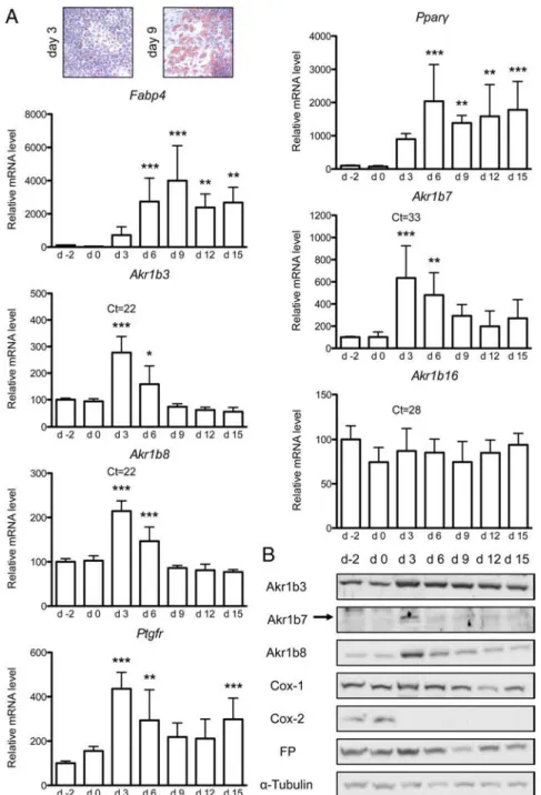
adi-pocyte fate. The mouse preadiadi-pocyte 3T3-L1 cell line is a well-characterized and widely used in vitro model to study adipocyte differentiation. To compare mouse aldose re-ductases expression during adipogenesis, 3T3-L1 differ-entiation was induced over a 2-week period. RT-qPCR analyses showed increased expression of Fabp4 and Ppar␥ genes in 3T3-L1 cells at differentiation days 3 and 6, con-firming adipocyte differentiation also attested by triglyc-eride accumulation using Oil-Red-O staining (Figure 2). In comparison with Akr1b3, b8, or b16, the Akr1b7 gene was expressed at very low levels in 3T3-L1 cells (detection threshold quantitative PCR cycle threshold (Ct) values at approximately 33 for Akr1b7 mRNA vs 22–28 for the other isoforms) (Figure 2A). Whereas Akr1b16 expression was unaltered throughout the culture period, Akr1b3, b7, and b8 mRNA levels increased from the onset of the adi-pogenic program (2.7-, 6-, and 2-fold, respectively) at dif-ferentiation day 3 and then progressively returned to basal expression levels found in undifferentiated cells. PGF re-ceptor gene (Ptgfr) mRNA levels (encoding the FP recep-tor) underwent a transient increase (4-fold) at differenti-ation day 3, paralleling Akr1b3, b7, and b8 expression patterns (Figure 2A). Moreover, aldose reductase-b3, -b7, -b8, and FP protein levels evaluated by Western blot paralleled their mRNA (Figure 2B). Finally, as previ-ously shown (35, 36), we confirmed that COX-1 protein was constitutively expressed throughout the differenti-ation program, whereas COX-2 was acutely down-reg-ulated from day 3 onward. We concluded that with the exception of Akr1b16, Akr1b3, b7, and b8, expression levels were all transiently increased during the early steps of adipogenesis with maximal expression at day 3 (at the onset of differentiation) and thereafter decreased during adipogenesis.
Differential expression of human aldose reductases during adipogenesis in hMADS cells
Involvement of human aldose reductases in adipose tis-sue homeostasis is poorly documented. Recent data re-ported a positive correlation between PGF2␣ release in
human preadipocytes isolated from obese women, BMI, and cytokine-induced AKR1B1 expression (26). In con-trast, the expression of the AKR1B10 gene as well as that of AKR1C3 and AKR1A1, two genes encoding enzymes (3␣-hydroxysteroid dehydrogenase type II and aldehyde reductase, respectively) with known PGF synthase activ-ity, has never been studied in fat cells (37, 38). We also monitored the mRNA levels for COX-2 (PTGS2 gene) and FPA/FPBreceptors (PTGFR1/PTGFR2 genes). RT-qPCR
analyses showed no correlation between BMI and expres-sion of these genes (Supplemental Figure 3). Western blot using specific antibodies (39) and RT-qPCR analyses
con-Figure 1. Differential expression of aldose reductase genes in
stromal vascular and adipocyte fractions. Aldose reductase relative mRNA expression was evaluated by RT-PCR after tissue
fractionation of gWAT, iWAT, and rWAT depots. A–C,
Quantifications of RT-PCR signals. RT-PCR analyses were conducted using four independent RNA pools, each resulting from the fractionation of two to six fat pads. Signal intensity was quantified for each PCR product and normalized according to an Actin signal. For each gene, the mRNA level was expressed relative to the signal obtained for SVF, which was given the value 1. n.d., not detected. Statistical analyses were performed to compare expression of each gene in SVF and adipocyte fraction by unpaired t test. *, P⬍ .05; **, P⬍ .01; ***, P ⬍ .001. D, Akr1b3 and Akr1b7 proteins were monitored by Western blot on extracts from SVF and adipocyte fraction.␣-Tubulin was used as a loading control.
firmed AKR1B1 expression in intact sc WAT from obese patients, but no correlation with BMI was shown in our study. The discrepancy with data from Michaud et al (26) could result from the small size of our cohort. Although
AKR1B10 transcripts were detected in rather low
amounts (average Ct 29.4), the pro-tein remained undetectable in these WAT samples. This suggests that
AKR1B10 expression could either
be absent or restricted to a minor population within sc WAT.
To analyze the expression of hu-man aldose reductase isoforms dur-ing adipogenesis, we used a hMADS cell line that can be differentiated into functional adipocytes under adi-pogenic culture conditions (40). Ad-ipogenesis was monitored by mea-suring changes at mRNA or protein levels of markers of the predifferen-tiation state (ZNF521, a regulator of adipose commitment ortholog to murine Zfp521) and early (PPAR␥) and late (LEPTIN, FABP4/aP2) dif-ferentiation (Figure 3B and Supple-mental Figure 4A). In agreement with progressive adipogenic differ-entiation, PPAR␥ mRNA expres-sion increased from day 3 to reach a plateau between day 9 and day 12. As previously described in other preadipocyte lines, a 1.5-fold in-crease in the ZNF521 mRNA level was transiently observed at day 0 in hMADS cells (41). Consistent with previous reports on leptin produc-tion, the highest LEPTIN mRNA level observed at day 6 was decreased 2-fold at day 9 (40). In agreement with full maturation of hMADS adi-pocytes, fatty acid binding protein 4 (FABP4)/adipose protein 2 (aP2) protein began to accumulate on day 9 (Supplemental Figure 4A). The lowest expression of AKR1B1 mRNA was observed at day 3 and progres-sively increased to reach 210% of basal level at day 15 (Figure 3A). This expression time course was tightly correlated with changes in al-dose reductase-B1 protein accumu-lation (Supplemental Figure 4A). In contrast, AKR1B10 mRNA levels had their highest ex-pression at day 0 and gradually decreased below 11% of the basal level throughout differentiation (Figure 3A). Un-like AKR1B1 and AKR1B10, AKR1A1 expression was only slightly and transiently increased (1.6-fold) between
Figure 2. Akr1b3, Akr1b7 and Akr1b8 expression levels are transiently increased during the
earlier step of 3T3-L1 adipogenesis. At day 0 (d0), 3T3-L1 preadipocytes were induced to differentiate into adipocytes (inset, Oil-Red-O staining showing progression of adipocyte differentiation of 3T3-L1 cells at days 3 and 9). A, From day 2 to day 15 of culture, mRNA levels of Fabp4, Ppar␥, Ptgfr, Akr1b3, Akr1b7, Akr1b8, and Akr1b16 were measured by RT-qPCR and normalized to Ppib. The qPCR Ct values from day 3 are indicated to compare relative expression level of the different Akr1b genes. mRNA quantifications were expressed as percentage of day 2 values. Statistical analyses were performed by one-way ANOVA followed by a Dunnett’s post hoc test. *, P⬍ .05; **, P ⬍ .01; ***, P ⬍ .001. B, Akr1b3, Akr1b8, Cox-1, Cox-2, and FP protein accumulations were monitored by Western blot throughout 3T3-L1 differentiation.␣-Tubulin was used as a loading control.
Figure 3. AKR1B1 and AKR1B10 display opposite expression profiles during hMADS cell adipogenesis. At day 0 (d0), hMADS preadipocytes
were induced to differentiate into adipocytes. A, AKR1B1, AKR1B10, AKR1A1, and AKR1C3 mRNA levels were measured by RT-qPCR throughout adipogenesis and values were normalized to TBP. B, To validate and follow the progress of adipogenic program, PPAR␥,
ZNF521, and LEPTIN gene expressions were analyzed by RT-qPCR and values were normalized to TBP. C, Level of PTGS2 transcripts were
measured by RT-qPCR and normalized to TBP. mRNA quantifications were expressed as a percentage of day 3 values. Statistical analyses were performed by one-way ANOVA followed by a Dunnett’s post hoc test, each value was compared with day 3 value. *, P⬍ .05; **, P⬍ .01; ***, P ⬍ .001.
differentiation day 3 and day 6. mRNA level for AKR1C3 was strongly induced (58-fold) from day 3 onward. Finally, COX-2 expression (PTGS 2 gene) was high in preadipocytes and rapidly turned off after the onset of adipogenesis between day 0 and day 3 (Figure 3C). Taken together, these data suggested that AKR1B10 and
AKR1B1 expression could be differentially involved in
adipogenesis.
Aldose reductase-mediated PGF2␣release during
hMADS cell adipogenesis
Previous in vitro enzymatic assays using human recom-binant proteins showed that aldose reductase-B1 had PGF synthase activity whereas B10 isoform was completely de-void of such activity (22). Therefore, we evaluated the functional link between AKR1B1 expression and PGF2␣
production during adipogenic differentiation of hMADS cells. Under differentiation conditions, PGF2␣production
rate of flow from hMADS cells increased progressively throughout adipogenesis to reach a 3-fold induction in mature hMADS adipocytes at day 15 (Figure 4A). This increase in PGF2␣ output was tightly correlated with changes in AKR1B1 expression at both mRNA and pro-tein levels (Figures 3A and 4A and Supplemental Figure 4B) and independent from changes in the gene expression of other PGF synthases, ie, AKR1A1 and AKR1C3 (Figure 3A). Consistent with the observations from mouse 3T3-L1 cells, expression of the rate-limiting enzymes COX was either constitutive (COX-1) or inhibited (COX-2) upon induction of differentiation and remained very low during hMADS cell adipogenesis (Figure 4B).
As shown in Figure 4B, expression of both FPAand FPB
isoforms of PGF2␣receptor was maximal in proliferative
preadipocytes (day ⫺3). It then decreased dramatically after day 3 (FPA, 17% of day⫺3 value) or day 6 (FPB, 6%
of day ⫺3 value) and remained low in maturing adi-pocytes. This suggested that PGF2␣released by adipocytes may essentially exert its antiadipogenic action in undif-ferentiated hMADS cells. Soon after differentiation commitment, FP receptors were turned off to trigger the adipogenic program and maintained at low levels throughout terminal differentiation. These low but detect-able amounts of FP proteins coexisting with high PGF2␣
levels would imply that PGF2␣could also have some
func-tion in mature adipocytes.
Exposure of hMADS cells to the specific aldose reduc-tase inhibitor Statil (Ponalrestat) over the 2-week differ-entiation period resulted in an 18% reduction in cumula-tive PGF2␣ production (Figure 4C). Remaining PGF2␣
accumulation could be attributed to constant expression of AKR1A1 or AKR1C3 (Figure 3A) whose PGF synthase activities are insensitive to Statil (38). The ability of
hMADS cells to accumulate intracellular lipids when ex-posed to adipogenic medium in the absence or presence of Statil was then examined by Oil-Red-O staining. As shown in Figure 4D, lipid accumulation was significantly increased by Statil treatment. As expected, Statil had no effect per se on AKR1B1, A1, B10, or C3 gene expression (Figure 4E). We thus concluded that increased PGF2␣
re-lease during hMADS adipogenesis was partially associ-ated with aldose reductase-B1 activity that might contrib-ute to delay adipocyte differentiation.
Interestingly, the reduction in PGF2␣production
result-ing from hMADS Statil treatment led to a concomitant decrease in COX-2/PTGS 2 expression (Figure 4E), whereas COX-1 was unaltered (not shown). This obser-vation was in agreement with previous demonstration of a PGF2␣-dependent up-regulation of COX-2 gene in
3T3-L1 cells (42). Our data thus confirm such a positive feedback loop for PGF2␣in human adipocytes.
Role of aldose reductase-dependent PGF2␣
production on the adipogenic program
Our kinetics expression data underscored the main changes occurring during the first 3 days of differentiation for most of the actors involved in PGF2␣
production/re-sponse. Further experiments were conducted to evaluate the effect of Statil over the first 3 days of differentiation and the impact of aldose reductase-dependent PGF2␣
pro-duction during this critical period (Figure 5). Compared with the vehicle condition, 10M Statil treatment resulted in a 27% decrease in PGF2␣release associated with a
con-comitant increase in the expression of the transcripts for proadipogenic regulators C/EBP and C/EBP␣ and for adipocyte marker ATGL, the lipolytic adipose triglyceride lipase. As expected, this treatment had no effect on AKR genes or on COX-1 expression and was too brief to alter
COX-2 mRNA levels (not shown). To evaluate the ability
of PGF2␣to reverse the Statil-dependent increase in
adi-pose conversion, hMADS cells cultured in adipogenic me-dium for 6 days were concomitantly exposed to the FP agonist cloprostenol in the presence of Statil. As shown in Figure 5, C and D, cloprostenol exposure counteracted Statil effects on both intracellular lipid accumulation and expression of transcripts for proadipogenic factors
C/EBP␣ and PPAR␥. Altogether our data indicate that, in hMADS cells, Statil exerts a primary proadipogenic effect at least by inhibiting aldose reductase-dependent PGF2␣
synthesis. Discussion
Previous data showing that certain aldose reductases are endowed with genuine PGF2␣synthase activity has
pro-Figure 4. PGF2␣flow is correlated to AKR1B1 expression during hMADS cell adipogenesis and is sensitive to Statil inhibitor. A, Culture media were collected every 3 days from day 0 to day 15 during hMADS cell differentiation and levels of released PGF2␣were determined by EIA. AKR1B1 protein was
detected by Western blot in hMADS cells extracts.␣-Tubulin was used as a loading control. B, COX-1, COX-2, FPA, and FPBaccumulation were analyzed
by Western blot during hMADS differentiation.␣-Tubulin was used as a loading control. C, Effects of aldose reductase Statil inhibitor on cumulative PGF2␣production of hMADS cells. hMADS cells were treated with Statil or DMSO (vehicle) during the entire 15-day differentiation period. Culture media
were collected every 3 days and PGF2␣levels measured by EIA. D, hMADS cells were differentiated for 9 days in the absence (vehicle) or presence of 10
M Statil, fixed, and stained for triglycerides with Oil-Red-O. To quantify staining, Oil-Red-O was extracted from differentiating adipocytes and
absorbance was then measured at 490 nm. E, hMADS cells were differentiated during 15 days in the presence of Statil or DMSO (vehicle) and expression of AKR1B1, AKR1B10, AKR1A1, AKR1C3, and PTGS 2 genes were analyzed by RT-qPCR, normalized to TBP, and expressed as a percentage of vehicle treatment values. Statistical analyses in panel A were performed by one-way ANOVA followed by a Dunnett’s post hoc test. Statistical analyses in panel C were performed by a two-way ANOVA followed by a Bonferroni post hoc test. In panels D and E, statistical analyses were performed by an unpaired t test. *, P⬍ .05; **, P ⬍ .01; ***, P ⬍ .001.
Figure 5. Statil enhances hMADS adipocyte differentiation by inhibiting aldose reductase-dependent PGF2␣synthesis. A, Effects of aldose
reductase Statil inhibitor on PGF2␣release in culture media were determined by EIA in hMADS cells treated with Statil or its vehicle during the first
3 days of differentiation. B, mRNA levels of C/EBP, C/EBP␣, and ATGL were monitored by RT-qPCR and normalized to TBP. Results were expressed as percentage of vehicle treatment values. C and D, Effects of Statil and cloprostenol (PGF2␣analog) treatments were examined after 6 days of
differentiation on C/EBP␣ and PPAR␥ expression by RT-qPCR (C) and on intracellular lipid accumulation by Oil-Red-O staining (D; bright field and contrast phase views). Statistical analyses in panels A and B were performed by one-way ANOVA followed by a Dunnett’s post hoc test, each value was compared with vehicle value. Statistical analyses in panel C were performed by a two-way ANOVA followed by a Bonferroni post hoc test. *, P⬍ .05; **, P ⬍ .01, ***, P ⬍ .001.
vided the first evidence for their possible influence on en-docrine or metabolic functions [(16, 22, 33); for review see reference 43]. More recently constitutive Akr1b7 gene knockout in mice allowed the in vivo demonstration that decreased PGF2␣production resulting from aldose
reduc-tase-b7 loss could affect adipose tissue homeostasis with-out compensation by other related isoforms (23). Here we performed comparative and quantitative analysis of the expression of aldose reductase (Akr1b subfamily) genes in a set of mouse tissues with a focus on adipose depots and adipose tissue fractions. Our data highlighted the unique adipose-specific features of mouse aldose reductase-b7, in line with its antiadipogenic action in vivo and revealed that in humans aldose reductase-B1 isoform might also regulate adipogenic differentiation at least by a PGF2␣
-dependent mechanism.
Aldose reductase isoforms in adipose tissues: minor expression sites for a probable major physiological function
Although aldose reductase-b3 remains the most abun-dant isoform in mouse adipose tissues, our quantitative studies reveal that fat represents a common expression site for all aldose reductase-like genes (at least one of their top 5 tissues is adipose tissue, Table 2). Previous in vitro ex-periments have anticipated the possible impact of these proteins on adipose tissue homeostasis. We reported that aldose reductase-b7 had antiadipogenic action when over-expressed in 3T3-L1 preadipocytes (31). Although the un-derlying mechanisms were not elucidated at that time, later evidence that it was endowed with PGF synthase activity suggested that aldose reductase-b7-mediated PGF2␣production was likely to support inhibition of
adi-pocyte differentiation (22, 33). This was definitively proven in vivo by using Akr1b7 knockout mice that de-veloped excessive adiposity in correlation with decreased accumulation of PGF2␣ (23). In the meantime, it was shown that endogenous aldose reductase-b3 could also fulfill the PGF2␣-dependent suppression of adipocyte
dif-ferentiation in the T3-L1 cell line (24). Interestingly, these authors reported that Akr1b3 expression was regulated throughout the 3T3-L1 differentiation program, mRNA levels showing a transient increase during the first day after the initiation of differentiation and then returning to low levels in maturing cells.
Although we broadly confirm these previous observa-tions, we reveal herein that all aldose reductase genes (ex-cept Akr1b16 that was constitutive) and to a lesser extent
Ptgfr gene (encoding FP receptor) are differentially
ex-pressed during 3T3-L1 adipogenesis with transient and maximal mRNA levels coinciding with the onset of dif-ferentiation and decreasing thereafter. Moreover, this
ob-servation was confirmed at the protein levels, suggesting that their coordinated down-regulation could be required for completion of the adipogenic program. Although both aldose reductases-b3 and -b7 were reported to negatively regulate 3T3-L1 differentiation through PGF synthase ac-tivity, we show that endogenous aldose reductase-b7 amounts are very low compared with that of -b3 (Figure 2). Conversely, Akr1b3 ablation in mice had no impact on adipose tissues, whereas loss of Akr1b7 induced, as ex-pected, an expansion of fat (23). Therefore, aldose reduc-tases function in adipose tissue should be investigated in vivo as much as possible. Here we report in vivo charac-terization of Akr1b genes expression within the two func-tional fractions of fat depots, ie, adipocytes and SVF. These data underscore the unique signature of Akr1b7 expression, which is the only gene of the four Akr1b genes in mouse to show a depot-specific pattern and absence of expression in mature adipocytes (Table 2 and Figure 1).
Akr1b3 shows higher expression in the adipocyte fraction,
thus contrasting with 3T3-L1 data (Figure 2 and reference 24). Altogether these observations suggest that aldose re-ductase-b7 functions in WAT are not overlapped by other isoforms, which is in good agreement with the adipose phenotype of Akr1b7⫺/⫺mice. The identification of the
cell population within SVF, which supports Akr1b7 ex-pression and relays its antiadipogenic action, is currently in progress.
Antiadipogenic action of aldose reductase-dependent PGF2␣production is conserved in
humans
Aldose reductase-B1 isoform was shown to be ex-pressed in primary preadipocytes isolated from human fat tissue, in close correlation with cytokine-induced PGF2␣
release. However, its impact on adipogenesis remains un-known (26). Our study suggests that the mode of action of these enzymes on adipose tissue homeostasis could differ in human and mouse. Using multipotent cells isolated from human adipose tissue (hMADS line), we show that expression of AKR1B1, the human homolog of mouse
Akr1b3, parallels hMADS adipogenic differentiation in
close correlation with PGF2␣ release. Importantly, we
demonstrate that inhibition of PGF2␣production using a
specific aldose reductase inhibitor (Statil) during hMADS adipogenesis enhances adipocyte differentiation, whereas exogenous PGF2␣ counteracts Statil effects (Figure 5).
Hence, in human as in mouse, the aldose reductase-de-pendent production of PGF2␣might participate in
restrict-ing differentiation of preadipocytes in a cell autonomous manner but the means of this restriction may differ. Unlike
Akr1b3 in 3T3-L1, the expression of AKR1B1 is not
adipo-genesis. However, PGF2␣receptors expression is turned
down after day 3 of hMADS differentiation (compare Fig-ures 2 and 4). This suggests that the need to overcome the repressive effect of PGF2␣ on adipogenesis might be
mainly achieved by limiting its signaling potential (by down-regulating FP receptors) in human cells and by lim-iting its synthesis (by down-regulating aldose reduc-tase-b3 and -b7 PGF synthases) in mouse preadipocytes. Nevertheless, we show that in both species, genes encod-ing aldose reductase-like devoid of PGF synthase activity, ie, Akr1b8 and AKR1B10 show maximum mRNA accu-mulation in undifferentiated adipocytes and are down-regulated in maturing adipocytes (Figures 2 and 3A).
In terms of structural conservations, substrate spectra and tissue distribution, murine aldose reductase-b8 is con-sidered as the ortholog of human aldose reductase-B10 (44). In addition to PGF2␣, other signaling molecules such
as those produced by the vitamin A (retinol) metabolism, retinoic acid and retinaldehyde (Rald) are known to neg-atively regulate adipogenesis (45, 46). Interestingly both aldose reductase-B10 and aldehyde reductase-C3 display the best Rald reductase activity among aldose and alde-hyde reductases (for review see reference 47). Once syn-thesized, Rald concentration depends both on its rate of irreversible oxidation to retinoic acid and on its rate of reduction back to retinol. Although we showed that
Akr1b8/AKR1B10 and AKR1C3 are differentially
ex-pressed after adipogenic stimulation, further experiments will be needed to unravel the possible impact of these genes as regulators of adipogenesis by influencing retinoid metabolism.
Taken together, our findings establish aldose reducta-ses as previously unrecognized contributors of adipose tis-sue homeostasis. By comparing human and mouse iso-forms in preadipocyte culture models, we propose that the two species have developed different but convergent strat-egies to control PGF2␣production and its biological
im-pact on fine-tuning early adipocyte differentiation. Fi-nally, our findings establish the background for further evaluation of pharmacological aldose reductase inhibi-tors, initially designed to treat diabetic complications, as potential obesogenic compounds.
Acknowledgments
We thank all the members of the team for helpful discussions. We also thank Sandrine Plantade, Khirredine Ouchen, and Philippe Mazuel for excellent animal care.
Address all correspondence and requests for reprints to:Anne-MarieLefrançois-MartinezandAntoineMartinez,CentreNationalde la Recherche Scientifique Unité Mixte de Recherche 6293, INSERM
Unité 1103, Génétique Reproduction and Développement, Clermont Université, BP80026, 24 Avenue des Landais, 63171 Aubière Cedex, France. E-mail:a-marie.lefrancois-martinez@univ-bpclermont.fr; or
antoine.martinez@univ-bpclermont.fr.
This work was supported by the Université Blaise Pascal, the Université d’Auvergne, the Centre National de la Recherche Sci-entifique, Institut National de la Santé et de la Recherche Médi-cale and by a grant from the Conseil Re´gional d’Auvergne Con-trat de Plan Etat Re´gion/Fonds Europe´ens de De´veloppement Re´gional 2010 Laboratoire d’Excellence and a PhD grant from the Société Française d’Endocrinologie.
Disclosure Summary: The authors have nothing to disclose.
References
1. Bohren KM, Bullock B, Wermuth B, Gabbay KH. The aldo-keto reductase superfamily. cDNAs and deduced amino acid sequences of human aldehyde and aldose reductases. J Biol Chem. 1989;264(16): 9547–9551.
2. Cao D, Fan ST, Chung SS. Identification and characterization of a novel human aldose reductase-like gene. J Biol Chem. 1998; 273(19):11429 –11435.
3. Hyndman DJ, Flynn TG. Sequence and expression levels in human tissues of a new member of the aldo-keto reductase family. Biochim
Biophys Acta. 1998;1399(2–3):198 –202.
4. Salabei JK, Li X-P, Petrash JM, Bhatnagar A, Barski OA. Functional expression of novel human and murine AKR1B genes. Chem Biol
Interact. 2011;191(1–3):177–184.
5. Gui T, Tanimoto T, Kokai Y, Nishimura C. Presence of a closely related subgroup in the aldo-keto reductase family of the mouse. Eur
J Biochem. 1995;227(1–2):448 – 453.
6. Pailhoux EA, Martinez A, Veyssiere GM, Jean CG. Androgen-de-pendent protein from mouse vas deferens. cDNA cloning and pro-tein homology with the aldo-keto reductase superfamily. J Biol
Chem. 1990;265(32):19932–19936.
7. Donohue PJ, Alberts GF, Hampton BS, Winkles JA. A delayed-early gene activated by fibroblast growth factor-1 encodes a protein re-lated to aldose reductase. J Biol Chem. 1994;269(11):8604 – 8609. 8. Fukumoto S-I, Yamauchi N, Moriguchi H, et al. Overexpression of the aldo-keto reductase family protein AKR1B10 is highly corre-lated with smokers’ non-small cell lung carcinomas. Clin Cancer
Res. 2005;11(5):1776 –1785.
9. Woenckhaus M, Klein-Hitpass L, Grepmeier U, et al. Smoking and cancer-related gene expression in bronchial epithelium and non-small-cell lung cancers. J Pathol. 2006;210(2):192–204.
10. Heringlake S, Hofdmann M, Fiebeler A, Manns MP, Schmiegel W,
Tannapfel A. Identification and expression analysis of the
aldo-ketoreductase1–B10 gene in primary malignant liver tumours.
J Hepatol. 2010;52(2):220 –227.
11. Kang MW, Lee ES, Yoon SY, et al. AKR1B10 is associated with smoking and smoking-related non-small-cell lung cancer. J Int Med
Res. 2011;39(1):78 – 85.
12. Schmitz KJ, Sotiropoulos GC, Baba HA, et al. AKR1B10 expression is associated with less aggressive hepatocellular carcinoma: a clini-copathological study of 168 cases. Liver Int. 2011;31(6):810 – 816. 13. Yadav UCS, Srivastava SK, Ramana KV. Aldose reductase inhibi-tion prevents endotoxin-induced uveitis in rats. Invest Ophthalmol
Vis Sci. 2007;48(10):4634 – 4642.
14. Yadav UCS, Ramana KV, Aguilera-Aguirre L, Boldogh I, Boulares
HA, Srivastava SK. Inhibition of aldose reductase prevents
experi-mental allergic airway inflammation in mice. Hartl D, ed. PLoS
ONE. 2009;4(8):e6535.
deficiency protects from autoimmune- and endotoxin-induced uve-itis in mice. Invest Ophthalmol Vis Sci. 2011;52(11):8076 – 8085. 16. Bresson E, Boucher-Kovalik S, Chapdelaine P, et al. The human aldose reductase AKR1B1 qualifies as the primary prostaglandin F synthase in the endometrium. J Clin Endocrinol Metab. 2011;96(1): 210 –219.
17. Barski OA, Tipparaju SM, Bhatnagar A. The aldo-keto reductase superfamily and its role in drug metabolism and detoxification.
Drug Metab Rev. 2008;40(4):553– 624.
18. Ramana KV, Yadav UCS, Calhoun WJ, Srivastava SK. Current pro-spective of aldose reductase inhibition in the therapy of allergic air-way inflammation in asthma. Curr Mol Med. 2011;11(7):599 – 608. 19. Srivastava SK, Ramana KV, Bhatnagar A. Role of Aldose Reductase and Oxidative Damage in Diabetes and the Consequent Potential for Therapeutic Options. Endocrine Reviews. 2005;26(3):380 –392. doi:10.1210/er.2004 – 0028.
20. Reginato MJ, Krakow SL, Bailey ST, Lazar MA. Prostaglandins promote and block adipogenesis through opposing effects on per-oxisome proliferator-activated receptor gamma. J Biol Chem. 1998; 273(4):1855–1858.
21. Liu L, Clipstone NA. Prostaglandin F2␣ inhibits adipocyte differ-entiation via a G␣q-calcium-calcineurin-dependent signaling path-way. J Cell Biochem. 2007;100(1):161–173.
22. Kabututu Z, Manin M, Pointud J-C, et al. Prostaglandin F2␣ syn-thase activities of aldo-keto reductase 1B1, 1B3 and 1B7. J Biochem. 2009;145(2):161–168.
23. Volat FE, Pointud J-C, Pastel E, et al. Depressed levels of prosta-glandin F2␣ in mice lacking Akr1b7 increase basal adiposity and predispose to diet-induced obesity. Diabetes. 2012;61(11):2796 – 2806.
24. Fujimori K, Ueno T, Nagata N, et al. Suppression of adipocyte differentiation by aldo-keto reductase 1B3 acting as prostaglandin F2␣ synthase. J Biol Chem. 2010;285(12):8880–8886.
25. Baba SP, Hellmann J, Srivastava S, Bhatnagar A. Aldose reductase (AKR1B3) regulates the accumulation of advanced glycosylation end products (AGEs) and the expression of AGE receptor (RAGE).
Chem Biol Interact. 2011:1–7.
26. Michaud A, Lacroix-Pépin N, Pelletier M, et al. Prostaglandin (PG) F2␣ synthesis in human subcutaneous and omental adipose tissue: modulation by inflammatory cytokines and role of the human aldose reductase AKR1B1. PLoS One. 2014;9(3):e90861.
27. Student AK, Hsu RY, Lane MD. Induction of fatty acid synthetase synthesis in differentiating 3T3-L1 preadipocytes. J Biol Chem. 1980;255(10):4745– 4750.
28. Rodriguez AM, Elabd C, Amri E-Z, Ailhaud G, Dani C. The human adipose tissue is a source of multipotent stem cells. Biochimie. 2005; 87(1):125–128.
29. Zaragosi L-E, Ailhaud G, Dani C. Autocrine fibroblast growth fac-tor 2 signaling is critical for self-renewal of human multipotent ad-ipose-derived stem cells. Stem Cells. 2006;24(11):2412–2419. 30. Mohsen-Kanson T, Hafner A-L, Wdziekonski B, et al.
Differenti-ation of human induced pluripotent stem cells into brown and white adipocytes: role of Pax3. Stem Cells. 2014;32(6):1459 –1467. 31. Tirard J, Gout J, Lefrançois-Martinez A-M, Martinez A, Begeot M,
Naville D. A novel inhibitory protein in adipose tissue, the aldo-keto
reductase AKR1B7: its role in adipogenesis. Endocrinology. 2007; 148(5):1996 –2005.
32. Pfaffl MW. A-Z of Quantitative PCR. La Jolla, CA: SA Bustin; 2004:87–112.
33. Lambert-Langlais S, Pointud J-C, Lefrançois-Martinez A-M, et al. Aldo keto reductase 1B7 and prostaglandin F2␣ are regulators of adrenal endocrine functions. Vella A, ed. PLoS One. 2009;4(10): e7309.
34. Cawthorn WP, Scheller EL, MacDougald OA. Adipose tissue stem cells meet preadipocyte commitment: going back to the future.
J Lipid Res. 2012;53(2):227–246.
35. Yan H, Kermouni A, Abdel-Hafez M, Lau DCW. Role of cyclooxy-genases COX-1 and COX-2 in modulating adipogenesis in 3T3-L1 cells. J Lipid Res. 2003;44(2):424 – 429.
36. Lu S, Nishimura K, Hossain MA, Jisaka M, Nagaya T, Yokota K. Regulation and role of arachidonate cascade during changes in life cycle of adipocytes. Appl Biochem Biotechnol. 2004;118(1–3):133– 153.
37. Komoto J, Yamada T, Watanabe K, Takusagawa F. Crystal struc-ture of human prostaglandin F synthase (AKR1C3). Biochemistry. 2004;43(8):2188 –2198.
38. Pépin NL, Chapdelaine P, Fortier MA. Evaluation of the prosta-glandin F synthase activity of human and bovine aldo-keto reduc-tases: AKR1A1s complement AKR1B1s as potent PGF synthases.
Prostagland Other Lipid Mediat. 2013:1–9.
39. Ma J, Yan R, Zu X, et al. Aldo-keto reductase family 1 B10 affects fatty acid synthesis by regulating the stability of acetyl-CoA car-boxylase-alpha in breast cancer cells. J Biol Chem. 2008;283(6): 3418 –3423.
40. Rodriguez A-M, Elabd C, Delteil F, et al. Adipocyte differentiation of multipotent cells established from human adipose tissue. Biochem
Biophys Res Commun. 2004;315(2):255–263.
41. Kang S, Akerblad P, Kiviranta R, et al. Regulation of early adipose commitment by Zfp521. Vidal-Puig AJ, ed. PLoS Biol. 2012;10(11): e1001433.
42. Fujimori K, Yano M, Ueno T. Synergistic suppression of early phase of adipogenesis by microsomal PGE synthase-1 (PTGES1)-pro-duced PGE2 and aldo-keto reductase 1B3-pro(PTGES1)-pro-duced PGF2␣. Giorgino F, ed. PLoS One. 2012;7(9):e44698.
43. Pastel E, Pointud J-C, Volat F, Martinez A, Lefrançois-Martinez
A-M. Aldo-keto reductases 1B in endocrinology and metabolism.
Front Pharmacol. 2012;3:148.
44. Joshi A, Rajput S, Wang C, Ma J, Cao D. Murine aldo-keto reduc-tase family 1 subfamily B: identification of AKR1B8 as an ortholog of human AKR1B10. Biol Chem. 2010;391(12):1371–1378. 45. Xue JC, Schwarz EJ, Chawla A, Lazar MA. Distinct stages in
adi-pogenesis revealed by retinoid inhibition of differentiation after in-duction of PPAR␥. Mol Cell Biol. 1996;16(4):1567–1575. 46. Ziouzenkova O, Orasanu G, Sharlach M, et al. Retinaldehyde
re-presses adipogenesis and diet-induced obesity. Nat Med. 2007; 13(6):695–702.
47. Ruiz FX, Porté S, Parés X, Farrés J. Biological role of aldo-keto reductases in retinoic acid biosynthesis and signaling. Front