HAL Id: hal-03105200
https://hal.archives-ouvertes.fr/hal-03105200
Submitted on 10 Jan 2021
HAL is a multi-disciplinary open access
archive for the deposit and dissemination of
sci-entific research documents, whether they are
pub-lished or not. The documents may come from
teaching and research institutions in France or
abroad, or from public or private research centers.
L’archive ouverte pluridisciplinaire HAL, est
destinée au dépôt et à la diffusion de documents
scientifiques de niveau recherche, publiés ou non,
émanant des établissements d’enseignement et de
recherche français ou étrangers, des laboratoires
publics ou privés.
Identification of Ablation Sites in Persistent Atrial
Fibrillation Based on Spatiotemporal Dispersion of
Electrograms Using Machine Learning
Amina Ghrissi, Fabien Squara, Johan Montagnat, Vicente Zarzoso
To cite this version:
Amina Ghrissi, Fabien Squara, Johan Montagnat, Vicente Zarzoso. Identification of Ablation Sites
in Persistent Atrial Fibrillation Based on Spatiotemporal Dispersion of Electrograms Using Machine
Learning. Computing in Cardiology, Sep 2020, Rimini, Italy. �hal-03105200�
Identification of Ablation Sites in Persistent Atrial Fibrillation Based on
Spatiotemporal Dispersion of Electrograms Using Machine Learning
Amina Ghrissi
1, Fabien Squara
1,2, Johan Montagnat
1and Vicente Zarzoso
11
Universit´e Cˆote d’Azur, CNRS, I3S Laboratory, Sophia Antipolis, France
2Universit´e Cˆote d’Azur, CHU Pasteur, Cardiology Department, Nice, France
Abstract
A recent patient-tailored ablation protocol to treat atrial fibrillation consists in identifying ablation sites based on their spatiotemporal dispersion (STD). STD represents a delay of the cardiac activation observed in intracardiac electrograms (EGMs) across contiguous leads. This work aims at automatically identifying ablation sites by classi-fying EGM data acquired by the PentaRay catheter into ablated vs. non-ablated groups using machine learning. More than 35000 multichannel recordings are acquired from 15 persistent AF patients. An annotation model is designed to label the dataset. The classifiers include: (1) multivariate logistic regression; (2) LeNet-STD, a shal-low convolutional neural network. A binary label identify-ing whether the mapped site contains STD pattern accord-ing to the interventional cardiologist is combined to raw EGMs as classifiers input. The LeNet-STD combined with data augmentation yields the best performance with an F1-score of 76%.
1.
Introduction
Atrial fibrillation (AF) represents the most widespread sustained arrhythmia experienced in clinical practice ris-ing in prevalence with advancris-ing age. AF is characterized by an irregular activation in the atria that start quivering causing the ventricular rate to become more rapid and dis-organized [1]. Among the existing treatments for persis-tent AF, ablation interventions overperform drug therapies in terms of efficiency.
Ablation is an invasive procedure that consists in burn-ing the cardiac tissues displayburn-ing irregularities with RF en-ergy delivered through catheters. Classical ablation proto-col is called stepwise and consists in: 1) burning the trig-gersaround the pulmonary veins thought to be responsible for initiating AF; 2) ablating cardiac areas exhibiting com-plex fractionated electrograms [2]. However, a growing number of reports show the limitations of the stepwise ap-proach [3].
A novel wholly patient-tailored ablation protocol, giving 95% of procedural success rate, consists in identifying ab-lation sites based on a pattern called spatiotemporal disper-sion (STD) [4]. Multipolar mapping catheters are used to record electrograms (EGMs) in the atria thus targeting ar-eas of STD as potential AF drivers. The high-density map-ping PentaRay catheter (Biosense Webster Inc, Irvine, CA, USA) is used for STD localization. It has a five-branch star design with two bipoles on each spline.
Before ablation with RF energy, interventional cardiol-ogists first position sequentially the PentaRay in different atrial sites. Ten bipoles are then simultaneously recorded per location by maintaining the catheter stable for at least 2.5 s. Finally, atrial sites displaying an irregular cardiac activity are annotated as dispersion points and tagged for ablation. According to guidelines in [4], dispersion areas refer to clusters of electrograms, either fractionated or not, displaying interelectrode time and space dispersion at a minimum of three contiguous leads, as shown in Fig. 1. Hence, STD-based ablation is a fully patient-tailored ther-apy. According to preliminary guidelines for STD identifi-cation from visual inspection, the 10-channel EGM record-ing acquired by the PentaRay would display a cardiac ac-tivation delay of 70% of AF cycle length (AFCL) on a minimum of three neighboring bipoles (channels) [4]. A recent study proposed to automatically detect STD EGMs using machine learning (ML) [5] and benchmarked a set of adapted data augmentation (DA) approaches to handle the high class imbalance ratio between STD and non-STD classes [6].
The present contribution proposes to design a decision-aid solution that helps interventional cardiologists iden-tify potential ablation sites automatically using ML tools. We classify ablated vs. non-ablated mapped sites using: (1) multivariate logistic regression (MLR); (2) LeNet-STD, a shallow convolutional neural network (CNN). A binary label identifying whether the mapped site contains STD pattern according to the interventional cardiologist is combined to raw EGMs as classifiers input. This ad-ditional input can be assimilated to a prior probability in
STD-guided ablation. Data augmentation is used to han-dle the dataset imbalance. ML techniques allow to au-tomatically identify ablated sites and guide cardiologists in patient-tailored catheter ablation procedures for treating persistent AF.
Figure 1. Dispersion areas are defined and delineated via a mapping approach [4]. Channels A 1-2, A 3-4, B 5-6, B 7-8, C 9-10 and C 11-12 display STD.
2.
Methods
2.1.
Study dataset
Data are provided by the Cardiology Department of Nice University Hospital Center (CHU) and are exported from the BioSense CARTO system. This intervention data come from 15 patients ablated for persistent AF based on STD pattern with the use of PentaRay catheter, shown in Fig. 2. They include electrocardiogram, EGM and
anno-Figure 2. PentaRay multi-spline catheter. tated dispersion points from both right and left atrial car-tographies. The study dataset includes a cohort of 35563 10-channel EGM signals of length 2.5 s composed of 1804 samples labeled STD and 33759 non-STD samples. Data annotation is performed by interventional cardiolo-gists such that EGMs presenting spatiotemporal dispersion are labeled as ‘STD’. We automatically merge the remain-ing labels into the ‘non-STD’ class.
2.2.
Annotation model
The intervention is a two-fold process: 1) mapping the atria and tagging STD sites; 2) ablating AF drivers (atrial locations) that have not necessarily been mapped in ad-vance. An annotated dataset is then needed to train super-vised ML classifiers that automatically detect the ablated mapped points among all the mapped ones (including STD
and non-STD). The labels are binary and should inform if the mapped point (data sample) has been ablated or not. This information is not provided by the CARTO system because: 1) the mapping and ablation phases are decou-pled; 2) the ablation sites are not restricted to the mapped points tagged as STD. Indeed, there might be STD posi-tions that are not ablated probably because they are iso-lated and do not form a cluster or because of anatomical reasons. Besides, there might be non-STD positions that are ablated because they are close enough to STD clusters or for anatomical reasons. For this purpose, we design a la-beling model based on the proximity between both mapped P and ablated V. Due to the limited precision of the abla-tion catheter and the manual ablaabla-tion process, the catheter might be slightly misplaced around the desired position (D). In practice, the catheter may slightly move around the desired position D during an ablation shot, also called a session. D might correspond or not to a mapped point P .
Figure 3. Sphere model of the ablation site. Based on these observations, we propose to consider each area ablated around D as a sphere (S) of center V and radius r. The Cartesian coordinates of V correspond to the average Cartesian coordinates of the points visited by the ablation catheter during the ablation session and r is the square root of the standard deviations of the corresponding coordinates. According to the partner interventional car-diologist, the distance between a mapped point P and the closest ablation zone S should be smaller or equal to 5 mm for P to be considered as ablated. This can be mathemati-cally translated into k−P V k−→ 2−r ≤ 5. Hence, for each
abla-tion session, the mapped points that lie at most 5 mm away from S are attributed the ‘ablated’ label (Pab), resulting in
the set:
Pab= {Pi∈ P : ∃Vj ∈ V, d(Pi, Vj) ≤ 5 + rj} (1)
Mapped points not fulfilling Eq. (1) form the non-ablated class (Pnon-ab). Based on this model, we obtain 15803
ab-lated and 19760 non-abab-lated points.
2.3.
Classification algorithms
Logistic regression model estimates the probability of a given class by using the logistic function. MLR is a base-line classifier in biomedical data analysis [7].
CNNs are commonly used in biomedical data classifi-cation [8]. A CNN is composed of convolutional (conv)
and pooling layers followed by fully connected (FC) ones. LeNet-STD is a CNN inspired by LeNet-5 architecture [9] which represents a good and computationally-affordable model. Let Z and kernel respectively refer to the number of filters and the receptive field of the filters. LeNet-STD is composed of the following layers: 1) 2-dimensional (2D) conv (Z=32, kernel=4×300); 2) average pooling; 3) 2D conv (Z=16, kernel=3×3); 4) average pooling; 5) FC (Z=128); 6) FC (Z=64); 7) final FC decision (Z=2).
During the mapping phase, the PentaRay catheter is maintained stable for 2.5 s at each location. A location refers to anatomical point inside the heart. Multichannel EGMs are sampled at 1 kHz, recorded in the hospital’s database then displayed through the CARTO system mon-itor, to be analyzed by the cardiologist. Hence, EGM sam-ples can be stored in a matrix of dimension 10 × 2500 and classified as images.
2.4.
Data Augmentation
DA applies transformations to original samples of the minority class in order to synthesize new samples. A re-cent study proved that random oversampling (ROS) is the best technique for the classification of multichannel EGMs into STD vs. non-STD [6]. ROS forms a balanced super-dataset by randomly replicating STD samples until they reach the number of the non-STD ones.
3.
Experiments and Results
Circularity transformation is applied to each data sam-ple in order to mimic the circularity of PentaRay branches. This transformation consists in replicating the first two rows of the 10 × 2500 matrix at its end to form a matrix of dimensions 12 × 2500 [6].
In order to identify potential ablation sites in STD-based ablation of persistent AF, we train baseline classifiers in-cluding MLR and LeNet-STD. The study dataset is anno-tated according to the model in Sec. 2.2 and is composed of 15803 ablated (Pab) and 19760 non-ablated (Pnon-ab)
sam-ples.
We conduct three types of experiments:
1) We train LeNet-STD and MLR with mini-batch gra-dient descent and early stopping criteria. The receptive field of the first convolutional layer of LeNet-STD is cho-sen large enough to cover the information between all three contiguous leads along one AFCL and a half thus fully cap-turing potential STD patterns.
2) The class imbalance ratio between labeled and non-labeled classes is equal to #Pab
#Pnon-ab = 80%, where #A
refers to the cardinality of the set A. As a result, classes are slightly imbalanced and the classifiers might face prob-lems learning characteristics from the minority class (ab-lated) due to the insufficient number of Pab samples.We
propose to use a DA method called ROS that proved effi-cient in [6]. The resulting models are denoted MLR-ROS and LeNet-ROS.
3) Based on the fact that both ablated and STD atrial areas should be spatially correlated, we concatenate the in-formation about whether a data point is labeled STD or not with the previous classifiers’ output. This additional input can be assimilated to a prior probability in STD-guided ablation. We build new architectures for MLR and LeNet-STD, denoted MLR-STDlab and LeNet-STDlab, by adding a sub-neural network (sub-NN) composed of a dense FC layer. The sub-NN gets the STD label as input and outputs a score function that will be concatenated with the output of the final decision layer of MLR and LeNet.
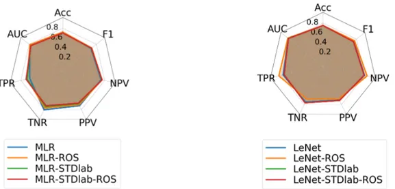
Five-fold cross-validation (CV) is used to asses the gen-eralization power of the trained classifiers. The classifica-tion results shown in Figures 4(a) and 4(b) represent the av-erage performance on the test dataset across the 5 folds of MLR and LeNet respectively. The metrics computed are: 1) accuracy (Acc); 2) area under the ROC curve (AUC); 3) sensitivity, also called true positive rate (TPR) or true rate of ablated samples; 4) specificity also called true negative rate (TNR) or true rate of non-ablated samples; 5) preci-sion, also called positive predictive value (PPV); 6) nega-tive predicnega-tive value (NPV); 7) F1-score. The results show that the overall performance of LeNet-STD improves than that of MLR with values of Acc, AUC and F1-score re-spectively equal to 75%, 82%, 73% vs. 72%, 78%, 67%. ROS allowed us to increase the TPR from 76% to 84% while maintaining TNR at 70% which further increases the F1-score to 76% with LeNet-STD architecture. Concate-nating with STD labels did not help improve neither de-crease the classification performance. Also, the standard deviation of the classification metrics through CV does not exceed 10−2. We conclude then that LeNet-STD-ROS is the best model.
4.
Conclusions and perspectives
The automatic identification of potential ablations sites in STD-based ablation of persistent AF can help guide interventional cardiologists in patient-tailored procedures. For this task, we design a model to annotate the multi-channel EGM dataset into ablated vs. non-ablated classes based on Euclidean distance between mapped and ablated points. ML tools are used to classify the labeled samples into ablated vs. non-ablated categories. MLR and LeNet-STD are benchmarked whether combined with DA or not. Moreover, we concatenate the supervised classifiers with sub-NN that takes as input the label of whether the con-sidered data sample is tagged STD or non-STD. Five-fold CV is also used to asses the robustness of trained models. Classification results on the test set show that LeNet-STD combined with ROS give the best performance with an
Figure 4. Classification performance on the test dataset. Values represent averages through 5-fold CV. Left: MLR classifier and variants. Right: LeNet-STD classifier and variants.
F1-score of 76%. Aggregating STD label did not help im-prove the model’s performance. This can be interpreted by the fact that ablation takes into consideration more anatom-ical factors than the STD pattern. Moreover, the sub-NN used to analyze STD labels is shallow and might be not efficient. Future work should aim at designing deeper ar-chitectures that might take further advantage of STD in-formation to improve the automatic detection of ablation sites.
Acknowledgments
This work is funded by the French government PIA pro-gram, IDEX UCAJEDIproject (ANR-15-IDEX-0001).
References
[1] C. T. January, et al. “2014 AHA/ACC/HRS guideline for
the management of patients with atrial fibrillation,” Journal of the American College of Cardiology, 64(21), 2014.
[2] K. Nademanee, et al. “A new approach for catheter
abla-tion of atrial fibrillaabla-tion: mapping of the electrophysiologic substrate,” Journal of the American College of Cardiology. Journal of the American College of Cardiology, 43(11), pp. 2044–2053, 2004.
[3] A. Verma, et al. “Approaches to catheter ablation for persis-tent atrial fibrillation,” New England Journal of Medicine, 372(19), pp. 1812–1822, 2015.
[4] J. Seitz, et al. “AF ablation guided by spatiotemporal
elec-trogram dispersion without pulmonary vein isolation: a wholly patient-tailored approach,” Journal of the American College of Cardiology, 69(3), pp. 303–321, 2017.
[5] I. Goodfellow, et al. Deep Learning, MIT Press, 2016.
www.deeplearningbook.org.
[6] A. Ghrissi, et al. “Data Augmentation for Automatic
Iden-tification of Spatiotemporal Dispersion Electrograms in Atrial Fibrillation Ablation Using Machine Learning,” in
42nd Annual International Conference of the IEEE
En-gineering in Medicine and Biology Society, EMBC 2020, Montreal, 2020.
[7] S. Dreiseitl, O. M. Lucila. “Logistic regression and artificial neural network classification models,” Journal of Biomedi-cal Informatics, 35(5-6), pp. 352–359, 2002.
[8] B. Pyakillya, et al. “Deep learning for ECG classification,”
Journal of Physics, 913(1), IOP Publishing, 2017.
[9] Y. LeCun, et al. “Gradient-based learning applied to
doc-ument recognition,” Proceedings of the IEEE, 86(11), pp. 2278–2324, 1998.
Address for correspondence: Amina Ghrissi
Universit´e Cˆote d’Azur, CNRS, I3S Laboratory
Les Algorithmes, Euclide B, 06103, Sophia Antipolis, France amina.ghrissi@univ-cotedazur.fr
![Figure 1. Dispersion areas are defined and delineated via a mapping approach [4]. Channels A 1-2, A 3-4, B 5-6, B 7-8, C 9-10 and C 11-12 display STD.](https://thumb-eu.123doks.com/thumbv2/123doknet/13430926.408857/3.918.102.401.227.366/figure-dispersion-defined-delineated-mapping-approach-channels-display.webp)