Anatomy of the Coiled Coil and Its Role in the
Conformational Change of Influenza Hemagglutinin
by
Chavela Marguerite Carr
Submitted to the Department of Biology in partial fulfillment of the requirements
for the degree of
DOCTOR OF PHILOSOPHY
in Biologyat the
Massachusetts Institute of Technology June 1995
C 1995 Chavela M. Carr. All rights reserved.
The author hereby grants to MIT permission to reproduce and to distrubute publicly paper
and electronic copies of this thesis document in whole or in part.Signature of Author
- 'Department of Biology
May 5, 1995 Certified byPeter S. Kim
Associate Professor, Department of Biology
Thesis Supervisor
Accepted byFrank Solomon
Chairman, Biology Graduate Committee
Dedication
In memory of
Lawrence Silas Carrand
Dwight Herrick Macduff
Acknowledgments
For impressing upon me their appreciation of nature and the importance of critical
thinking, I dedicate this thesis to my late grandfathers: Lawrence Silas Carr and DwightHerrick Macduff. I also thank my living family members for being patient with me and
forgiving my forgetfulness of birthdays and other special occasions during these seven
:years at MIT.I have been influenced by a great number of excellent teachers who believed in me and pushed me to excel beyond what I imagined possible. Most notable for my scientific and analytical development are my human-genetics high-school teacher, Mr. Gordon
Mendenhall; Mr. Dan Stiko, high-school teacher of several math courses; my
undergraduate mentor, Professor Douglas Cavener; and, of course, my graduate advisor,
Dr. Peter Kim. Toward my personal growth, I am indebted to my high-school choir and
dance director Ms. Sandra Butz; Mr. Gary Meyers, my high-school theater director, andMr. Michael Kim Howden, junior-high world-history teacher, enduring friend, counselor,
and confidant.The best years of my life have been spent in the exhilarating environment of MIT and the Whitehead Institute. The friends and colleagues I have met here I will treasure
forever. For their generosity, I especially thank my talented and beloved friends outside
the lab: Mattan Kamon, whose computer advice, encouragement and affection helped methrough the best and the most difficult years; Joel Phillips, for good food, good wine, and
a dear and lasting friendship; Christina Scherer, classmate and comrade in happiness andhardships from the first to the last day of graduate school; Shiufun Cheung, for his genuine
friendship and work on the graphic rendering of the HA conformations, which was so
widely publicized; Josephine Cheung, for her cheerful nature and friendship; Ignacio
McQuirk, for his appreciation of spicy food, good tea, excellent chocolate, and friendship;
Michelle and Bob Spina, that crazy couple who brought out my loud side and made sure I
enjoyed myself thoroughly, whenever I was with them; Meg Winberg, who kept my
musical and theatrical spirit alive; Amelia Shen, a terrific housemate and source of otherlasting friendships; David Shia, for sharing his computer, printer, and good humor, and
discovery described in Chapter 2. Special thanks goes to Eve Nichols, the Whitehead's
publicity director. Eve's enthusiasm and hard work in promoting my project has gone a
long way to encourage and inspire me.
The familial friendships inevitably formed when people work closely together for
several years added to the rich experience of graduate school. For their scientific and non-scientific contributions I thank "the old crew" and the "young turks" of the lab: ErinO'Shea, for the good-old days, the encouragement, and especially, the laughter. Jon
Staley, ("Studley") for so much support and friendship when I was a young graduate
student; Terry Oas, ("Oasage") for all manner of advice and so much help in the lab;Rheba Rutkowski (R
2), for her critical, expressive and outspoken appreciation of human
nature, and for her approval, which meant all the more for its difficulty to attain; David Litwack, for camaraderie during year-one in the lab, and especially for partnership in theoutstanding prank on Staley; Jonathan Weissman, for his great sense of humor, so much
scientific advice and a supportive friendship to endure all; Pehr Harbury (Harb) fornumerous lessons, for his generous help in reading and correcting numerous sections of
this thesis, and for a lasting friendship; Dan Minor (Major Minor) for practical and
scientific advice and for his efforts to unify the lab in group outings, like the hiking trip from Hell; Brenda Schulman, (N*) for sister-like friendship, for critical advice about my written work and for help digging through the literature for references for the introductionof this thesis; Bob Talanian (BT) for expert advice on DNase I footprinting (Appendix I);
David Lockhart, (Lock-jaw) my role model, my friend, my confidant; Jamie McKnight, whose help with vesicles and fluorescence was essential to chapter 3; Tom Alber and his group for their large contributions as the structural collaborators in the GCN4 project(Appendix I); Zeng-Yu Peng, for his candid and critical advice plus his humble and
good-natured spirit; Kevin Lumb whose contributions to this thesis deserve special
consideration, as the work of Appendix II is primarily his; Andrea Cochran, who provided
me with an alternative perspective on the world; Martha Oakley, for steadfast support,especially in my darkest hour; Deborah Fass, for her inspirational zeal for everything she
does; Lawren Wu, whose calm manner and easy sense of humor is good medicine in the
hectic environment of the lab; Charu Chaudhry, my hard-working, talented undergraduate
research student (UROP), who worked out the proteinase K assay used in Chapter 3, and
who was a great source of pride and joy; Steve Blacklow, for his enthusiasm; Min Liu, for
his personal and practical advice; Paul Matsudaira and his students Kathy Collins and DonDoering for many interesting scientific conversations and help during my early years; and Mitch Sanders, Michael Way and Navin Pokala for help with protein gels and immunoblot
protocols.
For expert technical assistance, I thank Mike Milhollen, who made the expression
vectors for prHMG and purified rHA2-X117; Mike Burgess, who made and analyzed the
masses of many peptides used in the studies descried here and some not described here;
Rheba Rutkowski, for the reasons mentioned above and for peptide synthesis; Sean Britt
for assistance with the GCN4 peptides; Christina Doetsch, the newest technician to the lab,
a true and lasting friend and treasure; Paul Matsudaira's technical associate Mark Chaffel,for help with tissue culture and use of photography supplies and Matt Footer for his
professional advice and for use of his photography skills and tools in preparing figures
used in Chapter 3.The most difficult acknowledgment to write is the one owed to my advisor,
Peter S. Kim. Though these words cannot convey the weight of my admiration and
gratitude, I thank you, Peter. I appreciate the exceptional facilities and opportunities thatPeter has provided for the members of the lab, including powerful and plentiful, high-tech
laboratory equipment, the large variety of scientific journals, expensive Silicon Graphics,
Inc. (SGI) computers for structual analysis; and especially the diverse group of highly
motivated students and post-docs who interacted to form a stimulating scientificcommunity. My success is a direct result of Peter's willingness to take risks in starting
new projects and his committment to provide everything I thought necessary for theexperiments, including new equipment, a hired computer consultant, and large quantities of
infectious flu virus! Peter's good judgment in hiring Charu Chaudhry, the talented
undergraduate student, who worked with me on the HA conformational assay (Chapter 3),
led to a deeply rewarding teaching experience. I am proud to be among the ever-growing league of distinguished students that have graduated from this lab.The 6 years I have spent in Peter's lab have confirmed the advice of sage professors
from both my undergraduate and graduate years, and I have since passed on this advice to prospective graduate students, who are hoping to build a career in science: You have to love it; its too hard!. So why did I spend 7 years of my young life in the laboratory? I didAnatomy of the Coiled Coil and Its Role in the Conformational Change of
Influenza Hemagglutinin
by
Chavela Marguerite Carr
Submitted to the Department of Biology on May 5, 1995 in partial fulfillment of the requirements for the
Degree of Doctor in Philosophy in Biology
Abstract
The identification of an unexpected coiled-coil sequence in influenza hemagglutinin (HA)
led to a model for the conformational change required for the membrane-fusion function of
this protein (Chapter 2). Analysis of coiled-coil peptides corresponding to the fusion-active
conformation of HA not only support the model, but led to a prediction for the mechanism of the conformational change. Chapter 3 describes evidence that the "native," fusion-inactive conformation of HA is trapped in a metastable state, which, when destabilized,undergoes the conformational change and induces membrane fusion. Chapter 4 discusses
recent advances that may shed some light on the steps for HA-mediated membrane fusion.The appendices are devoted to the leucine-zipper motif, a short coiled-coil region that forms
the dimerization domain of bZIP and bHLH-ZIP proteins. A conserved, buried hydrogen
bond in the hydrophobic coiled-coil interface destabilizes the GCN4 leucine zipper, yet itprovides specificity for the dimer conformation (Appendix I A; see also Harbury et al.,
1993). The stabilizing effects of increasing the hydrophobicity of a buried amino acid is quantified using non-natural amino acids for the incremental addition of methylene groups to the central position of the GCN4 leucine-zipper interface (Appendix I B). The
identification of a stable subdomain in the GCN4 leucine zipper has implications for the
mechanism of protein folding (Appendix II).
Thesis Supervisor: Dr. Peter S. Kim,
Tilte: Associate Professor of Biology
Table of Contents
Dedication 2 Acknowledgments 3 Abstract 6 Table of Contents 7 Chapter 1: Introduction 9 Chapter 2: 23Chapter 2 has been published as C. M. Carr and P. S. Kim, "A Spring-Loaded Mechanism for the Conformational
Change of Influenza Hemagglutinin." Cell 73, 823-832
(1993). © Cell Press.
Chapter 3: Evidence that Influenza Hemagglutinin is
Trapped in the Native Conformation 56
Chapter 4: 85
Chapter 4 has been published as C. M. Carr and P. S. Kim,
"Flu Virus Invasion: Halfway There." Science 73, 234-236
(1994).
American Association for the Advancement of
Science.Appendix
Appendix I: Structural and Functional Studies of the Coiled Coil
A.
A Conserved Asparagine Destabilizes the Leucine Zipper of GCN4
B. Effects of Increased Hydrophobicity at the Coiled Coil Interface Appendix II:Appendix 2 has been published as K. J. Lumb, C. M. Carr
and P. S. Kim, "Subdomain Folding of the Coiled Coil
Leucine Zipper from the bZIP Transcriptional ActivatorGCN4." Biochemistry 33, 7361-7367 (1994). © American
Chemical Society.Appendix II:
Bimolecular Equilibrium Equation Biographical Profile 9899
104 155 181 185 8 95Introduction
Proteins are the workhorses of life. The diversity of functions performed by
proteins is enormous. For example, proteins are required for the structural integrity of skin
and bones, the mechanical work performed by muscles, the efficient conversion of food
into energy, the transduction of signals during sensory perception, and the transcription
and translation of genes for the synthesis of other proteins. Fundamental to the description
of any one of these processes is a "nuts and bolts" understanding of the way proteins
work. Equipped with this knowledge, we may then begin to understand the nature of
diseases caused either by cellular proteins with altered or abolished functions, or by
parasites such as viruses, whose proteins function to invade and overtake the cell.This chapter begins by addressing the relationship between protein structure and function and by outlining the approaches used to discover how a protein works. Following a general introduction, this chapter proposes how these approaches can be applied to specific questions concerning the role of the coiled-coil motif in the structure and function
of hemagglutinin (HA), the membrane fusion protein from influenza virus. Chapters 2 - 4
describe how the application of our knowledge about the sequence determinants ofcoiled-coil structure leads to the discovery of a novel role for the coiled-coiled coiled-coil in membrane fusion.
The appendices describe the identification of specific determinants for the structure and stability of the coiled-coil dimerization domain of GCN4, a transcription factor from thebZIP family of proteins.
Protein Structure and Function
"Conformation---exemplified by the relationship between the three-dimensional structure of
proteins and their biological activity." --Lubert Stryer (Stryer, 1988).
The function of a protein is dictated by its conformation. Any attempt to reveal the
way a protein works requires knowledge of its three-dimensional structure. Yet
interpretation of the protein structure often requires additional information to highlight the
amino-acid residues of particular interest for protein function. Much progress has been
made toward understanding protein function by combining 2 complementary approaches:structural analysis and mutagenesis. Researchers have also had success in reconstituting
protein function from protein fragments, a method hereafter referred to as "protein
dissection." Ultimately, our understanding of protein structure and function will be tested
by our ability to design functional proteins.
Structure Determination
The most intuitive approach toward understanding a protein's function is to look at
its conformation. By far the best methods for high-resolution protein-structure
determination are x-ray crystallography (Blundell and Johnson, 1990), and more recently,
nuclear magnetic resonance spectroscopy (Wiithrich, 1986). But how does the structure
explain the function? For those proteins that bind to ligands or substrates (i.e.
enzyme-substrate complexes or transcription factor-DNA complexes), a co-crystal can reveal the details of specific interactions, explaining specificity of binding. However, in many cases the information conveyed by the structure is difficult to interpret in terms of protein
function or dynamics. The classic analogy is that a polaroid of a car's engine does not
explain how the car works. Nonetheless, examination of a protein's structure inspires
hypotheses for the mechanism of function, hypotheses which can be tested using a variety
of methods to perturb the protein and examine the effect of the perturbation on proteinstructure and function.
While the first step to understanding function is to look at the structure, proteins that share the same conformation may have different functions (for a review, see (Orengo et
al., 1994). For example, the four-helix bundle protein, Rop, is used for plasmid
replication (Banner et al., 1987), cyt b562, for electron transport (Mathews et al., 1979),
1L4, as a cytokine B (Smith et al., 1992; Powers et al., 1992) and as a component of thecoat protein of tobacco mosaic virus (Champness et al., 1976). The immunoglobin
superfold structure of antibodies is seen in many places, including the NF-KB transcription
factor (Ghosh et al., 1995; Mtiller et al.; 1995). The existence of protein families that share
a structural motif or modular domain yet perform different specific functions suggests that
there must be amino-acid sequence determinants for unique function superimposed on the
sequence determinants for a protein's three-dimensional structure. Thus, in order to
understand the specificity of protein function, these sequence determinants must be
Mutagenesis
A second and complementary approach toward discerning the functional regions of a protein is amino-acid substitution, or mutagenesis. Because the conformation of a protein is dictated by its amino-acid sequence (Epstein et al., 1963), alteration of the sequence can
change or abolish the structure and function of the protein. Random mutagenesis, followed
by a genetic screen or selection for functional protein, reveals that some sites in proteins are tolerant to substitution, while others are less so (Coplen et al., 1990; Hu et al., 1990). Those positions that are sensitive to substitutions may play an important role either in thestructure or specificity of the protein function. Mutations which abolish function without disrupting structure reveal amino acids which may play an important role in the specificity
of protein function. For example, mutations in the DNA-binding domain of the lambda
repressor and cro transcription factors led to the discovery of how these proteins recognize
and distinguish between different DNA sequences (Hochschild et al., 1986).
Similarly, structurally and functionally important residues are conserved between
proteins of the same family, and the role of these residues can be assessed by site-directed
mutagenesis (Reidhaar-Olson and Sauer, 1988). More recently, the introduction of "unnatural" amino acids into proteins has increased the repertoire of functional groups for substitution (Ellman et al., 1992; Mendel et al., 1992; Judice et al., 1993; Chung et al.,1993), allowing for a fine-tuned analysis of the functional groups important for protein
structure and function. In combination with high-resolution structure determination, alanine-scanning mutagenesis is another powerful tool for uncovering the determinants ofprotein structure and function (Cunningham and Wells, 1989).
Protein Dissection
A third approach that has met with surprising success is to reduce the
structure/function problem into parts by protein dissection (Oas and Kim, 1988; Goodman
and Kim, 1989; Peng and Kim, 1994). At first sight, the structure of proteins appears to
be unapproachably complex: a composite of interdependent interactions between amino acids both near and far from each other in sequence determines the overall 3-dimensionalstructure that is so essential to the function of the protein. Dissecting the protein into pieces by breaking the polypeptide chain would seem an unproductive approach.
Nonetheless, several observations suggest that proteins may be composed of
autonomous structural domains that can be studied separately: 1. Deletions or truncationsof proteins yield functional domains for some proteins, for example, DNA-binding
domains of bZIP proteins (for a review see Hu and Sauer, 1992). 2. Proteolytic digestion
of proteins into protease-resistant fragments has revealed that many proteins are composed
of more than one stable domain or subdomain, which folds as an autonomous unit (Wu etal., 1994; Matsudaira, 1992). 3. The discovery of structural motifs shared between
different families suggested that these evolutionarily conserved modules may be able to form independent structural units (for a review, see Cohen et al., 1995). Several structuralmotifs have been confirmed as autonomous folding units by protein dissection, for
example, the leucine-zipper dimerization domain of the bZIP family of transcription factors
(O'Shea et al., 1989a, b) and the SH2 and SH3 protein-binding domains of signalling
proteins (Cohen et al., 1995).Peptide models have been used successfully to study protein structure/function and
protein folding. Reconsititution of RNase A activity by mixing fragments of this protein
suggests that the whole is, in some cases, the sum of its parts (Richards and Vithayathil,1959). In addition, reconstitution of trypsin inhibition and native-like structure with
peptide models of bovine pancreatic trypsin inhibitor illustrate that the connectivity of the polypeptide chain need not remain intact for correct folding of single-domain proteins (Oasand Kim, 1988; Staley and Kim, 1990). The same conclusion comes from the result that
circularly permuted T4 lysozyme retains its native, stable fold (Zhang et al., 1993).
The implication of these discoveries is that otherwise intractable, insoluble or overly
large proteins can be manageably dissected into parts for studies of structure and functionin solution. Quick and easy methods to assay protein structure and stability, such as
circular dichroism, fluorescence and absorbance spectroscopy can then be applied. Trimming proteins into their minimal structural units also aids detailed structural analysisby crystallography and NMR. Of course, not all proteins will withstand dissection and
reconstitution, but progress has already been made on biologically important moleculessuch as SH3 proteins (Lim et al., 1994; Yu et al., 1994), tumor suppressor p53 (Jeffry et al., 1995), and Max (Ferre-D'Amare et al., 1993), the partner of the protooncogene, c-Myc.
Protein Design
The ultimate and most ambitious approach toward understanding a protein's
structure and function is protein design. Hypotheses for sequence patterns that specify a
given conformation can be tested by de novo design of a polypeptide chain, which should
fold into the predicted structure if the hypothesis is corrrect. This approach has met withlimited success (O'Shea et al., 1993), but as more is learned about determinants for
uniqueness of structure, this will be the ultimate test of our understanding of protein
structure and function.
Purpose
This thesis examines the role of the coiled-coil structural motif in the structure and
function of two very different proteins: hemagglutinin (HA), the membrane-fusion protein
from influenza virus and GCN4, a bZIP transcription factor. The primary approach used
in both studies is protein dissection: coiled-coil peptides were studied in solution to test predictions of protein stability and conformation. In addition to structural studies, thefunctions of these proteins have been reconstituted in vitro to test the effects of perturbation
on protein function.
I.
Role of a Coiled Coil in the Conformational Change of Influenza
Hemagglutinin Required for Membrane Fusion
The fact that specialized proteins are required for membrane fusion events, such as fertilization, neurotransmitter release and viral infection, has exciting implications for the
control of these events by inhibition of the fusogenic function of the proteins responsible.
Nonetheless, progress toward the rational design of inhibitors requires an understanding of
the mechanism of membrane fusion and the role of these specialized proteins in bilayermixing. In the simplest model for membrane fusion, there is one basic mechanism of
fusion with variation at the level of regulation of fusion activation for specificity of
targeting and timing of the fusion event. With this in mind, we hoped to learn some general principles about the mechanism of membrane fusion by studying the best characterized membrane fusion protein, influenza hemagglutinin (HA).
Several key questions are central to understanding membrane fusion: What is the structure of the fusogenic conformation? What is the mechanism of the conformational
change? What is the mechanism of membrane fusion? These three questions are addressed
in a discussion of experimental results, which shed light on these issues while raising new
questions. In addition, the possible generality of the principles learned from HA and
implications for the control of infection are discussed.A Modelfor the Fusogenic Conformation of HA
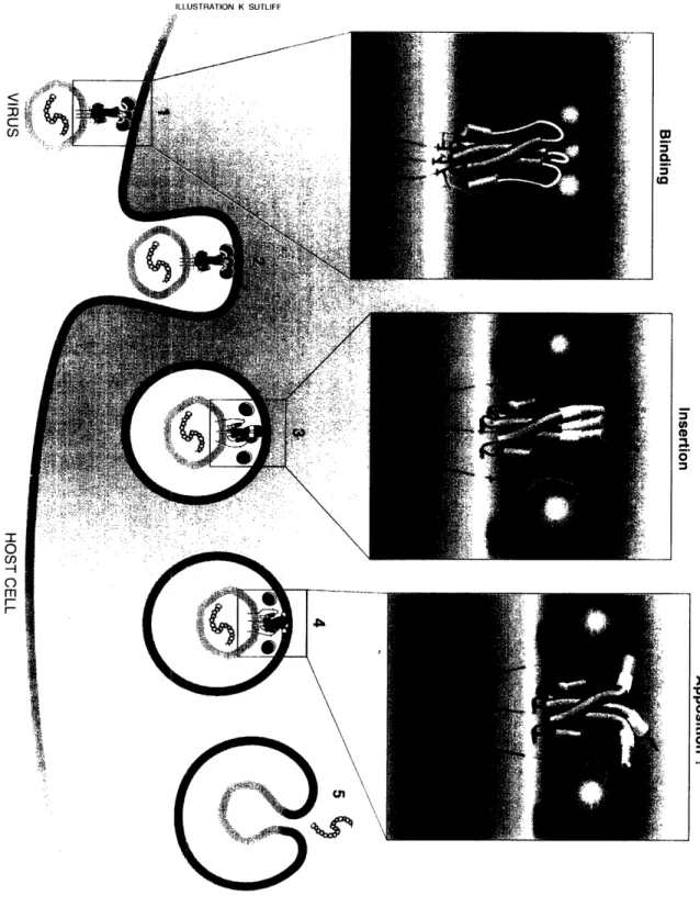
The HA envelope protein is responsible for the membrane fusion event that results
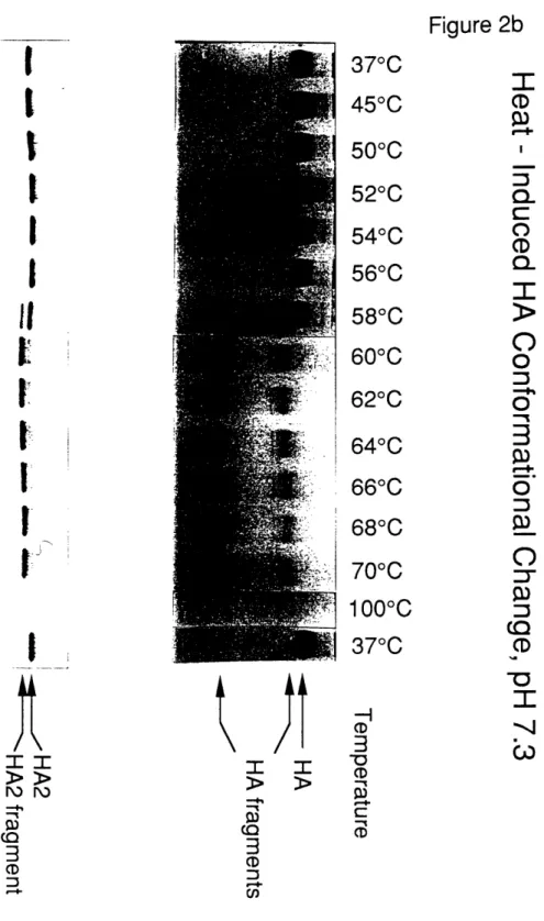
in entry of influenza virus into the host cell. HA is held together in the native conformation by a long, three-stranded coiled-coil core (Fig. 1). In response to the acidic environment ofthe cellular endosome, HA undergoes an irreversible conformational change that is required
for its function in membrane fusion. We propose that the dynamic nature of the HA coiled coil is critical for this conformational change (Chapter 2).Chapter 2 describes the identification of a "latent" coiled-coil region in HA. The coiled coil is latent because the structure of this region is extended to form a loop
conformation in the native state, though the sequence suggests that this region has a high propensity to form a coiled coil. We proposed that this region adopts a coiled coil as a
consequence of the conformational change required for membrane fusion. Evidence is
presented for the dramatically different "fusogenic" conformation of HA, and the relevance
of this conformation to its function in membrane fusion is discussed.
The Mechanism of the HA Conformational Change
Though mildly acidic pH induces the conformational change in vivo, several
observations suggested to us that the mechanism for HA-mediated membrane fusion is not
necessarily pH-dependent (see Chapter 2). These considerations led us to propose that the
conformational change that induces membrane fusion. The major prediction of this
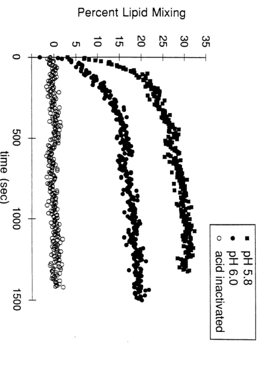
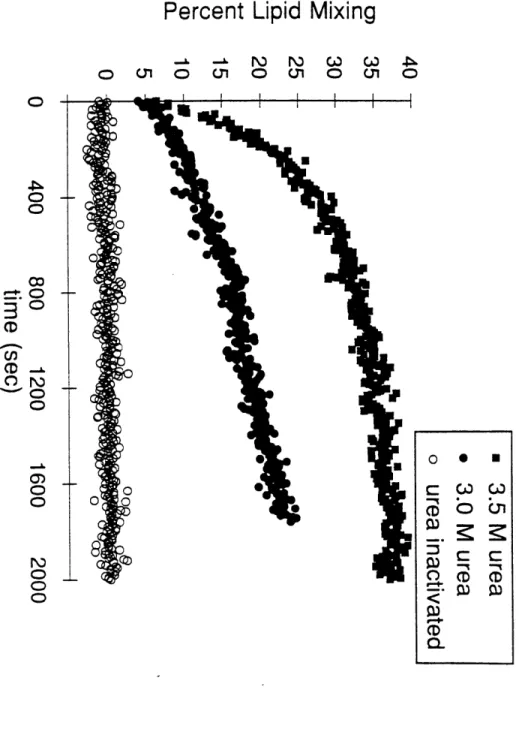
hypothesis is that the conformational change and membrane fusion can be induced at neutral pH in response to destabilizing conditions. In Chapter 3 we analyze theconformation and function of HA in intact influenza virus to test this prediction. Evidence is presented for a mechanism of the HA conformational change that may be relevant for
other membrane fusion events.
HIow do Membranes Fuse?
How the conformational change of HA is utilized for the final steps in membrane
fusion remains a mystery. Chapter 4 raises questions about the mechanism of membrane
fusion and summarizes some recent discoveries plus a model for the next step in
HA-mediated membrane fusion.II.
(Appendix) The Coiled Coil: Stability and Specificity of the
GCN4 Leucine Zipper
The "leucine-zipper," is a short coiled-coil dimerization motif of bZIP and
bHLH-ZIP transcription factors and a model system for the analysis of coiled coils. The structural
requirements of the coiled coil result in a conserved pattern of hydrophobic and hydrophilic
residues in the amino-acid sequence, known as the heptad repeat. The leucine-zipper
sequences also display this pattern, and in the case of GCN4 and the Fos/Jun heterodimer,
peptides corresponding to the leucine-zipper sequences form specific coiled-coil dimers insolution (O'Shea et al., 1989a, b).
The short coiled coil of the leucine-zipper motif faces the challenge of forming a stable, coiled-coil dimer while maintaining specificity for its correct partner to form a
functional transcription factor. For the Fos/Jun heterodimer, the determinants of partner
specificity have been localized to the predominantly charged amino-acid residues that flankthe hydrophobic interface of the leucine zipper dimer (O'Shea et al., 1992). We sought to
uncover additional determinants of leucine-zipper structure and function using the coiled
coil from GCN4. Appendix I addresses the role of a conserved asparagine in the
leucine-zipper coiled coil and the contribution of hydrophobicity to coiled-coil stability. Detailedcrystallographic analyses of the structures of GCN4 peptides were made possible by an
ongoing collaboration with Tom Alber's lab (O'Shea et al., 1991 and Appendix I).
Identification of a stably folded subdomain in an N-terminal deletion mutant of the GCN4leucine-zipper peptide (Appendix II), suggests that leucine-zipper folding may proceed
through this conformation as an intermediate in folding.References
Banner, D. W Kokkinidis, M., and Tsernoglou, D. (1987). Structure of the ColE1 rop
protein at 1.7 A resolution. Journal of Molecular Biology 196, 657-675.
Blundell, T. L., and Johnson, L. N. (1990). Protein Crystallography. (London: Academic
Press).
Champness, J. N., Bloomer, A. C., Bricogne, G., Butler, P. G., and Klug, A. (1976).
The structure of the protein disk of tobacco mosaic virus to 5A resolution. Nature 259,
20-4.Chung, H. H., Benson, D. R., Cornish, V. W., and Schultz, P. G. (1993). Probing the
role of loop 2 in Ras function with unnatural amino acids. Proc Natl Acad Sci U S A 90,10145-9.
Cohen, G. B., Ren, R., and Baltimore, D. (1995). Modular Binding Domains in Signal
Transduction Proteins. Cell 80, 237-248.
Coplen, L. J., Frieden, R. W., and Goldenberg, D. P. (1990). A genetic screen to identify
variants of bovine pancreatic trypsin inhibitor with altered folding energetics. Proteins 7,
116-31.
Cunningham, B. C., and Wells, J. A. (1989). High-resolution epitope mapping of
hGH-receptor interactions by alanine-scanning mutagenesis. Science 244, 1081-5.
Ellman, J. A., Mendel, D., and Schultz, P. G. (1992). Site-specific incorporation of novel
backbone structures into proteins. Science 255, 197-200.
Epstein, C. J., Goldberger, R. F., and Anfinsen, C. B. (1963). Cold Spring Harbor
Symp. Quant. Biol. 27, 439-449.
Ferre-D'Amare, A. R., Prendergast, G. C., Ziff, E. B., and Burley, S. K. (1993). Recognition by Max of its cognate DNA through a dimeric b/HLH/Z domain [see
comments]. Nature 363, 38-45.
Ghosh, G., Van Duyne, G., Ghosh, S., and Sigler, P. B. (1995). Structure of NF-kB
p50 homodimer bound to a kB site. Nature 373, 303-310.Goodman, E. M., and Kim, P. S. (1989). Folding of a peptide corresponding to the
alpha-helix in bovine pancreatic trypsin inhibitor. Biochemistry 28, 4343-7.
Harbury, P. B., Zhang, T., Kim, P. S., and Alber, T. (1993). A switch between two-,
three-, and four-stranded coiled coils in GCN4 leucine zipper mutants. Science 262,
1401-1407.
Hochschild, A., Douhan, J. 3., and Ptashne, M. (1986). How lambda repressor and
lambda Cro distinguish between OR1 and OR3. Cell 47, 807-16.Hu, J. C., O'Shea, E. K., Kim, P. S., and Sauer, R. T. (1990). Sequence requirements
for coiled-coils: analysis with lambda repressor-GCN4 leucine zipper fusions. Science
250, 1400-3.
Hu, J. C., and Sauer, R. T. (1992). The basic-region leucine-zipper family of DNA
binding proteins. Nucleic Acids and Molecular Biology 6, 82-101.
Jeffry, P. D., Gorina, S., and Pavletich, N. P. (1995). Crystal Structure of the
Tetramerization Domain of the p53 Tumor Suppressor at 1.7 Angstroms. Science 267,
1498-1502.
Judice, J. K., Gamble, T. R., Murphy, E. C., de Vos, A. M., and Schultz, P. G. (1993). Probing the mechanism of staphylococcal nuclease with unnatural amino acids: kinetic and
structural studies. Science 261, 1578-81.
Lim, W. A., Richards, F. M., and Fox, R. O. (1994). Structural determinants of
peptide-binding orientation and of sequence specificity in SH3 domains. Nature 372, 375-9.
Mathews, F. S., Bethge, P. H., and Czerwinski, E. W. (1979). The structure ofcytochrome b562 from Escherichia coli at 2.5 A resolution. J Biol Chem 254, 1699-706.
Matsudaira, P. (1992). Mapping structural and functional domains in actin-bindingproteins. (Oxford: IRL Press, Oxford University Press).
Mendel, D., Ellman, J. A., Chang, Z., Veenstra, D. L., Kollman, P. A., and Schultz, P.
(. (1992). Probing protein stability with unnatural amino acids. Science 256, 1798-802.
MUller, C. W., Rey, F. A., Sodeoka, M., Verdine, G., and Harrison, S. C. (1995).Structure of the NF-kB p50 homodimer bound to DNA. Nature 373, 311-317.
O'Shea, E. K., Rutkowski, R., and Kim, P. S. (1989a). Evidence that the leucine zipper
is a coiled coil. Science 243, 538-542.
O'Shea, E. K., Rutkowski, R., Stafford, W. F., and Kim, P. S. (1989b). Preferential
heterodimer formation by isolated leucine zippers from Fos and Jun. Science 245,
646-648.
C)'Shea, E. K., Klemm, J. D., Kim, P. S., and Alber, T. A. (1991). X-ray structure of
the GCN4 leucine zipper, a two-stranded, parallel coiled coil. Science 254, 539-544.
O'Shea, E. K., Rutkowski, R., and Kim, P. S. (1992). Mechanism of Specificity in the
Fos-Jun Oncoprotein Heterodimer. Cell 68, 699-708.
O'Shea, E. K., Lumb, K. J., and Kim, P. S. (1993). Peptide "Velcro": design of a
heterodimeric coiled coil. Current Biology 3, 658-667.
Oas, T. G., and Kim, P. S. (1988). A peptide model of a protein folding intermediate.
Nature 336, 42-48.
Peng, Z. Y., and Kim, P. S. (1994). A protein dissection study of a molten globule.
Biochemistry 33, 2136-41.
Powers, R., Garrett, D. S., March, C. J., Frieden, E. A., Gronenborn, A. M., and Clore,
G. M. (1992). Three-dimensional solution structure of human interleukin-4 by
multidimensional heteronuclear magnetic resonance spectroscopy. Science 256, 1673-7.
Reidhaar-Olson, J. F., and Sauer, R. T. (1988). Combinatorial cassette mutagenesis as aprobe of the informational content of protein sequences. Science 241, 53-7.
Richards, F. M., and Vithayathil, P. J. (1959). The Preparation of Subtilisin-modified
Ribonuclease and the Separation of the Peptide and Protein Components. J. Biol. Chem.
234, 1459-1464.
Smith, L. J., Redfield, C., Boyd, J., Lawrence, G. M., Edwards, R. G., Smith, R. A.,
and Dobson, C. M. (1992). Human interleukin 4. The solution structure of a four-helix
bundle protein. J Mol Biol 224, 899-904.
Staley, J. P., and Kim, P. S. (1990). Role of a subdomain in the folding of bovine
pancreatic trypsin inhibitor. Nature 344, 685-8.
Stryer, L. (1988). Biochemistry. (New York: W. H. Freeman and Company). Wu, L. C., Grandori, R., and Carey, J. (1994). Autonomous subdomains in protein
folding. Protein Sci 3, 369-71.
Wuithrich, K. (1986). NMR of Proteins and Nucleic Acids. (New York: Wiley). Yu, H., Chen, J. K., Feng, S., Dalgarno, D. C., Brauer, A. W., and Schreiber, S. L.
(1994). Structural basis for the binding of proline-rich peptides to SH3 domains. Cell 76,
933-45.
Zhang, T., Bertelsen, E., Benvegnu, D., and Alber, T. (1993). Circular Permutation of T4
Lysozyme. Biochemistry 32, 12311-8.
Figure 1
A three-stranded coiled coil forms the core of native HA. A ribbon-representation of the trimeric, native HA ectodomain (BHA) is depicted in purple, with the three-stranded
coiled coil highlighted in white. This structure represents HA in the native, fusion-inactive
conformation. A conformational change is required for the membrane fusion activity of
HA, and the coiled coil plays a central role (see Chapter 2).,
i
?P
-IA
00II P 22ir!
%4
1, .4io,
4t _
Ir~~~~~~r,
I m)l0 , 11~
~c
ZS~t71~L ItI l"1
4t,
-
N
,IA Spring-Loaded Mechanism for the
Conformational Change of Influenza Hemagglutinin
Summarx
Influenza hemagglutinin (HA) undergoes a conformational change that induces viral
fusion with the cellular membrane. The structure of HA in the fusogenic state is unknown.
We have identified a sequence that has a high propensity for forming a coiled coil.Surprisingly, this sequence corresponds to a loop region in the x-ray structure of native HA: the loop is followed by a three-stranded, coiled-coil stem. We find that a 36-residue
peptide (LOOP-36), comprised of the loop region and the first part of the stem, forms a
three-stranded coiled coil. This coiled coil is extended and stabilized in a longer peptide,corresponding to LOOP-36 plus the residues of a preceding, short a-helix. These findings
lead to a model for the fusogenic conformation of HA: the coiled-coil stem of the nativestate is extended, relocating the hydrophobic fusion peptide, by 100 A, toward the target
membrane.
Introduction
Membrane fusion is necessary for a large number of diverse processes in biology.
For example, protein trafficking, protein secretion, fertilization and neurotransmissionrequire the fusion of distinct membranes to form a single lipid bilayer. In general,
membrane fusion is very slow in the absence of specific proteins.The best characterized membrane fusion event occurs in the infection of animal cells by influenza virus. Infection begins with the binding of virus to sialic acid, a component of membrane proteins and lipids. The bound virion is then internalized into a cellular
endosome by receptor-mediated endocytosis. Ultimately, the viral envelope fuses with the
membrane of the mature endosome in response to a drop in pH (for reviews, see Wiley and
Skehel, 1987; Stegmann and Helenius, 1993). Membrane fusion is promoted by the
trimeric, viral-envelope glycoprotein, hemagglutinin (HA), which also functions in cell attachment (at neutral pH) by binding to sialic acid. At low pH, HA is sufficient for thefusion of membranes in vivo and in vitro (White et al., 1982).
Each HA monomer is synthesized as a protein precursor, denoted HA0. The HA0
polypeptide is processed proteolytically to form a pair of disulfide-bonded peptides,
denoted HA1 and HA2 (Lazarowitz et al., 1971; Skehel and Waterfield, 1975). This
proteolytic processing is essential for the infectivity of the virus (Klenk et al., 1975;
Lazarowitz and Choppin, 1975). Native HA binds sialic acid, but is dormant for
membrane-fusion activity.
There is considerable evidence for a large, irreversible conformational change in
HA which is required for membrane fusion (for reviews, see Wiley and Skehel, 1987;
White, 1992; see however, Stegmann et al., 1990). The conformational change is induced
by mildly acidic conditions (e.g., pH -5, the pH of the mature endosome) and results in a
trimeric structure that is remarkably thermostable (Doms and Helenius, 1986; Ruigrok et
al., 1986; 1988). At neutral pHI, a similar conformational change, leading to a stable, fusion-active state of HA, can be induced at temperatures above 600 C (Ruigrok et al.,Structure of the Native State of HA
HA can be proteolytically cleaved with bromelain to release the exoplasmic domain from the viral membrane. The resulting soluble domain, termed BHA, has sialic acid-binding and low-pH, liposome-acid-binding activities similar to intact HA (Brand and Skehel,
1972; Doms et al., 1985). The x-ray crystal structure of the BHA trimer, in the native state, has been determined with and without bound sialic acid (Wilson et al., 1981; Weis et al., 1988; 1990a). The interaction between HA and sialic acid has been well described (Weis et al., 1988; Glick et al., 1991; Sauter et al., 1992), and this interaction is separate
from the function of HA in membrane fusion.
The central region of the HA2 polypeptide folds into a helical-hairpin structure (see Fig. 1A): a short a-helix (red) is connected to a long a-helix (magenta) by an extended loop region (yellow). The long a-helix interacts with the corresponding long a-helices
from two other HA2 polypeptides to form an interwound rope of three helices, called a
three-stranded coiled coil. The three shorter a-helices are displayed on the outside of the
coiled coil. The sialic acid-binding domains of the HA 1 subunits assemble on top of the
fibrous stem, which is formed primarily by the three-stranded HA2 coiled coil (Fig. 1B).
'he length of the trimer, from the junction with the membrane to the distal tip of the HA1 subunits, is -135 A (Wilson et al., 1981).
HA2 is the transmembrane subunit, which spans the envelope membrane once. At
the amino terminus of HA2 is a highly conserved, hydrophobic sequence of -25 residues,
known to be necessary for membrane fusion (Daniels et al., 1985; Gething et al., 1986a).
Although buried in the hydrophobic interior in the native state (Fig. 1; light blue), theseresidues become exposed in the fusogenic state (for reviews, see Wiley and Skehel, 1987;
Stegmann and Helenius, 1993) and insert into the endosomal membrane (Stegmann et al.,
1991). For these reasons, this sequence is referred to as the "fusion peptide" (for a review, see White, 1992).The structure of HA in the fusogenic conformation is not known. An immediate
puzzle is to understand how the exposed fusion peptide facilitates fusion of the viral envelope with the endosomal membrane since, in the native conformation, the fusionpeptide is buried near the viral envelope, -100
A
from the distal tip of the HA molecule (Fig. 1).Model for the Structure of the Fusogenic State
We present a model for the fusogenic structure of HA. The model evolved from
our interest in the coiled-coil motif found in many proteins (for a review, see Cohen and
Parry, 1990) including the "leucine zipper" domain of some transcription factors(Landschulz et al., 1988; O'Shea et al., 1989; 1991; Ellenberger et al., 1992). The
coiled-coil motif consists of a-helices wrapped around each other with a left-handed superhelical
twist. In general, coiled coils contain either two (Crick, 1953) or three (Pauling and
Corey, 1953) parallel a-helices.
Coiled-coil sequences contain hydrophobic and hydrophilic amino-acid residues in
a repeating, heptad pattern (denoted positions a through g). Hydrophobic residues tend to
occur at positions a and d of the heptad repeat, and these residues form the interfacebetween helices. This feature is known as the "4-3 hydrophobic repeat" and is a hallmark of coiled-coil sequences (Hodges et al., 1972; McLachlan and Stewart, 1975).
We evaluated the coiled-coil propensity for the amino-acid sequences of proteins
with known three-dimensional structures. Each 28-residue segment was scored (Lupas et
al., 1991) according to the statistical preference of different residues for a specific position in the heptad (Parry, 1982). This analysis (see Experimental Procedures) revealed a sequence in HA2 that has an unusually high score (Fig. 2). Surprisingly, this high-scoringregion does not correspond to the known three-stranded coiled coil of HA, but to the
adjacent, extended loop region (Fig. 1; yellow).A closer examination of the HA2 sequence revealed a continuous, 88-residue
sequence with the 4-3 hydrophobic repeat characteristic of coiled coils (Fig. 3). The
sequence begins at the N-terminus of the short a-helix, includes the loop and continues
through the long a-helix of the coiled coil (Fig. 1; red, yellow and magenta). The heptad
repeat is maintained, in register, throughout the 88-residues. This sequence is highly
conserved among different strains of influenza virus (reviewed in Wiley and Skehel,These considerations lead to the hypothesis that HA2 folds into a long,
three-stranded coiled coil in the fusogenic state (Fig. 4): the existing three-three-stranded coiled coil (magenta) is extended to include the loop region (yellow) and the short, external a-helix (red). Thus, the 80 A coiled coil that exists in the native state forms a 135 A coiled coil in the fusogenic state.A protein conformational change of the magnitude proposed here, while rare, is not
unprecedented. A striking example, demonstrated by x-ray crystallography, involves the
serpin family of protease inhibitors. Upon cleavage of the reactive center peptide bond, a
segment of these proteins can undergo a dramatic conformational change in secondary and tertiary structure, resulting in the relocation of a short region by 70 A (Wright et al., 1990; Stein et al., 1990; Mottonen et al., 1992). In addition, a conformational change with aspects similar to our model for HA has been proposed recently for the monomer to trimertransition of yeast heat shock factor (Rabindran et al., 1993).
Results
In order to evaluate this model, we studied a 36-residue peptide called LOOP-36,
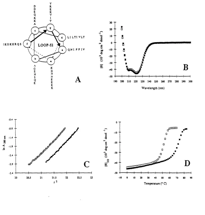
which corresponds to the 28-residue loop region, plus 8 residues of the long a-helix (Fig.
3; Fig. 5A). Most short peptides do not fold into stable structures in aqueous solution (for
reviews, see Wright et al., 1988; Kim and Baldwin, 1990), although coiled-coil peptides
containing four or five heptad repeats are a notable exception (Hodges et al., 1981; O'Sheaet al., 1991). Thus, a stringent test of the model is to determine if the LOOP-36 peptide
forms a three-stranded coiled coil.As determined by circular dichroism (CD) spectroscopy, LOOP-36 is highly helical
(:>90% at 100 pgM)
at pH 4.8, although it is unfolded at neutral pH (Fig. 5B). CD
experiments also reveal a reversible thermal unfolding transition for LOOP-36 at pH 4.8,
but there is no stable structure at neutral pH (Fig. 5C). Sedimentation equilibriumexperiments indicate that LOOP-36 is trimeric at pH 4.7, but primarily monomeric at pH
7.2 (Fig. 5D). We conclude that LOOP-36 folds into a trimeric, a-helical coiled coil at thepH of membrane fusion.
There is a sharp transition in the a-helical CD signal of LOOP-36 between pH 5 and pH 7 (Fig 5E). The predominance of acidic residues at particular heptad positions in the LOOP-1 sequence (see Fig. 2) is reminiscent of the arrangement seen in the Fos leucine
zipper homodimer, which displays a similar pH-dependence of stability (O'Shea et al.,
1.992). Protonation of acidic sidechains at low pH alleviates electrostatic repulsion that destabilizes the folded conformation at neutral pH. There is a second structural transition in
LOOP-36 helicity at even lower pH, but the conformation at pH 2 probably is not relevant
for membrane fusion.
At 370 C, however, LOOP-36 is unstable, even at pH 5. In addition, as mentioned earlier, the fusogenic conformation of HA can be induced at neutral pH, with elevated temperature. We therefore suspected that coiled-coil structure in the loop region would be stabilized in the context of the longer coiled coil. To test this notion, we studied a
52-residue peptide, LOOP-52, which begins at the N-terminus of the short a-helix and ends at
the C-terminus of LOOP-36 (Fig. 3; Fig. 6A).LOOP-52 forms a fully helical (-100%) structure at pH 7.0 and pH 4.8 (Fig. 6B).
At both neutral pH and pH 4.7, LOOP-52 is a trimer, as determined by equilibrium
sedimentation (Fig. 6C). The trimeric, helical structure in LOOP-52 is very stable: at pH 7.0, LOOP-52 unfolds with a transition midpoint (Tm) of 52 °C, and at pH 4.8, the Tm is 72 °C (Fig. 6D).
Comparison with Other Results
It is extremely unusual for a small protein fragment, in aqueous solution, to fold into a stable structure which is different from that found in the native protein. Our results: (i) demonstrate that the loop region can fold as a three-stranded coiled coil, (ii) provide strong support for the proposal that, in the fusogenic state, the coiled coil includes the external a-helix in addition to the loop region and (iii) indicate that the longer coiled coil is very stable, even at neutral pH.
Consistent with our model for the conformation of the fusogenic state, HA is
trimeric at pH 5 (Doms and Helenius, 1986). In addition, electron microscopic studies
membranes following treatment at low pH, or elevated temperatures at neutral pH (Ruigrok et al., 1986; 1988).
Earlier proteolysis experiments provide strong biochemical support for our model.
Although certain regions of the fusogenic state of HA are sensitive to proteolysis, much of
the HA2 subunit is resistant to degradation (Skehel et al., 1982; Ruigrok et al., 1988). The
final tryptic digestion product of HA2 starts at residue 40, and that obtained with
thermolysin starts at residue 38. In both cases, the residues of the short a-helix, loop
region and the long a-helix remain intact. The native state of HA is resistant toproteolysis. Nonetheless, if HA2 maintained the helical-hairpin structure in the fusogenic
state, one would expect the loop region to be sensitive to proteolysis upon dissociation and degradation of the HA1 subunits. In contrast, the loop region would be protected from proteolysis if it were part of a folded structure, such as the coiled coil in our model.A Mechanism for the Conformational Change
One hypothesis for the mechanism of the conformational change is that a decrease in pH shifts the thermodynamic equilibrium between the native and fusogenic states. It is striking that the following pH transitions coincide: (i) LOOP-36 helicity (and LOOP-52 stability), (ii) the known pH dependences for the conformational changes and (iii) the onset of membrane fusion activity in HA (see Fig. 7). All of these transitions occur
abruptly between pH 7 and pH 5. If the mechanism for the conformational change is under
thermodynamic control, it should be possible to design fusion mutants that alter the stability of the coiled coil, and hence the fusogenic state, without affecting the stability of the nativestate.
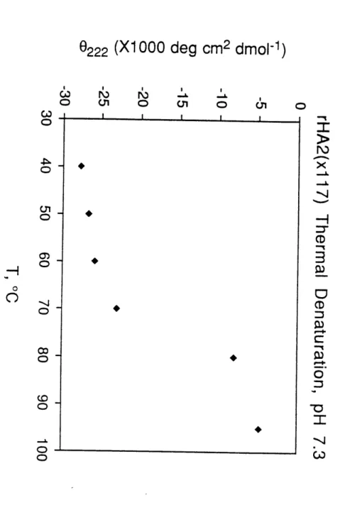
For several reasons, however, we favor a kinetically controlled mechanism for the
conformational change in HA, in which the native state of HA is metastable (i.e., although it is stably folded, it has the potential to form a thermodynamically more stable state). First, the conformational change, induced either by a decrease in pH or an increase in temperature at neutral pH, is irreversible (Ruigrok et al., 1986; for reviews, see Wiley and Skehel, 1987 and Stegmann and Helenius, 1993). Second, since LOOP-52 is very stable, even at neutral pH (Fig. 6C), we expect that the 88-residue coiled coil of the fusogenic conformation will be extremely stable. Third, there is a good correlation between thedecrease in stability of the native state and the increase in the pH of fusion by the HA mutants (Ruigrok et al., 1986).
In addition, previous biophysical studies provide strong evidence for a mechanism
that is under kinetic control. Circular dichroism experiments reveal an irreversible thermaltransition at -63
°C, correlated with the temperature of HA-mediated fusion at neutral pH,
followed by a second, reversible transition at even higher temperature (Ruigrok et al.,
1986; see, however, Ruigrok et al., 1988). The irreversibility of the first transition is
consistent with a transition from a metastable initial state to a more stable final state. Wewould assign the second transition to the unfolding of the long, stable coiled coil.
The notion of a metastable structure raises the question: how does HA fold into a
conformation that is not the thermodynamically most stable structure? The in vivo folding
of HA0 (Braakman et al., 1992) is known to be dependent on metabolic energy. A
minority of the HA0 molecules fail to fold into the native conformation, but instead form
stable, aberrant trimer species that do not leave the endoplasmic reticulum (Gething et al.,1986b; Copeland et al., 1986). These species may have an extended coiled coil structure
similar to our proposed fusogenic conformation. Thus, the native conformation of HA
may be the result of the in vivo folding pathway of the HA0 precursor. In addition, the
relative instability of the loop region of HA2 at neutral pH (see Figs. 5C and 6C) may beimportant for in vivo folding of HA0.
Regardless of the mechanism, there appear to be two types of interactions that stabilize the native state and thereby inhibit formation of the stable fusogenic state: (i) intersubunit HA1 protein-protein interactions and (ii) burial of the fusion peptide in the
hydrophobic core of the timer. As mentioned previously, in the fusogenic conformation,
the HA1 subunits dissociate and the fusion peptide becomes exposed (see, however,
Stegmann and Helenius, 1990).
In the structure of the native state (Wilson et al., 1981), extensive interactions are
apparent between the loop region of HA2 and its corresponding HA1 subunit (Fig. 1B).
Thus, the HA1 subunit may act as an inhibitor or "clamp" that binds to the loop region,
preventing the conformational change. In addition, multiple HAl-HA1 interactions arelikely to prevent the conformational change. Indeed, introducing disulfide bonds between
HA1 subunits inhibits the conformational change in HA and prevents membrane fusion
(Godley et al., 1992). Furthermore, many mutations that alter the pH of fusion appear to
destabilize HAl-HA1 or HA1-HA2 interactions in the native state (reviewed in Wiley and Skehel, 1987).The native conformation may also be stabilized by the fusion peptide, which makes significant hydrophobic interactions in the core of the native structure. The buried fusion
peptide resembles a hydrophobic "hook," which holds the helices together in a
helical-hairpin conformation (Fig. 1A). Many of the HA mutants that fuse under less acidic
conditions (Daniels et al., 1985; Gething et al., 1986a) contain amino acid substitutions near the region of the fusion peptide. For example, the x-ray crystal structure of the HA2 fusion mutant, D1 12G, reveals that 4 hydrogen bonds are lost (per monomer) between the aspartate side chain and residues of the fusion peptide. The pH of fusion for the mutant iselevated by 0.4 pH units, presumably because loss of the hydrogen bonds destabilizes the
native state (Weis et al., 1990b).
Speculation on the Relevance to Other Fusion Events
Although influenza hemagglutinin is the best characterized membrane fusion
protein, other viral (and non-viral) fusion proteins are known to be oligomeric (reviewed in
White, 1992), and some of these are candidates for further study in light of our model.
Like HA, these glycoproteins are derived proteolytically from a precursor; this proteolysis creates a new N-terminus with an adjacent fusion peptide sequence (for reviews, seeWhite, 1990; Stegmann and Helenius, 1993). Moreover, several of these fusion peptide
sequences are followed by a stretch of amino acids with a 4-3 hydrophobic repeat,suggesting a coiled-coil structure (Chambers et al., 1990). Our results suggest that some
of these 4-3 hydrophobic repeats may correspond to a coiled-coil structure that is found only in the fusogenic conformation of the glycoprotein.Proteins that mediate fusion at neutral pH (for example those involved in
fertilization, protein trafficking or HIV infection; reviewed in White, 1992) may also
unleash a latent structural conformation in response to an external stimulus, such as bindingof a receptor or interaction with another protein. For example, the binding of HIV to CD4
is known to induce conformational changes in the envelope glycoprotein that result inrelease of the extracellular subunit (gp 120) from the transmembrane subunit (gp41), exposure of the fusion peptide, and fusion of the viral membrane with the cell (reviewed in
Vaishnav and Wong-Staal, 1991). A peptide from gp41, corresponding to a sequence
adjacent to the N-terminal fusion peptide, has recently been shown to form a coiled coil(Wild et al., 1992). Moreover, expression of gp41 in the absence of gpl20 results in
syncytium formation (Perez et al., 1992), demonstrating that the interaction between gp120
and gp41 inhibits fusogenic activity, possibly in a manner similar to our proposed
inhibitory interaction of the HA 1 and HA2 subunits in the native state of HA.
Finally, if our model is correct, it may lead to new approaches for the discovery of
anti-viral drugs. For example, therapeutic agents might be designed to prevent the low-pH
activation of HA. Alternatively, since acid pretreatment of influenza virus can abolish infectivity (Doms et al., 1985; Stegmann et al., 1990; Puri et al., 1990), agents might be designed that cause the conformational change to occur prematurely. It is also interesting,in light of our results, that a synthetic coiled-coil peptide from gp41 of HIV inhibits viral
replication (Wild et al., 1992).Note added in Proof
Recently, a connection was made between fusion proteins involved in cellular
vesicle transport and vesicle-synaptosomal membrane fusion (S611ner,
T., Whiteheart, S.
W., Brunner, M., Erdjument-Bromage, H., Geromanos, S., Tempst, P., and Rothman, J.
E.(1993). SNAP receptors implicated in vesicle targetting and fusion. Nature 362,
318-324). The amino-acid sequences for some of these membrane proteins contain a long,
continuous region of 4-3 hydrophobic repeats.
Acknowledgments
We thank Rheba Rutkowski and Mike Burgess for synthesis of the LOOP-36 and
LOOP-52 peptides; Brad Stewart for the computer-aided sequence search; Jeff Stock for
the "coiledcoil" computer program; Shiufun Cheung for figure 4; Fred Hughson for help
with the HA literature and for in-depth discussions; Jonathan Weissman for discussion;Pehr Harbury and Tom Alber for discussions about oligomerization states of coiled coils;
Stan Watowich for the compilation of HA sequences for various strains of influenza A;
versions of the manuscript. C. M. C. is supported by an N. I. H. Training Grant (AI07348). P. S. K. is a Pew Scholar in the Biomedical Sciences. This research was
supported by the Howard Hughes Medical Institute.
Experimental Procedures
Coiled Coil Prediction
An algorithm (Parry, 1982) was used to predict coiled-coil propensities based on
the statistical preference of different amino acids for each position in the heptad repeat. Sequences from the Protein Data Bank (Brookhaven) were analyzed using a computer program ("coiledcoil"; Lupas et al., 1991) based on the algorithm. With a window size of28 residues, sequences with scores above 1.3 are thought to have a good probability for
coiled-coil structure, while scores between 1.1 and 1.3 generally correspond toamphipathic a-helices in globular proteins (Lupas et al., 1991). A score of 1.6 was
obtained for residues 54-81 of HA2.
Inspection of the complete HA2 sequence revealed a continuous heptad repeat from
residues 38 to 125. Although an earlier analysis had predicted a coiled coil structure formuch of the 88-residue sequence of HA2 identified here, the register of the heptad repeat
was not maintained throughout this sequence, and several interruptions in the coiled coil were also predicted (Ward and Dopheide, 1980; see also, Chambers et al., 1990).Peptide Synthesis and Purification
Peptides were synthesized on an Applied Biosystems model 430A peptide
synthesizer using Fmoc chemistry with Fastmoc reaction cycles modified to include acetic
anhydride capping (Fields et al., 1991). LOOP-36 corresponds to residues 54-89 and
LOOP-52 corresponds to residues 38-89 of HA2 from the X-31 strain of influenza virus
(Kilbourne, 1969). In each peptide, the N-terminus is acetylated and the C-terminus is
amidated. The peptides were cleaved using standard Fmoc protocols and desalted on aSephadex G- 10 or G-25 column (Pharmacia) in 5% acetic acid. Final purification was by
reverse-phase, high-performance liquid chromatography (HPLC, Waters, Inc.) at 25
°C
using a Vydac preparative or semipreparative C18 column (stock# 218TP1022 or
218TP510, respectively). A linear acetonitrile-H20 gradient of 0.1% buffer B increase per
minute was used with a flow rate of 10 ml/min (5 ml/min for the semipreparative column).Buffer A is 0. 1% trifluoroacetic acid (TFA) in water, buffer B is 90% acetonitrile and 0.1%
TFA in water. The identity of the peptides was confirmed by laser desorption mass
spectrometry (Finnigan MAT LASERMAT). The measured mass for LOOP-36 was 4451
Da (expected 4450 Da), and the measured mass for LOOP-52 was 6147 Da (expected
6146).
Circular Dichroism Spectroscopy
Circular dichroism (CD) spectroscopy (for a review, see Woody, 1985) was
performed on an AVIV CD spectrophotometer (model 62DS) equipped with a
thermoelectric temperature controller. The cuvettes used for thermal unfolding studies and pH studies were 1 cm and 0.1 mm in pathlength and the cuvettes used for wavelength spectra were 1 mm and 0.1 mm in pathlength. Peptide concentration was determined by tyrosine absorbance at 275.5 nm (Edelhoch, 1967) in 6 M guanidinium chloride
(Schwarz/Mann Biotech, Ultra-Pure grade).
The pH-dependence of LOOP-36 structure was determined by monitoring the CD signal at 222nm. Measurements were made at 0° C and 32 A.M peptide in a buffer of 150 mM NaCI, 20 mM (each) of sodium borate, sodium citrate and sodium phosphate. Both transitions observed in the pH-dependence studies are >95% reversible (data not shown).
For thermal unfolding experiments, the CD signal was monitored at 222nm as a
function of temperature. Samples at pH 7.0 contained 32 gM LOOP-36, 150 mM NaCI
and 10 mM sodium phosphate; or 500 pM LOOP-52, 50 mM sodium phosphate and 150
mM NaCl. Samples at pH 4.8 also contained 10 mM sodium citrate and 10 mM sodium borate for both LOOP-36 and LOOP-52. All thermal unfolding experiments are reversible (>95% for LOOP-36 samples and >85% for LOOP-52 samples) in the temperature rangefrom 0-60
°C for LOOP-36, and 0-850 C for LOOP-52 (data not shown).
CD spectra were obtained at 00 C, in the same conditions as used in thermal
unfolding experiments, except that the peptide concentration was 100 pM for LOOP-36. In
Fig. 7, percent helicity for LOOP-36 was calculated assuming that 100% helicitycorresponds to -33,000 deg cm 2 dmol -1, as has been found in studies of helical peptides of this length (Chen et al., 1974)
Equilibrium Sedimentation
Molecular weights were determined at 10 C for LOOP-36 and 40 C for LOOP-52, by analytical ultracentrifugation (reviewed in Laue et al., 1992) with a Beckman XL-A Optima Analytical Ultracentrifuge equipped with absorbance optics. An An-60Ti rotor was used at
22,000 and 27,000 rpm. Experiments were performed at pH 7.2 and pH 4.7. LOOP-36
samples were made at three concentrations (300, 100 and 33 IM) per pH and LOOP-52
samples were made at six concentrations (500, 167, 56, 130, 43 and 14 gM) per pH, and
all samples were dialyzed exhaustively (-24 hours) against buffer prior to the experiments. Buffer conditions were 150 mM NaCl, 50 mM sodium phosphate, pH 7.2; or 150 mM NaCl, 5 mM sodium acetate, pH 4.7. The molecular weights were determined by asimultaneous fit of each data-set using a non-linear least squares fit algorithm, HID-4000
(Johnson et al., 1981); no systematic residuals were observed.
References
Braakman, I., Helenius, J., and Helenius, A. (1992). Role of ATP and disulphide bonds
during protein folding in the endoplasmic reticulum. Nature 356, 260-262.
Brand, C. M., and Skehel, J. J. (1972). Crystalline antigen from the influenza virus
envelope. Nature New Biology 238, 145-147.
Chambers, P., Pringle, C. R., and Easton, A. J. (1990). Heptad repeat sequences are
located adjacent to hydrophobic regions in several types of virus fusion glycoproteins. J.
Gen. Virol. 71, 3075-3080.
Chen, Y.-H., Yang, J. T., and Chau, K. H. (1974). Determination of the helix and [3
form of proteins in aqueous solution by circular dichroism. Biochemistry 13, 3350-3359.
Cohen, C., and Parry, D. A. D. (1990). a-Helical coiled coils and bundles: how to
design an a-helical protein. Proteins 7, 1-15.
Copeland, C. S., Doms, R. W., Bolzau, E. M., Webster, R. G., and Helenius, A. (1986). Assembly of influenza hemagglutinin trimers and its role in intracellular transport.
J. Cell Biol. 103, 1179-1191.
Crick, F. H. C. (1953). The packing of a-helices: simple coiled coils. Acta. Cryst. 6,
689-697.
DIaniels, R. S., Downie, J. C., Hay, A. J., Knossow, M., Skehel, J. J., Wang, M. L.,
and Wiley, D. C. (1985). Fusion mutants of the influenza virus hemagglutinin
glycoprotein. Cell 40, 431-439.
Doms, R. W., Helenius, A., and White, J. (1985). Membrane fusion activity of the
influenza virus hemagglutinin. J. Biol. Chem. 260, 2973-2981.
Doms, R. W., Gething, M.-J., Henneberry, J., White, J., and Helenius, A. (1986).
Variant influenza virus hemagglutinin that induces fusion at elevated pH. J. Virology. 57,
603-613.
Doms, R. W., and Helenius, A. (1986). Quaternary structure of influenza virus
hemagglutinin after acid treatment. J. Virol. 60, 833-839.Edelhoch, H. (1967). Spectroscopic determination of tryptophan and tyrosine in proteins.
Biochemistry 6, 1948-1954.Ellenberger, T. E., Brandl, C. J., Struhl, K., and Harrison, S. C. (1992). The GCN4