Publisher’s version / Version de l'éditeur:
Proceedings of the Twelfth International Conference on Miniaturized Systems for
Chemistry and Life Sciences - MicroTAS 2008, pp. 507-509, 2008-10
READ THESE TERMS AND CONDITIONS CAREFULLY BEFORE USING THIS WEBSITE. https://nrc-publications.canada.ca/eng/copyright
Vous avez des questions? Nous pouvons vous aider. Pour communiquer directement avec un auteur, consultez la
première page de la revue dans laquelle son article a été publié afin de trouver ses coordonnées. Si vous n’arrivez pas à les repérer, communiquez avec nous à [email protected].
Questions? Contact the NRC Publications Archive team at
[email protected]. If you wish to email the authors directly, please see the first page of the publication for their contact information.
NRC Publications Archive
Archives des publications du CNRC
This publication could be one of several versions: author’s original, accepted manuscript or the publisher’s version. / La version de cette publication peut être l’une des suivantes : la version prépublication de l’auteur, la version acceptée du manuscrit ou la version de l’éditeur.
Access and use of this website and the material on it are subject to the Terms and Conditions set forth at
A multiple recording patch clamp chip with integrated subterranean
microfluidic channels for cultured neuronal networks
Py, Christophe; Mealing, Geoff; Denhoff, Mike; Charrier, Anne; Monette,
Robert; Comas, Tanya; Ahuja, Tarun; Martinez, Dolores; Krantis, Anthony;
Wingar, Simon
https://publications-cnrc.canada.ca/fra/droits
L’accès à ce site Web et l’utilisation de son contenu sont assujettis aux conditions présentées dans le site LISEZ CES CONDITIONS ATTENTIVEMENT AVANT D’UTILISER CE SITE WEB.
NRC Publications Record / Notice d'Archives des publications de CNRC:
https://nrc-publications.canada.ca/eng/view/object/?id=1136b426-c31e-47bb-b1db-848cc577953f https://publications-cnrc.canada.ca/fra/voir/objet/?id=1136b426-c31e-47bb-b1db-848cc577953f
A MULTIPLE RECORDING PATCH CLAMP CHIP
WITH INTEGRATED SUBTERRANEAN
MICROFLUIDIC CHANNELS FOR CULTURED
NEURONAL NETWORKS
Christophe Py1, Geoff Mealing1, Mike Denhoff1, Anne Charrier2, Robert Monette1, Tanya Comas1, Tarun Ahuja1, Dolores Martinez1, Anthony
Krantis3 and Simon Wingar1
1
National Research Council, CANADA, 2Centre National de la Recherche Scientifique, FRANCE and 3University of Ottawa, CANADA
ABSTRACT
We report on a new type of patch-clamp chip that can simultaneously monitor electrophysiological activity from several individual neurons grown in simple networks. As in automated patch-clamp chips used for isolated cells in suspension, cells are interrogated using a microhole integrated into a membrane. The novelty of our chip resides in its capacity to position, culture and guide neurons into organized networks over multiple interrogation sites, thereby permitting high resolution monitoring of pre and post synaptic activity in these networks.
ELECTROPHYSIOLOGICAL MONITORING OF BRAIN MODELS
In-vitro models are used extensively to study the molecular mechanisms that regulate the transmission and processing of information in the brain through synaptic communication. Neurochips have been developed to allow the interrogation of the electrophysiological activity of cells or tissues under various conditions. Multi-electrode arrays (MEAs) [1] enable stimulation and monitoring of neuronal networks taken from brain tissue or created when isolated brain cells reconnect in culture.
a
b
c
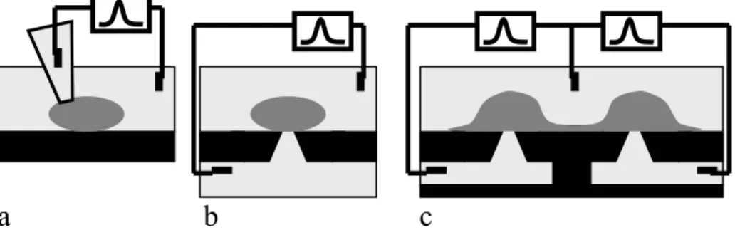
Figure 1: a: conventional glass pipette patch-clamp; b: patch-clamp chip, separating the culture medium from the electrochemically conductive solution; c:
Multiple probe patch-clamp chip with integrated subterranean microfluidics; the cells are cultured on-chip and have formed synaptic connections.
MEAs provide valuable information about synaptic function, but lack sufficient resolution to monitor electrical activity from individual cells or the ability to directly
measure ion channel activity. This is a significant limitation since ion channels play key roles in normal physiology and in many pathophysiologies. Ion channels are thus important targets for therapeutic intervention, and the way potential therapeutic compounds affects them is a necessary part of drug development. Conventionally, ion channel activity is monitored using a technique called “patch-clamp”, where a small-tipped glass pipette filled with an electrochemically conductive solution is used to electrically isolate a “patch” of the cell’s membrane; when the voltage across this patch is “clamped”, ion channel activity in the membrane is measured as small current bursts (Figure 1a). This is a difficult and laborious process that has inadequate throughput to be practical for pharmacological screening. Considerable effort has been invested in automating patch-clamping [2], including the development of patch-clamp chips, where the apex of the pipette is replaced by a microscopic hole in a membrane on which the cell is placed (Figure 1b) [3]. While this has resulted in increased throughput, the application is limited to individual suspended cells derived from cell lines. This has limited biological relevance since isolated cells lack synaptic connections and cell lines lack the cellular machinery linking ion channel activity to physiological function.
Microholes Microfluidic channels Membrane 10mV 1sec
Figure 2: patch-clamp chip with two microholes and dedicated subterranean microfluidic channels,
each with an input and an output to facilitate the injection of chemicals.
Figure 4: Whole-cell current-clamp recording of action potentials recorded from a neuron cultured over
a microhole.
MULTIPLE RECORDING PATCH CLAMP CHIP
Our chip is designed to combine the advantages of MEAs and patch-clamp technologies: simple organized networks of neurons cultured on the membrane are interrogated individually through micro holes that connect to dedicated subterranean microfluidic channels (Figure 1c and 2). To facilitate large-scale fabrication [4], our first generation of chips is integrated on a silicon platform, with a silicon nitride membrane and thicker SiO2 or SU8 films for the microfluidics. The chips are packaged in Plexiglas chambers with tubes to feed the fluidic channels, and sterilized
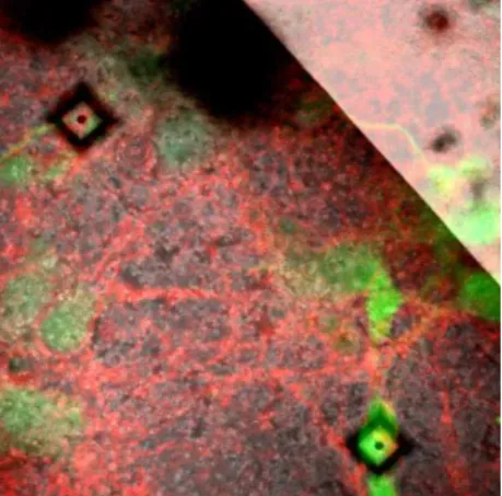
in an air plasma reactor. Cryopreserved rat primary cortical neurons [5] are cultured (2 - 3 weeks) on the substrate and develop into networks. Figure 3 shows a culture stained with Calcein for live cells (green) and a membrane dye (red). Neurons that have grown over the microhole and formed a seal around the microhole display excitable electrical activity, albeit with a high signal to noise ratio (Figure 4). While formation of a high resistance seal between the hole and the cell is critical, the primary cause of the poor signal is the high capacitance of the chip that couples the two electrochemical cells through the conductive silicon bulk and shunts the neurons. This problem is being addressed in our second generation of chips by patterning a thick SU8 photosensitive resin film with a small opening on top of the membrane to substantially reduce the shunt capacitance. Another important feature of the chip is the poly-D-lysine guidance patterns which influence cell placement, and guide process growth to immediate neighbors (Figure 5). These cues are efficiently stamped with silicone stamps replicated from microfabricated masters, aligned and pressed with the chips in a flip-chip bonder, such that synthesized organized networks of neurons can be aligned with the patch-clamp microholes.
Figure 3: Overlay of a reflection microscopy image and of a fluorescence microscopy picture
showing a cell body on top of a microhole.
Figure 5: Fluorescence microscopy image of cultured cells aligned on top
of stamped poly-D-lisine square patterns.
REFERENCES
[1] “Advances in Network Electrophysiology/ Using Multi-Electrode Arrays”, T. Makoto, M. Baudry, Springer 2006.
[2] “Automated Electrophysiology: High-Throughput of Art”, X. Wang and M. Li, Assay and Drug Development Technology 1(5), p695, 2003.
[3] “Microchip Technology in Ion-Channel Research”, F.J. Sigworth and K.G. Klemic, IEEE Transactions on Nanoscience 4(1), p121, 2005.
[4] Performed at the Canadian Photonics Fabrication Center, www.cpfc.nrc.gc.ca [5] QBM Cell Sciences, www.qbmcellscience.com