94
Nontoxigenic
Corynebacterium diphtheriae
Isolated from Intravenous Drug Users
Eva Gruner, Milos Opravil, Martin Altwegg, and Alexander von Graevenitz
From the Institute ofMedical Microbiology. University ofZurich; and the Division of Infectious Diseases. Department of Medicine. University Hospital. Zurich. Switzerland
During a prospective study 117 intravenous drug users were screened for infection with Coryne-bacterium diphtheriae.NontoxigenicC.diphtheriaewas found in 5 of 132 throat swab specimens and in 5 of 28 skin ulcer specimens taken from July 1991 to April 1992. When phenotypic and molecular typing methods were used, these 10 strains were shown to belong to a single clone. During the same period no strain was isolated from 200 controls. Clinical manifestations of infection were not clearly attributable toC.diphtheriae-no typical membranous pharyngitis was present. The presence of a single clone among homeless intravenous drug users in Zurich indicates the presence ofC.diphtheriaein parts of the population with poor standards of hygiene and low socioeconomic status.
Diphtheria is expected to be eliminated in immunized pop-ulations of industrialized countries, with toxigenic strains no longer circulating among indigenous inhabitants [I]. Never-theless, sporadic cases of diphtheria continue to be reported, with outbreaks probably limited to the lower socioeconomic levels of these populations [2-4]. The site of infection is of-ten the skin rather than the pharynx, and toxic manifesta-tions are unusual. Host resistance factors seem to influence susceptibility to infection with toxigenic as well as with non-toxigenic Corynebacterium diphtheriae [I, 2].
Recently we reported three cases of endocarditis and one case of septicemia that occurred among intravenous drug users during the two winter seasons of 1990 and 1991 in Zurich [5]. Molecular typing of these four non toxigenic iso-lates showed that they belonged to a single clone [6]. In consequence we wanted to know whether there was a reser-voir ofC.diphtheriaeamong the intravenous drug addicts living in Zurich. We present the results of our prospective study.
From July 1991 to April 1992, 117 intravenous drug users cared for at the infirmary for the homeless of the City of Zurich Medical Service and at the outpatient clinic of the Division of Infectious Diseases, Department of Medicine, University Hospital, Zurich, entered the study. Every person was examined with use of a pharyngeal swab and, in the presence of ulcerating skin lesions, an additional wound swab. Ifmultiple pharyngeal swab specimens were collected, the second swab was included in the study only if it was obtained after an intervalof~2months. A total of 160 swab specimens (pharyngeal, 132; superficial wounds, 28) were
Received 9 April 1993; revised 21 June 1993.
Reprints or correspondence: Dr. Milos Opravil, Division of Infectious Diseases. Department of Medicine. University Hospital. Ramistrasse 100. CH-8091 Zurich. Switzerland.
Clinical Infectious Diseases 1994;18:94-6
© 1994 by The University of Chicago. All rights reserved. 1058-4838/94/1801-00 I0$02.00
collected. The individuals (males, 68%; females, 32%) were 17-45 years of age. Approximately one-half of the patients were infected with the human immunodeficiency virus (HIV). Their immunization status with respect to diphtheria was not known.
Two hundred pharyngeal swabs were taken consecutively during the same period from patients with pharyngitis who were included as controls. These individuals had consulted their physicians about pharyngitis; belonged to the same age group (range, 18-40 years); and lived in the same area but were not drug users.
All swabs were plated onto Columbia agar base (Becton-Dickinson, Cockeysville, MD) with 5%sheep blood and onto cystine-tellurite blood agar (CTBA)
[7]
and were incubated aerobically at 37°C for 24 hours. Colonies suspected ofhar-boringC.diphtheriaewere identified according to the meth-ods of Saragea et al. [8]. Toxigenicity was tested in guinea pigs and with use of the polymerase chain reaction [9]. Fur-ther enzyme activities and carbohydrate fermentation reac-tions were assayed using the API ZYM and API Coryne sys-tems (API Biolvlerieux, La Balme, France). Susceptibility to the eight antibiotics commonly used to treat gram-positive infections (penicillin, erythromycin, netilmicin, streptomy-cin, tetracycline, chloramphenicol, rifampin, and vancomy-cin) was tested. For molecular typing genomic DNA was isolated, digested with three restriction endonucleases (EcoRI,HindlII,
orPvuII),
and hybridized with a biotin-la-beled pBR322 derivative containing a rDNA operon of Esch-erichia coli. All these typing methods have been described before [6].Five (4.3%) of 117 primary pharyngeal swab specimens and 5 (17.9%) of28 swab specimens from superficial wounds were positive for C.diphtheriae(table I). None of the pa-tients had C.diphtheriaeisolated in cultures of both throat and wound specimens. Two patients with positive pharyn-geal swab specimens suffered from tonsillitis without devel-oping the typical membranes and were successfully treated with erythromycin and flucloxacillin, respectively. One
pa-CID 1994; 18 (January) NontoxigenicC.di phtheriae in IV Drug Users 95
Table 1. Characteristics of patients withC.diphtheriaeinfection.
Patient no. 1 2 3 4 5 6 7 8 9 10 Age/sex 33/m 28/m 29/m 29/m 19/m 29/m 40/m 26/m 35/m 22/m HIV status Unknown Negative Negative Negative Negative Negative Negative Positive Negative Negative Culture specimen Wound (leg)* Wound (leg) Wound (arm) Wound (leg) Wound(hand)" Pharynx (pneumonia) Pharynx (tonsillitis) Pharynx (bronchitis) Pharynx (sore throat) Pharynx (tonsillitis)
Other organisms isolated S.aureus
E. coli, streptococcus group G. viridans streptococci. coagulase-negative
staphylococci
S.aureus.S p.l'OgClles.A. haemolvticum
S.aureus.S pFogellcl.viridans streptococci
S.aureus.S.jJFogellcs Viridans streptococci
Viridans streptococci. coagulase-negative staphylococci.Candida species Streptococcus agalact iac. viridans streptococci
Viridans streptococci. saprophytic Neisseria
Viridans streptococci. enterococci. saprophytic Neisseria
- - - _. ._ - - _.._ -* Wound swab positive again after 2 and 5 months. respectively.
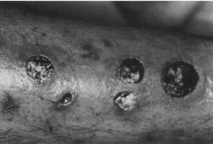
tient suffered from a common cold with concomitant sore throat, and the remaining two had bronchopulmonary infec-tions without pharyngeal symptoms. Both pharyngeal car-riers who were treated and reexamined were found to be culture negative 10 and 15 days later, respectively. The five patients with positive wound swab specimens had skin infec-tions and ulcers at the sites of drug injection. Figure 1 illus-trates multiple ulcers on the calf of a patient, cultures of which yieldedC.diphtherial' and Staphylococcus aureus. This patient was treated with amoxicillinjclavulanate for 2 weeks, followed by erythromycin for I week, resulting in clinical improvement; however, he relapsed 2 months later with in-fection due to the same flora. The other patients were treated with amoxicillinjclavulanate with variable success as a result of poor compliance and persistent drug abuse. Only one of them presented again after 5 months, at which time diphthe-ric bacilli were still detected in swab specimens. HlV serol-ogy was available for nine of the culture-positive patients; only one pharyngeal carrier was HIV -positive.
Figure 1. Calf of an intravenous drug user with multiple skin
ulcers ( 1- 3 em in diameter); culture of the wound swab yieldedC. diphtheriaeandS.aureus.
In pharyngeal swabs C. diphtherial' was found together with the normal flora of the oropharynx, whereas in skin lesions the organism was associated with j1-hemolytic strep-tococci (four of five), S. aureus (four), Arcanobacteriuni hac-molvticum (one), E. coli (one), and viridans streptococci (two) (table I). This corresponds to data reported earlier, in which the majority (73){) of diphtherial skin lesions was asso-ciated with Streptococcus pyogenes infection [2]. However, in our patients coinfection byS. aureus was less frequent(35~!() [2]. No C.diphtherial' strains were isolated from the pharyn-geal swabs of the 200 control patients who were not intrave-nous drug users.
The 10 isolates ofC.diphtherial' were identified as biotype mitis by virtue of their inability to ferment dextrin, starch, and glycogen [8]. They were all identical with respect to their biochemistry, and they were all resistant to tetracycline but susceptible to all other antibiotics tested.
The genome of biotype mitis strains has been described as particularly variable in contrast to biotype intermedius [10], and this variability was confirmed in our former study by comparison with rONA patterns of eight other mitis strains isolated in Switzerland over the past 5 years [6]. However. with each of the three restriction enzymes, the present iso-lates showed rONA patterns that were identical to those ob-served previously in the isolates from intravenous drug users wi th endocarditis and septicemia [6]. We believe, therefore. that they all belong to the same clone.
Although C.diphtherial' is only a human parasite, its isola-tion has become uncommon in microbiological laboratories of the western world. Its colonial morphology does not differ from that of many other gram-positive rods, and selective media such as CTBA have to be used if there is clinical suspi-cion of diphtheria. Otherwise, toxigenic as well as nontoxi-genic strains are simply reported as coryneform bacteria on the assumption that they belong to the normal flora of the skin or the upper respiratory tract. Thus. it is difficult to
popu-96 Gruner et al. CID1994; 18 (January)
lations. During the early 1980s Naumann et al. [3] foundC. diphtheriaein <0.1%of a German population
[3].
It has re-cently been reported that screening of> 500 healthy adults in an urban area of central Italy did not yield any pharyngeal carriers of either toxigenic or nontoxigenic diphtheric bacilli[11].
Similarly, none of the patients from our control group carriedC.diphtheriaein his throat.Our data reflect the continued decline in the incidence of diphtheria in areas where vaccination has been extensively practiced [1). However, the reason for the reduction in preva-lence of the bacterium itself is unclear, as vaccination is di-rected against the action of the toxin, and several studies in industrialized countries have demonstrated non protective levels of antitoxin titers in both children and adults despite vaccination
[1,
12]. The decline in diphtheria cases might, therefore, parallel development of the socioeconomic status, rather than the vaccination status, of the population. In fact, diphtheria or atypical manifestations of disease caused by non toxigenic strains continue to be reported [2, 4] but are mostly limited to persons living under poor socioeconomic conditions. Outbreaks among alcoholics and homeless peo-ple have been described in Seattle [2, 10] and in Stockholm [4], where pharyngeal or generalized diphtheria occurred in people with insufficient levels of immunity.Skin infections were described as chronic, nonhealing ulcers due to physical trauma and/or underlying dermatoses that were probably superinfected with predominantly non-toxigenic strains ofC.diphtheriae. Thus, poor hygiene and personal proximity seem to favor colonization; these factors may also be responsible for the often-cited higher con-tagiousness of skin vs. pharyngeal diphtheria [13]. Our pa-tients in whom C.diphtheriae was detected were homeless, except for one pharyngeal carrier who lived in his own apart-ment. However, they all slept in different homeless shelters and no close contacts between them could be identified. The transmission might have occurred among the intravenous drug users who congregated in one park (Platzspitz) of the city at that time. It is surprising that the HIV status of the patients did not seem to influence either the rate or the sever-ity (e.g., endocarditis or sepsis) of infection withC.
diphthe-riaein this setting.
This study illustrates the persistence ofC.diphtheriaein parts of the population. The presence of a single clone among intravenous drug users in Zurich indicates that spe-cific conditions have to be met to maintain colonization with C.diphtheriae. These conditions include poor standards of
hygiene and low socioeconomic status. Overcrowding in households and personal proximity seem to favor dissemina-tion ofcutaneous flora. Despite extensive immunizadissemina-tion pro-grams in developed countries, persons oflow socioeconomic status continue to be at risk for colonization and infection with diphtheric bacilli.
Acknowledgment
The authors express their gratitude to Dr. A. Studer, City of Zurich Medical Service, for the care of the patients.
References
I. Karzon DT, Edwards KM. Diphtheria outbreaks in immunized popula-tions. N Engl J Med1988;318:41-3.
2. Harnisch JP, Tronca E, Nolan CM, Turck M. Holmes KK. Diphtheria among alcoholic urban adults. A decade of experience in Seattle. Ann Intern Med1989;111:71-82.
3. Naumann P, Krech T. Maximescu P, et al. Phagenlysotopie und die Epidemiologie der Diphtherie-Erkrankungen 1975 bis 1984. Dtsch Med Wochenschr1986;111:288-92.
4. Rappuoli R. Perugini M, Falsen E. Molecular epidemiology of the 1984-1986 outbreak of diphtheria in Sweden. N Engl J Med 1988;318: 12-4.
5. Zuber PLF, Gruner E. Altwegg M, von Graevenitz A. Invasive infec-tion with non-toxigenic Corynebacterium diphtheriaeamong drug users. Lancet1992;339: 1359.
6. Gruner E, Zuber PLF, Martinetti Lucchini G, von Graevenitz A, Alt-wegg M. A cluster of non-toxigenicCorynebacterium diphtheriae in-fections among Swiss intravenous drug abusers. Med Microbiol Lett 1992;1:160-7.
7. Frobisher M Jr. Cystine-tellurite agar forC.diphtheriae. J Infect Dis 1937;60:99-105.
8. SarageaA.Maximescu P, Meitert E.Corynebacterium diphtheriae: mi-crobiological methods used in clinical and epidemiological investiga-tions. In: Bergan T, Norris JR. eds. Methods in microbiology. Vol13. New York: Academic Press.1979:61-176.
9. Martinetti Lucchini M. Gruner E. Altwegg M. Rapid detection of diph-theria toxin by the polymerase chain reaction. Med Microbiol Lett 1992; I:276-83.
10. Coyle MB, Groman NB. Russell JQ. Harnisch JP, Rabin M, Holmes KK. The molecular epidemiology of three biotypes of Corynebacte-rium diphtheriaein the Seattle outbreak, 1972-1982. J Infect Dis 1989; 159:670-9.
I I. Mencarelli M, Zan chiA.CellesiCRossoliniA.Rappuoli R, Rossolini GM. Molecular epidemiology of nasopharyngeal corynebacteria in healthy adults from an urban area where diphtheria vaccination has been extensively practiced. Eur J Epidemiol1992;8:560-7. 12. Bjorkholm B. Bottiger M, Christenson B, Hagberg L. Antitoxin
anti-body levels and the outcome of illness during an outbreak ofdiphthe-ria among alcoholics. ScandJInfect Dis1986; 18:235-9.
13. Belsey MA. Isolation ofCorynebacterium diphtheriaein the environ-ment of skin carriers. Am J Epidemiol 1970;91 :294-9.