342
Detection of
Septata intestinalis
in Stool Specimens and Coprodiagnostic
Monitoring of Successful Treatment with Albendazole
Rainer Weber, Barbel Sauer, Max A. Spycher, Peter Deplazes, Ruth Keller, Rudolf Ammann, Jakob Briner, and Ruedi Luthy
From the Departments ofMedicine (Divisions of Infectious Diseases and Gastroenterology) and Pathology. University Hospital. Zurich; and the Institute of Parasitology and Laboratory of Electron Microscopy. University of Zurich. Zurich. Switzerland We describe two patients with AIDS and chronic diarrhea in whom the microsporidian Septata
intestinaliswas detected with use of light and electron microscopic coprodiagnostic techniques. The ultrastructure of the microsporidian spores found in their stool specimens was distinctly different from that of Enterocytozoon bieneusi, another intestinal microsporidian found in pa-tients infected with human immunodeficiency virus. Electron microscopic examination of duo-denal biopsy specimens available from one of the patients enabled identification ofS. intestinalis and confirmed the similarity of spores found in feces and in duodenal tissue. Both patients' diarrhea stopped when they were treated with albendazole. Coprodiagnostic monitoring indi-cated disappearance of the parasites and allowed the diagnosis of a relapse in one patient, who responded well to a second course of treatment.
The use of novel histologic and coprodiagnostic tech-niques has improved the diagnostic yield for human immuno-deficiency virus (HIV)-infected patients with diarrhea; in particular, it has facilitated the identification of intestinal protozoa such as the microsporidia Enterocytozoon bieneusi and Septata intestinalis. which had been missed with use of routine examination methods [1-4]. Preliminary observa-tions have indicated that treatment of septata infection with albendazole may be curative [5]. Therefore, early identifica-tion of this parasite could be beneficial for patients. We de-scribe two patients with Hl V-associated chronic diarrhea in whom S.intestinalis was identified by means of coprodiag-nostic techniques. They were treated with albendazole and became asymptomatic. Coprodiagnostic monitoring indi-cated disappearance of the parasites and allowed detection of a relapse in one patient, who responded well to a second course of treatment.
A 27-year-old HIV-infected homosexual Swiss man was well until February 1992, when he began to have five to eight watery stools per day. Repeated stool examinations for ova and parasites (including cryptosporidia), a bacterial cul-ture, and a Clostridium difficile toxin assay were negative. His CD4+ cell count was 0.05 X 10⁹
fL.
Antiretroviral therapy and prophylaxis for Pneutnocvstis carinii pneumonia were started. Diarrhea persisted and led to a weight loss of 10 kg by May 1993, when his weight was 48 kg. Examination of
Received 29 October 1993; revised 24 January 1994.
Grant support: This work was supported in part by the Swiss National Program for AIDS Research (grant no. 91-7066).
Reprints or correspondence: Dr. Rainer Weber. Division of Infectious Diseases. Department of Medicine. University Hospital, CH-8091 Zurich. Switzerland.
Clinical Infectious Diseases 1994;19:342-5
© 1994by The University of Chicago. All rights reserved. 1058-4838/94/1902-0031$02.00
stool samples then revealed cysts of Entamoeba coli and En-tamoeba histolvtica. No amebic cysts were detected after treatment with metronidazole (500 mg three times daily for 10 days), but diarrhea persisted. At that time, light micro-scopic examination of chromotrope-stained stool specimens [ I] (previously not performed) revealed microsporidian spores measuring 1.2 to 1.5 urn X 2.5 to 3.0 ~m(figure I). No parasites were detected in urine specimens. In addition, adenovirus type 9 was cultured (with MRC-5 cells) from stool specimens and was identified by seroneutralization with use of polyclonal type-specific antibodies (American Type Culture Collection, Rockville, MD).
Electron microscopic examination of stool specimens con-firmed that the microsporidian spore ultrastructure was char-acteristic ofS.intestinalis [2-4]. The spores measured 0.97X 1.33 urn, and their polar tubules were arranged in one row (figure 2). We found differences between the spore sizes as-sessed by light microscopic and electron microscopic exami-nation techniques, possibly because of the different fixation procedures. Specimens were prepared as follows. Large fecal particles in the stool suspension were removed by gravity sedimentation performed for 2 minutes, and the resulting supernatant was centrifuged at 5,500gfor 3 minutes. The pellet was fixed for 30 minutes at 4°C with 3% glutaralde-hyde in phosphate buffered saline and was postfixed for 30 minutes at 4°C with 2% osmium tetroxide in O.IM cacodyl-ate buffer (pH, 7.2). After overnight incubation in a 2% aqueous solution of uranyl acetate, the material was dehy-drated with dimethoxypropane and embedded in an epoxy resin mixture (Epon 812; Fluka Chemic, Buchs, Switzer-land). The sections were stained with lead citrate and exam-ined with a transmission electron microscope (Philips EM-400 HM; Philips, Eindhoven, the Netherlands).
In June 1993 the patient was treated with albendazole (400 mg twice daily for 2 weeks); the diarrhea stopped within
CID1994;19(August) IntestinalSCplUIUintestinalisInfection 343
Figure 1. Smearof unconcentrated, formalin-fixed stoolshowed pinkish-red-stainedS. intestinalisspores on lightmicroscopic exami-nation (chromotrope-based stain; oil immersion; bar== 5Jim).
5
days, and stool examination findings became negative. Dis-seminated atypical mycobacteriosis and cytomegalovirus chorioretinitis were diagnosed in September 1993, and treat-ments were initiated. In January 1994 he had no complaints,his weight was 57 kg, and results of repeated stool examina-tions remained negative.
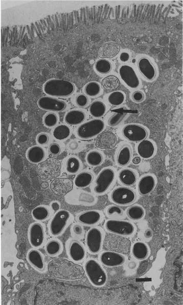
A 37-year-old HIV-infected heterosexual Swiss man had a history ofP. cariniipneumonia, cerebral toxoplasmosis, and Kaposi's sarcoma. His ('04+ cell count was 0.02 X 10⁹jL. He received antiretroviral therapy as well as maintenance therapy forP. cariniipneumonia and toxoplasmosis. In May 1993 the patient experienced watery diarrhea, weight loss. nausea, and vomiting. His weight was 50 kg. Microsporidian spores were found by light microscopic examination ofchro-motrope-stained stool specimens, and the microsporidian spore ultrastructure was confirmed by electron microscopic examination of stool specimens. An examination of urine specimens showed no parasites. Histologic examination of duodenal tissue sections obtained by gastroduodenoscopy (stained with hematoxylin-eosin and Brown-Brenn stain) showed mild inflammation (including slight intraepithelial infiltration by lymphocytes), villous atrophy, goblet cell de-pletion, and clusters of microsporidian spores within entero-cytes and within macro phages of the lamina propria mucosae [2, 3]. Microsporidia and small numbers ofGiardia lamblia organisms were also detected in the duodenal aspirate ..')'. intestinaliswas identified hy electron microscopic examina-tion of duodenal biopsy specimens [2-4] (figure3).
Treatment with alhendazole (400 mg twice daily) was initi-ated, and diarrhea ceased. One week later. however. the pa-tient was hospitalized because of sepsis syndrome. Empiric
Figure 2. Electron micrograph ofa stool specimen showing a sporeofS. intestinalis (A):a sporeofE. bieneusi(B.reproduced from[9])is shown forcontrast.The ultrastructural characteristics are the polar tubules(arrow)coiledwithin the sporeand a sporewall consistingofthe plasmamembrane. an electron-lucent endospore layer. and a dense outer sporecoat. The arrangement of the polar tubulesdiffers between S. intestinalis(tubules in one row[A, arrow])andE. bieneusi(tubules in two rows[B. arrOlI'l).Micrograph ofE.bieneusifrom reference[91.
344 Weber et al. CIO 1994; 19 (August)
intravenous antibiotic treatment (with imipenem) was started. Three blood cultures became positive for Staphylo-coccus aureus.Initially, the patient's condition remained criti-cal, and he developed dyspnea and diffuse interstitial infil-trates that were evident on a roentgenogram of the chest. Cultures and a parasitological examination ofbronchoalveo-lar lavage fluid (including a microsporidia-detection proce-dure [6, 7]) were negative. Although albendazole was with-drawn at the time of hospitalization (after a l-week course), the microsporidian spores in stool samples disappeared dur-ing hospitalization. After a 3-week course of therapy with imipenem, the patient was discharged; he had no pulmonary or gastrointestinal symptoms. In July 1993 the patient again experienced nausea, vomiting, and diarrhea, and examina-tion of stool samples again revealed S. intestinalis spores. A 2-week course of albendazole (400 mg twice daily) led to clinical improvement, and stool samples became negative. During a 4-month follow-up period, the patient had no com-plaints and gained 21 kg.
The identification of microsporidia depended on electron microscopic examination of biopsy specimens obtained by invasive procedures. However, initial detection of the para-site in tissue sections via light microscopy and in more easily obtainable bodily fluids (including stool specimens, duo-denal aspirates, urine, bronchoalveolar lavage fluid, and conjunctival smears) via cytological examination is now readily accomplished [1, 6-9]. Findings of light microscopic examination may even suggest a distinct microspordian spe-cies on the basis of the morphology of the developmental stages and spores as well as the pattern of organ, tissue, and cell involvement by the parasites. Nevertheless, electron mi-croscopy will remain necessary for classification by genus or species.
Our findings suggest that the intestinal microsporidiaE.
bieneusiand S. intestinalis can be distinguished with use of coprodiagnostic techniques. In the patients reported here, we found microsporidian spores by light microscopic examina-tion of stool specimens stained with chromotrope. The spores measuring 1.2 to 1.5 p,m X 2.5 to 3.0 urn had the typical staining pattern described for microsporidia but ap-peared consistently and significantly bigger than those ofE.
bieneusi, which measure --0.9 X 1.5 p,m[1].Therefore, we performed an electron microscopic examination ofstool spec-imens, which showed that the microsporidian spore ultra-structure differed from that ofE. bieneusi (particularly in regard to the configuration of the coiled tubules) and was comparable with that described for Encephalitozoon species in urine specimens [7, 8]. Since an Encephalitozoon species has never been identified in a human intestine and has not been associated with diarrhea in HIV -infected patients [7], we assumed that the patients had intestinal S. intestinalis infection; this was confirmed by electron microscopic exami-nation of duodenal tissue for one of our patients [4]. The similarity of spores found in stool specimens and in duodenal
Figure 3. Electron micrograph of a duodenal tissue specimen showing clusters of sporonts and spores of S. intestinalis within a
parasitophorous vacuole in an enterocyte. The parasitophorous vac-uole appears to be septate by a fibrillar network(arrow) surround-ing the developsurround-ing organisms (bar= 1JIm).
tissue also was documented. In contrast with the observa-tions of other investigators. who have found S. intestinalis to disseminate (particularly into the kidneys) in some patients [2-4], we found no clinical evidence of disseminated infec-tion in our patients, and examinainfec-tions of their urine samples were negative.
S. intestinalis has morphological and developmental char-acteristics comparable to those of Encephalitozoon species [2-4]. Indeed, spores of Encephalitozoon hellem identified in urine [7,8. 10] are very similar-perhaps identical-to those of S. intestinalis found in stool and urine specimens. The characteristics necessary to confirm that a microsporidian is unquestionably S. intestinalis have been described only in regard to tissue sections [2-4]. There are three distinctive
CID 1994;19 (August) IntestinalSeptata intestinalisInfection 345
features. (1) Both S.intestinalis and Encephalitozoon species develop intracellularly within a parasitophorous vacuole, but only S. intestinalis-infected cells show a specific parasite-secreted fibrillar network surrounding the developing organisms [4]. (2) Encephalitozoon species organisms are disporous: S.intestinalis is tetrasporous, i.e., elongated prolif-erative meronts are uninucleate, binucleate, or tetranucleate [4]. (3) In contrast with other microsporidia found in hu-mans,E.bieneusi develops in direct contact with the entero-cyte cell cytoplasm (no parasitophorous vacuole is present) and has a characteristic multinucleate merogonic stage.
Treatment ofE.bieneusi infection thus far has not resulted in disappearance of parasites in intestinal tissue, cessation of spore excretion, or lasting clinical improvement [II]. In con-trast, anecdotal observations of successful treatment of S. intestinalis infection have been reported [5]. In regard to our patients, we could demonstrate both clinical improvement and disappearance of parasites from stool specimens during a 2-week course of albendazole. Moreover, we have shown that light microscopic coprodiagnosis is very useful for moni-toring the course of treatment as well as for follow-up.
Stool examination not only is a means for diagnosis of intestinal microsporidiosis but also may allow the identifica-tion of different microsporidian species. Specific treatment with albendazole of diarrhea associated with Septata species appears to be curative. Further studies have to determine whether maintenance therapy is necessary and what dosage would be appropriate.
Acknowledgments
The authors are indebted to Prof. Thomas Bachi and Dr. Werner Wunderli for technical support and to Sandra Opravil for editorial assistance.
References
I. Weber R. Bryan RT. Owen RL. Wilcox CM. Gorelkin L. Visvesvara GS. Improved light-microscopic detection of microsporidia spores in stool and duodenal aspirates. N Engl J Med 1992;326: 161-6. 2. Orenstein JM. Tenner M. Cali A. Kotler DP. A microsporidian
previ-ously undescribed in humans. infecting enterocytes and macro-phages. and associated with diarrhea in an acquired immunodefi-ciency syndrome patient. Hum Pathol 1992;23:722-8.
3. Orenstein JM. Dieterich DT. Kotler DP. Systemic dissemination by a newly recognized intestinal microsporidia species in AIDS. AIDS 1992;6: 1143-50.
4. Cali A. Kotler DP. Orenstein JM. Septata intestinalis N. G .. N. Sp .. an intestinal microsporidian associated with chronic diarrhea and dis-semination in AIDS patients. J Eukaryot Microbiol 1993;40:
101-12.
5. Orenstein JM. Dieterich DT. Kotler DP. Albendazole as a treatment for disseminated microsporidiosis due to Septata intestinalis in AIDS patients [abstract]. In: Program of the Workshop on Intestinal Micro-sporidia in HIV Infection. Paris: Unite INSERM 313,1992. 6. Weber R. Kuster H. Keller R. et al. Pulmonary and intestinal
micro-sporidiosis in a patient with the acquired immunodeficiency syn-drome. Am Rev Respir Dis 1992; 146: 1603-5.
7. Weber R. Kuster H. Visvesvara GS. Bryan RT. Schwartz DA. Luthy R. Disseminated microsporidiosis due to Encephalitozoon hellem: pulmo-nary colonization. rnicrohematuria, and mild conjunctivitis in a pa-tient with AIDS. Clin Infect Dis 1993; 17:415-9.
8. Visvesvara GS. Leitch GJ. Moura H. Wallace S. Weber R. Bryan RT. Culture. electron microscopy. and immunoblot studies on a micro-sporidian parasite isolated from the urine of a patient with AIDS. J Protozool 1991 ;38: I05S-1 I IS.
9. Weber R. Sauer B. Luthy R. Nadal D. Intestinal coinfection with
Enter-ocvtozoon bieneusiand Crvptosporidiuni in a human immunodefi-ciency virus-infected child with chronic diarrhea. Clin Infect Dis 1993;17:480-3.
10. Didier ES. Didier PJ. Friedberg DN. et al. Isolation and characteriza-tion of a new human microsporidian, Encephalitozoon helleni (n. sp.). from three AIDS patients with keratoconjunctivitis. J Infect Dis 1991;163: 6 I 7- 2I.
II. BlanshardC.Ellis DS. Tovey DG. Dowell S. Gazzard BG. Treatn-ent of intestinal microsporidiosis with albendazole in patients with AIDS. AIDS 1992;6:311-3.