Tissue engineered cartilage generated from human trachea
using DegraPol
w
scaffold
q
Lin Yang
a, Stephan Korom
a, Manfred Welti
a, Simon P. Hoerstrup
b, Gregor Zu¨nd
b,
Florain J. Jung
a, Peter Neuenschwander
c, Walter Weder
a,*
aDivision of Thoracic Surgery, Laboratory of Tissue Engineering, University Hospital, Raemistrasse 100, CH-8091 Zurich, Switzerland b
Division of Cardiac Surgery, Laboratory of Tissue Engineering, University Hospital, Zurich, Switzerland
c
Department of Materials, Institute of Polymers, ETH, Zurich, Switzerland Received 20 September 2002; received in revised form 18 March 2003; accepted 4 April 2003
Abstract
Objective: To date numerous attempts have been undertaken to conquer the challenging problem of reconstructing long segmental tracheal defects, as yet without lasting success. Recently, employing concepts of tissue engineering in animals, cartilage-like constructs were transplanted in vivo. However, both the feasibility of fabricating tracheal replacements and the use of human tracheal chondrocytes (HTC) for tissue engineering are still under investigation. In this study, we optimized isolation and cultivation techniques for human tracheal cartilage, assessing the feasibility of seeding these cells onto a novel, three-dimensional (3-D) polyester-urethane polymer (DegraPolw
). Methods: Human tracheal cartilage was harvested from the trachea of lung donors, digested in 0.3% collagenase II, and the condrocytes serially passaged every 7 – 9 days. Cells were also cultivated over agar plate during the total 6 – 8 weeks expansion phase. Thereafter, chondrocytes were seeded onto DegraPolw
(pore sizes 150 – 200 mm) with a seeding density of 2:4 £ 107=ml, and chondrocyte-polymer
constructs maintained during in vitro static culture. Results: HTC displayed stable proliferation kinetics in monolayer culture with positive expression of collagen type II. Following polymer seeding, both cellular proliferation and extracellular matrix (ECM) production, as measured by MTT (3-(4,5-dimethylthiazol-2-yl)-2,5-diphenyltetrazolium bromide) and glycosaminoglycan assays, continued over extended culture. Active growth of HTC on DegraPolw
was further demonstrated by Alcian blue staining, with the histomorphological appearance of the construct resembling that of native cartilage. Scanning electron microscopy showed chondrocyte growth and ECM synthesis both on the surface and inside the porous scaffold, with a dense cell layer on the surface of the scaffold and a lower cell distribution in the scaffold’s interior. Conclusions: The harvested chondrocytes from human trachea cartilage expand well in vitro and possess the ability to form new cartilage-like tissue when seeded onto DegraPolw
matrix. However, improved culture conditions are needed to permit cellular growth throughout cell – polymer constructs.
q2003 Elsevier Science B.V. All rights reserved.
Keywords: Tissue engineering; Human trachea chondrocyte; DegraPolw
1. Introduction
During the past 50 years, several attempts have been made to cope with the challenging problems of reconstruct-ing long segmental tracheal defects. End-to-end anasto-mosis is still the most widely applied surgical procedure after tracheal resection. However, extensive tracheal defects comprising more than 40 – 50% of the total length of the
trachea require the use of grafting techniques[1]. To solve the problem of bridging long tracheal defects, several attempts have been made both clinically and experi-mentally, including the use of prostheses, autografts, and allografts [2]. Due to infection, extrusion, obstruction, stenosis and chronic rejection [1 – 3], replacement of substantial tracheal defects remains a challenge in con-temporary thoracic surgery.
The advances in tissue engineering during the past decade
[4 – 7] have opened a new approach toward the concept of functional tissue substitutes. To recreate cartilaginous tissue, a high-density chondrocyte solution is seeded onto a customized biocompatible and/or biodegradable scaffold.
1010-7940/03/$ - see front matter q 2003 Elsevier Science B.V. All rights reserved. doi:10.1016/S1010-7940(03)00263-X
www.elsevier.com/locate/ejcts
q
Presented at the 16th Annual Meeting of the European Association for Cardio-thoracic Surgery, Monte Carlo, Monaco, September 22 – 25, 2002. * Corresponding author. Tel.: þ41-1-255-8802; fax: þ41-1-255-8805.
While the parameters of the cellular components are limited by the nature of their source, intensive research has been focussed on the design of the ideal bed. Polymer scaffolds provide a three-dimensional (3-D) microenvironment which allows chondrocytes to anchor, permits exchange of gas and nutrients, and promotes the synthesis of extracellular matrix (ECM). Tracheal reconstruction based on tissue engineered constructs has been attempted by only a few groups[8 – 10]. Small chondrocyte – polymer patch contructs sutured into rabbit trachea have been shown to maintain structural architecture and intact epithelization for 6 weeks [10]. Survival for up to 1 week could be achieved by using seeded polyglycolic acid (PGA) cartilage-like cylindrical scaffolds to bridge tracheal defects in athymic rats, before the grafts stenosed [8]. Recently, employing a complex helical windpipe design, Kojima et al. were able to successfully reconstruct circumferential tracheal defects in sheep and keep them patent for up to 7 days[9]. Although these data support the concept of employing tissue engineered con-structs for the replacement of tracheal defects, its clinical applicability is hampered by several obstacles. First, extensive research is being devoted toward improving artificial matrices, however, the ideal scaffold has yet to be designed. A novel biocompatible, degradable and porous polyesterurethane polymer (DegraPolw) has recently been
shown to be a favorable scaffold for rat chondrocyte adhesion and cultivation[11]. Second, the knowledge on the specific in vitro characteristics of chondrocytes from different anatomical sites is fragmentary. Theoretically, chondrocytes isolated from tracheal cartilage should serve as an ideal cell source for fabricating artificial tracheal constructs. Interest-ingly, human tracheal chondrocytes (HTC) isolated from human trachea have never been analyzed for this purpose.
In this study we investigated the feasibility of culturing chondrocytes from human tracheal cartilage on DegraPolw
scaffold in vitro.
2. Materials and methods
2.1. Chondrocyte isolation and expansion
Human tracheal cartilage segments were harvested from the trachea or the main bronchus of donor lungs for lung transplantation (donor age ranged from 28 to 39 years). Immediately after surgery, samples were put into transport Ham’s F12 (PAN Biotech, Germany) solution containing 200 mg/ml gentamycin (PAN Biotech) and 0.5 mg/ml amphotericin (PAN Biotech). The perichondrium was removed under sterile conditions to prevent contamination with fibroblasts. Minced cartilage particle were then digested with 0.3% collagenase II (PAN Biotech) solution in fresh F12 at 37 8C for 16 – 18 h. The resulting suspension was centrifuged at 400 £ g for 5 min and the chondrocyte pellets resuspended in Ham’s F12 medium with 146 mg/l
L-glutamine, 50 mg/ml of gentamycin, and 10% fetal bovine
serum (PAN Biotech). Chondrocytes were plated onto 75-cm2vented polystyrene cell culture flasks (Costar 3376, Corning Inc., Corning, NY, USA) and supported in a humidified incubator at 37 8C with 5% CO2. Medium was
changed twice a week. Cells were trypsinized using trypsin/EDTA solution (0.05%/0.02%, PAN Biotech) for 2 – 5 min at 37 8C and serially passaged after reaching confluency. After the third or fourth passage, cells were collected and cultivated over 1% agar plates for 4 days to exclude contaminating fibroblast and to restimulate ECM secretion in chondrocytes. Following the agar-plating, cells were collected again in flasks and seeded on polymer when completing the 6th – 8th passage.
2.2. Preparation of polymer scaffold DegraPolw
, a biodegradable, porous and elastic poly-esterurethane foam, was processed into an open porous structure through a freeze-precipitation technique as previously described [12] The scaffolds obtained in this way had pore sizes in the range of 150 – 200 mm. DegraPolw
strips ð20 £ 5 £ 1 mm) were prepared and sterilized with ethylene oxide. Prior to use, the scaffolds were moistened with distilled water and degassed under vacuum.
2.3. Cell seeding and in vitro static culture
Chondrocytes were trypsinized from culture flasks and suspended to create a single cell suspension. After washing, a seeding solution of 2:4 £ 107=ml was created and 0.1 ml was distributed onto a DegraPolw strip in the bottom of a
polystyrene six-well flat-bottomed culture plate. Allowing for complete attachement, after 2 h, medium was changed and culture conditions maintained at 37 8C with 5% CO2.
2.4. Collagen type II expression
Qualitative expression of collagen type II protein was analyzed before seeding. Cells in solution were plated on a small slide for 16 h and fixed with 4% phosphate buffered formalin. A two-step indirect immunoenzymatic staining method was employed. Incubation with rabbit anti-rat collagen II polyclonal antibody (1:100, Chemicon Inc., USA) was carried out overnight at 4 8C followed by a 3-h incubation of peroxidose-labelled anti-rabbit immuno-globulin (1:200) at room temperature. Stains without first antibody of control specimens were also performed. 2.5. MTT
(3-(4,5-dimethylthiazol-2-yl)-2,5-diphenyltetrazolium bromide) assay for testing cell growth MTT assay determines viable cell numbers and is based on the mitochondrial conversion of the tetrazolium salt, 3-(4,5-dimethylthiazol-2-yl)-2,5-diphenyltetrazolium bro-mide (MTT). Briefly, a modified assay was employed to quantitatively assess the viable chondrocytes growing on
the scaffolds[13]. 5 £ 104 cells in 0.1 ml F12 were seeded on each DegraPolw disc (10 mm in diameter and 1 mm in
thickness) and placed in a 24-well culture plate. At different time points (2 h, and 3, 7, 21, and 42 days), 250 ml medium and 20 ml MTT solution were added to each well, incubated at 37 8C for 1 h, and the supernatant discarded and replaced by 400 ml isopropanol with 10% formic acid. Samples were incubated at 37 8C for an additional 5 min, vortexed for 10 min, and the MTT absorbency values of the resulting solution measured using an ELISA reader (Dynatech 5000, Dynatech, Billinghurst, UK) at a wavelength of 570 nm. 2.6. Glycosaminoglycan (GAG) content test
Samples of engineered chondrocyte-DegraPolw
con-structs were frozen, lyophilized, and digested with papain type III (Sigma) solution (25 mg/ml) containing 0.1 M NaH2PO4, 5 mM EDTA, 5 mM cysteine HCl at 56 8C for
15 h. Sulfated glycosaminoglycan content was determined spectrophotometrically at 590 nm wavelength after reaction with dimethylmethylene blue by using bovine chondroitin sulfate (Sigma, St. Louis, MO, USA) as a standard. Glycosaminoglycan was measured in mg/ml reported as a percentage of dry weight[14].
2.7. Histological and scanning electron microscopy (SEM) analysis
Samples were fixed for histological analysis with 4% phosphate-buffered formalin, embedded in paraffin and sectioned using standard histochemical techniques. Slide sections were stained with Alcian blue for chondroitin sulfate. Samples for SEM examination were fixed in 2% phosphate-buffered glutaraldehyde solution, then dehy-drated with a graded isopropanol series and air dried. Before analysis, the dried samples were mounted on alminum supports and sputter-coated with gold.
2.8. Statistics
The data shown were expressed as mean ^ standard deviation. We used SPSS 8.0 software for statistical analysis. An unpaired t-test was performed, considering a P-value of less than 0.05 as statistically significant.
3. Results
3.1. Successful cultivation and expansion of HTC
Following collagenase digestion and flask-culturing, HTC underwent a morphological change toward an elongated, fibroblast-like appearance in the 2-D environ-ment. Each monolayer passage led to a two- to threefold increase in cell numbers. When HTC were then seeded onto agar plates, cells regained their spherical morphology as
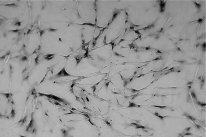
observed in all the cultures from various donors (n ¼ 9) by in vitro microscopy. During both cultivation modi, HTC constantly stained positive for the expression of collagen type II (Fig. 1).
3.2. Dynamic growth patterns displayed by DegraPolw
-seeded HTC
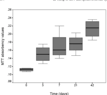
After seeding of the constructs, assessment of chondrocyte-DegraPolwin vitro static culture through MTT test showed
viable chondrocyte proliferation (Fig. 2). Compared with 2 h culture (MTT absorbency values ¼ 0:112 ^ 0:004), absor-bency value of MTT increased significantly to 0.176 ^ 0.023 at 21 days, and to 0.213 ^ 0.023 at 42 days, respectively (P , 0:05). Testing for GAG contents of constructs displayed a constant GAG increase up to 53 and 84% at 21 and 42 days, respectively, compared to day-3-culture values (Fig. 3). 3.3. Fabrication of a cartilage-like microstructure in HTC – DegraPolw
constructs When seeded onto 3-D DegroPolw
, HTC readily adhered to the polymer in multiple layers and regained a rounded configuration similar to the spherical appearance of chondrocytes in native cartilage, as observed with contrast phase microscope. Histological gross examination revealed a cartilage-like appearance of the HTC-seeded DegraPolw
constructs, and Alcian blue staining demonstrated intra-construct chondroitinsulfate production (Fig. 4).
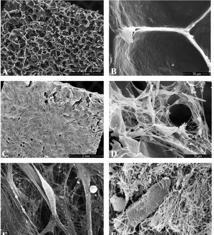
SEM pictures revealed that chondrocytes readily grow on the construct surface and into the open pores of the scaffold. Cells located on the surface of the scaffold showed fibroblast-like morphology with high numbers of cell-cell interactions. In contrast, cells located inside the scaffold demonstrated a different morphology with a rounded appearance and a less intimate cell-polymer attachment. Although sparsely, a 3-D network of thin collagen type II
Fig. 1. Immunohistochemical staining of HTC staining positive for collagen type II expression after 6 – 8 weeks in vitro monolayer and agar culture. Original magnification £100.
fibrils nevertheless occurred, and tethered the HTC within the DegraPolwpores (Fig. 5)[15].
4. Discussion
This is the first report to our knowledge on the feasibility of employing HTC in a concept of tissue engineering in neo-tracheal constructs.
Although chondrocytes isolated from varying sources have been successfully seeded onto different scaffolds, little attention has been focused on correlating anatomical origin of the cartilage with the functional requirements of the fabricated design. Several studies have investigated the ex vivo regeneration of cartilage from isolated human septal
[16], auricular[17,18], and articular chondrocytes[19], yet,
physiological properties of the cells differ substantially in regard to their anatomic origin. Cellular doubling times of human septal chondrocytes, for example, comprise 2.6 days
[16], in contrast to 10 days in cultures of human auricular cartilage cells [17]. Chondrocyte viability of human auricular after isolating was approximately 85% determined by a hemocytometer and trypan blue vital dye [17], yet, isolation efficacy of human septal chondrocytes was only 11.9% as determined by DNA assay [16]. Given the observed inter-tissue differences within the same species, and especially in attempting to recreate the complex functionality of the trachea, the anatomical origin of the cells employed will very likely influence the long time success of the tissue engineered replacement. Therefore, and in view of a subsequent in vivo employment, we explored the practicability of culturing HTC.
When placed in a 2-D culture milieu, chondrocytes characteristically undergo a process of de-differentiation: in addition to taking on a fibroblast-like shape, they loose their ability to secrete proteoglycans, and change from collagen type II to type I production [20]. The resulting matrix has inferior mechanical properties and lacks the functionality initially displayed at its original anatomical site. Yet, this loss of diversity can be reversed through cultivation in a 3-D medium, such as agarose, where chondrocytes regain spherical configuration and recover original matrix secre-tion patterns[21]. Restoration of spherical shape reflects a differentiated chondrocyte phenotype [20] and correlates with high fractions of collagen type II and the absence of collagen type I in tssue-engineered cartilage[15]. Collagen type II, interconnected with collagen type IX, is crucial in maintaining the internal framework of hyaline cartilage, thus preserving its shape, tensile strength and counter-balancing proteoglycan swelling pressure. Anderer et al. observed the re-expression of collagen type II in chondro-cytes cultured as spheroids during prolonged monolayer passages [19], and Rodriguez et al. were able to document
Fig. 2. MTT absorbency values at 570 nm showing viable HTC proliferation after seeding onto DegraPolw
discs and in vitro static culture.
Fig. 3. GAG content assay displayed an increase of GAG concentration in cell-polymer constructs over time.
Fig. 4. Three weeks after seeding of HTC onto DegraPolw
and in vitro static culture. Alcian blue staining displays a histomorphological architecture resembling native cartilage. Original magnification £100.
the same phenomenon in cultures of human ear chondro-cytes beyond the fourth week of passaging [17]. In our experiments, we observed the same phenomenon, where HTC cultured on agar plates re-expressed collagen type II after 6 – 8 weeks. Obviously, the process of re-acquiring the
former phenotype in terms of shape and synthesis patterns reflects a cell-specific reconstitution. However, quality and duration of this phenotypic change, especially in vastly expanded cell groups, have to be assessed in vivo. Possibly, following implantation the host of environmental stimuli
Fig. 5. Scanning electron micrographs (A,C: exterior; B,D,E,F: interior). (A,B) Plain DegraPolw
scaffold. (C,D) ECM synthesis 3 weeks after HTC were seeded onto DegraPolw
strips. Note that chondrocytes retained their spherical configuration, suggesting a well-differentiated phenotype. (E) Although sparsely, a 3-D network of thin collagen type II fibrils occurred in constructs at 3 weeks after seeding onto DegraPolw
. (F) In comparison, 3-D network of collagen type II fibrils in native human trachea cartilage.
toward reconstituted HTC may support their further re-differentiation.
In addition, we also assessed the growth dynamics of isolated HTC in 3-D DegroPolw scaffold. Expanding our
initial experiments and transferring HTC from a 2-D into a 3-D environment, they were able to retain their state of phenotypical reconstitution, thereby repopulating the poly-meric matrix. The fabricated construct resembled native cartilage both histologically and in SEM. Cellular distri-bution and attachment to the matrix during in vitro static culture are determined by gravity [22], and the spatially uniform distribution reflects porosity and surface charge of the scaffold. Attach rates for HTC to DegraPolwwere 80%
in comparison to the monolayer culture, with highest celluar densities on the construct surface. Although HTC with adjacent, well-spaced ECM frameworks could be detected within the central pores of the polymer, overall cellular concentrations diminished with distance from the surface. Probably, in static in vitro culture, gas exchange and diffusion of nutrients limit an even distribution of HTC throughout the construct. To overcome this obstacle, additional aspect of HTC – polymer cultivation should be considered. First, especially in regard of fabricating more massive, tubular designs, dynamic stress stimuli [23,24]
may be beneficial for inducing evenly structured construct to withstand in vivo mechanical impact. Thus, integration of a bioreactor with adjustable dynamic stress and shear stimuli for cultivation might improve the quality of the cellular growth pattern on/within the constructs. Second, in vivo conditioning of seeded cell/polymer designs before orthotopic implantation may serve as a natural bioreactor. Cell – cell interaction with the host and neovascularization of the design might improve the quality of the construct.
Taken together, our results demonstrate the successful harvest, cultivation, expansion and seeding of HTC in a tissue engineering concept. We have demonstrated that cultivated chondrocytes from human trachea can populate DegraPolw
, a novel biocompatible, degradable polymer, developing a cartilage-like histological architecture. In choosing tracheal chondrocytes for generating an orthotopic replacement structure, these preliminary results represent a basis for further research in the field. Employing tubular polymer designs, in vitro dynamic stimuli and/or in vivo conditioning, promising approaches for future design of a functional tracheal replacement can be envisioned.
Acknowledgements
This work was supported by the Olga-Mayenfisch-Stiftung, Zurich, Switzerland. We thank Mr. K. Marquardt for SEM analysis and Mrs. A. Morger for histological staining.
References
[1] Kon M, van den Hooff A. Cartilage tube formation by perichondrium: a new concept for tracheal reconstruction. Plast Reconstr Surg 1983; 72:791 – 7.
[2] Grillo HC. Tracheal replacement: a critical review. Ann Thorac Surg 2002;73:1995– 2004.
[3] Liu Y, Nakamura T, Yamamoto Y, Matsumoto K, Sekine T, Ueda H, Shimizu Y. Immunosuppressant-free allotransplantation of the trachea: the antigenicity of tracheal grafts can be reduced by removing the epithelium and mixed glands from the graft by detergent treatment. J Thorac Cardiovasc Surg 2000;120:108– 14.
[4] Langer R, Vacanti JP. Tissue engineering. Science 1993;260:920 – 6. [5] Vacanti CA, Langer R, Schloo B, Vacanti JP. Synthetic polymers seeded with chondrocytes provide a template for new cartilage formation. Plast Reconstr Surg 1991;88:753 – 9.
[6] Vacanti JP, Langer R. Tissue engineering: the design and fabrication of living replacement devices for surgical reconstruction and transplantation. Lancet 1999;354:SI32– S134.
[7] Fuchs JR, Nasseri BA, Vacanti JP. Tissue engineering: a 21st century solution to surgical reconstruction. Ann Thorac Surg 2001;72:577– 91. [8] Vacanti CA, Paige KT, Kim WS, Sakata J, Upton J, Vacanti JP. Experimental tracheal replacement using tissue-engineered cartilage. J Pediatr Surg 1994;29:201 – 5.
[9] Kojima K, Bonassar LJ, Roy AK, Vacanti CA, Cortiella J. Autologous tissue-engineered trachea with sheep dasal chondrocytes. J Thorac Cardiovasc Surg 2002;123:1177 – 84.
[10] Lee CJ, Moon KD, Choi H, Woo JI, Min BH, Lee KB. Tissue engineered tracheal prosthesis with acceleratedly cultured homolo-gous chondrocytes as an alternative of tracheal reconstruction. J Cardiovasc Surg 2002;43:275– 9.
[11] Saad B, Moro M, Tun-Kyi A, Welti M, Schmutz P, Uhlschmid GK, Neuenschwander P, Suter UW. Chondrocyte-biocompatibility of DegraPolw
-foam: in vitro evaluations. J Biomater Sci Polym Ed 1999;10:1107– 19.
[12] Saad B, Matter S, Ciardelli G, Uhlschmid GK, Welti M, Neuensch-wander P, Suter UW. Interactions of osteoblasts and macrophages with biodegradable and highly porous polyesterurethane foam and its degradation products. J Biomed Mater Res 1996;32:355 – 66. [13] Zu¨nd G, Ye Q, Hoerstrup SP, Schoeberlein A, Schmid AC,
Grunenfelder J, Vogt P, Turina M. Tissue engineering in cardiovas-cular surgery: MTT, a rapid and reliable quantitative method to assess the optimal human cell seeding on polymeric meshes. Eur J Cardiothorac Surg 1999;15:519– 24.
[14] Barrett AJ, Buttle DJ, Farndale RW. Improved quantitation and discrimination of sulphated glycosaminoglycans by use of dimethy-lene blue. Biochim Biophys Acta 1986;883:173– 7.
[15] Riesle J, Hollander AP, Langer R, Freed LE, Vunjak-Novakovic G. Collagen in tissue-engineered cartilage: types, structure, and cross-links. J Cell Biochem 1998;71:313– 27.
[16] Rotter N, Bonassar LJ, Tobias G, Lebl M, Roy AK, Vacanti CA. Age dependence of cellular properties of human septal cartilage: implications for tissue engineering. Arch Otolaryngol Head Neck Surg 2001;127:1248 – 52.
[17] Rodriguez A, Cao YL, Ibarra C, Pap S, Vacanti M, Eavey RD, Vacanti CA. Characteristics of cartilage engineered from human pediatric auricular cartilage. Plast Reconstr Surg 1999;103:1111 – 9. [18] van Osch GJ, van der Veen SW, Verwoerd-Verhoef HL. In vitro
redifferentiation of culture-expanded rabbit and human auricular chondrocytes for cartilage reconstruction. Plast Reconstr Surg 2001; 107:433 – 40.
[19] Anderer U, Libera J. In vitro engineering of human autogenous cartilage. J Bone Miner Res 2002;17:1420– 9.
[20] von der Mark K, Gauss V, von der Mark H, Muller P. Relationship between cell shape and type of collagen synthsised as chondrocytes lose their cartilage phenotype in culture. Nature 1977;267:531– 2.
[21] Benya PD, Shaifer JD. Dedifferentiated chondrocytes reexpress the differentiated collagen phenotype when cultured in agarose gels. Cell 1982;30:215 – 24.
[22] Sittinger M, Bujia J, Rotter N, Minuth WW, Burmester GR. Tissue engineering and autologous transplant formation: practical approaches with absorbable biomaterials and new culture techniques. Biomaterials 1996;17:237 – 42.
[23] Niklason LE, Gao J, Abbott WM, Hirschi KK, Houser S, Marini R, Langer R. Functional arteries grown in vitro. Science 1999;284: 489 – 93.
[24] Vunjak-Novakovic G, Martin I, Obradovic B, Treppo S, Grodzinsky AJ, Langer R, Freed LE. Bioreactor cultivation conditions modulate the composition and mechanical properties of tissue-engineered cartilage. J Orthop Res 1999;17:130– 8.