Human umbilical cord cells for cardiovascular tissue
engineering: a comparative study
Alexander Kadner
a,*, Gregor Zund
a, Christine Maurus
a, Christian Breymann
b,
Sidika Yakarisik
a, Gregor Kadner
a, Marko Turina
a, Simon P. Hoerstrup
aaClinic for Cardiovascular Surgery, University Hospital, Raemistrasse 100, CH 8091 Zurich, Switzerland b
Department of Gynecology and Obstetrics, University Hospital, Zurich, Switzerland Received 12 May 2003; received in revised form 20 November 2003; accepted 15 December 2003
Abstract
Objective: Tissue engineering of viable, autologous cardiovascular replacements with the potential to grow, repair and remodel represents an attractive approach to overcome the shortcomings of available replacements for the repair of congenital cardiac defects. Currently, vascular myofibroblast cells represent an established cell source for cardiovascular tissue engineering. Cell isolation requires the invasive harvesting of venous or arterial vessel segments prior to scaffold seeding, a technique which may not be preferable, especially in pediatric patients. This study evaluates cells isolated from human umbilical cord artery, umbilical cord vein and whole cord as alternative autologous cell sources for cardiovascular tissue engineering. Methods: Cells were isolated from human umbilical cord artery (UCA), umbilical cord vein (UCV), whole umbilical cord (UCC) and saphenous vein segments (VC), and were expanded in culture. All three expanded cell groups were seeded on bioabsorbable copolymer strips and grown in vitro for 28 days. Isolated cells were characterized by flow cytometry, histology, immunohistochemistry, proliferation assays and compared to VC. Morphological analysis of the seeded polymer strips included histology, immunohistochemistry, sodium dodecyl sulfate-polyacrylamide gel electrophoresis, transmission electron microscopy (TEM), scanning electron microscopy (SEM) and uniaxial stress testing. Results: UCA, UCV and UCC demonstrated excellent cell growth properties comparable to VC. Following isolation, all three cell groups showed myofibroblast-like morphology and characteristics by staining positive for a-smooth muscle actin (ASMA) and vimentin. Histology and immunohistochemistry of seeded polymers showed good tissue and extracellular matrix formation containing collagen I, III and elastin. TEM showed viable myofibroblasts and the deposition of collagen fibrils and progessive growing tissue formation, with a confluent surface, was observed in SEM. No difference was found among the mechanical properties of UCA, UCV, UCC and VC tissue engineered constructs. Conclusions: Tissue engineering of cardiovascular constructs by using UCA, UCV and UCC is feasible in an in vitro environment. Cell growth, morphology, characteristics and tissue formation were comparable between UCA, UCV, UCC and VC. UCC represent an attractive, readily available autologous cell source for cardiovascular tissue engineering offering the additional benefits of utilizing juvenile cells and avoiding the invasive harvesting of intact vascular structures.
q2004 Elsevier B.V. All rights reserved.
Keywords: Tissue engineering; Cardiovascular; Umbilical cord; Polymer scaffold
1. Introduction
The repair of congenital cardiac defects frequently requires the application of synthetic or bioprosthetic replacements, such as patch material or vascular conduits. All clinically available replacements demonstrate signifi-cant disadvantages which limit their long-term benefits. Shortcomings include the lack of a growth potential, an
obstructive tissue ingrowth, and calcification of the prosthetic replacements, which necessitates multiple oper-ations in pediatric patients[1 – 3]. Currently, cryopreserved homograft tissue appears to offer the most attractive solution by containing, theoretically, some viability with a potential growth ability [4 – 6]. Following implantation in children, these grafts are subject to enhanced calcification and donor scarcity remains a significant problem for pediatric patients [7 – 9].
The creation of tissue engineered viable replacement structures with the potential to grow, to repair and to
1010-7940/$ - see front matter q 2004 Elsevier B.V. All rights reserved. doi:10.1016/j.ejcts.2003.12.038
www.elsevier.com/locate/ejcts
* Corresponding author. Tel.: þ41-1-255-1111; fax: þ41-1-255-4369. E-mail address: [email protected] (A. Kadner).
remodel may offer a promising alternative for pediatric patients. Several groups demonstrated the feasibility of tissue engineering cardiovascular structures such as pul-monary artery conduits, patch material, trileaflet heart valves and vessel grafts by using three-dimensional scaffolds of synthetic polymer, collagen or xenogenic origin [10 – 13]. Recent results reported in the first clinical application of a tissue engineered pulmonary artery conduit, following implantation in a pediatric patient, are encoura-ging [14]. So far it remains unclear whether these tissue engineered constructs will function under physiologic conditions in the long-term.
Presently, autologous vascular-derived myofibroblasts represent an established cell source for the tissue engineering of functioning cardiovascular structures. Prior to scaffold seeding, arterial or venous-derived myofibroblasts are isolated from segments of aorta, carotis or saphenous vein segments. An approach, requiring the invasive harvesting of intact vascular structures.
The present study evaluates cells isolated from umbilical cord artery, umbilical cord vein and whole umbilical cord as alternative autologous cell sources for cardiovascular tissue engineering. The application of umbilical cord cells would provide autologous cells without the harvesting of intact vascular structures, which appears to be an advantage, especially in pediatric patients.
2. Material and methods 2.1. Cell isolation and cultivation
Human umbilical cords (20 – 30 cm length) ðn ¼ 4Þ and saphenous vein segments (2 – 3 cm length) ðn ¼ 4Þ were freshly harvested, washed with Dulbecco’s phosphate buffered saline (DPBS) (Gibco), and placed in Dulbecco’s modified Eagle’s medium (DMEM) (Gibco) supplemented with 10% fetal bovine serum (HyClone) and Gentamycin (25 mg/ml) (Gibco). Umbilical cord artery and venous segments were isolated from whole cord. The tissue sections were processed for subsequent tissue cultivation between 30 and 60 min after harvest and following, pieces of umbilical cord artery, umbilical cord vein, whole umbilical cord, and saphenous vein were minced into 1 mm pieces and distributed in 100 £ 15 mm Petri dishes (Gibco BRL, Rockville, MD). Tissue sections were cultured with DMEM (Gibco) supplemented with 10% fetal bovine serum (HyClone) and Gentamycin (25 mg/ml) (Gibco). Medium was replaced at 24 and 72 h and every six days following. Daily progress was monitored by phase-contrast microscopy. The cells were serially passaged and expanded in a humidified incubator at 37 8C with 5% CO2. Sufficient cell numbers for seeding
of polymer scaffolds were obtained after a period of 18 – 21 days.
2.2. Polymer scaffolds
Non-woven polyglycolic-acid mesh (PGA) (Albany Int.) was coated with a thin layer of poly-4-hydroxybutyrate (P4HB, MW: 1 £ 106, PHA 4400, Tepha Inc., Cambridge, MA) by dipping into a tetrahydrofuran solution (1% wt/vol P4HB). Following solvent evaporation, a continuous coating and physical bonding of adjacent fibers was achieved. PH4B is a biologically derived biopolymer which shows a complete biodegradation after 6-8 weeks. Polymer scaffold strips (5 £ 2 cm; n ¼ 14s) were cut from the PGA/P4HB compo-site material and cold gas sterilized with ethylene oxide. 2.3. Cell seeding and in vitro culture of polymer constructs
Bioabsorbable polymer scaffolds were seeded with an approximate density of 4.5-5.5 £ 106UCC or VC per cm2 and cultured in DMEM supplemented with 10% fetal bovine serum and Gentamycin (25 mg/ml) in a humidified incu-bator (37 8C, 5%CO2) for 28 days.
2.4. Flow cytometry
Single cell suspensions of UCA, UCV, UCC and VC were prepared for flow cytometry. 0.5 £ 106cells in 100 ml HANKS solution plus 1%BSA were incubated with saturating concentrations of monoclonal antibodies CD 31-FITC (Clone LCI4 þ 6 þ 7, kindly provided by P. Kilshaw), ASMA (Clone 1A4, Sigma, St Louis, MI), and vimentin (Clone V9, NeoMarkers, Fremont, CA). For intracellular staining (ASMA, vimentin) cells were permea-bilized with ethanol for 60 min at room temperature and incubated with monoclonal antibodies for 30 min. Follow-ing washFollow-ing, stainFollow-ing with a secondary FITC-conjugated IgG goat-anti-mouse antibody (Chemicon, Temecula, CA) was performed for 30 min. Forward and side scatters were set to exclude debris and 10,000 gated events were counted per sample. Corresponding irrelevant isotype-matched and positive controls were performed for each antibody. Cells were analyzed with the flow cytometer FACS-Calibur (Becton Dickinson Immunocytometry Systems, San Jose, CA). Data analysis was performed with the CELL QUEST software program (Becton Dickinson Immunocytometry Systems, San Jose, CA). Expression levels were calculated as mean fluorescence intensity ratio (MFIR) defined as mean fluorescence intensity of the studied antibodies divided by mean fluorescence intensity of corresponding isotype controls.
2.4.1. Histology and immunohistochemistry of umbilical cords
Sections of umbilical cords ðn ¼ 4Þ were fixed in 4% phosphate-buffered formalin and embedded in paraffin. Paraffin sections were cut at 5-mm thickness and studied by hematoxylin – eosin and Trichrome-masson stain. Immu-nohistochemistry was performed by incubation with
monoclonal mouse antibodies for ASMA (Sigma, St Louis, MI), vimentin (NeoMarkers, Fremont, CA) and a secondary FITC-labeled goat-anti-mouse IgG antibody (Sima, St Louis, MI). Prior to intracellular staining, tissue sections were treated with 0.1% Triton (Sigma, St Louis, MI) for 10 min. 2.4.2. Histology and Immunohistochemistry
of UCA, UCV, and UCC cultures
Samples of UCA, UCV and UCC were cultivated onto glass coverslips in DMEM. After 2 – 3 days cells were washed with DPBS and fixed in methanol for 10 min. Cells were examined histologically by hematoxylin – eosin and Trichrome-masson stain. Immunohistochemistry was per-formed by incubation with monoclonal mouse antibodies for ASMA (Sigma, St Louis, MI), vimentin (NeoMarkers, Fremont, CA), elastin (Sigma, St Louis, MI), and collagen I, III (Oncogen, Boston, MA). Incubation with a secondary FITC-labeled goat-anti-mouse IgG antibody (Sima, St Louis, MI) and a biotin-labeled goat-anti-mouse IgG antibody (Sima, St Louis, MI) to elastin, and collagen I and III was performed. The biotin-labeled antibody signal was developed with the avidin – peroxidase system (ABC kit, Vector Lab, Burlingame, CA). Prior to intracellular staining for ASMA and vimentin permeabilization of the cells was performed by incubation with 0.1% Triton (Sigma, St Louis, MI) for 10 min.
2.4.3. MTT assay
Following isolation from umbilical cord and saphenous vein segments, UCA ðn ¼ 4Þ; UCV ðn ¼ 4Þ; UCC ðn ¼ 4Þ and VC ðn ¼ 4Þ were seeded in 24-well plates with a density of 5 £ 104cells/ml per well and cultured with DMEM. MTT assays were performed on days 1, 3, 6 and 8. Two hundred and fifty microliters medium and 20 ml MTT solution (Sigma, St Louis, MI) were added to each sample and incubated for 1 h at 37 8C followed by isopropanol for 5 min at RT. The optical density of the supernatants were measured at 570 nm and growth curves developed. 2.5. Analysis of UCA, UCV, and UCC-seeded polymer constructs
2.5.1. Histology and Immunohistochemistry
Sections of UCA, UCV, and UCC-seeded polymer scaffolds were fixed in 4% phosphate-buffered formalin and embedded in paraffin. Paraffin sections were cut at 5 mm thickness and studied by hematoxylin – eosin and Trichrome-masson stain. Immunohistochemistry was performed as described above by incubation with monoclonal mouse antibodies for ASMA, vimentin, elastin and collagen I and III. 2.5.2. SDS-PAGE
Following lyphilization, samples of UCA, UCV and VC were homogenized and treated with a collagenase digestion procedure as described by Hayashi et al.[15]. Denaturated samples, standards of collagen I and III (Calbiochem, La
Jolla, CA) and a high range molecular weight marker (Bio-rad, Hercules, CA) were loaded onto a 10% sodium dodecyl sulfate-polyacrylamide gel (SDS-PAGE). A 100 V potential was applied across the gel for 80 – 90 min. The protein bands were stained with Coomassie blue (Bio-rad, Hercules, CA). 2.5.3. Scanning and transmission electron microscopy
Additional samples of UCA, UCV, and UCC-seeded polymer strips were fixed in 2% glutaraldehyde (Sigma, St Louis, MI) and studied by transmission electron microscopy (TEM) and scanning electron microscopy (SEM) at days 7, 14, 21 and 28.
2.5.4. Biomechanical testing
Longitudinal strips (20 £ 5 £ 1 mm3, n ¼ 5 of each group) of UCA, UCV, UCC, and VC-seeded polymer strips were uniaxial stress tested with an Instronw
tensile analyzer (model 4411) equipped with a 100 N load cell and pneumatic clamps (max. pressure 75 psig) (Instron Corp., Canton, MA). The cross head speed was 0.5 inch/min corresponding to a linear strain rate of 1 min21. The mechanical properties were analyzed for maximum stress and strain at maximal load.
3. Results
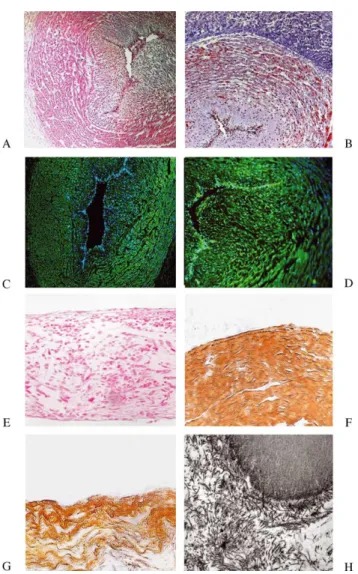
3.1. Histology and immunophenotyping of umbilical cords Hematoxylin – eosin and Trichrome-masson staining of sections of umbilical cord demonstrated the typical formation of umbilical cords with two arteries and one vein surrounded by Wharton’s jelly. The cells showed a fibroblast-like morphology and good homogenous depo-sition of extracellular matrix, predominantly in Wharton’s jelly (Fig. 1A and B).
Staining with monoclonal antibodies against ASMA and vimetin showed strong positive signals in the media of umbilical artery and vein, while the signals were detected at a lower level in cells of Wharton’s jelly (Fig. 1C and D). 3.2. Flow cytometry
Results of the FACS analysis of umbilical cord derived cells and saphenous vein derived cells are shown inFig. 2 and reveale comparable values for cell granularity and size. Analysis for the expression of ASMA and vimentin by UCA, UCV and UCC shows also comparable levels to those of VC. No positive signal was detected for the endothelial cell marker CD 31 among the isolated cell populations. 3.3. Histology and immunophenotyping of isolated umbilical cord cells
Hematoxylin – eosin and Trichrome-masson staining of fixed UCA, UCV and UCC cells showed elongated cells
with fibroblast-like morphology and the deposition of extracellular matrix throughout the cell culture. Immuno-histochemistry revealed the intracellular expression of ASMA and vimentin, both characteristic intracellular filaments of myofibroblast. Cultured UCC did not stain positive for CD 31, an endothelial cell marker.
3.4. MTT assay
Umbilical cord cells showed excellent cell growth. No differences in cell growth potential between cultivated UCA and UCV were detected by MTT assay analysis and compared to previously investigated UCC and VC (Fig. 3) [15].
3.5. Histology and immunohistochemistry of seeded polymer strips
Hematoxylin – eosin and Trichrome-masson staining of UCA, UCV and UCC-seeded polymer strips showed good tissue formation (Fig. 1E). During the cultivation period, a progessive cellular ingrowth and extracellular matrix deposition was observed in the polymer strips. Immunohis-tochemistry showed positive staining for ASMA and vimentin, while the extracelluar matrix analysis demon-strated the deposition of elastin and collagen I and III by all three cell groups (Fig. 1F and G).
Fig. 1. Hematoxylin – eosin staining of a umbilical cord vein (A) and Trichrome-masson staining of a umbilical cord artery (B) shows cells with a fibroblast morphology and deposition of extracellular matrix proteins, predominantly in Wharton’s jelly. Staining with monoclonal antibodies against ASMA (C) and vimentin (D) shows strong positive signals in umbilical cord vessels. Hematoxylin – eosin staining of a UCA strip shows good tissue formation and cellular ingrowth into the polymer strips (E). Immunohistology demonstrates the formation of elastin in a umbilical cord vein strip (G). TEM image shows the deposition of collagen fibrils in a umbilical cord artery seeded strip (H).
Fig. 2. Flow cytometry demonstrated no significant difference in the cell morphology for cell size and granularity of UCA and UCV compared to VC population (A). No difference was detected for the expression level of vimentin, ASMA or CD 31 between UCA, UCV, UCC and VC (B). The corresponding MFIR values are shown in each graph.
3.6. SDS-PAGE
Results of the SDS-PAGE analysis are shown inFig. 4. The banding pattern of UCA and UCV probes are comparable to VC probes. Corresponding bands are studied at the range of collagen chains a1ðIÞ;a1ðIÞ; b and g as described by Weston et al.[16].
3.7. Transmission and scanning microscopy
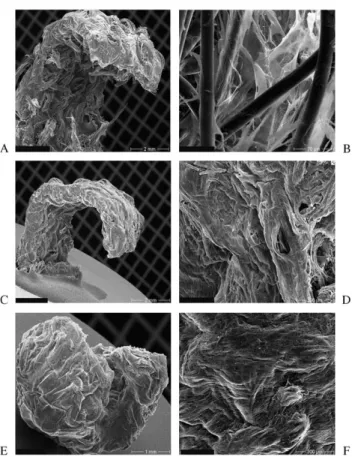
TEM of UCA, UCV and UCC sections of seeded patches showed secretionally active myofibroblasts with deposition of collagen fibrils (Fig. 1H). SEM analysis demonstrated good cell-polymer attachment with a subsequent progress-ive tissue formation during the cultivation period of 28 days. No difference could be observed between UCA, UCV or UCC seeded strips. Cells of all three groups formed a confluent surface on both sides and grew well into the deeper parts of the polymer. However, a continuous degradation of the polymer scaffold was studied by multiple breakages and fragmentation of the polymer fibers (Fig. 5). 3.8. Biomechanical testing
The biomechanical testing revealed comparable results for UCA, UCV, UCC, and VC-seeded constructs in regard to maximal uniaxial tensile stress testing (UCA: 0.158 ^ 0.024 MPa), UCV: 0.190 ^ 0.088 MPa, UCC: 0.264 ^ 0.093 Mpa, VC: 0.373 ^ 0.054 Mpa and to distensibility (UCA: 0.0.389 ^ 0.212 mm/mm, UCV: 0.523 ^ 0.055 mm/mm, UCC: 0.385 ^ 0.047 mm/mm, VC: 0.43 ^ 0.056 mm/mm).
4. Discussion
The results of the present study demonstrate that human umbilical cord cells represent an attractive autologous cell source for tissue engineering of cardiovascular constructs, especially for the repair of congenital defects. Umbilical cords were easy to obtain in segments between 20 and 30 cm length, which allows for the generation of a sufficient
cell number in a relatively short period of time. In order to obtain a similar result by using cells of saphenous vein origin, segments of a similar length would have to be harvested from pediatric patients or higher cell passages would be required prior to cell seeding.
The human umbilical cord is composed of two arteries and one vein surrounded by embryonic connective tissue— Wharton’s jelly [17]. Cells of umbilical cord artery, umbilical cord vein and Wharton’s jelly, exhibit myofibro-blast-like characteristics by co-expressing ASMA and vimentin. Following immunohistological analysis, Kobaya-shi et al. also observed no qualitative differences concerning cytoskeletal differentiation and extracellular matrix mol-ecules of arterial and venous umbilical cord vessel walls, and further analysis of segments isolated from the distal, middle, and proximal part of the umbilical cord shows no significant differences in the longitudinal axis of the umbilical cords[18].
Following isolation from segments of umbilical cord artery, umbilical cord vein and whole cord, cultivated cells continued to express myofibroblast morphology with a cell size and granularity which was comparable to vascular cells of saphenous vein origin. Furthermore, no difference was detected in the expression levels of the myofibroblast typical microfilament ASMA, or the intermediate filament
Fig. 5. SEM analysis of UCA, UCV and UCC strips show a good cell-polymer attachment of the cells with a subsequent progressive tissue formation during the cultivation period of 28 days. A seeded strip is shown after 14 days (A and B), 21 days (C and D) and after 28 days (E and F). Fig. 4. SDS-PAGE shows corresponding banding pattern at the range of
collagen chains a1(I), a1(2), b and g of UCA, UCV, and VC probes after
vimentin by cultivated UCA, UCV and UCC compared to VC. Following seeding, cells of all three groups demon-strated a good attachment and good growth pattern on the polymer strips. A continuous formation of a confluent, smooth tissue surface, and the deposition of extracellular matrix, consisting of collagen I, III and elastin, was studied. Further similarity of the extracellular matrix formation of umbilical cord cells and vascular cells was demonstrated by SDS-PAGE protein separation which revealed a comparable collagen chain formation between UCA, UCV, UCC and VC probes.
The importance of using arterial versus venous myofi-broblasts for tissue engineering is, as yet, unclear. Schnell et al. compared polymer strips seeded with fibroblasts isolated from human aortic and saphenous vein segments in an in vitro study [19]. They concluded that the saphenous vein seeded strips showed a higher tissue maturity, collagen formation and mechanical stability, compared to aortic constructs. However, the authors do not provide any information of the original cell sources, such as number of patients, patient age, and existing comorbidities, such as artherosclerosis, which may have a tremendous impact on myofibroblast morphology and extracellular matrix for-mation. In contrast, by using arterial and venous cell lines, several groups report the successful generation of heart valves, pulmonary conduits, and patch material[14,20 – 24]. There was no difference in performance or tissue formation between the in vivo tested constructs.
The results of the present study did not show any particular differences among UCA, UCV and UCC compared to VC, concerning cell characteristics and tissue formation under static cultivation conditions. It appears that in regard to practical clinical application, cell isolation could be performed from whole term umbilical cords. However, further experiments exposing UCA, UCV and UCC seeded constructs to a dynamic environment are required to confirm these findings.
Umbilical cord cells represent an attractive cell source for tissue engineering of cardiovascular structures for the repair of congenital defects by offering the advantages of avoiding the invasive harvesting of intact vascular struc-tures from pediatric patients and the availability of approximately 20 – 30 cm vascular tissue sections, which allows the isolation of a large amount of juvenile, fast growing cells for the generation of a sufficient cell number for scaffold seeding in a short period of time.
Additional advantages of using umbilical cords as a cell source for cardiovascular tissue engineering present the possibility to preserve the post-partal cords by standard cell and tissue banking technology to obtain an autologous cell pool for the patient’s lifetime, and the isolation of endothelial cells from the cord vessels to create an endothelial layer on the tissue engineered constructs, which may be crucial for their long-term function. The evaluation of these concepts will be areas of future studies.
Acknowledgements
The authors wish to thank, Jay Tracy, Astrid Fleisch-mann, MS, Christina Guenther, MD, and Sirpa Price, Laboratory for Tissue Engineering and Cell Transplan-tation, University Hospital Zurich, for their valuable work on cell culture and Klaus Marquard, Department of Surgical Research, University Hospital Zurich, for providing the SEM pictures. Finally we are grateful to Annegret Bittermann and Oliver Hoechli, Electron Microscopy Laboratory, University Zurich for their advice and technical assistance with the fluorescence microscopy imaging.
References
[1] Mayer Jr JE. Uses of homograft conduits for right ventricle to pulmonary connections in the neonatal period. Semin Thorac Cardiovasc Surg 1995;7:130– 2.
[2] Ben-Shachar G, Edwards JE. Separation of neointima from Dacron graft causing obstruction. J Thorac Cardiovasc Surg 1981;82:268. [3] Endo S, Saito N, Misawa Y, Sohara Y. Late pericarditis secondary to
pericardial patch implantation 25 years prior. Eur J Cardiothorac Surg 2001;20:1059– 60.
[4] Molina JE, Edwards J, Bianco R, Clack R, Rasmussen T, Moss G, Lang G. Growth of fresh-frozen pulmonary allograft conduit in growing lambs. Circulation 1989;80(Suppl):I183 – 90.
[5] Schoen FJ, Mitchell RN, Jonas RA. Pathological considerations in cryopreserved allograft heart valves. J Heart Valve Dis 1995;4:72 – 6. [6] Rajani B, Mee RB, Ratliff NB. Evidence for rejection of homograft cardiac valves in infants. J Thorac Cardiovasc Surg 1998;115:111 – 7. [7] Cleveland DC, Williams WG, Razzouk A. Failure of cryopreserved homograft valved conduits in the pulmonary circulation. Circulation 1992;86:II150 – 3.
[8] Kirklin JK, Smith D, Novick W, Naftel DC, Kirklin JW, Pacifico AD, Nanda NC, Helmcke FR, Bourge RC. Long-term function of cryopreserved aortic homografts. J Thorac Cardiovasc Surg 1993; 106:154 – 66.
[9] Turrentine MW, Ruzmetov M, Vijay P, Bills RG, Brown J. Biological versus mechanical aortic valve replacement in children. Ann Thorac Surg 2001;71:356 – 60.
[10] Shinoka T, Shum TD, Ma PX, Tanel RE, Isogai N, Langer N, Langer R, Vacanti JP, Mayer Jr JE. Creation of viable pulmonary artery autografts through tissue engineering. J Thorac Cardiovasc Surg 1998; 115(3):536 – 45. discussion 545 – 6.
[11] Bader A, Steinhoff G, Strobl K, Schilling T, Brandes G, Mertsching H, Tsikas D, Froelich J, Haverich A. Engineering of human vascular aortic tissue based on a xenogeneic starter matrix. Transplantation 2000;70:7 – 14.
[12] Stock UA, Sakamoto T, Hatsuoka S, Martin DP, Nagashima M, Moran AM, Moses MA, Khalil PN, Schoen FJ, Vacanti JP, Mayer Jr JE. Patch augmentation of the pulmonary artery with bioabsorbable polymers and autologous cell seeding. J Thorac Cardiovasc Surg 2000;6:1158– 67.
[13] Hoerstrup SP, Zund G, Sodian R, Schnell AM, Grunenfelder J, Turina MI. Tissue engineering of small caliber vascular grafts. Eur J Cardiothorac Surg 2001;20:164– 9.
[14] Shinoka T. Transplantation of a tissue-engineered pulmonary artery. N Engl J Med 2001;344:532– 3.
[15] Kadner A, Hoerstrup SP, Tracy J, Maurus C, Melnitchouk S, Kadner G, Zund G, Turina MI. Human umbilical cord cells: a new cell source for cardiovascular tissue engineering. Ann Thorac Surg 2002;74: S1422 – 8.
[16] Weston MW, Goldstein S, Epting RE, He S, Mauldin JM, Yoganathan AP. Establishing a protocol to quantify leaflet fibroblast response to physiologic flow through a viable heart valve. Am Soc Artif Intern Organs J 1997;43:M377 – 82.
[17] Schoenberg MD, Moore RD. Studies on connective tissue III: enzymatic studies on the formation and nature of carbohydrate intermediate of the connective tissue polysaccharides in the human umbilical cord. Arch Pathol 1958;65:115 – 24.
[18] Kobayashi K, Kubota T, Aso T. Study on myofibroblast differen-tiation in the stromal cells of Wharton’s jelly: expression and localization of alpha-smooth muscle actin. Early Hum Dev 1998;51: 223 – 33.
[19] Schnell AM, Hoerstrup SP, Zund G, Kolb S, Sodian R, Visjager JF, Grunenfelder J, Suter A, Turina M. Optimal cell source for cardiovascular tissue engineering: venous vs. aortic human myofi-broblasts. Thorac Cardiovasc Surg 2001;49:221– 5.
[20] Hoerstrup SP, Sodian R, Daebritz S, Wang J, Bacha EA, Martin DP, Moran AM, Guleserian KJ, Sperling JS, Kaushal S, Vacanti JP,
Schoen FJ, Mayer Jr JE. Functional living trileaflet heart valves grown in vitro. Circulation 2000;102(Suppl 3):III44– 9.
[21] Stock UA, Nagashima M, Khalil PN, Nollert GD, Herden T, Sperling JS, Moran A, Lien J, Martin DP, Schoen FJ, Vacanti JP, Mayer Jr JE. Tissue engineered valve constructs in the pulmonary circulation. J Thorac Cardiovasc Surg 2000;119:732 – 40.
[22] Sodian R, Hoerstrup SP, Sperling JS, Daebritz S, Martin DP, Moran AM, Kim BS, Schoen FJ, Vacanti JP, Mayer Jr JE. Early in vivo experience with tissue-engineered trileaflet heart valves. Circulation 2000;102(Suppl 3):III22 – 9.
[23] Steinhoff G, Stock U, Karim N, Mertsching H, Timke A, Meliss RR, Pethig K, Haverich A, Bader A. Tissue engineering of pulmonary heart valves on allogenic accellular matrix conduits. Circulation 2000; 102(Suppl 3):III50 – 5.
[24] Shum-Tim D, Stock U, Hrkach J, Shinoka T, Lien J, Moses MA, Stamp A, Taylor G, Moran AM, Landis W, Lander R, Vacanti JP, Mayer Jr JE. Tissue engineering of autologous aorta using a new biodegradable polymer. Ann Thorac Surg 1999;68:2298 – 304.