Video-intuboscopy: a new aid to routine and difficult tracheal intubation
3
0
0
Texte intégral
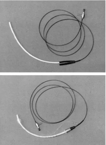
(2) 526. British Journal of Anaesthesia Intubating mode (guided tracheal intubation) Intubation was guided in two patients with unexpected difficult laryngoscopy (grade 3 view).9 In the first patient the video-optical intubation stylet was already loaded in the tracheal tube (standby mode) so that the anaesthetist had only to change his view to the video-display and tracheal intubation was carried out under video visual control within another 15 s. In the second patient, after initial direct laryngoscopy without intubation attempts, mask ventilation was performed for a short period to prepare the tracheal tube with the video-optical stylet. The patient was then intubated within 30 s with another conventional laryngoscopy and using the video-view from the tracheal tube tip. In both patients intubation was performed successfully at the first attempt, in a rapid and safe manner, each by a different anaesthetist with no previous experience of this method. There was no need for head, neck or laryngeal manipulation, or help from an assistant.. Discussion REVIEW. Figure 1 Malleable, video-optical intubation stylet (od 5 mm) with proximal tracheal tube (TT) connector, 2 m of transmitting cable (od 2.3 mm) and video camera–light source (top). TT (id 7.5 mm) loaded with the video-optical intubation stylet (bottom).. video display to confirm tracheal tube position and adjust the tracheal tube as required using the video view transmitted from the stylet tip. The videooptical stylet is then removed from the tracheal tube. Intubation mode (guided tracheal intubation) If a view of the vocal cords cannot be obtained during tracheal intubation, the best possible direct laryngoscopic view is maintained with the laryngoscope blade in one hand. The tracheal tube loaded with the videooptical intubation stylet is introduced into the oropharynx with the other hand. The anaesthetist then uses the video view from the tracheal tube tip to guide the pre-formed tracheal tube between the vocal cords and into the trachea, and confirm the correct position, before the stylet is removed. FIRST EXPERIENCES. Video-intuboscopy is a new indirect laryngoscopic intubation technique and combines the three following elements: (1) intuboscopy (transmission of the view from the tracheal tube tip to a proximal viewfinder), (2) conventional laryngoscopy and (3) video-endoscopy. Intuboscopy Intuboscopic-guided intubation was first described by Murphy in 1967.10 He used a fibreoptic choledoscope in a nasally passed tracheal tube. Both tracheal tube and flexible endoscope were advanced together and steered into the trachea by twisting the proximal tracheal tube while watching through the viewfinder.10 Conventional laryngoscopy Conventional direct laryngoscopy combined with fibrebronchoscopic intubation in anaesthetized patients, performed by two anaesthetists, has been reported as a useful intubation technique, providing enough room in the pharynx for steering the endoscopic instrument to the vocal cords, particularly if the pharynx is obscured by copious secretions, blood or soft tissue swelling.11. Standby mode (confirmation of tracheal intubation) Six operators used the video-optical intubation stylet in 30 patients undergoing elective surgery, and reported no disadvantages. The low weight, malleable nature and thin, flexible, transmitting cable to the video camera–light source unit allowed the video-optical intubation stylet to be used in the same way as a conventional intubation stylet. Removal of the video-optical intubation stylet was uneventful and any time delay from stylet removal was no longer than that with conventional gum elastic bougies or intubation stylets.. Video-endoscopy Video-endoscopy implies that the anaesthetist does not have to look into an eyepiece, and facilitates endoscopic procedures.12 The video display aids dexterity and enables nearly simultaneous observation of the video monitor, patient and monitors in a comfortable and efficient position for intubation. Video-endoscopy for tracheal intubation is used mainly for demonstrating and teaching direct laryngoscopy, fibrebronchoscopy or the Bullard intubation technique.12–15.
(3) Video-intuboscopy VIDEO-INTUBOSCOPY. Standby mode The video-optical stylet provides a useful means of monitoring tracheal tube position, and teaching and supervising tracheal intubation. Airway monitoring. Airway imaging allows accidental oesophageal intubation to be detected and avoids endobronchial intubation during the intubation procedure.16 Furthermore, subglottic airway pathology such as laryngeal or tracheal tumours, stenosis, compression, webs or an aberrant tracheal bronchus could be detected during intubation, so that airway management may be modified appropriately. The costs for a compact video-intuboscopic imaging unit ($6000) are relatively low compared with median payments for claims for respiratory adverse events ($200 000) and are in the range of other anaesthetic monitoring equipment.17 Further development of the device to provide a disposable guide and covering element, containing the reusable fibreoptic part, would allow it to be used as a cost effective device. Teaching direct laryngoscopy is usually limited because the instructor cannot see the view of the laryngoscopist during the procedure. Headframemounted video camera systems and video transmission from the laryngoscope blade have been developed for video visualization direct laryngoscopy.13 18 Video-intuboscopy in the “standby mode” is another means of allowing demonstration, teaching and supervising of intubation using the video view from the tracheal tube tip. Intubating mode Limited experience (in two patients with grade 3 laryngoscopy) indicated that video-intuboscopicguided tracheal intubation can allow simple, rapid, safe and gentle intubation. Looking at a video-display in critical circumstances is much more comfortable than looking into a viewfinder of an endoscopic device, because the operator remains in the usual intubation position and can change the view from the oropharynx to monitor and vice versa. Teaching and supervising is facilitated with the video monitor. Most anaesthetists are skilled in conventional laryngoscopy and in steering an intubation guiding stylet. Video-intuboscopic-guided tracheal intubation is almost the same procedure, except that the tracheal tube is guided by the video view from the stylet tip. This makes the video-optical intubation stylet an easy device to handle. If the device is being used to confirm intubation, when a patient is encountered with unpredicted difficult intubation, the method can be changed promptly to aid intubation. There is no delay in preparing intubation equipment, no need to interrupt the intubation procedure and no need for additional help. The method may be beneficial in patients with cardiovascular diseases, dental disorders and in patients with cervical spine pathology. However, these possibilities need to be confirmed by further investigations. Difficult tracheal intubation complicated by oropharyngeal secretions, swelling and bleeding, in. 527 which fibrebronchoscopic intubation may fail, could be managed by simultaneous direct laryngoscopy and video-intuboscopic-guided intubation, as has been done with fibrebronchoscopes.11 19 Lifting the tongue with the blade enables suction with large bore catheters and provides enough pharyngeal room for direction of the tracheal tube into the glottic opening. Training in video-intuboscopic-guided intubation may be obtained by simulating difficult intubation.19 20 This would allow initial direct laryngoscopy and video-guided tracheal intubation while lowering the laryngoscope blade so that the epiglottis descends and conceals the cords. In this way, training in video-intuboscopic-guided intubation can be done without jeopardizing patient safety and without loss of experience in direct laryngoscopic intubation. Clinical studies are required to confirm simplicity and efficacy, to define the further role and to explore other applications of this new intubation technique.. References 1. Caplan RA, Posner KL, Ward RC, Cheney FW. Adverse respiratory events in anesthesia: A closed claims analysis. Anesthesiology 1990; 72: 828–833. 2. Pierce EC, Cooper JB. Analysis of anesthetic mishaps. International Anesthesiology Clinics 1984; 22: 43–49. 3. Samsoon GLT, Young JRB. Difficult tracheal intubation. Anaesthesia 1987; 42: 487–490. 4. Siker ES. A mirror laryngoscope. Anesthesiology 1956; 17: 38–42. 5. Huffman JP. The application of prisms to curved laryngoscopes. A preliminary study. Journal of the American Association of Nurse Anesthetists 1968; 36: 138–129. 6. Talor PA, Towey RM. The broncho-fiberscope as an aid to endotracheal intubation. British Journal of Anaesthesia 1972; 44: 611–612. 7. Katz RL, Berci G. The optical stylet–A new intubation technique for adults and children with specific reference to teaching. Anesthesiology 1979; 51: 251–254. 8. Bjoraker DG. The Bullard intubating laryngoscopes. Anesthesia Review 1990; 17: 64–70. 9. Cormack RS, Lehane J. Difficult tracheal intubation in obstetrics. Anaesthesia 1984; 39: 1105–1111. 10. Murphy P. A fibre-optic endoscope used for nasal intubation. Anaesthesia 1967; 22: 489–491. 11. Couture P, Perrealut C, Girard D. Fibreoptic bronchoscopic intubation after induction of general anaesthesia: another approach. Canadian Journal of Anaesthesia 1992; 39: 99. 12. Popat MT, Howells H. Miniature screen for fibreoptic intubation using a camera. Anaesthesia 1997; 52: 802–803. 13. Henthorn RW, Reed J, Szanfranski JS, Ganta R. Combining the fiberoptic bronchoscope with a laryngoscope blade aids teaching direct laryngoscopy. Anesthesia and Analgesia 1995; 80: 433. 14. Pittman SK, Parnass SM, El-Ganzouri A, Braverman B. Video-assisted fiberoptic endotracheal intubation. Anesthesia and Analgesia 1994; 78: 197. 15. Crosby ET. Techniques using the Bullard laryngoscope. Anesthesia and Analgesia 1995; 81: 1314. 16. Szekely SM, Webb RK, Williamson, Russell WJ. Problems related to the endotracheal tube: An analysis of 2000 incident reports. Anaesthesia and Intensive Care 1993; 21: 611–616. 17. Cheney FW. The ASA closed claims project: Lessons learned. ASA Annual Refresher Course Lectures 1996; 422: 1–6. 18. Higgins MS, Deshphande JK, Badr A. New video system improves teaching of direct laryngoscopy. Anesthesiology 1996; 84: 1010–1011. 19. Ovassapian A, Sharon JY, Dykes MH, Brunner EE. Fiberoptic nasotracheal intubation–incidence and causes of failure. Anesthesia and Analgesia 1983; 62: 692–695. 20. Cormack RS, Lehane J. Simulating difficult intubation. British Journal of Anaesthesia 1983; 55: 1155..
(4)
Figure
Documents relatifs
