Appropriate placement of intubation depth marks in a
new cuffed paediatric tracheal tube
{
M. Weiss*, A. C. Gerber and A. Dullenkopf
Department of Anaesthesia, University Children’s Hospital Zurich, Steinwiesstrasse 75,
CH-8032 Zurich, Switzerland
*Corresponding author. E-mail: [email protected]
Background. The aim of this study was to evaluate the appropriateness of intubation depth marks on the new Microcuff paediatric tracheal tube.
Methods. With local Institutional Ethics Committee approval and informed parental consent, we included patients from birth (weighing >3 kg) to 16 yr who were undergoing general anaesthesia requiring orotracheal intubation. Tracheal intubation was performed using direct laryngoscopy, the intubation depth mark was placed between the vocal cords, and the tube was taped to the lateral corner of the mouth. The distance between the tube tip and the tracheal carina was assessed by flexible bronchoscopy with the patients in supine, and their head in neutral positions. Tube sizes were selected according to the formula: internal diameter (ID; mm)=(age/4)+3.5 in children >2 yr. In full-term newborns (>3 kg) to less than 1 yr ID 3.0 mm tubes were used and in children from 1 to less than 2 yr ID 3.5 mm tubes were used. Endoscopic examination was performed in 50 size ID 3.0 mm tubes, and in 25 tubes of each tube size from ID 3.5 to 7.0 mm. Tracheal length and percentage of the trachea to which the tube tip was advanced were calculated. Results. 250 patients were studied (105 girls, 145 boys). The distance from the tube tip to the carina ranged from 1.4 cm in a 2-month-old infant (ID 3.0 mm) to 7.7 cm in a 14-yr-old boy (ID 7.0 mm). Mean tube insertion into the trachea was 53.2% (6.3) of tracheal length with a minimum of 40% and a maximum of 67.6%.
Conclusions. The insertion depth marks of the new Microcuff paediatric tracheal tube allow adequate placing of the tracheal tube with a cuff-free subglottic zone and without the risk for endobronchial intubation in children from birth to adolescence.
Br J Anaesth 2005; 94: 80–7
Keywords: children; equipment, cuffs tracheal; intubation, tracheal; monitoring, depth mark Accepted for publication: June 26, 2004
Correct insertion depth of tracheal tubes in children is essen-tial to avoid accidental bronchial intubation, irritation of the carina, and accidental extubation. The length of the trachea in neonates and infants (39.4–60.5 mm) is short, leaving little margin for error.1 Thus, intubation depth marks at the tube tip have been introduced for optimal placement of the tube tip in the mid-tracheal position.2
However, as reviewed recently by Goel and Lim, a large disparity exists in the position and the presence of depth marks, bands, and lines between different types of uncuffed and cuffed tracheal tubes.3Similarly, the lack of intubation depth marks and inappropriately high positioned depth marks in cuffed paediatric tubes have been reported.3–6In the latter, the tube tip will become positioned critically deep in the trachea, when placed according to the depth marks. Further, even with the upper cuff border positioned directly below the vocal cords, a small margin of safety regarding
endobronchial intubation has been reported in cuffed pae-diatric tubes because of long tube cuffs and Murphy eyes.7If placed with the tip in the mid-tracheal position, in many tracheal tubes the cuff will lie within the larynx, again par-ticularly in those with long cuffs and a Murphy eye.4
Recently, a new cuffed paediatric tracheal tube (Micro-cuff Paediatric Tracheal Tube, Microcuff GmbH, Weinheim, Germany) with a high volume-low pressure cuff has been introduced. The thin-walled cuff is made {Declaration of interest. In the authors’ institution cuffed tracheal tubes
have been routinely used in children from birth to adolescence since 2000. Dr Weiss and Dr Gerber are involved in the design and evaluation of a new cuffed tracheal tube in co-operation with Microcuff GmbH, Weinheim, Germany. No agreements or financial benefits arise from this co-operation. Dr A. Dullenkopf has been supported by a Clinical Research Grant provided from Microcuff GmbH, Weinheim, Germany for this study.
from polyurethane, which is thought to improve sealing characteristics, allowing shorter cuffs.8The short cuff and the avoidance of a Murphy eye allows appropriate intubation depth with a cuff-free subglottic tube shaft.
The aim of the present study was to evaluate the approp-riateness of the intubation depth marks in the new Microcuff paediatric tracheal tube in a large population of patients ranging from neonates to adolescence.
Methods
The intubation depth marks in the Microcuff paediatric tra-cheal tube are based on tratra-cheal dimensions published by Griscom,1 9 potential tube tip displacement distances as reported in the literature,10–13 and the formula described by Motoyama for selection of cuffed tracheal tubes in chil-dren aged >2 yr (internal diameter [ID, in mm]=[age in yr/ 4]+3.5).14For patients below 2 yr of age tubes were chosen according to the recommendations of Steward and Khine, respectively (Table 1).15 16
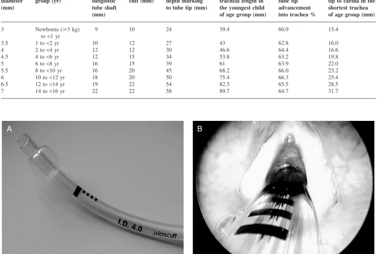
The depth marks are placed so that the tube tip can be advanced to 60–65% of the shortest trachea of the related age group (Table 1). This results in a safe margin for caudal tube displacement during head flexion of at least 15 mm in a neonate, and of 32 mm in a 14-yr-old child (smallest child considered for an ID 7.0 mm tube) (Table 1).10 13The short cuff allows a cuff-free subglottic tube shaft (distance between intubation depth mark and upper border of the cuff) of 9 mm in a 3.0-mm ID tracheal tube and of 22 mm for a 7.0-mm ID tracheal tube.17 18The semi-circular glottic intubation depth mark, placed on the concave side of the tube, is placed between the vocal cords during direct laryngoscopy (Fig. 1). Four points proximal to the semi-circular mark indicate the distance to it (in total 8 mm) and are useful in adjusting the placement of a tube.
After obtaining local Institutional Ethics Committee approval and informed parental consent, paediatric patients from birth (weighing >3 kg) up to 16 yr of age undergoing general anaesthesia requiring oro-tracheal intubation were included in this study. Children with known airway
A B
Fig 1(A) Microcuff paediatric tracheal tube with high volume-low pressure cuff, semi-circular intubation depth mark, cuff-free subglottic tube shaft. (B) Glottic intubation depth mark placed between the vocal cords. The four points can be used to estimate the distance to the intubation depth mark in case of an obstructed view to the vocal cords or correction of too deep tracheal tube insertion.
Table 1Tube sizes and age-related anatomical and technical measures8 9 Internal diameter (mm) Intended age group (yr) Cuff-free subglottic tube shaft (mm) Length of cuff (mm) Distance from depth marking to tube tip (mm) Shortest (95% CI) tracheal length in the youngest child of age group (mm)
Percentage of tube tip advancement into trachea %
Distance from tube tip to carina in the shortest trachea of age group (mm) 3 Newborns (>3 kg) to<1 yr 9 10 24 39.4 60.9 15.4 3.5 1 to<2 yr 10 12 27 43 62.8 16.0 4 2 to<4 yr 12 12 30 46.6 64.4 16.6 4.5 4 to<6 yr 12 15 34 53.8 63.2 19.8 5 6 to<8 yr 16 15 39 61 63.9 22.0 5.5 8 to<10 yr 16 20 45 68.2 66.0 23.2 6 10 to<12 yr 18 20 50 75.4 66.3 25.4 6.5 12 to<14 yr 19 22 54 82.5 65.5 28.5 7 14 to<16 yr 22 22 58 89.7 64.7 31.7
anomalies, expected or previous difficult intubation, and an ASA physical status of more than III were excluded. Pre-medication and induction of anaesthesia (inhalation or i.v.) depended upon the patient’s medical condition and prefer-ence. Monitoring included precordial stethoscope, pulse oximetry, ECG, and non-invasive blood pressure recording. After adequate mask ventilation was achieved, a non-depolarizing neuromuscular blocking agent was admi-nistered and anaesthesia was maintained with sevoflurane in oxygen. The tracheal tube size was selected according to Table 1. Tracheal intubation was performed by direct laryngoscopy, the glottic intubation depth mark placed between the vocal cords, and the tube taped at the right corner of the mouth. The correct tube position was initially confirmed by capnography and auscultation of the lungs. Adequate size of the tracheal tube was tested by the presence of air leakage at a maximum of 20 cm H2O airway pressure
with the cuff not inflated. If no air leakage was obtained, the tube was exchanged. The cuff was inflated to prevent audible air leakage with the cuff pressure not exceeding 20 cm H2O,
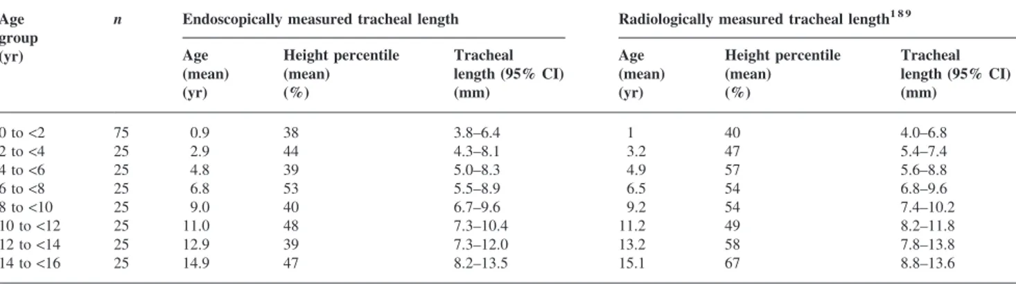
using a cuff manometer (Cuff Pressure Manometer, Mallinckrodt, Athlone, Ireland). The correct position of the intubation depth mark was confirmed by one of the two investigators using direct laryngoscopy, and adjusted if required. Subsequently, the distance from the tube tip to the tracheal carina was assessed by means of flexible video-endoscopy (Flexible Airway Endoscopes, Acutronic Medical Systems, Baar, Switzerland) using the drawback technique. A clip was placed on the fibrescope at the level of the swivel adapter as the crest of the tracheal carina
was just visualized on the monitor. Then the endoscope was drawn back until the proximal tube tip was visualized, and the distance between the clip and the level of the swivel adapter measured (Fig. 2).
Endoscopic examination was performed in 50 patients receiving a 3.0 mm ID tube and in 25 patients receiving a tube varying from ID 3.5 to 7.0 mm, with the patients in supine and their head in a neutral position. Neutral position of the head was defined as a vertical line from the external ear channel to the superior orbital margin (ear-eye-line). In addition, patient characteristics, tracheal tube insertion depth at the lateral corner of the mouth, and minimal cuff pressure required to seal the trachea were noted.
Statistical analysis
Data are presented as mean (SD) and/or median and range as appropriate. Tracheal length (vocal cords to carina distance) was calculated by adding the distance from depth mark to tube tip to the measured distance from tube tip to carina. The percentage of the trachea to which the tracheal tube tip was advanced within the trachea was calculated. Linear and/or logarithmic regression models were calculated for the rela-tionship of the distance from the tube tip to the carina, calculated tracheal length and tube insertion depth to age, weight, and length. In patients >2 yr of age, the distances from the tube tip to carina were compared with those derived from standard formulae for oral tube insertion (insertion depth [cm]=11.5+[age(yr)·0.5] and 12+[age(yr)·0.5], respectively).19 95% confidence intervals (CI) were
Fig 2 Measurement of the distance from tube tip to carina is performed using the fibreoptic drawback technique: a clip is placed on the fibrescope at the level of the swivel adapter as the crest of the carina is just visualized on the monitor (left). Then the endoscope is drawn back until the proximal tube tip (arrow) is just visualized, and the distance between the clip and the level of the swivel adapter is measured (right).
calculated for tracheal length and compared with those reported from radiological examination.1 9
Results
250 patients (105 female, 145 male) were studied. Median height percentile was 43.1% (IQR 11.9–72.9%)20(Table 2). In two patients the selected tube (ID 3.5 mm and ID 5.5 mm) had to be replaced because of no air leakage at more than 20 cm H2O airway pressure (height and weight in both
patients below the third percentile).20 In the remaining 248 patients, sufficient tracheal sealing was achieved with the cuff inflated to a pressure of <20 cm H2O (median 10 cm
H2O [4–18]). The distance from the tube tip to the tracheal
carina ranged from 1.4 cm in a 2-month-old infant to 7.7 cm in a 14-yr-old boy (Table 3). Calculated tracheal length ranged from 3.8 to 13.5 cm and demonstrated a good corre-lation with age (r=0.923), height (r =0.926), and less so with weight (r=0.890) (Fig. 4).
Mean tube tip advancement into the trachea was 53.2% (SD6.3) of the tracheal length, with a minimum of 40% in a 3.5-yr-old boy and a maximum of 67.6% in a 10-yr-old boy (Table 3). Overall tube insertion depth from the lateral corner of the mouth correlated well with age (insertion [cm]=10.6+[age (yr)·0.5]; r=0.956), height (r=0.960), and less so with weight (r=0.887) for all patients (Fig. 3). For children >2 yr oral tube insertion depth (cm) corresponded to 11.5+[age (yr)·0.5] (r=0.870).
With correction of the distances the from the tube tip to the tracheal carina in children >2 yr according to an oral tube insertion depth (cm) of 11.5+(age [yr]·0.5) or 12+(age [yr]·0.5), the cuffs would have become placed within the larynx or even between the vocal cords in 56 and 20 patients, respectively. Furthermore, with the 12+(age [yr]·0.5) formula, 10 tubes would have been advanced below the margin of safety for caudal tube tip displacement during head-neck flexion (Fig. 3).
Calculated 95% CIs for measured trachea length, and those reported from radiological examination1 9 are pre-sented in Table 4. Our data are comparable with those reported by Griscom.1
Discussion
In this study, we evaluated the appropriateness of the intu-bation depth marks of the new Microcuff paediatric tracheal tube with regard to the distance from tube tip to the tracheal carina, and with regard to the tracheal insertion depth. The main finding was that the intubation depth marks provided a safe margin regarding inadvertent endobronchial intubation and were an improvement over a theoretical formula for oral tube insertion depth (Fig. 3). The mean tube tip position corresponded to a mid-tracheal position.
Intubation depth marks in paediatric tracheal tubes were introduced for safe positioning of tracheal tubes, particularly in the emergency situation when tracheal
Table 3Endoscopically measured distance from tube tip to tracheal carina, calculated tracheal length, and percentage of the trachea to which the tube tip was advanced. Data are mean (SD) [range]. (n=250 patients)
Tube size ID (mm)
n Intended age group (yr)
Distance from tube tip to tracheal carina (cm)
Calculated tracheal length (cm)
Percentage of the trachea to which the tube tip is advanced 3 50 Birth to<1 2.3 (0.6) [1.4–3.5] 4.7 (0.6) [3.8–5.9] 51.5 (6.0) [40.7–63.2] 3.5 25 1 to<2 2.8 (0.6) [1.8–4.0] 5.5 (0.6) [4.5–6.7] 49.9 (5.8) [40.3–60.0] 4 25 2 to<4 3.1 (0.8) [2.0–4.5] 6.1 (0.8) [5.0–7.5] 49.7 (6.4) [40.0–60.0] 4.5 25 4 to<6 3.2 (0.8) [1.9–4.5] 6.6 (0.8) [5.3–7.9] 52.3 (6.7) [43.0–64.2] 5 25 6 to<8 3.3 (0.8) [2.0–4.7] 7.2 (0.8) [5.9–8.6] 54.4 (6.1) [45.3–66.1] 5.5 25 8 to<10 3.7 (0.8) [2.4–5.6] 8.2 (0.8) [6.9–10.1] 55.4 (5.1) [44.6–65.2] 6 25 10 to<12 3.9 (0.7) [2.4–5.5] 8.9 (0.7) [7.4–10.5] 56.4 (4.6) [47.6–67.6] 6.5 25 12 to<14 4.2 (1.0) [2.8–6.6] 9.6 (1.0) [8.2–12.0] 56.8 (5.7) [45.0–65.9] 7 25 14 to<16 5.2 (1.4) [2.8–7.7] 11.0 (1.4) [8.6–13.5] 53.6 (6.7) [43.0–67.4] Table 2Patient characteristics. Data are mean (SD) [range]. (n=250 patients)
Tube size ID (mm)
n Intended age group (yr)
Age (yr) Height (cm) Weight (kg)
3 50 Birth to<1 0.4 (0.3) [0.0–0.9] 62.1 (8.4) [48.0–84.0] 6.4 (2.1) [3.2–11.1] 3.5 25 1 to<2 1.5 (0.3) [1.1–1.9] 79.4 (5.4) [67.0–88.5] 10.5 (1.6) [7.7–14.6] 4 25 2 to<4 2.9 (0.6) [2.0–3.9] 93.9 (4.7) [87.5–102.0] 13.7 (2.1) [10.5–19.2] 4.5 25 4 to<6 4.8 (0.5) [4.0–5.9] 107.0 (6.6) [93.0–121.0] 17.1 (1.9) [12.5–20.0] 5 25 6 to<8 6.8 (0.6) [6.0–7.9] 122.0 (8.6) [104.0–139.5] 24.5 (5.6) [16.2–39.5] 5.5 25 8 to<10 9.0 (0.7) [8.0–9.8] 131.6 (8.6) [115.0–147.0] 27.0 (6.2) [18.6–45.5] 6 25 10 to<12 11.0 (0.6) [10.0–11.8] 144.3 (9.1) [125.0–163.0] 37.1 (8.6) [25.3–58.0] 6.5 25 12 to<14 12.9 (0.5) [12.0–13.9] 153.0 (6.4) [142.0–164.0] 42.3 (8.1) [31.0–65.7] 7 25 14 to<16 14.9 (0.8) [14.1–15.9] 163.1 (11.7) [132.0–187.0] 54.3 (12.9) [31.0–83.8]
intubation often has to be performed by inexperienced personnel. Correctly positioned intubation depth marks on tubes should allow a cuff-free subglottic tube shaft,4–7 21 appropriate tracheal tube insertion depth to avoid
endobronchial intubation,22 and inadvertent extubation during manipulation of the head.10–13 Unfortunately, there are no British, European, or American standards for tracheal tube markings23 24 and each manufacturer
A B C –2 0 2 4 6 8 0 2 4 6 8 10 12 14 16 18 Age (yr)
Distance tube tip to car
ina (cm) –2 0 2 4 6 8 0 2 4 6 8 10 12 14 16 18 Age (yr) T
ube tip correction
/distance
tube tip to car
ina (cm)
T
ube tip correction
/distance
tube tip to car
ina (cm) –2 0 2 4 6 8 0 2 4 6 8 10 12 14 16 18 Age (yr)
Tracheal tube insertion based on intubation depth marks (children 0 –16 yr)
Oral tube insertion depth correction to cm=11.5+[age (yr)×0.5] (children ≥2 yr)
Distance of tube tip correction within trachea
Oral tube insertion depth correction to cm=12+[age (yr)×0.5] (children ≥2 yr)
Distance of tube tip correction within trachea
Fig 3 Fibrebronchoscopically measured tube tip to carina distances with indicated margin of safety for endobronchial intubation during head–neck flexion. The thin line indicates caudal tube tip displacement in case of head–neck flexion (neonate 8 mm; adult patient 19 mm).10–13(A) Tracheal tube tip position above the tracheal carina based on intubation depth markings (n=250). (BandC) Formula-based corrected tube tip position above the carina in children aged >2 yr (n=175). (B) Oral tube insertion depth (cm)=11.5+(age [yr]·0.5); (C) oral tube insertion depth (cm)=12.0+(age [yr]·0.5). Negative correction values indicate a new cuff position in the subglottic area or even between the vocal cords.
has its own intubation depth marks.3 4 25 Based on our measurements, the intubation depth marks of the Microcuff paediatric cuff tracheal tube guarantee a cuff-free subglot-tic area, allow adequate placing of the tracheal tube and
minimize the risk of endobronchial intubation or accidental extubation, even with caudal and cranial tube tip displace-ment because of head–neck flexion and extension (Fig. 3).10–13
Age and trachea length
y=0.155x+10.441 R2=0.7874 y=0.0778x+5.4496 R2=0.9214 y=0.0565x+0.9425 R2=0.8586 y=0.3992x+4.6898 R2=0.8526 y=0.1169x+4.4618 R2=0.7924 y=2.5682Ln(x)–0.2171 R2=0.8035 y =3.6005Ln(x)+3.6667 R2=0.8938 T rachea length (cm) T rachea length (cm) 0 10 10 30 40 50 Weight (kg) 60 70 80 90 0 2 4 6 8 10 Age (yr) 12 14 16 18 0 10 20 30 40 50 Weight (kg) Weight and trachea length
Height and trachea length Weight and oral tube insertion depth
Height and oral tube insertion depth
60 70 80 90 2 4 6 8 10 12 14 21 19 17 15 13 11 9 7 Or al inser
tion tube depth (cm)
21 19 17 15 13 11 9 7 Or al tube inser tion depth (cm) 21 19 17 15 13 11 9 7 0 2 4 6 8 10 Age (yr)
Age and oral tube insertion depth y=0.5493x+10.612 R2=0.9136 Or al tube inser tion depth (cm) 12 14 16 18 40 60 80 100 120 140 Height (cm) 160 180 200 40 60 80 100 120 140 Height (cm) 160 180 200 T rachea length (cm) 2 4 6 8 10 12 14 2 4 6 8 10 12 14
Fig 4 Linear/logarithmic regression plots for the comparison of tube insertion depth at the lateral corner of the mouth based on intubation depth marks and calculated tracheal length with age, height, and weight (n=250).
Table 4Tracheal length assessed by fibreoptic endoscopy and chest radiography. (n=250 patients) Age
group (yr)
n Endoscopically measured tracheal length Radiologically measured tracheal length1 8 9
Age (mean) (yr) Height percentile (mean) (%) Tracheal length (95% CI) (mm) Age (mean) (yr) Height percentile (mean) (%) Tracheal length (95% CI) (mm) 0 to<2 75 0.9 38 3.8–6.4 1 40 4.0–6.8 2 to<4 25 2.9 44 4.3–8.1 3.2 47 5.4–7.4 4 to<6 25 4.8 39 5.0–8.3 4.9 57 5.6–8.8 6 to<8 25 6.8 53 5.5–8.9 6.5 54 6.8–9.6 8 to<10 25 9.0 40 6.7–9.6 9.2 54 7.4–10.2 10 to<12 25 11.0 48 7.3–10.4 11.2 49 8.2–11.8 12 to<14 25 12.9 39 7.3–12.0 13.2 58 7.8–13.8 14 to<16 25 14.9 47 8.2–13.5 15.1 67 8.8–13.6
The age-related formula for oral tube insertion depth in children >2 yr of age calculated on the basis of our data resulted in an overall tube insertion depth of 0.5 cm less than the conventionally used formula for children aged more than 2 yr (insertion depth [mm])=12+(age [yr]·0.5).14 19 The
main reason for this is, that the intubation depth marks of the Microcuff tracheal tube were placed so that the tube tip becomes situated at 60–65% of the shortest trachea of the intended age group while still leaving a safe margin for caudal tube displacement with head flexion (Table 1). Con-sequently, in a larger patient receiving a similar sized tube, the tube would be advanced to a shorter percentage of the trachea, resulting in a reduced oral insertion depth (com-pared with standard formulae) and an increased distance from tube tip to carina. This is not a shortcoming of the intubation depth marks, but reflects a consistent problem with paediatric tracheal tubes, that outer diameter, pre-formed bend, and depth marks will not be appropriate for each individual in an age range of 2 yr. Multiple intubation depth marks could be used to indicate age-dependent inser-tion depth. However, multiple markings on the distal end of a tube could be confusing during intubation. Nevertheless, the proposed intubation depth marks allowed safe placement of the cuffed tracheal tube in all children in our study (Fig. 3). Several techniques, other than depth marks, have been proposed for determining the appropriate tube insertion depth: palpation of the tube tip or the cuff in the jugular fossa,26 27endobronchial intubation followed by tube draw-back until bilateral breath sounds are heard or inspiratory pressure decreases,28 endoscopic control29 or lighted stylet,30 chest X-ray, and formula-based insertion depth.14 19 These techniques may be appropriate for uncuffed tubes; however, in many conventional cuffed tubes, the subglottic and the intra-glottic position of the tube cuff still can occur.3–7 Thus, cuffed paediatric tubes should be initially inserted according to an appropriately placed intubation depth mark to guarantee a cuff-free subglottic airway.
In conclusion, intubation depth marks are useful in cuffed paediatric tubes to guarantee adequate tracheal tube place-ment with a cuff-free subglottic airway and a sufficient margin for preventing inadvertent endobronchial intuba-tion, or tracheal extubation. Based on our findings, the intubation depth marks of the Microcuff paediatric tracheal tube allowed the safe placement of a cuffed tracheal tube in children from a wide age range and were an improve-ment on the age-based formulae for oral tube insertion depth.
References
1 Griscom NT, Wohl MEB. Dimensions of the growing trachea related to age and gender. Am J Roentgenol 1986; 146: 233–7
2 Loew A, Thibeault D. A new and safe method to control the depth of endotracheal intubation in neonates. J Pediat 1974; 54: 506–8
3 Goel S, Lim SL. The intubation depth marker: the confusion of the black line. Paediatr Anaesth 2003; 13: 579–83
4 Weiss M, Dullenkopf A, Gysin C, Dillier C, Gerber AC. Short-comings of paediatric cuffed endotracheal tubes. Br J Anaesth 2004; 92: 78–88
5 Holzki J. Tuben mit cuff im neugeborenen- und kleinkindesalter ein risiko. Anaesthesist 2002; 51: 321–3
6 Dillier CM, Trachsel D, Baulig W, Gysin C, Gerber AC, Weiss M. Laryngeal web due to an inappropriately designed cuffed paediatric endotracheal tube. Can J Anaesth 2004; 51: 72–5
7 Ho AMH, Aun CST, Karmakar MK. The margin of safety asso-ciated with the use of cuffed paediatric tracheal tubes. Anaesthesia 2002; 57: 169–82
8 Dullenkopf A, Schmitz M, Gerber AC, Weiss M. Sealing characteristics of pediatric cuffed tracheal tubes. Pediatr Anesth 2004; 14: 825–30
9 Pettersson H, Ringertz H. Measurements in Pediatric Radiology. London: Springer, 1991
10 Todres ID, deBros F, Kramer SS, Moylan FMB, Shannon DC. Endotracheal tube displacement in the newborn. J Pediatr 1976; 89: 126–7
11 Donn SM, Kuhns LR. Mechanism of endotracheal tube movement with change of head position in the neonate. Pediatr Radiol 1980; 9: 37–40
12 Sugiyama K, Yokoyama K. Displacement of the endotracheal tube caused by change of head position in pediatric anesthesia. Evaluation of fiberoptic bronchoscopy. Anesth Analg 1996; 82: 251–3
13 Conrady PA, Goodman LR, Lainge F, Singer MM.
Alteration of endotracheal tube position. Crit Care Med 1976; 4: 8–12
14 Motoyama EK. Endotracheal intubation. In: Motoyama EK, Davis PJ, eds. Smith’s Anesthesia for Infants and Children, 5th Edn. St Louis: CV Mosby Co., 1990; 272–5
15 Steward DJ, Lerman J. Techniques and procedures of pediatric anesthesia. In: Steward DJ, Lerman J, eds. Manual of Pediatric Anesthesia, 5th Edn. New York: Churchill Livingstone, 2001; 69–127
16 Khine HH, Corddry DH, Kettrick RG, et al. Comparison of cuffed and uncuffed endotracheal tubes in young children during general anesthesia. Anesthesiology 1997; 86: 627–31
17 Schild JA. Relationship of laryngeal dimensions to body size and gestational age in premature neonates and small infants. Laryngoscope 1984; 94: 1284–92
18 Kahance JC. Growth of the human prepubertal and pubertal larynx. J Speech Hear Res 1982; 25: 446–55
19 Hatch DJ. Paediatric anaesthetic equipment. Br J Anaesth 1985; 57: 672–84
20 Prader A, Largo RH, Molinari L, Issler C. Physical growth of Swiss children from birth to 20 years of age. Helv Paediat Acta 1989; (Suppl.) 52
21 Cavo JW. True vocal cord paralysis following intubation. Laryngoscope 1985; 95: 1352–9
22 Kuhns LR, Poznanski AK. Endotracheal tube position in the infant. J Pediat 1971; 78: 991–6
23 British Standard. British Standard BS EN 1782. 1998; ISBN 0 580 29716 0:8
24 American Society for Testing and Materials. Standard specifica-tions for cuffed and uncuffed tracheal tubes (ASTM F1242–89). Philadelphia: ASTM, 1989
25 Wallace CJ, Bell Graham TB. Tracheal tube markings. Pediatr Anesth 2004; 14: 283–5
26 Bednarek FJ, Kuhns LR. Endotracheal tube placement in infants determined by suprasternal palpation: a new technique. Pediatrics 1975; 56: 224–9
27 Pollard RJ, Lobato EB. Endotracheal tube location verified reliably by cuff palpation. Anesth Analg 1995; 81: 135–8
28 Bloch EC, Ossey K, Ginsberg B. Tracheal intubation in children: a new method for assuring correct depth of tube place-ment. Anesth Analg 1988; 67: 590–2
29 Heller RM, Cotton RB. Early experience with illuminated endotracheal tubes in premature and term infants. Pediatrics 1985; 75: 664–6
30 Dietrich KA, Strauss RH, Cabalka AK, Zimmerman JJ, Scanlan KA. Use of flexible fiberoptic endoscopy for determination of endo-tracheal tube position in the pediatric patient. Crit Care Med 1988; 16: 884–7