Detection of missense mutations by
single-strand conformational
polymorphism (SSCP) analysis in five
dysfunctional variants of coagulation
factor VII
Osamu Takamlya1-2, Geoffrey Kemball-Cook1-3, David M.A.Martin1, David N.Cooper4, Arthur von Fatten8, Esther Melll5, Ian Hann8, Dennis R.Prangnell7, Hilary Lumley", Edward G.D.Tuddenham1 and
John H.McVey1-*
'Haemoatub Research Group. CkHcai Research Centre, Watford Road, Harrow, Mddtoaex HA1 3UJ, UK, *Scnool of Alted Medical Science, SMnehu Urtvenfty, Aaahl 3-1-1, Matsumoto, 390, Japan, 3NafloneJ Institute for Btotoglcal Standards and Control, DMeton of Haematolooy, Blanche Lane, South MImms, Potters Bar, Hertfordshire EN6 3QG, 'Charter Molecular Genetics Laboratory, Thromboeie Research Institute, Manreaa Road, London SW3 6LR UK, ^Laboratory of Blood Coagulation, Room CH.AB.2e, University HoaptaJ, CH-8081, Zurich, Switzerland, •Department of Haamatotogy, Hospital for 9ck CMdren. Great Omond Street, London WC1N 3JH, 'Department of Haematology, County Hospital, Greetwel Road, Lincoln LN2 5QY and •Department of Haematology, May Day Hospcal, May Day Road, Thornton Heath, Surrey CR4 7YE, UK
Received June 9 1993; Revised and Accepted July 14, 1993
Five unrelated subjects with dysfunctional coagulation factor VII (FVII) were studied In order to Identify missense mutations affecting function. Exons 2 to 8 and the Intron-oxon Junctions of their FVII genes were amplified from peripheral white blood cell DNA by PCR and screened by SSCP analysis. DNA fragments showing aberrant mobility were sequenced. The following mutations were Identified: In case 1 (FVIhC < 1 % , FVIhAg 18%) a heterozygous A to G transition at nucteotlde 8915 In exon 6 results In the amlno add substitution Lys-137 to Glu near the C-termlnus of the FVIIa light chain; In case 2 (FV1I:C 7%, FVIIrAg 47%) a heterozygous A to G transition at nucleotide 7834 In exon 5 results In the substitution of Gln-100 by Arg In the second EGF-IIke domain; In case 3 (FVII:C 20%, FVILAg 76%) a homozygous G to A transition at nudeotlde position 6055 In exon 4 was detected resulting In substitution of Arg-79 by Gin In the first EGF-IIke domain; In case 5 (FVII:C 10%, FVIhAg 52%) a heterozygous C to T transition at nucleotide position 6054 In exon 4 also results in the substitution of Arg79, but In this case It Is replaced by Trp; case 4 (FVIhC < 1 % , FVIhAg 100%) was homozygous for a previously reported mutation (G to A) at nucleotide position 10715 In exon 8, substituting Gin for Arg at position 304 in the protease domain. Cases 1, 2 and 5 evidently have additional undetected mutations.
INTRODUCTION
Factor VII (FVII) is a trace vitamin K-dependenl plasma glycoprotein that circulates in blood as a single-chain zymogen composed of 406 amino acid residues (Mr 50,000) (1). Upon vascular injury and in the presence of calcium, FVH forms a one-to-one stiochiometric complex with its cell surface co-factor tissue factor (TF). Once complexed to TF, FVH is cleaved to its active form, factor Vila (FVIIa) and rapidly converts zymogen factor X and factor EX to the active enzymes. The formation of an active complex between TF and FVKa is widely thought to represent the primary stimulus for blood coagulation.
FVII zymogen is converted to its active form by proteolytic cleavage at a single site (Arg 152-De 153), resulting in a two chain molecule composed of an N-tenninal light chain, linked by a single disulphide bridge to a C-terminal heavy chain. The light chain largely consists of an amino terminal y-carboxyglutamic acid rich domain, followed by two epidermal
growth factor (EGF)-like domains. The heavy chain consists of the serine protease catalytic domain. The FVK gene consists of nine exons; exons la and lb encode the 5' untranslated region and most of the pre-pro leader sequence whereas exons 2 - 8 encode the mature protein (2).
Hereditary FVK deficiency is a rare coagulopathy, with an estimated incidence of 1 in 500,000 and only about 150 cases have been reported. The clinical expression of FVK deficiency is variable, and despite its key role in coagulation initiation there often appears to be little correlation between residual FVEt activity measured in vitro and haemorrhagic symptoms in cases of congenital deficiency: the reasons for this discrepancy are not understood (3,4). In the present study, we have sought missense mutations in the FVK genes of five unrelated subjects with dysfunctional factor VK variants not associated with bleeding symptoms, by PCR-SSCP analysis and DNA sequencing.
1356 Human Molecular Genetics, 1993, Vol. 2, No. 9 RESULTS
Exons 2 to 8 and the intron-exon junctions of the FVII genes of five unrelated patients with dysfunctional FVII variants were amplified by PCR. The PCR products with the exception of exon 7 snowed one distinct band of appropriate size on agarose gel electrophoresis, and no differences were detected between the subjects and normal controls. The PCR products from exon 7 showed one major DNA band and either one or two minor bands when analyzed by agarose gel electrophoresis. A polymorphic repeat at the 3' exon-intron junction of exon 7 has previously been described and may account for the minor bands observed (5,6). SSCP analysis of PCR products from exon 7, however, revealed no difference in mobilities between FVII variant samples and normal subjects analysed in parallel.
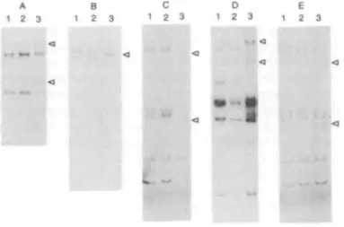
SSCP analysis identified only a single PCR product with aberrant mobility relative to the normal control in each of the FVII variants. In case 1, two extra bands with slower migration were observed for exon 6 (Fig. 1 A). In case 2, a band with slower migration than the corresponding normal control was seen for exon 5 (Fig. IB). Exons 3 and 4 were amplified and analyzed as a single product, and in cases 3 and 5 these products showed
A B C D E 1 2 3 1 2 3 1 2 3 1 2 3 1 2 3
Figure 1. PCR-SSCP analysis in the FVII gene in five unrelated dysfunctional F VII variants. A: c u e 1, B: c u e 2, C: case 3, D: case 4 and E: case 5 snow the results of PCR-SSCP analysis of exon 6, exon 5, exons 3 and 4, exon 8 and exons 3 and 4 mrfmting intron/exon junctions respectively. PCR-SSCP analysis of exons 3 and 4 followed digestion wkfa Dde I digestion, and exon 8 after Nar I digestion. Arrows indicate extra bands wfch altered migration, when compared with normal control bonds. 1 and 2: normal controls, 3: FVH variant
aberrant mobility. To further localize the mutation, the PCR products were digested with the restriction endonuclease Dde I which cuts within intron 3. Subsequent SSCP analysis showed the shifted fragment to be associated with exon 4 in both cases (Fig. 1C, E). Exon 8 was amplified as a single product, but being too large for SSCP analysis was digested with various restriction endonucleases prior to analysis. The primary PCR product was 640bp and the fragment sizes following digestion with the restriction endonucleases were: Nar I, 289 and 351bp; BstX I, 299 and 341bp; and Pst I, 275 and 365bp. In case 4, two extra bands with slower migration than the normal control were observed following digestion with Nar I (Fig. ID). However, no difference in migration was observed following digestion with either BstX I or Pst I (data not shown).
To determine the nucleotide substitution responsible for the altered electrophoretic mobilities detected by SSCP analysis, each of the PCR-amplified DNA fragments was cloned and sequenced (Table 1).
Substitution of A by G at position 8915 was detected in exon 6 of case 1. As a result, the AAA codon for Lys-137 is replaced with the codon GAA for Glu. 9 of the 10 clones sequenced contained the mutant FVQ sequence and 1 the wild type FVH sequence. Based on the SSCP analysis (Fig. 1A) this individual is most probably heterozygous for the mutation. The sequence was confirmed on both sense and antisense strands but no restriction enzyme site is created or destroyed to enable direct confirmation of heterozygosity.
In case 2, an A to G substitution was identified at nucleotide position 7834 in exon 5. The sequence of 5 of the 10 clones had the A to G substitution and the remainder corresponded to the wild type sequence. To confirm this mutation, the amplified DNA fragment including exon 5 and adjacent intron junctions was digested with the restriction endonuclease Sea I. The mutation abolishes a Sea I restriction site by changing the sequence from AGTACT to GGTACT. Three bands were observed corresponding to undigested DNA and the appropriate size fragments diagnostic for the presence of the Sea I site. It was concluded from the sequence and restriction analysis that this patient was heterozygous for the A to G transition at position 7834, which results in the substitution of Gin-100 by Arg.
In case 3, a point mutation of G to A was found in exon 4 at nucleotide position 6055, resulting in the substitution of Arg-79 by Gin. All ten clones sequenced corresponded to the mutant FVII sequence. The hctnozygosity of this mutation was confirmed by loss of an Msp I restriction endonuclease site.
In case 4, transition of G to A was detected in exon 8 at nucleotide position 10715. This results in the substitution of Arg Table 1. Phenotype and genotype daa from five (actor VH variants
Case No. FVTIC* u.dl"1 FVHAg+ u.dl- 1 Exon SSCP analysis* Nucleotide position' Base change AA change Restriction enzyme analysis!
7 20 10 18 47 76 100 52 6 5 4 8 4 Hctcro 7 Homo Homo Homo 8915 7834 6035 10715 6054 AAA to GAA CAGtoCGG COG toCAG CGGtoCAG CGGtoTOG Lys-137-Ohi Gln-100-Arg Arg-79—On Arg-304-Gln Arg-79-Trp N.D. Hetero (Scu I) Homo(MpI) Homo (M*P I, Pv n Hetero (Mqp I, Hoe 'Determined by one stage rifting assay using FVH tWirUn^ pi««m« m j rabbit bi nboplasti
+Assayed using an ELJSA kit (Diagnostica Stago.France) based on a porydonal antibody to human FVH. A normal pooled plasma, assumed to contain lOOu.dl-1 of FVH:C and FVH:Ag wai used u control in both mays.
INumbered according to O'Hara et al (2).
•Hetero - heterozygous mutant/wild type; Homo - homorygous mutation. N.D. - Not determined. ? - Indeterminate. Restriction nn*nnndf*w* used to confirm the imriaipni are given in parentheses.
by
Gin
at p i t i o n 304 in thcatalytic domain ofFW.
All
tenclones displayed the mutam
FM
sequence, strongly suggestingthat this individual is homoygous for the mumion. This was
confirmed
by mtriction endonuclease d i g d o n , the mumion resulting in h ecrtation
of aPvu
I1 site and the l a of an MspI site.
In case 5, a C to T transition was identified in exon 4 at nucltotide 6054, d t i n g in h e substitution of Arg-79 by Trp.
In t i i s case, the individual was heteroygous for the mutation
as determined by sequence analysis and by the haerozygau loas of a Hue
III
restriction r lease site.DISCUSSION
Determination of mutations in dysfunctional
FW
~l~oleculesprovidesimpormtinformahmtoaccompanypro&inmodelling
and structurofunction audks. In this study, we have
undacaLen
gem& analysis of five unrelated subjects who pcwsess
d y s f u m o n a l F W v a r i a n t s . I n d c a s e s , ~ w u e d a e a e d
in exom using PCRSSCP analysis Fable 1). Tbc m m h m werc
then idcntifitd by sequencing cloned DNA and
confirmed
by resaiction endonucltasc d i g d o n in 4 out of 5 c . . Three ofthe
mutarions detected have wt previously btcn rcportal(cases
1, 2 and
9,
om (case 3)bas
been described ia a compouodhomozygote (7) and we have previously d e s c n i
the
mutationin case
4in
an unrelated iodividual (8). TIE three mucationsin
axhm 79and
304 occur in CpG dinwleatidts, a h w n hocepotfor human g c a mutation, and are either C-T or G-A
transitions amsistcnt
with
a model of methylation-mdiated deaminaeion of 5-mdhylcytosim (reviewed by Cooper &K m w d (9))
The rqllacermrt of Lys-137 by Glu (case 1) in
the
connecting
domain of FW is a radical subseitlbion, since the native lysineside chain is
largc
and positively charged at physiologxal pH,wbereas the glutamate group is smaller with a negative charge.
This residue is adjacent to Cys-135 which forms a disulphidc
bridge
with
Cys-262 linking the light and h v y chains ofF W .
It is difkadt to predict
the
e .of such an amino acid s a b d u t hbut the alteration of charge
and
shape and the resultant cadonnational change of this region of the molecule mi@interfere with chain folding and subsequent disulphide bod
formation.
The mutation identified in case 2 results in the substitution of
Arg for G h 1 0 0 in the second
EGF*
domain (EGF2). Girt-100 is highly conserved withinthe
EGF2 Qomain of the vieamin K depenQent scrim protease family. In the f& edition of the haemophilia B database (lo), amongst 22 distinct misscmc1358 Human Molecular Genetics, 1993, Vol. 2, No. 9
Table 2. PCR condition uied to amplify the bmnan FVII gene
B
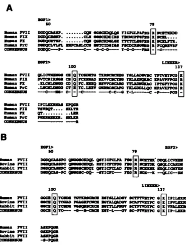
nrci •orla* m i uu>lt m i BoBaa FVII •orina m i •abblt m i onicrcLn ra nxicrcroo no gmricnuD ra -mere— ra •aMsa m i •ovlaa m i •abbit m iFigure 3. A. Comparison of the predicted **"inn acid sequences of lnTimn factors VD (17), DC (18), X (19) and protein C (PrQ (20) beginning at resklue 46:
EOF-UlPB domailM 1 and 2, pirn thr ltnlrfng rryinn m thrr»i+»-ny li*||||iini| nf (far HgM
rlu*BM m shown. B, Comparison of tbc predicted aminoacid sequences of human (17), bovine (21) and rabbit (22) FVII. Srrpiffirf IUIMMIISUUS were displayed wing the programs Lineup and Pretty (Genetics Computer Group, 1991). The •w^iwiH »^ tjQ numbered according to d v mature hi "nun p v n Kquence. The coosenauf wpinrg for each comparison is shown.
corresponding position (Gln-97 to Pro) has been described, but no phenotype data are available.
The mutations identified in cases 3 and 5 result in the substitution of Arg-79 by Gin or Tip respectively in the first EGF domain. Chaing and High have reported a FVII variant, FVII Charlotte, which was a compound homozygote, homozygous for both Arg-79 to Gin and for Arg-152 to Gin substitutions (7). It has been reported that the recombinant Arg-79 to Gin mutant does not bind its cofactor TF (11,12). In contrast, Kazama et al (13) have recently expressed and characterized recombinant FVII with the Arg-79 to Gin mutation and report that it exhibits TF-dependent activity mriiaigiiiriinhlff from plasma derived FVII. Whilst these conflicting results require further investigation, our own data support a role for Arg-79 in factor VII structure or function, since we have iH*ntifii-H two independent mutations at this residue associated with altered factor VII activity. We have modelled the EGF1 domain of FVII using the coordinates of the highly homologous EGF1 domain of FIX previously established by NMR spectroscopy. The side chain of Arg-79 does not form part of the integral structure of EGF 1, but is in full solvent contact on the surface of the domain (Fig. 2A). The replacement of Arg-79 by Gin results in a change of shape and charge,
Exoo Denaturation AnDealing 2 3 a n d 4 5 6 7 8 96°C, 45 i 95.5'C, 30 i 96'C, 30 s 96°C, 60 i 96°C, 45 s 96*C, 45 s 62*C, 15 65°C, 15 58°C, 60 60-C, 60 70°C, 60 72°C, 135 72-C, 120 72'C, 120 72°C, 180 70*C, 128 70*C, 128
The PCR reaction was initially incubated at the denaturation l a n a m i r t for each exon for 3 nrira before undergoing 30 cycles of PCR using a PCH3 Techne cyder (Techne, Cambridge).
futhermore, the energy minimised model structure indicates a change in the orientation of the amino acid side chain (Fig 2B). Similarily, the substitution of Arg-79 by Trp results in a change in charge and the presentation of a bulky aromatic side chain on the surface of the domain (Fig. 2Q. It has been suggested that the EGF domains of FVII are involved in determining its binding to its cofactor TF, however, h is not known whether EGF1, EGF2, or both domains are required to mediate this interaction (14). A monoclonal antibody whose epitope has been mapped to residues 51 - 8 8 of FVII has, however, been shown to inhibit FVH activation and its binding to TF, thus Arg-79 may be part of a binding she important in mediating the FVH/TF interaction (12).
The DNA sequences of other members of the family of vitamin K-dependent serine proteases, factor IX, factor X and protein C, have been determined. A comparison of the predicted amino acid sequence of EGF1, EGF 2 and the linker region between the light and heavy chains (Fig 3A) demonstrates that residues Arg-79, Gln-100 and Lys-137 are highly conserved. The equivalent residues in the 4 proteins are either identical or there is a conservative substitution. Furthermore, a comparison of the human with the rabbit and bovine FVH sequences demonstrates complete evolutionary conservation (Fig. 3B).
In conclusion, we have detected missense mutations in five independent dysfunctional human FVII variants, and we infer that these amino acid substitutions occur in regions of the molecule that are functionally important. Consistent with this view, each mutation results in radical changes of shape and charge in an amino acid side chain which is highly conserved both within the family of vitamin K-dependent serine proteases and between different species. In vitro expression and characterization of the mutant proteins are in progress in our laboratory in order to further define the functional effect of these mutations. MATERIALS AND METHODS
Subjects
Five unrelated individuals from various countries were stndied whose plasmas rontnin dysfunctional FVH as ^rfiiif^ by reAnrrA FVH activity in dotting tests using rabbit thmmhi^^min None of these nibjwti has any significant bleeding tendency sod all were detected *"^i routine i*** ^IMIBI JTI twn& screeosg tor reasoos nri»T thin m«nifr«t t w i n . May FVH activities and antigen levds of these subjects are shown in Table 1. Genomic DNA was prepared from peripheral blood leukocytes by «f*»*»iifh*H NXUHI; (15).
DNA ampmVatloii
Exons 2 to 8 of each subject's FVH genes were amplified by PCR using the otigonudeotides described previously (8). Five hundred nanograms of g^awniir DNA, 0.5/ig of each oligonodeotide, and 1.5U Taq DNA polymerase (Promega) were added to 90pl buffer couaiuiug 1.5 mM MgC^, lOmM Tris-HQ (pH 8.3),
50mM KCL 0.01 * gelatin, 200*M each deoxynudeotide triphosphaie
of cr-^P-dATP (3000 Ci/mmol, 10 mCi/ml). The reaction mixture v m men
SSCP analyst
SSCP analysis w n pa fanned according to the method of Hayasbi tt al (16). 3.5^1 of the PCR product were duuted 10-fbU in 83% fimuuuide, 20mM EDTA, 0.03% bromopbenoi blue md 0.05* xylene cyanol. The samples were then baled at 80°C for 3 ntm to denature die DNA, imp-cooled on ice for 5 mini, before loading onto a 4.3% non-denaturing acryUmide gd (%T, 4.5%;%C, 2.23%). Ekcuopborerii w u pei-fbimed at 40W for 1.3 to 3 noun at 4°C. The gels were dried before mt""Hi"fl"ill'y AmrJifiwi produca from eion 8 were with the reatriction endonudeaset Afar I, Pa I and flaX I according to the manuiactiiFGn hulliMiw »€ prior to SSCP analysB. SmiMyily. PCR prodocts of exon 3 + 4 were drgrilrd with the restriction endonucleaac Ddt I digmwu before SSCP analysis.
DNA dotting and feqnendng
The amplified DNA fragments were gd purified on a 2% agarose gd and doned tt the EcoR V BIB of the p1"**"'1 vector pBSK (Stratagene, Cambridge, UK). The intern were stquenced by the dideoxy chain tnmiintinn method using a T7 DNA sequencing kit (Pharmacia, U.K.). Ten independent clone* were seojuenced for each fragment.
Moleeatai dung
The first epidermal growth factor like domain of FVII was "vHlfd on die hOCQOlOflOUS fifTIIU*** ID F I X n»ww* fltVIHH' COOtuIDfltBS DOT t"f- StTUCtOTC QdnVDu from nuclear magnrrir. resonance spectroscopy (kindly supplied by Prof. I.D.Campbell, Department of Biochenustiy, Oxford). No insertions or <ifif^mi were necessary, tnerefore non-conserved residues could be replaced directly using the Bioporymer mn^itf of Tmigh* n (Biosym Techootogy Inc., San Diego, Calif.) on a SJHcoo Graphics Personal Iris. The resulting structure was subjected to molecular dynamics f^'lw^*1 and energy minimiiwi by die method of steepest descent. Residue snbstiUMkius were dien made in mil modd corresponding to die Arg-79-Trp and Arg-79-Gan nmMations described and the resulting f^ifflirff finally subjected to Authei euei
13. Kunkel, L.M., Smith, K.D. and Boyer, S.H. (1977) Proc.Natl.AcKLSci.USA, 74, 1243-1249.
16. Hayashi, K. (1991) PCR Meth.Appl., 1, 3 4 - 3 8 .
17. Hagen, F.S., Gray, C.L., Ohm, P., Grant, FJ.. Saari, G.C., Woodbury, R.G., Hart, C.E., msley, M., iOnd, W., KnncM, K. and Davie, EW. (1986) Proc.Nad.AcacLSd.USA, 83, 2412-2416.
18. YosUtake, S., Schach, B.G., Foster, D.C., Davie, E.W. and KuncM, K. (1983) Biochemistry, 24, 3736-3730.
19. Leytus, S.P., Poser, D.C., Kunchi, K. and Davie, E.W. (1986) Biochemistry, 23, 3098-3102.
20. Faster, D.C., Yosnkake, S. and Davie, E.W. (1983) Proceedings Of The National Academy Of Sciences Of The United States Of America, 82, 4673-4677.
21. Takeya, H., Kawabaa, S., Nakagawa, K., Yamamkni, Y., Miyata, T., Iwanagm, S., Takao, T. and giimnniihi^ Y. (1988) J.Biol.Chem., 263, 14868-14877.
22. Brothen, A.B., Clarke, BJ., Sheffield, W.P. and Blajcnman, M A (1993) Thrombosis Res., 69, 231-238.
ACKNOWLEDGEMENTS
We *Hiik Pnofcuor Icn D.Campbell, Depntmcnt of Biochemistry, Unhtnity of Oxford, for the tnmlr coonliiutes of the EOF1 «^f"Tn*in of FIX.
REFERENCES
1. Bajaj, S.P., Rapaport, S.I. and Brown, S.F. (1981) J.Biol.Chem., 236, 233-239.
2. OTiara, PJ., Grant, FJ., Haldeman, B.A., Gray, C.L., Insley, M.Y., Hagen, F.S. and Murray, MJ. (1987) Proc.Nad.Acad.Sd.USA, 84, 5158-5162.
3. Ragni, M.V., Lewis, J.H., Spero, J.A. and Hanba, U. (1981) AmJ.Haematol., 10, 79-88.
4. Tripiett, D A , Brandt, J.T., Batard, M A , Dixon, J^. and Fair, D.S. (1985) Blood, 66, 1284-1287.
5. OUara, PJ. and Gram, FJ. (1988) Gene, 66, 147-158.
6. Muchetti, O., Owiwiiiti, D., Paatnocfamit P., Piuoui, M. tnd Bemrdi, F. (1991) Nud.Add.Res., 19, 4370.
7. Sridhara, S, Clarke, BJ., Ofosu, F.A., High, K.A. and Blajcfaman, M.A. (1993) Thrombosis Res., 70, 307-316.
8. O'Brien, D.P., Gale, K.M., Anderson, J.S., Mcvey, J.H., Miller, GJ., Meade. T.W. and Tuddenham, E.O.D. (1991) Blood, 78. 132-140. 9. Cooper DN, Krawczak M (1993) Single base-pair Eibtliuilimt*. In: Human
Gene Mutation. BIOS Scientific Publishers l i m i t s . Oxford: 109-161 10. GJanndli, F., Green, P.M., High, K.A., Sommer, S., Poon, M.C., Ludwig,
M., Schwaab, R., Rehsma, P.H., Goossens, M., YosUoka, A. and Brownke, G.G. (1993) Nud.Add.Res., (submitted)
11. Sridhara, S., Clarke, B., Chaing, S., High, K., Ofosu, F. and Blajcnman, M. (1992) FASEB J.. 6, 2219.
12. Clarke, BJ., Ofosu, F.A., Sridhara, S., Bona, R.D., RicUes, F.R. and Blajchman, M.A. (1992) FEBS Letters, 298, 206-210.
13. Kazama, Y., Foster, D.C. and Kisid, W. (1992) Blood Cotg.Fibrinoi.. 3, 697-702.
14. Toomey, JR., Smith, KJ. and Stafford, D.W. (1991) J.Biol.Chem., 266, 19198-19202.