The Journal of
Laryngology and Otology
(Founded in 1887 by MORELL MACKENZIE and NORRIS W O L F E N D E N )
August 1940
THE PARANASAL APPROACH TO
, INTRASELLAR TUMOURS
SEMON LECTURE, 1939
By PROFESSOR F. R. NAGER (Zurich)
BEFORE entering upon the subject of my lecture I should like to express my best thanks to the Semon Lectureship Board for the honour of their invitation to me. The fact that once again this honour has been bestowed on a foreign Rhinologist instead of on one of the many eminent British confreres is a new proof how faithfully the Selection Committee follows the ideas of Sir Felix Semon, who was a very successful promoter of inter-national scientific interchange. We are convinced that only a common devotion here to medical science can effectively bind people. To further this aim I have tried to pay my contribu-tion by this study. Moreover, I should like to pay my respects to the holder of the Lectureship—to the University of London. There are special relations between this institution and our country, as among one of the founders and first senators there was a Swiss, whose name is not quite forgotten, Peter Marc Roget, the editor of the Thesaurus of English Words and Phrases. I have great pleasure, being at the present time Dean of the medical faculty, in conveying the greetings of the Alma Mater Turicensis.
Motto : " . . . there is only one thing that can effectively bind people and that is a common devotion."—Consecratio Medici, by Harvey Cushing, to whose memory this paper is dedicated.
Professor F. R. Nager
In view of the successful progress of neuro-surgery in the treatment of the pituitary tumours by the transcranial method it would seem presumptuous or even superfluous to deal to-day with the extracranial approach which, according to the opinion of well-known authors is supposed to be superseded.
In this exposure I should like to prove that even nowadays these less dangerous methods are not only justified but are even indicated for special cases.
If general or local symptoms such as intracranial pressure or diminishing vision necessitate an operation in the hypophysal region the essential condition is a large visible exposure of the sella without danger to the neighbouring structures. In consequence of the localization in the depth at the base of the skull a funnel-like approach is necessary. This funnel should be at the same time large and short so that the depth may be easy of survey and without danger to the surrounding neigh-bourhood.
From the historical development of the surgery of this region it may be seen how this postulate was tried.
There are two ways of exposure of the sella : transcranial with formation of osteoplastic flaps of the bony capsule or
extracranial through the nose. To-day the frontal craniotomy
is especially in favour, its technique being very much improved and the danger reduced. According to the viewpoint of eminent neurosurgeons it is supposed to be the method of choice.
The extracranial method started first transnasally by opening the nose in making a flap, resection of the septum and the turbi-nates—excentration nasi—to expose the sphenoid sinus and the sellar region. The modification of Kocher of the superior nasal route was in so far a considerable progress that after splitting the nose in the median line he proceeded to isolate the septum submucously, removed it with the rostrum and reached the sphenoid sinus, after which the sella was easily exposed. The facial scars, which occurred by these methods, were avoided by the method of Cushing, and that of Halsted with his sub-labial incision and isolation of the septum after Kocher's procedure. In the meantime Killian's submucous resection of the septum was becoming generally known and led to the septal method of Hirsch, which differs from that of Kocher and Cushing only by the fact that the incision is purely endonasal. The difficult}' of these methods lies in the length and the
Paranasal Approach to Intrasellar Tumours
narrowness of the funnel by reason of which the survey of the sellar region is unsatisfactory.
FIG. I .
Cushing's method approaching the sellar region by sublabial incision and submucous resection of the septum.
The last modification of the submucous septal routes to the sphenoid sinus is the transpalatine approach of Preysing which we formerly used and will therefore be described.
Under local anaesthesia the mucous membrane of the hard palate is split and opened by a T-shaped incision. The bone is carefully removed so that the mucous membrane of the nose remains quite intact. From the inferior edge of the septum which is now visible, we proceed as in the method of approach just mentioned to the sphenoid sinus and so to the sella. After the removal of the tumour the septal mucous membrane may be pressed together by filling the nose with gauze and the palatal wound is primarily sutured. There is no doubt that this way represents a very short and relatively large and at the same time safe access, without external scar. Moreover, the sella will be exposed from below where is the least danger. On the other hand the possibility of infection from the mouth has prevented this method from being used more frequently. Personally we have not seen any fatal complication in cases of pituitary tumours, but having had more experience we have latterly used only Chiari's method.
All the above mentioned extracranial methods exposed the sellar region intentionally in the median line, in consequence
Professor F. R. Nager
of which—with the exception of the transpalatal way—the funnel of access was rather long and narrow so that the survey of the deeper parts was unsatisfactory. Based on anatomical studies and increased experience in the surgical treatment of the ethmoid and sphenoid affections Chiari published in 1911 the transethmoidal and transsphenoidal approach which we have used in all cases with the exception of three.
The operation is performed under local anaesthesia eventually combined with evipan. The injections of \ per cent. Novocain solution with adrenalin under the soft tissues are also directed alongside the inner wall of the orbit to reach the ethmoid nerve, then following the lateral nasal wall submucously to the sphenoethmoidal region. Moreover the highest end of the septum will be injected on both sides whilst to reach the anterior part of the sphenoid sinus, a piece of gauze saturated with 10 per cent, cocaine is inserted and remains till this region is exposed during the operation. With a curved incision as in the external ethmoid operation the soft tissues are removed following the line of the lamina papyracea and extending deeplyinto the orbit. With special spatulas the contents of the orbit are retracted outwards. All the ethmoid cells are removed in the usual way so that in the depth of this short and larger funnel the anterior wall of the sphenoid sinus is visible. By its resection the posterior wall becomes exposed, especially if also the inter-sphenoidal septum and the rostrum including the end of the nasal septum are removed. Now the whole anterior wall of the sella is clear. Very often pituitary tumours lead to alteration of its colour or shape in the form of bulging, etc. This very thin lamella has to be removed very carefully. The floor and the anterior wall should be removed as much as possible but the upper part avoided because of the danger of haemorrhage from the cavernous sinus. The sensitiveness of the now exposed dura may be ameliorated by applying gauze saturated with 10 per cent, cocaine for a few minutes. After painting with iodine solution the dura is punctured. If on aspiration a clear liquid-cerebrospinal fluid issues it proves that there is no tumour but an internal hydrocephalus is present, and further procedure would be contra-indicated. If, on the other hand, it results in a dry puncture or in a little brownish-reddish liquid a tumour is present and the dura should be opened by a cruci-form incision. Very often the semi-liquid masses of the tumour ooze out of the opening. Now the contents of the sella will be removed as much as possible either by long curettes or by suction until the posterior wall of the sella can be felt by the probes. At this instant the dura collapses and the pulsation of small drops of blood is distinct. For purposes of X-ray-control—a small silver wire may
THE PARANASAL APPROACH TO INTRASELLAR TUMOURS—F. R. XAGER
FIG. 2.
Hirsch's method differs from Cushing's only by intranasal incision—as in Killian's submucous resection of the septum.
FIG. 3.
Preysing's transpalatinal method in different stages. A, B and C.
s = osseous septum visible after resection of palatum durum. m=mucous membrane of nasal floor.
r = Rostrum, postero-superior end of the septum.
FIG. 4.
Chiari's transethmoidal and transsphenoidal approach to the sella.
P= Pituitary gland.
S = Posterior wall of sphenoid sinus. £ = Rest of ethmoid cells.
D S
-Ife
FIG. 5.
jD = I)ura of the pituitary body. -S" — Posterior wall of sphenoid sinus.
FIG. 6.
Spatula to keep aside the contents of the orbit.
FIG. 7.
Glass tube for suction of tumour masses (and blood), which are collected in the bulb and may be examined by expulsion after turning the bulb upwards.
Paranasal Approach to Intrasellar Tumours
be inserted for some days into the cavity. After spraying some iodoform powder and applying a strip of gauze, the end of which protrudes from the nostril, the external wound can be sewn up. Following the operation the patient is given urotropin and calcium to prevent bleeding, although we have not observed any considerable haemorrhage. The course is usually normal and the stitches are removed on the third day and the strip of gauze on the eighth. The scar is almost invisible even if a secondary operation should be necessary. If the local anaesthesia is successful the bleeding is insignificant, and the operation is not more difficult than any external ethmoidectomy. It may be easily performed in about one hour and the physical as well as the mental strain of the patient is not severe, especially if compared with any transcranial procedure. This method of approach which we have performed thirty-eight times in tumours gives always in a simple and safe way a good survey of the sellar region. As there is furthermore no damage to the surrounding parts this route would seem to fulfil the above mentioned postulate. Unfortunately it is not generally known. In Vienna where it has been first described the Hirsch method prevailed and only a few pupils of Chiari as Kahler, Marschik, etc., adhere to it. As time went on even this procedure—according to the opinion of several neuro-surgeons—met the same fate as any extracranial approach.
In regard to this Cushing's attitude was of great importance. In 1912 based on his large experience, he considered the trans-sphenoidal route as very promising, especially if by further close study of the early symptomatic manifestations a more precocious diagnosis should be obtainable. The surgical removal of the floor of the sella might have the same favourable effect as when the tumour breaks spontaneously into the sphenoidal region so that the brain be not exposed to high pressure. At this time this was his method of choice. But in 1935 Cushing concluded that his former procedure should be replaced by the transfrontal approach especially in cases of tumour which did not lead to dilatations of the sella, i.e. with suprasellar evolution and which therefore are not accessible through the nose. A radical operation was therefore only possible by frontal craniotomy. He adds that comparison of these two methods is not possible because his transsphenoidal funnel is long and narrow and does not give a good survey. He admits, however, that the immediate danger of the transnasal operation is less than that following the transfrontal route, whereas the definite visual restoration might be better by this way. Therefore he preferred the access by the frontal craniotomy to any other one.
Professor F. R. Nager
Dandy's opinion on the matter is very definite. The transnasal route according to him is impracticable and can never be otherwise. The actual area of exposure of the sella by any nasal approach is scarcely larger than a lead pencil. Moreover only the inferior part of the tumour can be removed and that without" eye control. Possibly in cystic and very soft tumours a good and lasting effect may be obtained, but these cases are very exceptional. Moreover there is danger of ascending infection from the nose, especially when cerebrospinal liquid issues. The only practical attack according to Dandy must be and is now the transcranial route. Further, he opines that if the tumour has previously been operated from below, a secondary transfrontal operation is no longer possible because of the danger of infection from the rest of the growth which is in connection with the nose. In fact no instances are present to justify a transsphenoidal attack of these tumours.
As previously mentioned Kahler and Marschik remained faithful to Chiari's method, the latter has just published an observation of a patient who was successfully operated two years ago. Based on personal observations I myself have had some papers published showing the expediency and advantages of this procedure. Lately several publications appeared in which Dandy's standpoint is not accepted in its extreme form. The importance of Cairns' and Henderson's papers lies in the fact that their conclusions are based on Cushing's own observa-tions of both methods. In his chapter on the treatment of pituitary tumours Gilbert Horrax in 1938 mentions that almost all neuro-surgeons have relinquished the transsphenoidal approach in favour of the transfrontal one. However, he quotes the opinion of Cairns who points out, that given the advantages of the transcranial route there are several cases (big tumours and severe damage of the optic nerve) where the exposure of the sellar region gives better results and better chances of visual restoration. A similar view is taken by Henderson in his most elaborate publication, The Pituitary
Adenomata, a follow-up study of the surgical results in 338
cases (Dr. Harvey Cushing series). According to Henderson the method of choice to operate on the chromophobe adenomata is the transfrontal exposure of the chiasma. By it a radical removal of the suprasellar part of such tumours is possible. It gives the best prospects of visual restoration. But the transsphenoidal route has its " definite advantages " especially in old people and in those in bad general condition with high blood pressure. It represents also a palliative decompression in cases
Paranasal Approach to Intrasellar Tumours
of a big sellar tumour with extensive intracranial development and consequent alterations of the general state of health. Moreover the big frontal sinuses in acromegaly render the transfrontal way more difficult. In special forms of the reduced visual field, namely, when central scotomata are present, there arises the suspicion of a compression of the chiasma from behind. It happens in cases where the chiasma is prefixed and lies in the sulcus chiasmatis, so that a growing pituitary tumour reaches
Topographic relations between chiasma and pituitary body and its infundibulum—after Henderson.
and first affects the posterior part of the chiasma, which produces this typical reduction of the visual field. As only little or no space is left for the transfrontal exposure without severe danger to the optic nerve in these cases the trans-sphenoidal way is also preferable. After painstaking compari-son Hendercompari-son concludes that the operative mortality in both methods is not essentially different. The danger of meningitis after the exposure of tumours from below was over-estimated. On the other hand there is no danger of blood-clot-formation which may occur after the transfrontal operation. The early results as to visual restoration are generally better with the latter method only in cases of homonymous hemianopia in which the tract is involved or with bilateral scotoma in which the transfrontal approach gives better results. Here lies, according to Henderson, a chief indication for the inferior, i.e. trans-sphenoidal exposure which therefore should not be abandoned. Henderson's conclusions have a definite importance from the fact, that all the cases were operated and controlled by Cushing who was an expert in both methods. In conclusion I would mention also Philipp Gilbert who based on his experience,
Professor F. R. Nager
advocates that the transsphenoidal approach should be given more consideration especially when the tumour originates in the sella and when the bad conditions of the surrounding regions necessitates the operation.*
Personal Observations
As until a short time ago there was no neurosurgical department in Zurich, patients who were diagnosed as suffering from pituitary tumours and with severe eye troubles were sent to us after our first successful treatment of one patient in 1918. To relieve these cases we had no other choice than the trans-sphenoidal method. In all there were 39 patients and 47 operations were performed, 4 of which were by Preysing's transpalatal method, and the rest (43) by Chiari's modification of the transnasal procedure. It was found that five of these patients were not suffering from pituitary tumours but from internal hydrocephalus. Their histories will be given in a short report after the tumour cases.
In grouping our observations we followed the method of Henderson according to the results of the microscopic investiga-tions performed at the Pathological Institute of our University.
A. Chromophobe Adenoma.
These were the most frequent—15 cases (7 men and 8 women) The principal symptoms were increasing visual troubles—the chief indication for operation. Temporal hemianopia was present in 13 eyes, total blindness in 8 eyes, the rest had central scotoma or other defects. With one exception the sella was always distinctly dilated even to atrophy of the clinoid processes. Acromegalic symptoms were distinct in four patients whereby the clinical diagnosis of a chromophobe tumour was not certain. In two cases the operation was per-formed by Preysing's method while in the others the modifica-tion of Chiari was used. At the puncture of the dura there showed in two cases a cystic softening of the tumour, also twice a haemorrhage was slightly present but without real hindrance to the continuation of the operation. Fourteen * The author gladly mentions here a personal observation of W. O. LODGE, F.R.S.C.Edin., Halifax, England. By Chiari's method he removed successfully a chromophobe adenoma of a lady, 28 years of age who some years ago had undergone a first operation only with temporary result. After the second operation by Dr. L., the papilloedema and bitemporal hemianopsia disappeared and normal vision returned on one eye, the general condition being much improved.
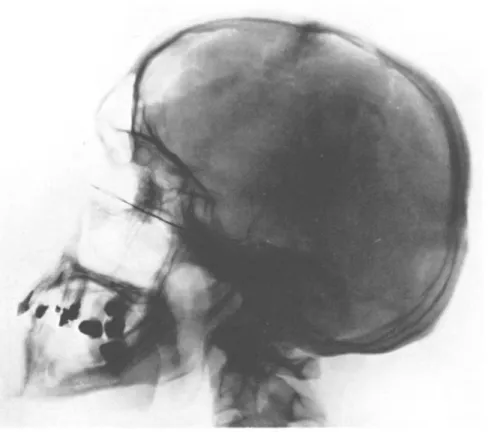
T H E rARANASAI. APPROACH TO I N T R A S E U . A R ' I Y M O T R S — F . R. NAGEK
FIG. 9. II.G. 40 vears. Chromophobe adenoma, ante op.
FIG. IO.
M.G. Q . 40 years. Chroniophobe adenoma, post. op.
7.Z-3V. Ocalo»;
FIG. I I .
Visual field of chromophobe adenoma ; A, before, and B, after operation.
F I G . 12.
L.M. £. 37 years. Acidophil adenoma, post, operation.
Paranasal Approach to Intrasellar Tumours
patients stood the operation well, only one who was admitted with a recurrence of tumour symptoms after having an opera-tion four years previously (in another hospital) and who was in a very bad general condition, died eight days after the operation of pneumonia and extensive alteration of the heart muscle. At autopsy a large adenoma with destruction of the skull and the cerebral peduncle was found, but there were no signs of meningitis. Another case had shown slight signs of meningeal irritation but the lumbar puncture gave only the symptoms of a tumour. On later transfrontal exposure of the chiasma our neurosurgeon, Dr. Krayenbuhl, found a large tumour-rest, extending to the region of the third ventricle which could not be removed. The patient is still alive two years after the last operation and is working to a certain extent, the eye trouble being unchanged. (Figs. 9 to 12.)Of these fourteen patients one died after four years and a big tumour of the size of a fist with hydrocephalus was found at the post mortem. Another patient, 46 years of age, had ten years after the operation a sudden stroke, having worked very hard up to the day before. I was called to the case shortly before death and made an endonasal puncture of the sella region through the scar tissue and was able to draw off about 10 cm. of liquid blood in which pituitary cells could be demonstrated. Presumably the apoplexia had occurred in the region of the rest of the tumour. This is in accordance with the remark of Henderson and Cushing that patients with pituitary tumours had a certain predisposition to premature stroke ; it might be due to endocranial hypertension. Among our patients there are five who had the operation more than five years ago. The late results in the other cases cannot be judged, the time since the operation being too short.
In three cases of this group there was a recurrence, the symptoms being observed at intervals of between six months and four years after the operation. The above-mentioned patient who died of pneumonia eight days after the second operation was one of the recurrences, a second died six months after the second operation in consequence of a ski accident, the third has already been mentioned as having had an inoperable tumour even by the transfrontal route but who is still alive. It should be mentioned that the immediate course of the re-operation was the same as in the first. X-ray treat-ment was given after the first and second operation in almost
Professor F. R. Nager
all cases, as it is found that this combined therapy is thought to be the best.
B. The Acidophil Adenoma.
There were four cases in this group who showed typical acromegalic symptoms which generally started from one to five years before the visual troubles. This is due to the fact that the tumour originates in the sella and affects the eye-nerve only in later stages. Chiari's method was used in three cases and Preysing's in the other. The course of these opera-tions was also uneventful. All the patients had good visual restoration whereas the alterations of the face and extremities did not change much. As to the late results one patient showed signs of recurrence after four years but refused re-operation and died later on. Of the three others one is abroad. We saw him thirteen years after the operation able to work. The other two with 3^ and 15 years respectively after the operation are working in good general condition.
C. Atypical Adenoma.
Among our series this group of thirteen cases is considerable, this may be due to a different interpretation of the microscopic findings, some belong undoubtedly to the adenocarcinomata which in Henderson's work fill one special group. To judge only from the clinical symptoms some of these cases might belong to one of the former mentioned groups. From the microscopic viewpoint the cells of these growths differed and were immature showing an irregular structure which led to the diagnosis of atypical adenoma. In several cases there was a distinct infiltration of the surrounding tissue so that in three of them the diagnosis was carcinoma. As the general clinical symptoms, also the X-ray findings and the visual troubles were different, so before the operation definite diagnosis as to the nature of the tumour was not possible.
Only in one case there was no impairment whatever of the visual function, but he complained of attacks of severe headache and very marked giddiness in blowing his nose. At the same time there were acromegalic symptoms and during four years an always increasing dilatation of the sella. As the X-ray treatment was not successful an operation was necessary. To-day, eighteen years after the operation, the acromegalic symptoms are still present but the patient who holds an important position as engineer is fully able to work.
T H E PARAXASM. AI'PROACH T U M O U R S — F . R. NAGER () IxlkASEI.LATi FIG. D.A. 13-38 years.
Atypical adenoma with large cyst formation, ante operation.
D.A.
Paranasal Approach to Intrasellar Tumours
Of the other patients four had a bi-temporal and five a unilateral hemianopia, the other eye being almost totally blind.
One patient was blind in both eyes and the last had only small central scotoma, the other eye being normal. A con-sequent control of the visual field and function was extremely important because conclusions could be drawn as to the condition of the rest of the tumour. The following case may illustrate this.
D.A., a man of 38 years, was sent to me in 1934 by Prof. Vogt because of his pituitary tumour. His eye trouble of nine years' standing had resulted in total blindness of the left eye and a temporal hemianopia on the right one in spite of having had different X-ray treatment. To judge from the X-ray findings (Figs. 13 and 14) the enlargement of the sella was like a shallow bowl in shape which did not appear favourable for the transsphenoidal exposure. As the patient refused point blank any transfrontal operation we proceeded to the exposureof thesellafollowingChiari's method fromtheleft (blind)side on March 24th, 1934. On puncture of the dura there came a consider-able quantity, first of brownish, then of clear liquid. This fact led to the supposition that the cyst was empty and continuation of the operation seemed not necessary. He made a good recovery and his visual function returned so far that he could write again without difficulty. But six weeks later headaches and sleepiness returned together with reduction of his vision so that on May 2nd Chiari's operation was performed on the right side. On opening the dura considerable masses of a brownish tumour were removed, the microscopic examination of which showed that there was an atypical adenoma. Four weeks after a good healing there was again a decrease of visual function which after the second operation had returned to a certain extent so that an endonasal puncture through the scar field was performed, and about 15 cm. liquid were aspirated. After a temporary collapse his sight returned, but only for six months, so that we were forced to operate again on the left side. On this occasion big masses of very soft tissue were removed After a good recovery on May 8th, 1936, a total blindness of th« right eye occurred, so that a new puncture had to be made. The following days he was very sleepy and during the next months we had to repeat this sort of decompression whereby very considerable quantities of brownish liquid were removed. Since then, five years after the first operation, and three years after the last puncture, the patient is able to work partially. The visual function of the right side is almost normal. He writes and reads normally and is occupied in the construction of wireless sets. A certain tendency to slight
Professor F. R. Nager
sleepiness combined with obesity which may be due to hypofunction of the pituitary gland is treated with different gland extracts.
It can be concluded from the history of this case and the X-ray findings that the extension of the tumour was mostly suprasellar, so that the radical removal of it was not possible by Chiari's method. On the other hand the preliminary operations allowed of a decompressing-puncture being made at any moment as soon as signs of refilling of the cyst were noticed. Cases of this kind necessitate a very close co-operation between the oculist and the operating surgeon. Only thereby was the preservation' of the visual function of his right eye possible, every decrease being immediately notified and correspondingly treated. Our observation corresponds to those of Henderson and dishing of cystic adenoma which independently of the operation had frequently a tendency to re-fill. There is even danger of post-operative haemorrhage into the cyst, which by endonasal aspiration can be more easily removed than by re-opening the bone flap.
In another case, B.R., a woman of 23, we were forced to perform Chiari's operation three times within one year because of the constant recurrence of severe local and general symptoms. She had been before in the surgical department and had been treated with X-rays without definite result. Every operation was followed by temporary improvement but a radical cure was not possible owing to the fact that histologically the tumour proved to be an adeno-carcinoma. Thirteen months later she died of cachexia. It should be mentioned that the patient stood all three operations without complications.
There was no post-operative mortality amongst all these 13 patients on whom 17 operations were performed following Chiari's method. The number is, of course, too small for definite conclusions.
To-day 7 of these 13 patients are still alive. Three died at an interval of one month to a year of adenocarcinoma, the prognosis of which is definitely a very bad one. One patient succumbed seven years after the operation from accidental bronchopneumonia. The last of the series died of unstanch-able haemorrhage of the nose after having worked intensively for three years as a nurse in an asylum. The autopsy showed an erosion of the internal carotid on the roof of thesellaprobablv due to a late necrosis in consequence of the application of
FIG.
F.B. 27 years.
hvdrocephalus-\ Same case, FIG. F.B. (J. hydrocephalus 16. 27 years, dilatation of
Paranasal Approach to Intrasellar Tumours
radium after the operation. This is the first and only case treated locally with radium, the use of which was recommended at this time. An analogous observation caused Cushing-Henderson to warn against any post-operative radium implantation, whereas the X-ray treatment has always been used.
In our series we had no basophilic adenoma nor a cranio-pharyngioma. There was, however, a case of Neuroglioma.
His history will be given.
M.F., a man of 60, suffered for five years from severe headaches, increasing lassitude and visual troubles.. All symptoms were in favour of pituitary tumour, and he was treated intensively three times with X-rays. His diminishing visual decrease necessitated an operation. His general condition was rather poor, the sella proved to be extensively enlarged and eroded. The right eye had a temporal hemianopia with V = o - i . In the left eye the visual field was reduced to a very small sphere in the superior nasal angle. The transsphenoidal operation was normal, the tumour was soft but not so semi-liquid as in the other adenoma. Histologically it was a neuroglioma. After ten months improvement general symptoms especially cachexia occurred which in spite of every glandular treatment ended in the death of the patient.
As no autopsy was performed the case cannot be considered as totally cleared up. Possibly his death was due to a total loss of function of the glandular part of the hypophysis.
Appendix
In this section five cases will be shortly reported which show the limitations of every transsphenoidal procedure. They were sent under the diagnosis of pituitary tumour.
(1) S.M., a woman of 34, suffered for several years from very severe headaches, starting after childbirth. At the same time the face and extremities increased in size as in acromegaly. At X-ray examination it was found that the sella had taken the form of a shallow bowl and at the same time there were signs of increased intracranial pressure. The visual function did not show consider-able reduction but at this time (1921) the more elaborate method of perimetry was not yet known or used. Chiari's operation was performed (1921) and it was found that after removal of the anterior wall of the sella pulsating cerebrospinal fluid issued. The operation was immediately stopped. Nine days later the patient died of meningitis. At post mortem purulent meningitis and
Professor F. R. Nager
ependymitis hypophyseos, hydrocephalus internus and pneumonia were found, the pituitary gland itself not being enlarged.
(2) F.B., a man of 27, complained of severe headaches, lassitude and visual troubles : bitemporal hemianopia and bilateral papillitis. By X-ray (Figs. 15 and 16),dilatation of the sella was found together with signs of increased intracranial pressure. As the patient suffered from suppuration of the ethmoid cells and sphenoid sinus, endonasal operations were performed so t-hat suppuration was cured. But in spite of it the more severe troubles persisted so that an operation was necessary. To avoid the nasal cavity as much as possible the transpalatal exposure was performed. On the puncture of the dura clear cerebrospinal fluid was found which proved the presence of an internal hydrocephalus and the operation stopped. Two days later the patient died suddenly. At post mortem were found : purulent leptomeningitis and ependymitis hypophyseos with internal hydrocephalus. The infundibulum of the hypophysis was enlarged.
(3) S.P., a woman of 64, suffered'since 1903 from almost unbear-able headaches with increasing visual troubles. As dilatation of the sella was found, a pituitary tumour was diagnosed and X-ray treatment applied. As this failed, an operation was indicated. The presence of a clear liquid on puncturing the dura after Chiari's method proved the certainty of internal hydrocephalus. The only anomalous feature of the post-operative course was a slight oozing of liquid, but headaches and visual troubles diminished. A year later these symptoms recurred and the visual function was in severe danger. Endonasal puncture was tried through the scar-tissue which proved to be bony. A local and general improvement followed. The latest report of her (five years after the operation) is that she is able to read large print and her general condition is satisfactory.
(4) G.A., a man of 58, was referred to us because of general cerebral symptoms, double vision and weakness of sight. At the eye examination, the right eye was found to be blind and the left eye hemianopic. By X-ray extended destruction both of the floor and the back of the sella was established. The puncture of the dura (Chiari's method being followed) showed a clear liquid. The healing was normal, but on examining him five years later the general and local troubles had not changed very much.
(5) L.W., a woman of 43, suffered since 1929 from headaches and severe visual troubles leading to bi-temporal hemianopia. The X-ray examination showed a dilated sella and symptoms of increased intracranial pressure. After the simple puncture of the dura (6.10.37) cle a r liquid appeared. Four days later she died. At autopsy were found meningitis, multiple tumours—meningioma—of the dura and hydrocephalus internus.
Paranasal Approach to Intrasellar Tumours
We felt compelled to report even these cases so as to show how difficult the differential diagnosis may be in certain cases, and how very necessary is further research to avoid an uncertain diagnosis. If our series of operations have shown that in
tumours the transsphenoidal operation is comparatively safe, the
same operation in hydrocephalus proved very dangerous, the post-operative mortality being three-fifths. The cause of hydro-cephalus could only be found in one case in the form of multiple meningioma of the dura, one of which was situated in the foramen magnum compressing the fourth ventricle. By closer study of the history of these five cases we have learnt to interpret several symptoms to be more typical of internal hydrocephalus than of pituitary tumour. Among these symptoms we should mention especially increased intra-cranial pressure, shown bythe X-ray examination. Although it must be admitted that in the later stages of big tumours these symptoms may also be observed. In this connection it is to be hoped that the more extended research work of the pathology of the pituitary gland together with further improvement in deter-mining the alteration in the visual field will contribute to a better differentiation of the two diseases.
We have only used ventriculography till now in two cases of tumour without finding a dilated extension of the 3rd ventricle into the infundibulum, so that a tumour was probable whereas an internal hydrocephalus could be excluded. But it is known that in addition or in consequence of a tumour or even hydro-cephalic dilatation of the ventricle may occur. The positive finding in ventriculography therefore does not always exclude a tumour. Another difficulty lies in the fact that according to modern neurosurgery the operation—if indicated by ventri-culography—should immediately follow this examination, which is only possible if X-ray and surgical departments are in the same building or adjacent. In the last three of these cases I asked my colleagues who sent the patients for operation if they could definitely exclude internalhydrocephalus, which they could not do, which shows the necessity for further research.
Conclusions
Our own practical experience in the operation of thirty-nine pituitary tumours combined with others already quoted lead to a definite viewpoint as to the value of the trans-sphenoidal approach to pituitary tumours.
Professor F. R. Nager
As mentioned, there were in the beginning accidental factors which led us to perform the operation. With increasing experience I was convinced that the transnasal approach had definite advantages, especially as regards post-operative complication, but the categorical rejection of these methods by eminent neurosurgeons would naturally cause hesitation in taking up these methods. For that reason the publications of Cairns and especially of Henderson are so precious because of their conciliatory attitude towards the principle of transnasal approach. Therefore to-day the question is no longer : is there only one method for operative treatment of these tumours, viz. the transfrontal one, but, are there certain pituitary tumours which would be better approached by the trans-sphenoidal method ? It was never pretended that by the transnasal route all hypophyseal tumours could be removed, especially the suprasellar ones ; these can only be removed by transfrontal craniotomy. But if the tumour is mainly or entirely localized in the dilated sella or its tendency is to grow downward, the transsphenoidal route has undoubtedly definite
advantages. By using Chiari's modification of the extracranial
exposure of the sella we get a short rather large funnel-like access to the sellar region with a good survey and without danger to the surrounding parts.
As has been proved by our experience the operative mortality in tumours is very small and also the danger of meningitis from the nose has been over-estimated ; in fact in tumours it may be considered slight. Our cases were not picked out, the immediate danger of increasing blindness made operation absolutely necessary. Even in such cases where suprasellar development was more certain we were forced to relieve the patient. This fact will .naturally affect the results. We lost within one year five patients, but in three of them the adenoma was of a decidedly malignant character. Within from two to five years three patients died, one of a big intercerebral eosinophilic tumour, one of neuroglioma, and the third of haemorrhage in consequence of late radionecrosis. Within five to ten years post-operative one patient succumbed through a stroke (ten years) and one from bronchial pneumonia, both without autopsy. As far as we know nineteen patients are still alive.
The late results cannot be judged yet, the time being too short since the operation. However, seven patients were
Paranasal Approach to Intrasellar Tumours
operated five to ten years ago and two between eleven to twenty years.
If the eye affections were not too far advanced before the operation, recovery has been observed almost in every case. Without entering into details, generally speaking the good effect of the operation on visual troubles could be seen ; often the restoration came very early, even quite suddenly in one case, as during the puncture of the cystic tumour the patient could see objects in the room which before had been invisible to him. The value of easy decompression of the sella by the transsphenoidal approach therefore cannot be denied.
In our series four cases of recurrence were observed and six re-operations by Chiari's method were necessary. The symptoms occurred from one to four years after the first operation. Of these the most interesting was the case of cystic adenoma, which was arrested only after three operations and different punctures. From Henderson's report recurrences were observed independently of the operation. In his series there were forty-five operations for recurrences, the first operation had been the transsphenoidal in twenty-two cases, and in eleven the transfrontal approach. The fact of recurrence depends therefore on the development and the nature of the tumour.
Our experience proved that if the necessity of re-operation is present, it can be easily performed by following the same method. So far as we saw the healing process was normal and there was no danger of meningitis. Furthermore, a preliminary exposure by the transnasal route does not interfere with a secondary transfrontal approach, because, as we have seen, the rest of the tumour does not become infected, and the opening heals by fibrous or even bony tissue. Through this scar-tissue a decompressing puncture can be performed if necessary.
The possible danger of Chiari's modification is increased if, instead of a tumour, or together with it, an internal hydro-cephalus is present. Here the simple puncture may lead to purulent infection and meningitis.
Dandy's other objections to the transnasal route can be easily allayed. We admit that the approach to the hypophysis by Cushing's and Hirsch's methods is only possible by means of a long and narrow funnel, which does not give a satisfactory survey. In regard to this Chiari's modification represents a
Professor F. R. Nager
very remarkable progress. By starting on the level of the
ethmoid the access will be shorter and, by moving the orbital
contents to the side, also much larger, so that a better survey of the deep regions will be obtained. By the use of a good head-light there will be no difficulty at all in making a large opening of the sella and of the dura, so that the removal of the tumour is possible under the operator's constant eye control. Only the tumours which are mainly or exclusively located above the sella cannot be removed from below by the transsphenoidal approach and here Chiari's method will not and can not be successful. This route—by the way—was not intended for these cases, but we know that even by the transfrontal route the radical removal is in some cases not possible. On the other hand even the reduction of a certain part of the tumour may be very successful by the decompression of the chiasma.
It seems, therefore, that by using Chiari's modification Cushing's recommendation of the transsphenoidal methods in 1912 is even to-day of value.
Based on these conclusions it is now possible to determine not only the advantages, the indications, but also the limits of the transsphenoidal approach to the sella.
Chiari's paranasal approach to the sella corresponds best to the postulate of a small and large funnel with a good survey and no danger for the surrounding regions. Without great strain on the patient a decompression of the chiasma can easily be obtained if danger of loss of sight be present. By the removal of the floor and anterior wall of the sella a pituitary tumour can more easily grow downwards so that the risk of the affection of the chiasma will be greatly diminished. Chiari's method offers the possibility of a biopsy by which the prognostication of the. tumour as well as the post-operative X-ray treatment will be more certain. Intrasellar cystic tumour can be easily emptied without risk and if necessary re-emptied either by puncture or by secondary operation.
A primary transsphenoidal approach does not render a secondary transfrontal one impossible. On the contrary we believe that this may be the method of choice if the tumour is at the same time located intra- and supra-sellar.
The chief indication for the transsphenoidal approach may, therefore, be fixed in about the following way : Pituitary tumours with mainly intrasellar development, with the tendency to extend downwards which is shown by following up
Paranasal Approach to Intrasellar Tumours
the X-ray findings and with incipient eye troubles should be approached from below. Moreover, this method is preferable in tumours which lead to central scotoma. Here the chiasma is supposed to be affected from behind so that the transfrontal access to the tumour is much more difficult.
The transsphenoidal approach has the advantage that it does not impair a secondary operation whether performed by the same or by the transfrontal route. On the contrary it seems that in cases of tumours with intra- and suprasellar extension the less dangerous procedure of Chiari might be used, whereas in recurrences the transfrontal would be preferable. Where an immediate decompression of the chiasma is urgent the transsphenoidal exposure by Chiari's modification of the extracranial methods seems to be the best and least dangerous.
On the other hand this method should not be employed in cases of pure suprasellar tumours without dilatation of the sella. Here lies the chief indication for the frontal craniotomy with the best chances of radical removal. Moreover, every transsphenoidal approach to the sella is strictly contra-indicated where internal hydrocephalus is present or supposed. But as has been previously shown the differential diagnosis may be difficult in some cases.
It is hoped that in future more intense research and loyal co-operation of all workers in this field will contribute not only to better and less faulty diagnosis but to the decision of the method to be used with the best chance of recovery and the least danger to the patient.
Summary
In this paper the question is discussed as to whether, according to the opinion of some well-known neurosurgeons the total rejection of extracranial approach to pituitary tumours is justified to-day.
A short survey of the development of surgery of the pituitary tumours is given and special attention is called to Henderson's quite recent study of Cushing's large series of these tumours.
Chiari's method of approach to the sellar region by trans-ethmoidal and transphenoidal route is described. A survey of 39 transsphenoidal exposures performed on 34 patients with pituitary tumours is given. With one exception—death after
Professor F. R. Nager
eight days of pneumonia with heart failure—there was no direct post-operative mortality. This corresponds to Henderson's and Kahler's observations.
Chiari's method may, therefore, be declared to be without real danger and big strain for the patient. The early and late results of these cases are given. According to Henderson's, Kahler's and the author's experience the transsphenoidal approach to pituitary tumour is not only not superfluous or even obsolete ; but in certain cases this route has its definite advantages. The chief indications for this paranasal route are big intrasellar tumours and special forms of visual troubles, viz., central scotoma and homonymous hemianopia, whereas the tumours with suprasellar extension will be better reached by the transfrontal method. Moreover the transphenoidal exposure of the sella is contra-indicated in internal hydro-cephalus. Further research is necessary for the differential diagnosis of pituitary tumour versus hydrocephalus internus.
LITERATURE
DANDY, The Brain in Practice of Surgery. Dean Lewis, 1936, 579. CUSHING, H., Consecratio Medici.
The Pituitary Body and its Disorders.
Amplification of the Harvey Lecture. 1912, Philadelphia.
" Intracranielle Tumoren." Deutsche Ausgabe, 1936, Berlin. HORRAX, GILBERT, " Symposion Pituitary Gland." Proceed, of the
Assoc.f. Research in nerve and mental diseases, 1938, xvii. Williams
Wilkins Co., Baltimore.
CAIRNS, " The ultimate results of Operations for intracranial Tumours."
Yale Journ. Biol. and Med., 1936, viii, 421, cit. n. G. Horrax.
HENDERSON, " The Pituitary Adenomata." Brit. Journ. of Surgery, 1939, xxvi, 104.
PHILLIPS, GILBERT, " Transsphenoidal Decompression for Pituitary Adenoma." Brit. Journ. of Surgery, 1938, xxvi, 102.
MARSCHIK, H., " Ueber einen geheilten Fall von epithelialem Vorder-lappentumor." Mschr. f. Ohkde, 1939, lxxiii, 170.
NAGER, F. R. and Prof. HENSCHEN, " Die paranasale (transethmoidale) Operation des Hypophysistumors nebst Bemerkungen zur Chirurgie d.Schadelbasis." Corr. Bl. f. Schweiz Aerzte, 1919, 35.
NAGER, F. R., " Die extrakraniellen Operationen der Hypophysen-affektionen u. ihre Operationserfolge, nach eigenen Erfahrungen."
Schweiz. Archiv. f. Neurologie u. Psychiatrie, 1929, xxiv, 1.
NAGER, F . R., " L'operation des tumeurs intrasellaires par la voie trans-ethmoidale de Chiari." Extrait des comptes—rendus du
Paranasal Approach to Intrasellar Tumours
DE MONTMOLLIN, CLAUDE, " Les indications de l'operation de Chiaridans les tumeurs de l'hypophyse." Revue de Laryngologie, Otologie,
Rhinologie, avril 1933, 4.
KAHLER, " Zur Operation der Hypophysentumoren." Zschr. f.
OHkde., 1917, Ixxv, 287.
KAHLER, " Die Krankheiten der Hypophyse in Denker-Kahler."
Hdb. d. Hals, Nasen-u. Ohren-Krankheiten, 1929, v, 645.