BRIEF COMMUNICATIONS
for lymph node – negative invasive breast
cancer in premenopausal (Trial VIII) and
postmenopausal (Trial IX) women. Trial
VIII (
14 ) evaluated whether sequential
treatment with six 28-day courses of
com-bination chemotherapy with
cyclophos-phamide, methotrexate, and 5-fl uorouracil
(CMF) followed by 18 monthly
subcuta-neous implants of goserelin (CMF
→
goserelin) improved disease-free survival
compared with either six 28-day courses of
CMF alone or 24 monthly implants of
goserelin alone. Trial IX (
15 ) evaluated
whether sequential treatment with three
28-day courses of CMF followed by
tamox-ifen for 57 months (CMF
→ tamoxifen)
improved disease-free survival compared
with tamoxifen alone for 60 months. During
The percentage of proliferating cells in a
tumor (ie, the tumor proliferation fraction)
is an established predictor of breast cancer
prognosis (1,2). The proliferation antigen
Ki-67 is detectable in cells at all phases of
the cell cycle except G
0( 3 ), and the Ki-67
labeling index (the percentage of cells with
Ki-67 – positive nuclear immunostaining) is
a measure of tumor proliferation ( 4 , 5 ) that
has been associated with breast cancer
out-come in several studies (6 – 10). Other
stud-ies ( 11 , 12 ) have suggested that a high Ki-67
labeling index is predictive of
responsive-ness to neoadjuvant (primary)
chemother-apy, but, to our knowledge, there are no
such reports concerning Ki-67 labeling
index as a predictor of responsiveness to
adjuvant chemotherapy.
In 2005, the ninth St Gallen consensus
conference on primary therapy for early
breast cancer ( 13 ) emphasized the
impor-tance of the endocrine responsiveness of
the tumor in selecting adjuvant therapy for
early breast cancer and acknowledged the
existence of a group of patients whose
responsiveness to endocrine therapy is
uncertain even though their tumors express
hormone receptors. Because these patients
may benefi t from chemoendocrine therapy,
we examined whether the Ki-67 labeling
index could identify patients who might
particularly benefi t from the addition of
chemotherapy to endocrine therapy in the
adjuvant setting in two International Breast
Cancer Study Group (IBCSG) trials.
IBCSG Trials VIII ( 14 ) and IX ( 15 )
were randomized clinical trials that were
conducted between 1988 and 1999; the
median follow-up for each trial is 10
years. The trials compared adjuvant
endo-crine therapy alone with sequential
che-motherapy followed by endocrine therapy
Predictive Value of Tumor Ki-67
Expression in Two Randomized Trials of
Adjuvant Chemoendocrine Therapy for
Node-Negative Breast Cancer
Giuseppe Viale , Meredith M . Regan , Mauro G . Mastropasqua , Fausto Maffini ,
Eugenio Maiorano , Marco Colleoni , Karen N . Price , Rastko Golouh ,
Tiziana Perin , R. W . Brown , Anikó Kovács , Komala Pillay , Christian Öhlschlegel ,
Barry A . Gusterson , Monica Castiglione-Gertsch , Richard D . Gelber ,
Aron Goldhirsch , Alan S . Coates
On the behalf of the International Breast Cancer Study Group
Several small studies have reported that having a high percentage of breast tumor cells that express the proliferation antigen Ki-67 (ie, a high Ki-67 labeling index) predicts better response to neoadjuvant chemotherapy. However, the predictive value of a high Ki-67 labeling index for response to adjuvant chemotherapy is unclear. To investigate whether Ki-67 labeling index predicts response to adjuvant chemoendocrine therapy, we assessed Ki-67 expression in tumor tissue from 1924 (70%) of 2732 patients who were enrolled in two randomized International Breast Cancer Study Group trials of adjuvant chemoendocrine therapy vs endocrine ther-apy alone for node-negative breast cancer. A high Ki-67 labeling index was associ-ated with other factors that predict poor prognosis. Among the 1521 patients with endocrine-responsive tumors, a high Ki-67 labeling index was associated with worse disease-free survival but the Ki-67 labeling index did not predict the relative efficacy of chemoendocrine therapy compared with endocrine therapy alone. Thus, Ki-67 labeling index was an independent prognostic factor but was not predictive of better response to adjuvant chemotherapy in these studies.
J Natl Cancer Inst 2008;100: 207 – 212
Affiliations of the authors: Divisions of Pathology
and Laboratory Medicine (GV, MGM, FM) and Medical Oncology (MC, AG), European Institute of Oncology, University of Milan, Milan, Italy; International Breast Cancer Study Group (IBCSG) Statistical Center, Dana-Farber Cancer Institute, Frontier Science and Technology Research Foundation, Harvard School of Public Health, Boston, MA (MMR, KNP, RDG); Department of Pathological Anatomy, University of Bari, Bari, Italy (EM); The Institute of Oncology, Ljubljana, Slovenia (RG); Division of Pathology, Centro di Riferimento Oncologico, Aviano, Italy (TP); Melbourne Pathology, Collingwood, Victoria, Australia (RWB); Department of Pathology, Göteborg/Sahlgrenska University Hospital, Göteborg, Sweden (AK); Division of Anatomical Pathology, Department of Clinical Laboratory Sciences, University of Cape Town, National Health Laboratory Services and Groote Schuur Hospital, Cape Town, South Africa (KP); Kantonspital, St Gallen, Swiss Group for Clinical Cancer Research (SAKK), Bern, Switzerland (CO); Division of Cancer Sciences and Molecular Pathology, Western Infirmary, University of Glasgow, UK (BAG); IBCSG Coordinating Center, Bern, Switzerland (MCG); Oncology Institute of Southern Switzerland, Bellinzona, Switzerland (AG); IBCSG, Bern, Switzerland (ASC); University of Sydney, Australia (ASC) .
Correspondence to: Giuseppe Viale, MD, FRCPath,
Division of Pathology and Laboratory Medicine, European Institute of Oncology, University of Milan, Via Ripamoniti 435, Milan, Italy (e-mail: giuseppe.viale@ieo.it ).
Reprint requests to: International Breast Cancer
Study Group Coordinating Center, Effingerstrasse 40, CH-3008 Bern, Switzerland (e-mail: giuseppe. viale@ieo.it ).
See “Funding” and “Notes” following “References.” DOI: 10.1093/jnci/djm289
© The Author 2008. Published by Oxford University Press. All rights reserved. For Permissions, please e-mail: journals.permissions@oxfordjournals.org.
the conduct of these trials, the estrogen
receptor (ER) and the progesterone
recep-tor (PgR) status of the tumor and tumor
size and grade were locally assessed and
noted on trial case report forms. Tumors
were graded 1 – 3 either according to Bloom
and Richardson ( 16 ) or according to overall
differentiation as well differentiated (grade
1), moderately differentiated (grade 2), or
poorly differentiated (grade 3).
In 2003, the IBCSG completed the
retrospective collection of formalin-fi xed,
paraffi n-embedded primary breast tumor
tissue samples that were obtained from
Trials VIII and IX participants. The
collec-tion program was conducted in accordance
with institutional guidelines and national
laws. The samples were subjected to
immu-nohistochemical assessment of ER, PgR,
and HER2 expression and Ki-67 labeling
index at the IBCSG Central Laboratory
in Milan, Italy, by personnel who were
blinded to participant treatment
assign-ment and outcomes, as previously described
( 17 ). Whole tumor sections were incubated
with the specifi c primary mouse
monoclo-nal antibodies to ER (clone 1D5, 1 : 100
dilution) or PgR (clone 1A6, 1 : 800
dilu-tion) (both from Dako, Glostrup, Denmark).
HER2 immunoreactivity was assessed using
a HercepTest kit (Dako) as recommended
by the manufacturer and scored for the
intensity of immunostaining, the
complete-ness of cell membrane staining, and the
percentage of immunoreactive neoplastic
cells by using a four-tier scale from 0 to 3+,
as previously described ( 18 ). The centrally
assessed values of ER, PgR, and HER2
status were used in this report.
Reassessment of the trial conclusion
based on the centrally assessed hormone
receptor values confi rmed the fi nding that
the benefi t of chemotherapy was limited
to patients whose breast tumors expressed
little or no ER or PgR ( 17 , 19 ).
Tumor material was available and
assessable for Ki-67 labeling index for 758
(71%) of 1063 Trial VIII patients and 1166
(70%) of 1669 Trial IX patients. Ki-67
labeling index was assessed using mouse
monoclonal antibody MIB-1 (1 : 200
dilu-tion; Dako); the percentage of cells that
showed defi nite nuclear immunoreactivity
with MIB-1 among 2000 invasive
neoplas-tic cells in randomly selected high-power
(×400) fi elds at the periphery of the tumor
was recorded.
Centrally reviewed ER and PgR status
were classifi ed as present ( ≥ 1%
immunoreac-tive cells) or absent (<1% immunoreacimmunoreac-tive
cells). Samples were considered to be positive
for HER2 overexpression if the staining
intensity score was 3+ and negative for HER2
overexpression if the staining intensity score
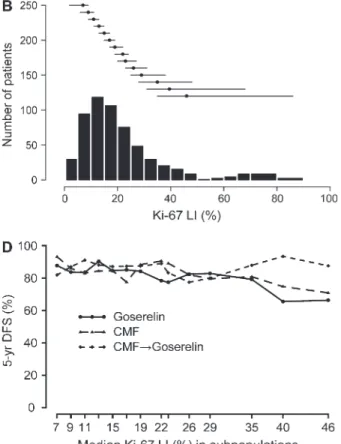
was 0, 1+, or 2+ ( 20 ). The Ki-67 labeling
index was dichotomized to high (
≥ 19%
immunoreactive cells) and low (<19%
immu-noreactive cells) groups by using the median
value of Ki-67 immunoreactivity as the cut
point, which was based on the frequency
dis-tributions of the Ki-67 labeling index in the
two trial cohorts ( Fig. 1, A and B ).
Univariate and multivariable logistic
regression modeling was used to assess the
association of other tumor features with
high vs low Ki-67 labeling index. Analyses
were undertaken separately for the two
trials. These analyses revealed that in both
trials, higher tumor grade, larger tumor
size, and the absence of tumor expression
of ER and of PgR were associated with a
high Ki-67 labeling index (
P < .001 for
each) (Supplementary Table 1, available
online). HER2 overexpression was
associ-ated with a high Ki-67 labeling index in
postmenopausal patients (ie, Trial IX;
P < .001) but not in premenopausal patients
(ie, Trial VIII;
P = .61) (Supplementary
Table 1, available online).
We next examined the association of
high and low Ki-67 labeling indices with
disease-free survival among patients with
endocrine-responsive breast cancer (ie, 923
patients with ER-present tumors on Trial
IX and 598 patients with ER- and/or
PgR-present tumors on Trial VIII). Cox
pro-portional hazards modeling was used to
examine interactions of Ki-67 labeling
index and other tumor characteristics with
disease-free survival. To check assumptions
of proportionality, curves of the log of the
cumulative hazard function for each value
of a covariate adjusted for other covariates
in the model were plotted and assessed
visually to determine if the vertical shift
between the curves was constant over time.
The data appeared to meet the assumptions
of proportionality. All P values are
two-sided, and statistical signifi cance was
defi ned as P less than or equal to .05.
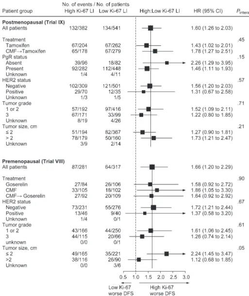
Among postmenopausal patients who
were treated in Trial IX, a high Ki-67
labeling index was associated with worse
disease-free survival (hazard ratio [HR] of
recurrence or death = 1.60, 95% confi
-dence interval [CI] = 1.26 to 2.03, P < .001).
A multivariable analysis adjusting for other
tumor features confi rmed that high Ki-67
labeling index was an independent
prog-nostic feature ( P ≤ .05) (data not shown).
We examined the pairwise interactions of
the other tumor features and Ki-67
label-ing index with disease-free survival to
investigate whether the association between
Ki-67 labeling index and disease-free
sur-vival varied as a function of specifi c tumor
characteristics. There was no evidence of
an interaction for any factor: a high Ki-67
labeling index was consistently associated
with worse disease-free survival ( Fig. 2 ). As
was previously reported for Trial IX as a
whole ( 15 ), treatment arm was not
associ-ated with disease-free survival in this
endo-crine-responsive subgroup of patients.
There was no interaction of Ki-67 labeling
index and treatment arm with disease-free
survival ( P
interaction= .45; Fig. 2 ), indicating
that patients whose tumors had a high
Ki-67 labeling index had worse disease-free
C O N T E X T A N D C A V E A T S
Prior knowledge
Some studies have suggested that having a high percentage of breast tumor cells that label with an antibody against the prolifera-tion antigen Ki-67 predicts a better response to primary (ie, neoadjuvant) chemotherapy. Study design
A retrospective assessment of the predic-tive value of a high Ki-67 labeling index for response to therapy among women enrolled in two randomized trials of adjuvant che-moendocrine therapy vs endocrine therapy alone for node-negative breast cancer. Contribution
A high Ki-67 labeling index did not predict which women would benefit from further treatment with chemotherapy added to endocrine therapy.
Limitations
Only women with node-negative breast cancer were included in this study.
Implications
Other biomarkers are needed to define which women with endocrine-responsive node-negative early breast cancer could benefit from the addition of adjuvant che-motherapy to endocrine therapy.
survival than patients whose tumors had a
low Ki-67 labeling index regardless of the
treatment they received. The relative
treat-ment effect of CMF → tamoxifen vs
tamoxi-fen alone among patients whose tumors
had a high Ki-67 labeling index (HR =
1.03, 95% CI = 0.73 to 1.45) was consistent
with that among patients whose tumors
had a low Ki-67 labeling index (HR = 0.86,
95% CI = 0.61 to 1.20). This homogeneity
in the treatment effect was confi rmed in a
multivariable analysis that adjusted for
tumor grade, tumor size, and PgR and
HER2 status: in patients with
ER-express-ing tumors, a high Ki-67 labelER-express-ing index did
not predict resistance to tamoxifen alone;
nor did it predict benefi t from sequential
CMF → tamoxifen ( P
interaction= .41).
Among premenopausal patients who
were treated in Trial VIII, high Ki-67
labeling index was associated with worse
disease-free survival (HR of recurrence
or death = 1.66, 95% CI = 1.20 to 2.29;
P = .002). A multivariable analysis that
adjusted for other tumor features
con-fi rmed that a high Ki-67 labeling index was
an independent prognostic marker ( P < .05)
(data not shown). We examined the
pair-wise interactions of other tumor features
and Ki-67 labeling index with disease-free
survival. There was no evidence of an
inter-action for any factor: a high Ki-67 labeling
index was consistently associated with
worse disease-free survival ( Fig. 2 ). As was
previously reported for Trial VIII as a
whole ( 14 ), treatment arm was not
associ-ated with outcome in this
endocrine-responsive subgroup of patients. There was
no interaction of Ki-67 labeling index and
treatment arm with disease-free survival
( P
interaction= .90;
Fig 2
) , indicating that
patients whose tumors had a high Ki-67
labeling index had worse disease-free
sur-vival than patients whose tumors had low
Ki-67 labeling index regardless of
treat-ment received. The relative treattreat-ment
effect of each pairwise comparison of the
three treatment arms among patients whose
tumors had a high Ki-67 labeling index was
consistent with that among patients with
low tumor Ki-67 labeling index (CMF →
goserelin vs goserelin alone in patients with
low [HR = 0.78, 95% CI = 0.44 to 1.40] and
high [HR = 0.81, 95% CI = 0.48 to 1.38]
Ki-67 labeling index; CMF → goserelin vs
CMF in patients with low [HR = 1.05, 95%
CI = 0.56 to 1.99] and high [HR = 0.92,
95% CI = 0.55 to 1.52] Ki-67 labeling index;
CMF vs goserelin in patients with low
[HR = 0.74, 95% CI = 0.41 to 1.35] and high
Fig. 1 . Distribution and subpopulation treatment effect pattern plots(STEPP) analysis of breast cancer tumor Ki-67 labeling index. Frequency distribution of Ki-67 labeling index in postmenopausal patients (Trial IX) with estrogen receptor (ER) – expressing tumors ( A ) and in premenopausal patients (Trial VIII) with ER- and/or progester-one receptor (PgR) – expressing tumors ( B ). The black circles indicate the median Ki-67 labeling index, and the horizontal lines indicate the range for subpopulations of patients that were used for the STEPP analysis. C and D ) STEPP analysis of 5-year disease-free survival by treatment arm according to Ki-67 labeling index in postmenopausal patients (Trial IX) with ER-expressing tumors ( C ) and in premeno-pausal patients (Trial VIII) with ER- and/or PgR-expressing tumors ( D ).
Overlapping subpopulations of patients were defi ned on the basis of Ki-67 labeling index, and the resulting patterns of the treatment effects estimated within each subpopulation are displayed. The sub-populations have a fi xed number of patients (approximately 120 for Trial IX and approximately 100 for Trial VIII); each subsequent sub-population changed by 20 patients. The x-axis indicates the median Ki-67 labeling index value for patients in each subpopulation; the y-axis indicates the treatment effects, expressed as the 5-year disease-free survival percentage estimated by using the Kaplan-Meier method. DFS = disease-free survival; LI = labeling index; CMF = com-bination chemotherapy with cyclophosphamide, methotrexate, and 5-fl uorouracil.
[HR = 0.89, 95% CI = 0.53 to 1.48] Ki-67
labeling index). This homogeneity in the
treatment effects was confi rmed in a
multi-variable analysis that adjusted for tumor
grade, tumor size, and PgR and HER2
status: in patients with ER-expressing
tumors, a high Ki-67 labeling index did not
predict resistance or benefi t to any of the
treatments ( P
interaction= .69).
We further examined the pattern of
treatment effects across the continuum of
Ki-67 labeling indices using the
nonpara-metric subpopulation treatment effect
pat-tern plot (STEPP) method (
21 ), which
avoids the need to select a cut point in the
distribution of a continuous feature such as
Ki-67 labeling index. The STEPP method
uses a sliding-window approach to defi ne
several overlapping subpopulations of
patients according to Ki-67 labeling index
and plots
the resulting treatment effects
estimated within each subpopulation. The
subpopulations have a fi xed number of
patients (approximately 120 for Trial IX
and approximately 100 for Trial VIII); each
subsequent subpopulation changed by 20
patients. The plot
’ s x-axis indicates the
median Ki-67 labeling index for patients in
each subpopulation; the y-axis indicates
the treatment effects, expressed as the
5-year disease-free survival percentage
estimated by using the Kaplan-Meier
method. We observed no evidence of any
association between Ki-67 labeling index
and the relative effi cacy of the trial
thera-pies across the continuum of Ki-67
label-ing indices ( Fig. 1, C and D ).
Our primary goal was to determine
whether the Ki-67 labeling index of a
tumor can be used to identify
endocrine-responsive breast cancer patients who
would benefi t from adjuvant
chemother-apy. Ki-67 has been previously evaluated as
a prognostic factor (6 – 10), and our fi nding
that Ki-67 is a prognostic factor in early
breast cancer is, in general, consistent with
the conclusions of a recent meta-analysis
( 22 ) that included more than 12 000 patients
and our previous study ( 9 ) and those of
oth-ers ( 2 , 6 – 8 , 10 , 23 ) that suggest that higher
values of Ki-67 indicate a worse prognosis.
Several other studies have examined the
value of using tumor Ki-67 expression to
predict response to neoadjuvant
chemo-therapy. Chang et al. (
11 ) and Archer
et al. ( 12 ) reported an association between
high pretreatment Ki-67 labeling index
and better response to chemotherapy in
the neoadjuvant setting. A 2005 review
article (
24 ) cited fi ve small studies (the
number of patients per study ranged from
106 to 399) (25 – 29) that investigated the
predictive value of Ki-67 labeling index in
Fig. 2 . Association between Ki-67label-ing index and disease-free survival according to other tumor features among postmenopausal patients (Trial IX) and premenopausal patients (Trial VIII) with endocrine-responsive tumors. The box size is inversely proportional to the SE of the hazard ratio (HR); the extending hori-zontal lines indicate the 95% confi dence intervals (CIs); an arrow indicates that the confi dence interval extends beyond the limits of the x-axis. The vertical solid lines provide a reference for the hazard ratio of the overall cohorts, and the dashed line provides a reference for haz-ard ratio = 1.0. Disease-free survival was defi ned as the length of time from the date of randomization to any relapse (including ipsilateral breast recurrence), the appearance of a second primary cer (including contralateral breast can-cer), or death, whichever occurred fi rst. Cox proportional hazards modeling was used to estimate hazard ratios and 95% confi dence intervals and two-sided P values for pairwise interactions. Unknown values are excluded from P value calculations. LI = labeling index; CMF = cyclophosphamide, methotrex-ate, fl uorouracil chemotherapy; PgR = progesterone receptor; DFS = disease-free survival.
the neoadjuvant setting; two of these
stud-ies (
25 , 26 ) concluded that a high Ki-67
labeling index is associated with response
to chemotherapy, whereas the other three
studies ( 27 – 29 ) found no such association.
Our data indicate that Ki-67 labeling
index does not predict which patients will
benefi t from adding chemotherapy to
endocrine therapy in the adjuvant setting.
Instead, our data indicate that a high Ki-67
labeling index is associated with worse
disease-free survival in all treatment groups
and that the association between type of
treatment and disease-free survival is
inde-pendent of Ki-67 labeling index. Thus, in
this study, Ki-67 labeling index was a
prognostic factor, not a predictive factor.
A limitation of this study is that it
included only patients with node-negative
breast cancer; results may differ in other
populations.
Our results suggest that although tumor
proliferation fraction as assessed by Ki-67
labeling index is a valuable prognostic
indi-cator, other biomarkers will be required
to defi ne which patients with
responsive, node-negative early breast cancer
would benefi t from the addition of adjuvant
chemotherapy to endocrine therapy.
References
1. Mandard AM , Denoux Y , Herlin P , et al . Prognostic value of DNA cytometry in 281 premenopausal patients with lymph node neg-ative breast carcinoma randomized in a con-trol trial: multivariate analysis with Ki-67 index, mitotic count, and microvessel density . Cancer . 2000 ; 89 : 1748 – 1757 .
2. Clahsen PC , Van de V , Duval C , et al . The utility of mitotic index, oestrogen receptor and Ki-67 measurements in the creation of novel prognostic indices for node-negative breast cancer . Eur J Surg Oncol . 1999 ; 25 : 356 – 363 .
3. Gerdes J , Schwab U , Lemke H , Stein H . Production of a mouse monoclonal antibody reactive with a human nuclear antigen associ-ated with cell proliferation . Int J Cancer . 1983 ; 31 : 13 – 20 .
4. Lehr HA , Hansen DA , Kussick S , et al . Assessment of proliferative activity in breast cancer: MIB-1 immunohistochemistry versus mitotic fi gure count . Hum Pathol . 1999 ; 30 : 1314 – 1320 .
5. Thor AD , Liu S , Moore DH , Edgerton SM . Comparison of mitotic index, in vitro bromo-deoxyuridine labeling, and MIB-1 assays to quantitate proliferation in breast cancer . J Clin Oncol . 1999 ; 17 : 470 – 477 .
6. Sahin AA , Ro J , Ro JY , et al . Ki-67 immunos-taining in node-negative stage I/II breast
car-cinoma. Signifi cant correlation with prognosis . Cancer . 1991 ; 68 : 549 – 557 .
7. Domagala W , Markiewski M , Harezga B , Dukowicz A , Osborn M . Prognostic signifi -cance of tumor cell proliferation rate as deter-mined by the MIB-1 antibody in breast carcinoma: its relationship with vimentin and p53 protein . Clin Cancer Res . 1996 ; 2 : 147 – 154 .
8. Pietilainen T , Lipponen P , Aaltomaa S , Eskelinen M , Kosma VM , Syrjanen K . The important prognostic value of Ki-67 expres-sion as determined by image analysis in breast cancer . J Cancer Res Clin Oncol . 1996 ; 122 : 687 – 692 .
9. Trihia H , Murray S , Price K , et al . Ki-67 expression in breast carcinoma . Cancer . 2003 ; 97 : 1321 – 1331 .
10. Jansen RL , Hupperets PS , Arends JW , et al . MIB-1 labelling index is an independent prognostic marker in primary breast cancer . Br J Cancer . 1998 ; 78 : 460 – 465 .
11. Chang J , Ormerod M , Powles TJ , Allred DC , Ashley SE , Dowsett M . Apoptosis and prolif-eration as predictors of chemotherapy response in patients with breast carcinoma . Cancer . 2000 ; 89 : 2145 – 2152 .
12. Archer CD , Parton M , Smith IE , et al . Early changes in apoptosis and proliferation follow-ing primary chemotherapy for breast cancer . Br J Cancer . 2003 ; 89 : 1035 – 1041 .
13. Goldhirsch A , Glick JH , Gelber RD , Coates AS , Thürlimann B , Senn HJ . Meeting high-lights: international expert consensus on the primary therapy of early breast cancer 2005 . Ann Oncol . 2005 ; 16 : 1569 – 1583 .
14. International Breast Cancer Study Group . Adjuvant chemotherapy followed by goserelin versus either modality alone for premeno-pausal lymph node-negative breast cancer: a randomized trial . J Natl Cancer Inst Cancer Spectrum . 2003 ; 95 : 1833 – 1846 .
15. International Breast Cancer Study Group . Endocrine responsiveness and tailoring adju-vant therapy for postmenopausal lymph node-negative breast cancer: a randomized trial . J Natl Cancer Inst . 2002 ; 94 : 1054 – 1065 . 16. Bloom H , Richardson W . Histological
grad-ing and prognosis in breast cancer; a study of 1409 cases of which 359 have been followed for 15 years . Br J Cancer . 1957 ; 11 ( 3 ): 359 – 377 .
17. Regan MM , Viale G , Mastropasqua MG , et al . Re-evaluating adjuvant breast cancer trials: assessing hormone receptor status by immu-nohistochemical versus extraction assays . J Natl Cancer Inst . 2006 ; 98 : 1571 – 1581 . 18. Jacobs TW , Gown AM , Yaziji H , Barnes MJ ,
Schnitt SJ . Specifi city of HercepTest in deter-mining HER-2/neu status of breast cancers using the United States Food and Drug Administration-approved scoring system . J Clin Oncol . 1999 ; 17 : 1983 – 1987 .
19. Viale G , Regan MM , Maiorano E , et al . Chemo-endocrine versus endocrine adjuvant therapies for node-negative breast cancer: predictive value of centrally reviewed
expres-sion of estrogen and progesterone receptors . J Clin Oncol . In press .
20. Birner P , Oberhuber G , Stani J , et al . Evaluation of the United States Food and Drug Administration-approved scoring and test system of HER-2 protein expression in breast cancer . Clin Cancer Res . 2001 ; 7 : 1669 – 1675 .
21. Bonetti M , Gelber RD . A graphical method to assess treatment-covariate interactions using the Cox model on subsets of the data . Stat Med . 2000 ; 19 ( 19 ): 2595 – 2609 .
22. de Azambuja E , Cardoso F , de Castro G Jr , et al . Ki-67 as prognostic marker in early breast cancer: a meta-analysis of published studies involving 12 155 patients . Br J Cancer . 2007 ; 96 : 1504 – 1513 .
23. Offersen BV , Sorensen FB , Knoop A , Overgaard J . The prognostic relevance of estimates of proliferative activity in early breast cancer . Histopathology . 2003 ; 43 : 573 – 582 .
24. Colozza M , Azambuja E , Cardoso F , Sotiriou C , Larsimont D , Piccart MJ . Proliferative markers as prognostic and predictive tools in early breast cancer: where are we now? Ann Oncol . 2005 ; 16 : 1723 – 1739 .
25. MacGrogan G , Mauriac L , Durand M , et al . Primary chemotherapy in breast invasive car-cinoma: predictive value of the immunohisto-chemical detection of hormonal receptors, p53, c-erbB-2, MiB1, pS2 and GST pi . Br J Cancer . 1996 ; 74 : 1458 – 1465 .
26. Assersohn L , Salter J , Powles TJ , et al . Studies of the potential utility of Ki67 as a predictive molecular marker of clinical response in pri-mary breast cancer . Breast Cancer Res Treat . 2003 ; 82 : 113 – 123 .
27. Bottini A , Berruti A , Bersiga A , et al . Relationship between tumour shrinkage and reduction in Ki67 expression after primary chemotherapy in human breast cancer . Br J Cancer . 2001 ; 85 : 1106 – 1112 .
28. Colleoni M , Zahrieh D , Gelber RD , et al . Preoperative systemic treatment: prediction of responsiveness . Breast . 2003 ; 12 : 538 – 542 . 29. Chang J , Powles TJ , Allred DC , et al . Biologic
markers as predictors of clinical outcome from systemic therapy for primary operable breast cancer . J Clin Oncol . 1999 ; 17 : 3058 – 3063 .
Funding
This research was funded by the International Breast Cancer Study Group, which is supported by the Swiss Group for Clinical Cancer Research (SAKK); Frontier Science and Technology Research Foundation; The Cancer Council Australia; Australian New Zealand Breast Cancer Trials Group (National Health Medical Research Council grants 920876, 950328, 980379, and 100925); United States National Cancer Institute (CA75362); Swedish Cancer Society; Foundation for Clinical Cancer Research of Eastern Switzerland (OSKK); Cancer Association of South Africa (for South African participation); Oncosuisse/Cancer Research Switzerland (for collection of tumor blocks within Switzerland).
Notes
We thank the many pathologists who submitted tumor blocks and slides, Rosita Kammler and the pathology team in Bern, and Stefania Andrighetto and data management team at the pathology offi ce in Milan. We thank the patients, physicians, nurses, and data managers who participate in the IBCSG trials.
A. Goldhirsch received honoraria from Novartis, Pfi zer, and AstraZeneca.
The authors and the IBCSG were responsible for the trial design and conduct; collection, analy-sis, and interpretation of data; writing of the manu-script; and the decision to submit the manuscript for publication.
International Breast Cancer Study Group Participants and authors
Scientifi c Committee: A. Goldhirsch, A. S. Coates (Co-chairs).
Foundation Council: B. Thürlimann (President), M. Castiglione-Gertsch, A. S. Coates, J. P. Collins, H. Cortés Funes, M. de Stoppani, R. D. Gelber, A. Goldhirsch, M. Green, A. Hiltbrunner, S. B. Holmberg, D. K. Hossfeld, I. Láng, J. Lindtner, C.-M. Rudenstam, R. Stahel, H.-J. Senn, A. Veronesi. Coordinating Center, Bern, Switzerland: M. Castiglione-Gertsch (CEO and Study Chair), A. Hiltbrunner (Director); G. Egli, M. Rabaglio, R. Maibach, R. Studer, B. Ruepp, E. Marbot; Pathology Offi ce: R. Kammler (Head Pathology Coordinating Offi ce), H.-R. Pauli, A. Aeschbacher, S. Oelhafen.
Statistical Center, Harvard School of Public Health and Dana-Farber Cancer Institute, Boston, MA: R. D. Gelber (Group Statistician), K. Price (Director of Scientifi c Administration), M. Regan, D. Zahrieh, S. Gelber, A. Keshaviah, Z. Sun, B. Cole, L. Nickerson.
Data Management Center, Frontier Science and Technology Research Foundation, Amherst, NY: L. Blacher (Director), R. Hinkle (Trial Data Manager), S. Lippert, J. Celano.
Pathology Offi ce, European Institute of Oncology, Milan, Italy: G. Viale, E. Maiorano, M. Mastropasqua, S. Andrighetto, G. Peruzzotti, R. Ghisini, E. Scarano, P. Dell’Orto, B. Del Curto.
Pathology Offi ce, University of Glasgow, Scotland, UK: B. Gusterson, E. Mallon.
The Ontario Cancer Treatment and Research Foundation, Toronto Sunnybrook Regional Cancer Centre, Toronto, Canada: K. Pritchard, D. Sutherland, C. Sawka, G. Taylor, R. Choo, C. Catzavelos, K. Roche, H. Wedad.
National Institute of Oncology, Budapest, Hungary: I. Láng, E. Hitre, E. Juhos, I. Szamel, J. Toth, Z. Orosz, I. Peter.
Centro di Riferimento Oncologico, Aviano, Italy: D. Crivellari, S. Monfardini, E. Galligioni, M. D. Magri, A. Veronesi, A. Buonadonna. S. Massarut, C. Rossi, E. Candiani, A. Carbone, T. Perin, R. Volpe, M. Roncadin, M. Arcicasa, F. Coran, S. Morassut.
Spedali Civili & Fondazione Beretta, Brescia, Italy: E. Simoncini, G. Marini, P. Marpicati, M. Braga, P. Grigolato, L. Lucini.
General Hospital, Gorizia, Italy: S. Foladore, L. Foghin, G. Pamich, C. Bianchi, B. Marino, A. Murgia, V. Milan.
European Institute of Oncology, Milano, Italy: A. Goldhirsch, M. Colleoni, G. Martinelli, L. Orlando,
F. Nolé, A. Luini, R. Orecchia, G. Viale, G. Renne, G. Mazzarol, F. Peccatori, F. de Braud, A. Costa, S. Zurrida, P. Veronesi, V. Sacchini, V. Galimberti, M. Intra, S. Cinieri, G. Peruzzotti, U. Veronesi.
Ospedale Infermi, Rimini, Italy: A. Ravaioli, D. Tassinari, G. Oliverio, F. Barbanti, P. Rinaldi, L. Gianni, G. Drudi.
Ospedale S. Eugenio, Roma, Italy: M. Antimi, M. Minelli, V. Bellini, R. Porzio, E. Pernazza, G. Santeusanio, L. G. Spagnoli.
Ospedale S. Bortolo, Vicenza, Italy: M. Magazu, V. Fosser, P. Morandi, G. Scalco, M. Balli, E. S. G. d’Amore, S. Meli, G. Torsello.
The Institute of Oncology, Ljubljana, Slovenia: J. Lindtner, D. Erzen, E. Majdic, B. Stabuc, A. Plesnicar, R. Golouh, J. Lamovec, J. Jancar, I. Vrhovec, M. Kramberger.
Groote Schuur Hospital and University of Cape Town, Cape Town, Republic of South Africa: D. M. Dent, A. Gudgeon, E. Murray, G. Langman, I. D. Werner, P. Steynor, J. Toop, E. McEvoy.
Sandton Oncology Center, Johannesburg, Republic of South Africa: D. Vorobiof, M. Chasen, G. Fotheringham, G. de Muelenaere, B. Skudowitz, C. Mohammed, A. Rosengarten, C. Thatcher.
Madrid Breast Cancer Group, Madrid, Spain: H. Cortés-Funes, C. Mendiola, J. Hornedo, R. Colomer, F. Cruz Vigo, P. Miranda, A. Sierra, F. Martinez-Tello, A. Garzon, S. Alonso, A. Ferrero. West Swedish Breast Cancer Study Group, Göteborg, Sweden: C. M. Rudenstam, M. Suurküla, Ö. Sjukhuset, G. Havel, S. Persson, J. H. Svensson, G. Östberg, S. B. Holmberg, A. Wallgren, S. Ottosson-Lönn, R. Hultborn, G. Colldahl-Jäderström, E. Cahlin, J. Mattsson, L. Ivarsson, O. Ruusvik, L. G. Niklasson, S. Dahlin, G. Karlsson, B. Lindberg, A. Sundbäck, S. Bergegårdh, H. Salander, C. Andersson, M. Heideman, Y. Hessman, O. Nelzén, G. Claes, T. Ramhult, A. Kovacs, P. Liedberg.
Swiss Group for Clinical Cancer Research (SAKK) member institutions — Inselspital, Bern, Switzerland: M. F. Fey, M. Castiglione-Gertsch, E. Dreher, H. Schneider, S. Aebi, J. Ludin, G. Beck, A. Haenel, J. M. Lüthi, L. Mazzucchelli, J. P. Musy, H. J. Altermatt, M. Nandedkar, K. Buser.
Kantonsspital, St Gallen, Switzerland: H. J. Senn, B. Thürlimann, C. Oehlschlegel, G. Ries, M. Töpfer, U. Lorenz, O. Schiltknecht, B. Späti, A. Ehrsam, M. Bamert, W. F. Jungi.
Istituto Oncologico della Svizzera Italiana, Bellinzona, Switzerland: F. Cavalli, O. Pagani, H. Neuenschwander, L. Bronz, C. Sessa, M. Ghielmini, T. Rusca, P. Rey, J. Bernier, E. Pedrinis, T. Gyr, L. Leidi, G. Pastorelli, G. Caccia, A. Goldhirsch.
Kantonsspital, Basel, Switzerland: R. Herrmann, C. F. Rochlitz, J. F. Harder, S. Bartens, U. Eppenberger, J. Torhorst, H. Moch.
Hôpital des Cadolles, Neuchâtel, Switzerland: D. Piguet, P. Siegenthaler, V. Barrelet, R. P. Baumann, B. Christen.
University Hospital, Zürich, Switzerland: B. Pestalozzi, C. Sauter, D. Fink, M. Fehr, U. Haller, U. Metzger, P. Huguenin, R. Caduff.
Centre Hospitalier Universitaire Vandois, Lausanne, Switzerland: L. Perey, S. Leyvraz, P. Anani, F. Gomez, D. Wellman, G. Chapuis, P. De Grandi, P. Reymond, M. Gillet, J. F. Delaloye, C. Genton, M. Fiche.
Hôpital Cantonal, Geneva, Switzerland: P. Alberto, H. Bonnefoi, P. Schäfer, F. Krauer, M. Forni, M. Aapro, R. Egeli, R. Megevand, E. Jacot-des-Combes, A. Schindler, B. Borisch, S. Diebold, M. Genta, M. Pelte.
Kantonsspital Graubünden, Chur, Switzerland: F. Egli, P. Forrer, A. Willi, R. Steiner. J. Allemann, T. Rüedi, A. Leutenegger, U. Dalla Torre, H. Frick. Australian New Zealand Breast Cancer Trials Group member institutions — Operations Offi ce, University of Newcastle: J. F. Forbes, D. Lindsay.
The Cancer Council Victoria (previously Anti-Cancer Council of Victoria), Clinical Trials Offi ce, Melbourne: J. Collins, R. Snyder, B. Brown, E. Abdi, H. Armstrong, A. Barling, R. Basser, P. Bhathal, W. I. Burns, M. Chipman, J. Chirgwin, I. Davis, R. Drummond, D. Finkelde, P. Francis, D. Gee, G. Goss, M. Green, P. Gregory, J. Griffi ths, S. Hart, D. Hastrich, M. Henderson, R. Holmes, P. Jeal, D. Joseph, P. Kitchen, P. Kostos, G. Lindeman, B. Mann, R. McLennan, L. Mileshkin, P. Mitchell, C. Murphy, S. Neil, I. Olver, M. Pitcher, A. Read, D. Reading, R. Reed, G. Richardson, A. Rodger, I. Russell, M. Schwarz, S. Slade, R. Stanley, M. Steele, J. Stewart, C. Underhill, J. Zalcberg, A. Zimet, C. Dow, R. Valentine.
Flinders Medical Centre, Bedford Park, South Australia: T. Malden.
Mount Hospital, Perth, Western Australia: G. Van Hazel.
Newcastle Mater Misericordiae Hospital Waratah, Newcastle, Australia: J. F. Forbes, S. Braye, J. Stewart, D. Jackson, R. Gourlay, J. Bishop, S. Cox, S. Ackland, A. Bonaventura, C. Hamilton, J. Denham, P. O’Brien, M. Back, S. Brae, R. Muragasu.
Prince of Wales, Randwick, New South Wales, Australia: M. Friedlander, B. Brigham, C. Lewis.
Royal Adelaide Hospital, Adelaide, Australia: I. N. Olver, D. Keefe, M. Brown, P. G. Gill, A. Taylor, E. Yeoh, E. Abdi, J. Cleary, F. Parnis.
Sir Charles Gairdner Hospital, Nedlands, Western Australia: M. Byrne, G. Van Hazel, J. Dewar, M. Buck, G. Sterrett, D. Ingram, D. Hastrich, D. Joseph, F. Cameron, K. B. Shilkin, P. Michell, J. Sharpio, G. Harloe, J. Lewis, B. Snowball, P. Garcia Webb, J. Harvey, W. D. De Boer, P. Robbins, N. Buxton, M. N. I. Walters. University of Sydney, Dubbo Base Hospital and Royal Prince Alfred Hospital, Sydney, Australia: J. Beith, M. H. N. Tattersall, A. S. Coates, F. Niesche, R. West, S. Renwick, J. Donovan, P. Duval, R. J. Simes, A. Ng, D. Glenn, R. A. North, R. G. O’Connor, M. Rice, G. Stevens, J. Grassby, S. Pendlebury, C. McLeod, M. Boyer, A. Sullivan, J. Hobbs, D. Lind, J. Grace, P. McKenzie.
W. P. Holman Clinic, Launceston: D. Boadle, T. Brain, I. Byard, D. Byram.
Auckland Breast Cancer Study Group, Auckland, New Zealand: V. J. Harvey, R. G. Kay, P. Thompson, D. Porter, C. S. Benjamin, A. Bierre, M. Miller, B. Hochstein, A. Lethaby, J. Webber, J. P. Allen, M. Allon, J. F. Arthur, M. Gurley, P. Symmans, M. Christie, A. R. King.
Waikato Hospital, Hamilton, New Zealand: I. Kennedy, G. Round, J. Long.
Manuscript received July 5 , 2007 ; revised November 21 , 2007 ; accepted November 27 , 2007 .