inheritance in a Plasmodium falciparum genetic cross
The MIT Faculty has made this article openly available.
Please share
how this access benefits you. Your story matters.
Citation
Gupta Ankit et al. "Complex nutrient channel phenotypes despite
Mendelian inheritance in a Plasmodium falciparum genetic cross."
PLoS Pathogens 16, 2 (February 2020): e1008363
As Published
https://doi.org/10.1371/journal.ppat.1008363
Publisher
Public Library of Science (PLoS)
Version
Final published version
Citable link
https://hdl.handle.net/1721.1/126012
Terms of Use
Creative Commons Attribution 4.0 International license
Complex nutrient channel phenotypes
despite Mendelian inheritance in a
Plasmodium falciparum genetic cross
Ankit Gupta1¤a, Abdullah A. B. Bokhari1, Ajay D. Pillai1¤b, Anna K. Crater1,
Jeanine Gezelle1, Gagandeep SagguID1, Armiyaw S. NasamuID2, Suresh M. Ganesan2, Jacquin C. NilesID3, Sanjay A. DesaiID1*
1 Laboratory of Malaria and Vector Research, National Institute of Allergy and Infectious Diseases, National
Institutes of Health, Rockville, Maryland, United States of America, 2 Department of Biological Engineering, Massachusetts Institute of Technology, Cambridge, Massachusetts, United States of America, 3 Division of Comparative Medicine, Massachusetts Institute of Technology, Cambridge, Massachusetts, United States of America
¤a Current address: Department of Biochemistry, All India Institute of Medical Sciences, Raebareli, Uttar Pradesh, India,
¤b Current address: National Centre for Cell Science NC, NCCS Complex, Savitribai Phule Pune University Campus, Pune, Maharashtra, India
*sdesai@niaid.nih.gov
Abstract
Malaria parasites activate a broad-selectivity ion channel on their host erythrocyte mem-brane to obtain essential nutrients from the bloodstream. This conserved channel, known as the plasmodial surface anion channel (PSAC), has been linked to parasite clag3 genes in P. falciparum, but epigenetic switching between the two copies of this gene hinders clear understanding of how the encoded protein determines PSAC activity. Here, we used linkage analysis in a P. falciparum cross where one parent carries a single clag3 gene to overcome the effects of switching and confirm a primary role of the clag3 product with high confidence. Despite Mendelian inheritance, CLAG3 conditional knockdown revealed remarkably pre-served nutrient and solute uptake. Even more surprisingly, transport remained sensitive to a CLAG3 isoform-specific inhibitor despite quantitative knockdown, indicating that low doses of the CLAG3 transgene are sufficient to confer block. We then produced a complete CLAG3 knockout line and found it exhibits an incomplete loss of transport activity, in con-trast to rhoph2 and rhoph3, two PSAC-associated genes that cannot be disrupted because nutrient uptake is abolished in their absence. Although the CLAG3 knockout did not incur a fitness cost under standard nutrient-rich culture conditions, this parasite could not be propa-gated in a modified medium that more closely resembles human plasma. These studies implicate oligomerization of CLAG paralogs encoded by various chromosomes in channel formation. They also reveal that CLAG3 is dispensable under standard in vitro conditions but required for propagation under physiological conditions.
a1111111111 a1111111111 a1111111111 a1111111111 a1111111111 OPEN ACCESS
Citation: Gupta A, Bokhari AAB, Pillai AD, Crater
AK, Gezelle J, Saggu G, et al. (2020) Complex nutrient channel phenotypes despite Mendelian inheritance in a Plasmodium falciparum genetic cross. PLoS Pathog 16(2): e1008363.https://doi. org/10.1371/journal.ppat.1008363
Editor: Joe Smith, Seattle Children’s Research
Institute, UNITED STATES
Received: July 25, 2019 Accepted: January 28, 2020 Published: February 18, 2020
Copyright: This is an open access article, free of all
copyright, and may be freely reproduced, distributed, transmitted, modified, built upon, or otherwise used by anyone for any lawful purpose. The work is made available under theCreative Commons CC0public domain dedication.
Data Availability Statement: All relevant data are
within the manuscript and its Supporting Information files.
Funding: S.A.D. was supported by the Intramural
Research Program of National Institutes of Health, National Institute of Allergy and Infectious Diseases (https://www.niaid.nih.gov/). The funders had no role in study design, data collection and analysis, decision to publish, or preparation of the manuscript.
Author summary
Malaria, a globally important infectious disease, is caused by parasites that invade and grow in circulating red blood cells to avoid host immune attack. Infected red blood cells have increased uptake of diverse nutrients, fueling parasite growth; this uptake is mediated by an ion channel that transports essential nutrients across the red blood cell membrane. Three proteins made by the parasite have been linked to this channel, but how they increase uptake is unknown. Here, we used mapping in a genetic cross of two strains of the virulent human malaria parasite to confirm a primary role of one protein known as CLAG3. We then used gene editing to produce a parasite that has reduced CLAG3 levels when a stabilizing chemical is removed; surprisingly, solute transport was minimally changed despite a 90% reduction in CLAG3. Gene editing was also used to make a parasite without any CLAG3. This knockout parasite had reduced nutrient uptake, but it still grew normally in media with high nutrient levels; it was unable to grow when nutrient levels were lowered to levels like those in the human bloodstream. The complex effects of chan-nel inhibitors on these genetically modified parasites suggests that CLAG3 and the two other proteins interact with each other to form large protein clusters in the red blood cell membrane; these clusters may form the nutrient uptake pore. Our studies indicate that CLAG3 is required for parasite survival and growth in the bloodstream and that the chan-nel it produces can be targeted to make new antimalarial drugs.
Introduction
Malaria remains an important global health concern as effective vaccines are unavailable and acquired resistance to approved drugs may undermine progress made in the last two decades [1]. As much of the recent progress is attributed to mosquito control [2], increasing rates of insecticide resistance in these disease vectors is also worrisome for achieving the goal of malaria eradication [3]. There is therefore a concerted effort to identify new drug and vaccine targets [4].
Most of the symptoms and sequalae of malaria result from parasite growth and replication within circulating erythrocytes in the animal host. The virulent human parasite,P. falciparum,
significantly remodels its host cell by exporting hundreds of effector proteins into erythrocyte cytosol [5,6]. These proteins establish membranous structures in the host cell [7], alter erythro-cyte deformability [8], mediate cytoadherence and immune evasion [9,10], and facilitate uptake of nutrients and essential ions [11].
An unusual, small conductance ion channel known as the plasmodial surface anion channel (PSAC) is now established as the primary uptake mechanism for a broad range of nutrients, anions, and organic cations [12], but does not account for an increased Ca++permeability of infected cells [13]. Target-based chemical screens have identified potent PSAC inhibitors that sterilize parasite cultures and are being pursued for antimalarial drug development [14]. Inter-estingly, while PSAC inhibitors have relatively poor efficacies againstin vitro parasite growth
when examined using standard RPMI 1640 media containing supraphysiological nutrient con-centrations, they kill parasites at low nanomolar concentrations if tested with a modified medium that has nutrient levels closer to those in human plasma [14]. These findings have established that PSAC serves an essential role in parasite nutrient acquisition and stimulated antimalarial drug development against this target.
PSAC inhibitors have also proven to be important tools for identifying the parasite genes responsible for this channel. Inhibitor screens identified an isolate-specific PSAC antagonist,
Competing interests: The authors have declared
ISPA-28, that selectively blocks channels associated with the Dd2 parasite line, but not those from unrelatedP. falciparum lines [12]. Genetic mapping of channel block in a Dd2 x HB3 genetic cross and DNA transfection experiments implicated twoclag3 genes from
chromo-some 3 in the parasite genome. Consistent with a role in PSAC formation, the encoded CLAG3 protein localizes to the host membrane with a variant surface-exposed motif required for ISPA-28 binding [15]. CLAG3 and its paralogs from other chromosomes traffic to the host membrane and appear to be the only conserved surface-exposed antigens on primate, rodent, and avian erythrocytes infected with their respectivePlasmodium spp. [16–18], paralleling functional conservation of PSAC activity in malaria parasites [19]. RhopH2 and RhopH3, unrelated proteins that associate with CLAG proteins to form the RhopH complex, are also strictly conserved inPlasmodium spp. and have been linked to PSAC activity [20–22]. Finally, additional independent support for CLAG3 involvement has come fromin vitro selection of
functional mutants carrying either a CLAG3 mutation or near-complete silencing ofclag3 and
a chromosome 2 paralog termedclag2 [23–25].
Despite these independent lines of evidence, there are still many unknowns about these proteins and their contributions to PSAC activity. None of the identified proteins (CLAGs, RhopH2, and RhopH3) have detectable homology to known ion channels; even when consid-ered together, they appear to lack the number of transmembrane domains required to form a stable aqueous pore [26]. The roles ofclag paralogs on other chromosomes—clag2, clag8, and clag9—in the P. falciparum genome are largely unexplored; expansion of this multigene family
appears to be ongoing [27]. It is also unclear whyclag genes show variable expansion in other Plasmodia, with some species having up to 35 copies [28,29]. The twoclag3 genes in P. falcipa-rum undergo epigenetic switching [12,30,31], but the benefit to the parasite is unclear. Although parasite multigene families typically use gene switching for immune evasion [32], an interesting alternative proposal is that switching between the twoclag3 genes alters channel
properties and enables optimal nutrient uptake that can respond to the human host’s nutri-tional status [33]. Because these proteins are broadly acknowledged drug and vaccine targets [14,21,34,35], these and various other questions about the RhopH complex and the roles of the member proteins in intracellular parasite development will require concerted study.
Here, we examined CLAG3 requirement and functional roles in parasites that have either one or two copies of theclag3 gene. We began with linkage analysis for inheritance of
differen-tial PSAC block by a novel inhibitor in the GB4 x 7G8P. falciparum genetic cross [36], where GB4 and many progeny clones have only oneclag3 gene instead of the two copies present in
most field and laboratory lines. This copy number reduction permitted high confidence genetic mapping of theclag3 locus, with a logarithm of odds (LOD) score exceeding those
pre-viously reported for anyP. falciparum phenotype. We then used DNA transfection to produce clag3 conditional knockdown and knockout lines, uncovering the complex contributions of
these proteins in channel formation and nutrient acquisition. These studies provide additional validation of a conserved, essential antimalarial drug target and clarify possible models for PSAC composition, structure and function.
Results
Copy number reduction permits high confidence genetic mapping of
CLAG3 in PSAC inhibitor phenotype
While most field and laboratory lines ofP. falciparum have two clag3 genes referred to as clag3.1 and clag3.2, some parasite lines have undergone copy number reduction through
homologous recombination between the two paralogs located 16 kB apart on chromosome 3 [34]. As the singleclag3 gene in these lines carries the 5’ UTR of clag3.2 and the 3’ UTR of
clag3.1, this hybrid gene is termed clag3h. We used primers specific to the UTRs of each
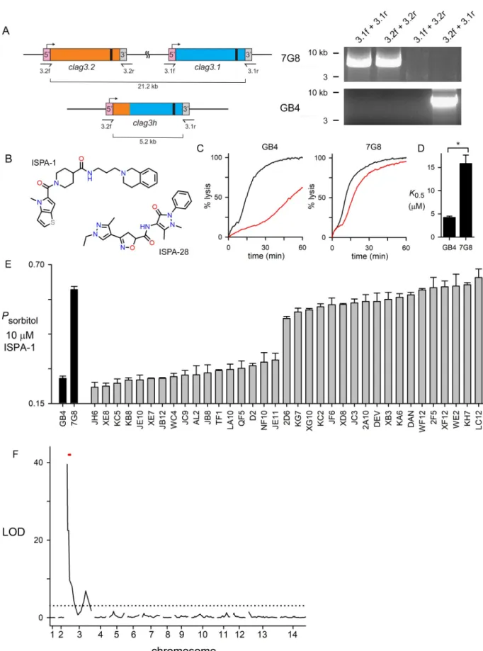
para-log to determine that GB4 and 7G8, the parents of aP. falciparum genetic cross [36], carry one and two copies, respectively (Fig 1A). The proteins encoded by theseclag3 alleles are more
than 90% identical, with most of the variation present in a small hypervariable region near the C-terminus (HVR,S1A Fig). The unique HVR sequence of the Dd2 CLAG3.1 protein is responsible for PSAC block by ISPA-28 in this laboratory clone and lack of activity against channels from other parasite lines [15,37]. In keeping with this, we found that ISPA-28 is inef-fective against 7G8 and GB4 channels (Fig 1BandS1B Fig). While ISPA-28 was instrumental in mapping studies using the Dd2 x HB3 to implicate CLAG3 [12], this compound does not produce heritable differences in PSAC phenotype between the 7G8 and GB4 lines and is, therefore, not useful for linkage studies in their genetic cross.
Because reducedclag3 copy number in GB4 should be inherited in a subset of progeny, we
reasoned that the GB4 x 7G8 cross may provide unique insights into the role of theclag3
prod-uct. We therefore surveyed hits from previous PSAC inhibitor screens to identify ISPA-1, a rel-atively low affinity inhibitor that blocks GB4 channels with 4-fold greater affinity than those associated with 7G8 parasites (Fig 1B–1D). Using a kinetic assay that continuously tracks osmotic lysis resulting from PSAC-mediated sorbitol uptake, theK0.5values for channel block
were 4.2± 0.3 and 15.8 ± 1.8 μM for GB4 and 7G8 parasites, respectively (P < 10-4
,n = 5 dose
response experiments each). We then examined inheritance of PSAC block in the cross prog-eny using a 10μM ISPA-1 concentration in osmotic lysis experiments and found that all 35 progeny clones matched one or the other parental line (Fig 1E), suggesting monogenic inheritance.
We then used quantitative trait locus (QTL) mapping to identify possible parasite genomic loci that define ISPA-1 block of channels in this cross (Fig 1F). A primary scan using defined microsatellite markers and single nucleotide polymorphisms revealed a single locus at the 5’ end of chromosome 3 with a logarithm of odds (LOD) score of 40.0, exceeding previously reported scores for examinedP. falciparum phenotypes. The mapped locus of 139.5 kB
con-tains 32 annotated genes (S1 Table) including twoclag3 genes mapped by previous linkage
analysis studies of PSAC activity in other genetic crosses [12,14,38]. This result provides inde-pendent evidence for this locus in solute and nutrient trafficking into infected erythrocytes.
Because the progeny clones inFig 1Esegregated into two groups of essentially equal size, these data also argue against a survival advantage of either parental allele at the responsible locus duringin vivo expansion in chimpanzees, as required to produce this genetic cross;
absence of preferential inheritance also argues against epistatic interactions between this locus and other parasite genomic loci [39].
Conditional CLAG3 knockdown reveals an unexpected dose effect on PSAC
activity and inhibitor efficacy
Because CLAG3 plays a pivotal role in solute transport and parasite propagation, we used DNA transfection to produce parasite lines with conditional reductions in CLAG3 protein. Prior studies have reported reduced growth phenotypes with CLAG3 knockdown achieved through either epigenetic silencing orin vitro selection [24,25,40], but none have clearly linked loss of CLAG3 to compromised parasite survival. Because exported proteins such as CLAG3 may be less effectively knocked down by conventional translation-level repression methods [41], we selected the recently developed TetR-DOZI system for conditional expression [42]. This system utilizes an RNA-aptamer sequence in the 3’ UTR of a target gene’s mRNA to recruit a fusion protein consisting of the Tet repressor protein (TetR) and DOZI (development of zygote inhibited), a DDX6 helicase protein that represses translation [43]. This
two-Fig 1. Mendelian inheritance of a PSAC phenotype in the GB4 x 7G8 cross. (A) Schematic showing the twoclag3 genes in 7G8 and a single clag3h in GB4; gene ribbons are color-coded to show site of recombination to produce copy number reduction in GB4. Black bar near the 3’ end
component fusion improves the effectiveness of knockdown and is suitable for targeting both soluble and membrane-associated proteins inP. falciparum [42,44].
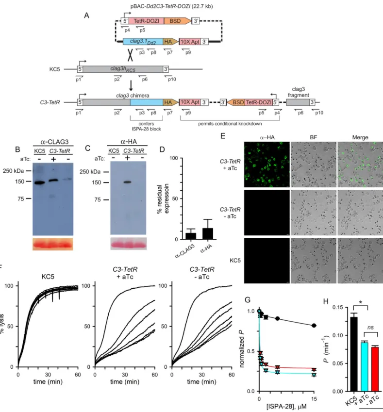
We designed and produced a single plasmid strategy for allelic exchange recombination to achieve efficient CLAG3 knockdown (Fig 2A). ThepBAC-Dd2C3-TetR-DOZI plasmid carries
a 3.2 kB fragment from the end of the Dd2clag3.1 gene with an in-frame C-terminal HA
epi-tope tag and a 10x aptamer sequence in the 3’UTR for recruitment of TetR-DOZI. We selected KC5, a progeny clone from the GB4 x 7G8 cross, for transfection because it carries a single
clag3h gene and has been successfully used in prior studies [37]; a singleclag3h gene avoids
epigenetic switching between the desired integrated gene and a second unmodifiedclag3 gene
[12].
We anticipated homologous recombination between the KC5clag3h genomic site and the
Dd2clag3.1 gene fragment on the plasmid as these clag3 genes are nearly identical. This
recombination adds a C-terminal HA epitope tag to the encoded protein and, importantly, confers PSAC block by ISPA-28, an inhibitor specific for Dd2clag3.1-associated channels;
ISPA-28 sensitivity has been mapped to a variant motif near the C-terminus of the encoded protein [15]. As the transfection plasmid carries only aclag3 fragment without a promoter
sequence, changes in PSAC phenotype can only be observed after in-frame integration into the genomicclag3h. Upon transfection, anhydrotetracycline (aTc) was continuously applied
together with selection for thebsd marker to preserve conditional CLAG3 expression and
avoid TetR-DOZI-aptamer mediated suppression. After parasite outgrowth under this selec-tion, limiting dilution cloning yielded theC3-TetR clone. PCR and DNA sequencing
con-firmed faithful integration at the desired locus (S2 Fig).
We used immunoblotting to evaluate expression of the chimeric transgene product. With a CLAG3 antibody that recognizes the protein’s C-terminus in all examinedP. falciparum lines,
lysates fromC3-TetR cultures yielded a band of somewhat slower mobility than observed in
the KC5 parent, consistent with an expected increase of ~ 4 kDa based on addition of a C-ter-minal HA epitope tag and a larger variant motif on Dd2 CLAG3.1 (Fig 2B). When probed with anti-HA antibody,C3-TetR yielded a band of indistinguishable size but absent from the KC5
parent, as predicted by our transfection strategy (Fig 2C).
Immunoblotting with these antibodies also revealed robust CLAG3 knockdown upon
C3-TetR cultivation without aTc (Fig 2B and 2C). Quantification of band densities from inde-pendent trials revealed an approximately 90% knockdown (Fig 2D). Immunofluorescence microscopy confirmed homogeneous knockdown, as expected for clonal parasites carrying the conditional regulation cassette (Fig 2E). Despite effective knockdown, we could propagate
C3-TetR without aTc indefinitely and did not detect reduced growth under standard in vitro
conditions.
We next examined the effect of transfection and CLAG3 knockdown on PSAC phenotypes and measured osmotic lysis kinetics in sorbitol, a sugar alcohol taken up by infected cells pri-marily via PSAC [45]. While 15μM ISPA-28 had negligible effect on uptake into KC5-infected
of each gene represents a variant region that encodes a CLAG3 extracellular loop implicated in binding of isolate-specific inhibitors. Primer binding sites are indicated. Ethidium-stained gel at right shows PCR confirming the differences inclag3 copy number. (B) Structures of ISPA-1
and ISPA-28. (C) Sorbitol-induced osmotic lysis kinetics for GB4 and 7G8 without and with 10μM ISPA-1 (black and red traces in each panel, respectively). ISPA-1 produces greater block of GB4 channels, as indicated by slower sorbitol-induced lysis. (D) Mean± S.E.M. half-maximal inhibitory concentrations,K0.5, for ISPA-1, determined from dose response experiments as in panel C.�,P < 10−4. (E) Mean± S.E.M. sorbitol permeability with 10μM ISPA-1, normalized to 1.0 for matched trials without ISPA-1. Results are shown for indicated parental lines and progeny clones from the genetic cross (n = 13–16 and 2–5 for parental and progeny clones, respectively). Each progeny’s phenotype matches that
of a parent. (F) Logarithm of odds (LOD) scores from a primary scan of QTL for ISPA-1 affinity. The peak (LOD of 40) maps to the 5’ end of chromosome 3 and includes theclag3 locus (red dash). Dashed line indicates the P = 0.05 significance threshold, calculated from 1000
permutations.
Fig 2. CLAG3 conditional knockdown reveals complex effects on channel pharmacology and transport rates. (A) Allelic exchange strategy for production of a
chimericclag3 gene and conditional knockdown. The TetR-DOZI conditional regulator and blasticidin S deaminase (BSD) are expressed under a single promoter and
separated by a viral 2A skip peptide [75, not illustrated]. A C-terminal HA epitope tag (HA) is appended to theclag3 gene and is followed by a stop codon and a 10x
aptamer sequence (10X Apt) embedded in the 3’ UTR. Primer positions are shown as used for PCR results inS2 Fig; primer sequences are listed inS2 Table. (B-C) Immunoblots using total cell lysates from indicated parasites cultivated with or without 2μM aTc, probed with CLAG3- or HA epitope tag-specific antibody (panels B and C, respectively). Bottom shows Ponceau S staining of hemoglobin as a loading control. (D) Digital quantification of residual CLAG3 expression upon aTc removal
cells, channels inC3-TetR were potently blocked by ISPA-28, with affinity comparable to that
seen in Dd2 and in other transfections using the Dd2clag3.1 gene [37,Fig 2F–2H], as
expected. Remarkably, however, aTc removal to knockdown CLAG3.1 only modestly reduced ISPA-28 sensitivity despite a near-complete knockdown of the block-enabling transgene (Fig 2D and 2E). Although we could detect a modest reduction in ISPA-28 sensitivity (red vs. blue triangles,Fig 2G;P = 0.005 in comparisons at a 15 μM concentration, n = 7 independent trials
each), the effect of knockdown was much less than expected by models invoking a single CLAG3 monomer that directly forms a pore. Retained ISPA-28 sensitivity despite a ~90% knockdown of the Dd2 CLAG3.1 protein is surprising because this inhibitor is inactive against channels associated with all other CLAG proteins in KC5 and its parental lines (Fig 2Fand
S1B Fig); this result indicates that a remarkably low dose of the Dd2 CLAG3.1 protein can con-fer ISPA-28 block on these parasite-induced channels.
A direct 1:1 relationship between CLAG3 and channel activity was also contradicted by measurements of PSAC-mediated permeability, which is inversely proportional to the inhibi-tor-free osmotic lysis halftime [46]. The halftime for KC5 parasites in sorbitol, 7.7 min, corre-sponds to an apparent sorbitol permeability of 0.13± 0.007 min-1, which is similar to values obtained with other wild-type parasites [45]; osmotic lysis ofC3-TetR-infected cells was
signifi-cantly slower both with and without aTc addition (Fig 2H). Although the reduced permeability without removal of the aTc stabilizer might reflect expression of an altered CLAG3 protein, we consider incomplete protection of translation by aTc to be a more conservative explanation. Experiments using parasites cultured with a higher aTc concentration of 4μM did not signifi-cantly affect transport rates, but were limited by increasing toxicity of aTc on parasite cultures. Another possibility,clag3 and clag2 silencing due to transfection under blasticidin S selection
[24,25,47], could also contribute to reducedC3-TetR permeabilities. Because clag gene
silenc-ing and transport phenotypes recover quantitatively within 4 weeks of blasticidin S removal [47], a significant effect of drug selection is unlikely. Surprisingly, cultivation ofC3-TetR
with-out aTc produced only a modest reduction in the lysis halftime that did not reach statistical significance (P = 0.7, n = 7 trials each), again inconsistent with a 1:1 relationship between
CLAG3 expression and PSAC formation.
We also transfected 7G8 parasites withpBAC-Dd2C3-TetR-DOZI, obtained an integrant
clone by limiting dilution, and observed similar changes in transport phenotypes that further argue against a simple stoichiometric relationship between CLAG3 expression and PSAC for-mation (S3 Fig). PCR indicated that the7G8-TetR limiting dilution clone had undergone
homologous recombination of the plasmid into the 7G8 parasite’sclag3.1 gene while
preserv-ing the nativeclag3.2 gene (S3A and S3B Fig). Immunoblotting with HA epitope tag anti-bodies confirmed transgene expression that could be effectively abolished upon aTc removal; in contrast to theC3-TetR line, however, the total amount of CLAG3 was largely unaffected by
conditional knockdown in7G8-TetR, as revealed with anti-CLAG3 antibodies that bind to a
conserved C-terminal epitope (S3C and S3D Fig). RT-PCR revealed that the chimericclag3.1
gene is preferentially expressed in the presence of aTc and confirmed epigenetic switching to
to produce knockdown. Mean± S.E.M. band densities from 3 independent harvests and immunoblots as in panels B and C. (E) Indirect immunofluorescence images of trophozoite-stageC3-TetR and KC5 wildtype parasites probed with anti-HA antibody (green). C3-TetR parasites are shown after cultivation with or without aTc. Scale
bar, 5μm. (F) Continuous recordings of sorbitol-induced osmotic lysis kinetics for the parental KC5 line or the C3-TetR line cultivated with or without aTc, as indicated. Traces reflect lysis kinetics with 0, 0.3, 0.6, 1.8, 5, or 15μM ISPA-28 (top to bottom, respectively in each panel). (G) Dose responses for ISPA-28 inhibition for KC5 (black circles) andC3-TetR grown with or without aTc (blue and red triangles, respectively). Symbols represent mean ± S.E.M. permeabilities, normalized to 1.0
for matched traces without inhibitor;n = 5–7 trials at each concentration. Solid lines represent best fits to y = a/(1 + (x/b)) + (1-a)/(1 + (x/c)). (H) Mean ± S.E.M.
apparent sorbitol permeabilities for indicated lines, calculated as the reciprocal of the inhibitor-free lysis halftime;n = 5–7 trials each.�,p < 10−4;
ns, not significantly
different.
increaseclag3.2 expression by >100-fold upon conditional knockdown of clag3.1 (S3E Fig); this switching accounts for the sustained production of CLAG3 in7G8-TetR. As with the C3-TetR clone, this transfectant also had preserved block by ISPA-28 despite quantitative
reductions in the expression of the Dd2clag3.1 element required for ISPA-28 block (S3F and S3G Fig). Although still less than predicted by simple models of block, aTc removal reduced ISPA-28 efficacy to a greater extent in the7G8-TetR parasite than in C3-TetR (S3H and S3I Fig.), presumably because of switching and expression of the unmodifiedclag3.2 gene
associ-ated with ISPA-28 insensitive channels.
A viable CLAG3-null parasite has reduced, but partly preserved PSAC
activity
In light of the modest effects of CLAG3 knockdown, we next explored whether a viable
clag3-null parasite could be produced and used CRISPR/Cas9 transfection with a guide RNA that targets exon 1 ofclag3h in KC5 and clag3.2 in other P. falciparum lines. Although transfection
has previously been used to knockoutclag3.2 [40], the resulting parasite retained an intact
clag3.1 gene. While those authors used drug selection to silence the remaining clag3.1 gene,
low-level CLAG3 expression is likely with such silencing and would yield transport phenotypes similar to those described above forC3-TetR.
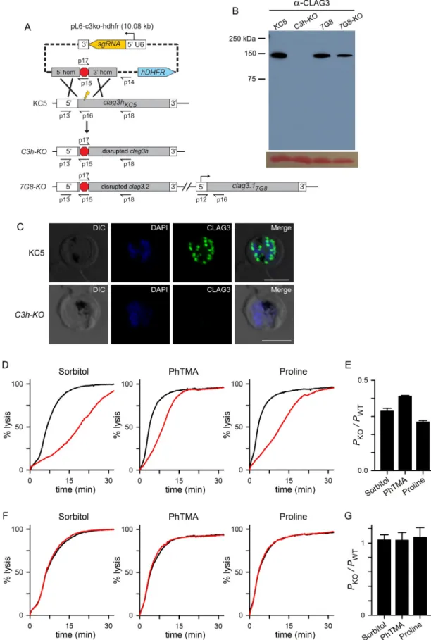
The KC5 line and a two-plasmid CRISPR/Cas9 strategy was used to address this possibility and avoid a dependence on gene silencing for the null phenotype (Fig 3A). We successfully generated theC3h-KO limiting dilution clone that lacks an intact clag3 gene (S4A Fig). Immu-noblotting and immunofluorescence imaging confirmed complete absence of CLAG3 protein in this parasite (Fig 3B and 3C).
We then examined PSAC-mediated transport and found significant, but incomplete reduc-tions in the permeabilities of sorbitol, the organic cation phenyl-trimethylammmonium (PhTMA+), and proline, solutes representing the broad selectivity of PSAC (Fig 3D). Each sol-ute’s permeability was reduced by 60–75%, as determined from the inverse relationship between permeability and time to osmotic lysis [46]; interestingly, ANOVA comparisons and post-hoc testing revealed differing relative decreases for each solute (Fig 3E,P < 0.01, n = 3–4
trials for each solute).
Transfection of the 7G8 parasite with the samepL6-c3ko-hdhfr plasmid yielded the 7G8-KO
line with a disruptedclag3.2 gene (Fig 3AandS4B Fig). This knockout line retains a full length
clag3.1 gene that produces an unmodified CLAG3 protein (Fig 3B). Continued expression of this paralog resulted in unchanged permeabilities for sorbitol, PhTMA+, and proline (Fig 3F and 3G). Thus,7G8-KO serves an important control to show that loss of CLAG3 protein is
responsible for the reduced permeability inC3h-KO. We have not pursued simultaneous
dis-ruption of bothclag3 genes in the 7G8 background; such a double knockout would have
reduced PSAC activity quantitatively similar to that ofC3h-KO because these lines have nearly
identical sequences for RhopH2, RhopH3, and other CLAG proteins.
Whileclag genes on other P. falciparum chromosomes have not been unambiguously
impli-cated in PSAC activity [16], it is possible that successful production of aclag3 null parasite and
partly preserved host cell permeability result from compensatory increases in expression of these paralogs. We therefore used quantitative RT-PCR to measure transcript levels forclag
paralogs,rhoph2 and rhoph3 in the knockout lines. In the KC5 wild-type parent, clag3h was
found to be the most highly expressed member of theclag gene family; its knockout produced
only modest changes in expression of otherclags, rhoph2, and rhoph3 (Fig 4A). Correcting for family-wise errors that result from multiple comparisons, the increase inclag9 expression, but
Fig 3. Production of a viable CLAG3-null parasite with incomplete reduction in solute permeabilities. (A) CRISPR/Cas9
respectively, Student’st test with the Holm-Bonferroni correction; n = 3 independent trials
each);clag8 and rhoph2 were not significantly upregulated in C3h-KO. None of these genes
were upregulated in7G8-KO (Fig 4B). Thus, both lines tolerateclag3 knockout without
marked compensatory changes in other genes linked to PSAC.
Preferential expression ofclag3.1 in the 7G8 line results from unbalanced switching rates
between the twoclag3 paralogs, as previously reported [12,30]. Because the7G8-TetR
condi-tional knockdown confirms competence for epigenetic switching (S3E Fig), we did not pursue production of aclag3.1 knockout in the 7G8 background: that transfectant would also retain
unaltered transport properties without changes in otherclag and rhoph genes.
C3h-KO requires supraphysiological levels of essential nutrients
Although transfection to produceC3h-KO indicates that CLAG3 is not needed for in vitro
propagation, the conservation and variable expansion of theclag3 clade in Plasmodium spp.
suggest a more essential role underin vivo conditions in vertebrate infections [27,29,33]. In light of PSAC’s role in nutrient uptake [14], we hypothesized that the high concentrations of most nutrients in standard RPMI 1640-based media may permit growth of the CLAG3-null parasite but that the lower levels in host plasma may prove inadequate. We therefore compared
shown (bottom ribbon). Primer positions are shown as used for PCR results inS4 Fig; primer sequences are listed inS2 Table. (B) Anti-CLAG3 immunoblot using total cell lysates from indicated parasites; bottom shows Ponceau S hemoglobin staining as a loading control. (C) Indirect immunofluorescence images of schizont stage KC5 wildtype andC3h-KO parasites probed with
anti-CLAG3 antibody (green). Scale bar, 5μm. (D) PSAC-mediated osmotic lysis kinetics for indicated solutes. Black and red curves reflect kinetics for KC5 andC3h-KO. (E) Mean ± S.E.M. permeabilities of indicated solutes in the knockout line, normalized to
1.0 for matched experiments with the wild-type parental line (PKO/PWT). The residual permeabilities of these solutes differed from one another (P < 0.01, one-way ANOVA). (F) Preserved PSAC-mediated transport of each solute in 7G8-KO when compared to
7G8 (red and black traces, respectively). (G) Mean± S.E.M. permeabilities in 7G8-KO relative to matched experiments with 7G8 confirming no detectable reduction in each solute’s transport (n = 3 trials each).
https://doi.org/10.1371/journal.ppat.1008363.g003
Fig 4. Modest effects on expression ofclag paralogs and rhoph genes in the knockout lines. (A) Mean ± S.E.M. normalized
expression ofclag and rhoph genes in KC5 and C3h-KO (black and red bars, respectively; n = 3), calculated according to 2-ΔCt
using PF07_0073 as an internal control. Onlyclag9 exhibited a statistically significant upregulation in the Ch3-KO knockout
(asterisk,P = 0.007). (B) Insignificant changes in expression of these genes in the 7G8-KO knockout when compared to 7G8
(red and black bars, respectively;n = 2–3 for each gene). The msp2 control exhibits stage-specific transcription similar to clag
andrhoph genes, and is not affected in these knockout lines.
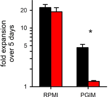
growth in RPMI 1640-based media to that in PGIM, a modified medium with more physiolog-ical concentrations of key nutrients [14]. Using SYBR Green to measure nucleic acid produc-tion, 5 day expansion ofC3h-KO was indistinguishable from that of its wild-type parent when
cultures were maintained in RPMI 1640-based media (Fig 5;P = 0.47, n = 3 trials with
tripli-cate measurements). In PGIM, however,C3h-KO growth was drastically compromised, with
nucleic acid production reduced by 17± 7 fold when compared to that of the KC5 parent in the same medium (P = 0.006, n = 3). While wild-type cultures also grow slower in PGIM than
in RPMI 1640 (compare black bars,Fig 5), their PGIM expansion rates match those seen with pooled human serum [14], suggesting that this modified medium better reflectsin vivo parasite
expansion than RPMI 1640-based media. Although circulating nutrient levels in human sub-jects are difficult to measure and standardize between laboratories [48,49], the nominal con-centrations of isoleucine and hypoxanthine in PGIM more closely resemble levels in healthy human volunteers. Plasma levels for these and other nutrients have not been accurately mea-sured in malaria-endemic countries, but limited access to quality nutrition suggests that they may be lower. Thus, while we successfully produced a viable CLAG3-null parasite underin vitro culture conditions, our findings suggest that CLAG3 is required for P. falciparum survival
and expansion in human infections, where parasite nutrient acquisition is rate-limiting.
Inhibitors also support direct CLAG3 contribution to PSAC formation
Finally, we examined PSAC block by known small molecule inhibitors and found that theC3h-KO knockout line exhibits marked changes in pharmacology. Phloridzin, a nonspecific,
low affinity inhibitor that acts at the intracellular face of the channel [50,51], was markedly less effective in inhibiting sorbitol uptake inC3h-KO than in the wild-type parent (Fig 6A and 6B; half-maximal blocking concentrations,K0.5, of 4200± 1100 and 45 ± 10 μM, P = 0.01, n = 4
dose responses each). ISG-21, a potent and specific inhibitor found through high-throughput screening that appears to act at an extracellular site on the channel [14], also exhibited a signifi-cantly reduced efficacy in the CLAG3 knockout (Fig 6C,K0.5of 15.0± 2.4 nM vs. 2.6 ± 0.7 nM
in the wild-type parent,P = 0.008, n = 3). ISPA-1, the inhibitor used to map the clag3 locus in
the 7G8 x GB4 cross (Fig 1), was also less effective (Fig 6D,P < 0.001, n = 3), further
support-ing direct interaction with CLAG3. Finally, consistent with its specificity for the Dd2 CLAG3.1 protein, knockout of CLAG3 did not alter the lack of ISPA-28 activity against channels on the KC5 line (Fig 6E); this finding also further excludes ISPA-28 blocking activity against channels linked toclag paralogs on other chromosomes. These complex changes in pharmacology in the C3h-KO knockout parasite support a critical role of CLAG3 in establishing and modifying
PSAC activity at the host membrane.
Discussion
Here, we used linkage analysis and distinct reverse genetic approaches to study the increased permeability of erythrocytes infected with malaria parasites. Described initially with macro-scopic flux measurements [52,53], these changes are mediated by the broad-selectivity plasmo-dial surface anion channel (PSAC) and are conserved on human and other vertebrate
erythrocytes infected with malaria parasites [19,54]. PSAC activity is essential for intracellular parasite growth and replication as it functions in nutrient uptake from host plasma [14,20]. Although originally proposed to result from upregulation of transporters endogenous to the erythrocyte, many studies have now linked PSAC activity to a parasite-encoded RhopH pro-tein complex consisting of CLAG3, RhopH2, and RhopH3 [12,20,21,37]. The present studies provide three new insights into the role of CLAG3 in PSAC formation and intracellular growth of malaria parasites. First, we used DNA transfection to produce the first CLAG3-null parasite
and show that loss of CLAG3 does not compromise growth under standardin vitro culture
conditions; growth in PGIM, a modified medium that more closely resemblesin vivo nutrient
availability [14], was dramatically compromised, implicating CLAG3 requirement for parasite survival and expansion in human infections. Second, we found that loss of CLAG3 compro-mises but does not abolish PSAC activity, which is surprising given the multiple lines of evi-dence supporting this protein’s central role and our genetic mapping studies (Fig 1). Third, we used simultaneous CLAG3 allele replacement and conditional knockdown to determine that remarkably low levels of CLAG3 protein can confer block by ISPA-28, a PSAC inhibitor spe-cific for the engineered transgene. In addition to providing independent evidence for a CLAG3 role in channel-mediated nutrient uptake, our studies reveal the complex behavior of knockdown and knockout lines despite simple Mendelian inheritance in aP. falciparum cross.
We propose a conservative model that accounts for these and other observations from previ-ous studies.
While loss of RhopH2 or RhopH3 abolishes PSAC activity in sensitive whole-cell patch-clamp measurements [20], our CLAG3-null parasite retains 25–30% of the wild-type perme-ability. Does this suggest that RhopH2 and RhopH3 are more important PSAC subunits? What mechanisms might account for the differing effects of individual subunit knockdowns? In one model, CLAG3 plays a lesser role in PSAC formation while RhopH2 and RhopH3 serve strictly essential roles, possibly contributing directly to the nutrient pore. This model is exem-plified by maxi-K potassium channels, where the pore is formed entirely byα subunit oligo-mers but a non-essentialβ subunit remains associated with the channel to serve important roles in activation at the cytoplasmic face [55]. In contrast to this model’s predictions for PSAC, protease susceptibility experiments have revealed that CLAG3 spans the host mem-brane while RhopH2 and RhopH3 are primarily or exclusively intracellular [15,20].
Fig 5. Comparison ofKO and wild-type parasite growth rates. Mean ± S.E.M. expansion of wild-type and
C3h-KO cultures over 5 days (black and red bars, respectively; n = 3). Expansion in indicated media is shown on a
logarithmic scale. Asterisk,P = 0.006.
Furthermore, computational analyses, various inhibitor binding studies and site-directed mutagenesis all suggest CLAG3 carries a critical pore-lining transmembrane domain and argue against a non-essential accessory role for CLAG proteins [15,23]. To incorporate these findings, we propose an alternate model wherein CLAG proteins primarily form the channel pore while RhopH2 and RhopH3 serve essential, but poorly understood functions at the chan-nel’s intracellular face (Fig 7). This model posits that CLAG3-null parasites still make func-tional channels because other CLAG paralogs derived fromclag2, clag8, or clag9 gene
expression can associate with RhopH2 and RhopH3 to allow pore formation [16,56]. Because
rhoph2 and rhoph3 are single copy genes in all Plasmodium spp., their knockouts cannot be
compensated in a similar fashion and are, therefore, unable to sustain nutrient uptake.
Fig 6. Altered pharmacology inC3h-KO. (A) Sorbitol-induced osmotic lysis kinetics for wild-type and C3h-KO. Traces are shown for cells suspended with the
inhibitor phloridzin (0, 10, 50, 100, 250, and 500μM in the wild-type panel; 0, 100, 250, 500, 5000 μM for C3h-KO). Increasing inhibitor concentrations produce monotonic increases in transport block (top to bottom traces in each panel); note that there is less effective block ofC3h-KO channels despite use of higher
[phloridzin]. (B—E) Mean± S.E.M. residual permeabilities from dose response experiments as in A for phloridzin, ISG-21, ISPA-1, and ISPA-28, respectively. In each panel, wild-type parent andC3h-KO are shown with black and red circles, respectively (up to 4 trails at each concentration). Solid lines represent best fits to y = a/(1 + (x/b)) + (1-a)/(1 + (x/c)).
Further supporting a critical role for CLAG proteins is the observation thatclags appear to
be the only multigene family strictly conserved throughoutPlasmodium spp. [16,57–59]. Fam-ily size varies significantly with some plasmodial species having only twoclags and others
hav-ing up to 35 copies [29]. Recent studies in the humanP. falciparum pathogen suggest ongoing
expansion of this family [27], with active evolution of diversifying mutations in the twoclag3
genes [24]. It is not clear why this family has undergone variable expansion and why only some members are under epigenetic control [30]. One possibility is evasion of host immunity targeting the surface-exposed domain on CLAG proteins [12]. Several observations are consis-tent with this explanation. RhopH2 and RhopH3, PSAC components that are not surface-exposed (Fig 7), can serve their essential functions as single-copy genes without expansion. Immune evasion is also supported by the highly variant sequence at the exposed CLAG3 domain, which appears to be under immune selection [34,60]. Indeed, ISPA-28 and ISPA-1 were identified as inhibitors specific for certain CLAG3 alleles because these compounds block PSAC through interactions with this variant region. An important problem with the immune evasion proposal, however, is that 2 or 3 paralogs, as present in manyPlasmodium spp., are
generally considered insufficient to allow repeated cycles of switching and immune evasion [32].
An interesting alternative hypothesis is thatclag gene expansion allows for production of
distinct channels with solute selectivities that fine-tune nutrient uptake; this fine-tuning would be advantageous as nutritional status may vary significantly amongst humans and other hosts. Distinct channel subpopulations may also facilitate uptake of diverse nutritive solutes while still maintaining a low Na+permeability and erythrocyte osmotic stability [61,62]. Supporting this hypothesis, a recent study found preferential expression ofclag3.2 in parasites drawn from
humans with malaria; some, but not all, of these parasites switched toclag3.1 upon in vitro
cul-ture, suggesting regulation by extracellular nutrient availability [33]. This possibility is also supported by their observation that low concentrations of blasticidin S, a toxin that enters
Fig 7. Proposed model for CLAG3 contribution to increased permeability of infected erythrocytes. Illustration shows the host erythrocyte membrane with
channels composed of complexes that have several CLAG monomers in association with RhopH2 and RhopH3 proteins at their cytosolic face. Our model incorporates the observed 90% reduction of CLAG3 protein inC3-TetR when aTc is removed (middle vs. left panel), the 60–75% reduction in permeability in C3h-KO (right panel), and the greater transcription of clag3 genes than of other clags. We propose that ISPA-28 block of channels in the C3-TetR conditional
knockdown is preserved because channel oligomers that contain at least one CLAG3 subunit retain sensitivity to this inhibitor; the precise stoichiometries of CLAG, RhopH2, and RhopH3 subunits and their relative positions within functional channels remain unknown. TheC3h-KO knockout retains PSAC activity at a
reduced level because other CLAG paralogs may oligomerize to form channels. Ribbon legend shows subunit color-coding with breaks to reflect expression from distinct chromosomes.
infected cells through PSAC [47], selects forclag3 switching in some, but again not all, lines
[25,33]. Counter to this hypothesis, our transport measurements have not revealed significant changes in solute selectivity uponclag3 expression switching (data for 7G8-TetR inS3 Figand data in [12]). In the present studies,clag3 knockout reduced permeability of sorbitol, proline
and the organic cation PhTMA+by similar amounts (Fig 3D and 3E), further countering the proposal thatclag3-associated channels transport specific solutes while other solutes are
acquired by channels linked toclag2, clag8 or clag9.
Our CLAG3-null parasite grows without detriment in a standard RPMI 1640 medium that contains supraphysiological concentrations of amino acids and vitamins [48]. Remarkably, however,C3h-KO growth was largely abolished in PGIM, a modified medium with more
phys-iological levels of key nutrients (Fig 5). As parasite growth rates in PGIM match those in pooled human serum without addition of synthetic media products [14], our findings suggest that CLAG3 is needed underin vivo conditions but dispensable under nutrient-rich culture
conditions. PGIM may also underestimate thein vivo efficacy of inhibitors that target parasite
nutrient acquisition because it has unchanged, high levels of many nutrients acquired via PSAC [48]. Becausein vitro parasite growth is compromised for many of these permeant
nutri-ents in single solute elimination studies [63], we suspect that various other modifications of the RPMI 1640 formulation to reduce key nutrients to more physiological levels would also compromise or abolish growth of the CLAG3-null parasite. As no modified medium can con-fidently replicate the conditions encountered in human infections, we propose that ongoing drug discovery targeting PSAC should be guided byin vivo studies of transport rates and
para-site survival and propagation. As potent inhibitors have only been validated underin vitro
con-ditions to date [14,64], these inhibitors may prove even more efficacious in future animal and human infection studies.
While conditional knockdown has been primarily used to study genes refractory to knock-out [41,65], our combined use of knockdown and knockout strategies provided important insights not possible with either approach alone. Our knockdown studies reveal that remark-ably low doses of CLAG3, such as those that persist in previously generated silencing mutants and conditional knockdowns [24,40], are sufficient to preserve PSAC phenotypes. Combined use of conditional knockdown and allele replacement to confer ISPA-28 sensitivity permitted sensitive detection of this dose effect on channel phenotypes in our study. The observed reten-tion of ISPA-28 sensitivity despite a 90% knockdown of transfected CLAG3 allele is inconsis-tent with models invoking a single CLAG3 subunit per functional channel because this arrangement would yield a direct linear relationship between transgene expression and ISPA-28 potency. Higher order stoichiometries, e.g. two or more CLAG molecules per channel, can produce retained ISPA-28 sensitivity despite quantitative knockdown if we assume that CLAG2, CLAG8 and/or CLAG9 also produce channels and that CLAG paralogs aggregate ran-domly to form functional channels in the host membrane. In general, ifk CLAG molecules are
required to form the channel, the number of distinct channels that can be formed from combi-nations of the four CLAG paralogs in theC3-TetR parasite is given by the binomial coefficient
3 þk k
!
; this corresponds to 10, 20, and 35 distinct complexes that could be formed with 2, 3, or 4 CLAG molecules per channel, respectively. As the number of possible complexes increases, the fraction that contain one or more CLAG3 monomers also increases: 40, 50, and 57.1% of channels will contain at least one CLAG3 monomer in dimeric, trimeric, and tetra-meric arrangements, respectively, if each of the paralogs is equally abundant. (If CLAG3 is present at higher copy number than other paralogs, as our data suggests, then this fraction will increase even more rapidly with subunit stoichiometry.) Now, if channels containing one or
more CLAG3 monomers are blocked by ISPA-28 with similar affinities, then ISPA-28 block will be preserved to greater extents in theC3-TetR CLAG3 knockdown for higher
stoichiome-tries (Fig 7). Thus, our pharmacological studies using theC3-TetR knockdown line exclude
models where a single CLAG monomer directly contributes to PSAC formation; instead, they implicate higher order complexes at the host membrane and provide experimental evidence for contributions from CLAG paralogs encoded from otherP. falciparum chromosomes.
Larger complexes are also supported by the recognition that a 1:1:1 complex of CLAG3, RhopH2, and RhopH3, as suggested by immunoprecipitation studies [66], lacks the number of predicted transmembrane domains generally needed to form a stable aqueous pore in biologi-cal membranes [16,26]. Anomalous migration of these membrane proteins in native PAGE experiments, as often seen for integral proteins [67], has prevented an accurate size determina-tion for this complex [37]. While coimmunoprecipitation has detected associations between two CLAG3 isoforms in a transfected parasite [37], our transport measurements provide the first experimental evidence for functional oligomerization to form the nutrient channel at the host membrane.
Our observations on knockdown phenotypes parallel those in another pharmacological study that expressed mixtures of sensitive and resistant alleles of K+channel subunits [68]; by varying the amounts of the two alleles in a heterologous expression system, that study correctly determined that only a tetrameric arrangement of the K+channel could account for their observed phenotypes. Unfortunately, several complexities intrinsic to our work prevented us from a similar precise determination of the number of CLAG subunits required to form a channel. When compared to this K+channel study, the larger number of CLAG paralogs and an inability to control their amounts with the precision of a heterologous expression system are obvious problems for precise quantification. Additionally complicating is that the level of ISPA-28 block depends on the precise amounts of each CLAG paralog faithfully exported and delivered to the host membrane; we are unable to estimate this with any accuracy but recog-nize that it is only partly determined by each gene’s relative transcription (Fig 4). Another unknown is that RhopH2 and RhopH3 also appear to contribute directly to the channel as they remain associated with CLAGs from the time of their co-translational assembly and rhop-try packaging up through their eventual insertion into the erythrocyte membrane [20]. Indeed, the stoichiometries and contributions of these other conserved proteins to PSAC phenotypes remain elusive.
Because enzymes that modify other proteins or lipids may also have largely preserved phe-notypes upon quantitative knockdown, we also wondered whether our knockdown phenotype could reflect a model where CLAG3 does not directly contribute to the pore, but instead enzy-matically activates other proteins to form the channel, as has been frequently proposed [69]. While such enzymatic or regulatory roles for CLAG3 remain formally possible, a point muta-tion introduced in the surface-exposed variant loop of the Dd2 CLAG3.1 allele dramatically alters PSAC’s protease susceptibility and ISPA-28 block [15]. Recognizing that ISPA-28 and other inhibitors block these channels instantly when added in whole-cell patch-clamp experi-ments performed with non-physiological solutions [12,51], we consider an enzymatic or regu-latory CLAG3 role unlikely: we are unaware of any precedent for such a model in the
electrophysiology literature.
The model proposed here conservatively accounts for the phenotypes of our CLAG3-null and conditional knockdown lines as well as those from prior studies. Nevertheless, refinements or new structure-function models remain an important direction for future molecular and bio-chemical studies, which are critical for understanding host cell remodeling and for advancing antimalarial development against parasite nutrient acquisition.
Materials and methods
Parasite culture
P. falciparum laboratory lines were cultivated at 37o
C under 5% O2, 5% CO2, 90% N2in O+ human erythrocytes obtained from Interstate Blood Bank (Memphis, TN) at 5% hematocrit in RPMI 1640 medium (KD Medical) supplemented with 25 mM HEPES, 50μg/mL hypoxan-thine, 0.5% NZ Microbiological BSA (MP Biomedicals), gentamicin and 28.6 mM NaHCO3 (Gibco). A modified medium, PGIM [14], follows the RPMI 1640 medium with supplements but has reduced concentrations of isoleucine and hypoxanthine (11.4μM and 3.01 μM, respec-tively). Primers specific to 5’ and 3’ UTR regions ofclag3.1 and clag3.2 were used to detect the
number and possible combinations ofclag3 alleles in the GB4 and 7G8 parental lines (S2 Table; SpeedSTAR HS DNA polymerase, Takara).
Osmotic lysis measurements
The increased permeability of infected erythrocytes to organic solutes and block by strain-spe-cific inhibitors was quantified using a kinetic assay that tracks 700 nm light transmittance through a suspension of cells [45]. Osmotic lysis resulting from uptake of sorbitol, a sugar alco-hol with high PSAC permeability, produces increased light transmittance. Synchronized para-site cultures were enriched at the trophozoite stage with the Percoll-sorbitol method, washed, and resuspended at 37oC in lysis buffer (280 mM sorbitol, 20 mM Na-HEPES, 0.1 mg/ml bovine serum albumin, pH 7.4) to initiate sorbitol uptake and cell swelling that leads to osmotic lysis. Iso-osmotic replacement of sorbitol with either 280 mM proline or 145 mM PhTMA+Cl-was used to measure permeabilities of these solutes; these solutes are representa-tive of the broad range of solutes with PSAC permeability [62]. Inhibitors, where present, were added from DMSO stock solutions without preincubation; control experiments excluded DMSO effects on uptake measurements. 700 nm light transmittance was then continuously tracked in a DU640 or DU800 spectrophotometer (Beckman Coulter). Inhibitor dose-responses and reductions in permeability in transfectant parasites were then calculated from the time required to reach a fractional lysis threshold, based on a conservative two-compart-ment model of infected cell osmotic lysis in permeant solutes [46]. This method produces esti-mates of permeability coefficients and inhibitor affinities that quantitatively match those obtained with patch-clamp methods [70].
ISPA-1 was identified as a strain-specific PSAC inhibitor in prior high-throughput screens using four clonal laboratory strains ofP. falciparum (Dd2, HB3, 3D7, and Indo 1; [12]). Trans-port surveys identified this compound as suitable for genetic mapping in the GB4 x 7G8 cross. Dose response studies revealed that ISPA-1 block was adequately fitted by a single Langmuir isotherm with normalized sorbitol permeability,P, given by P = a/ [1 + (x/b)] where a and b
are constants. A 10μM concentration of ISPA-1 produced the greatest difference in parental phenotypes and was used to examine block in progeny clones. Dose responses for block by ISPA-28, a strain-specific inhibitor with potent and specific block of channels linked to the Dd2clag3.1 gene [12], required fitting to a sum of two Langmuir isotherms, as reported previ-ously [37].
QTL analysis
Associations between ISPA-1 block of solute uptake and 290 known microsatellite markers from the GB4 x 7G8 cross were sought by quantitative trait loci (QTL) analysis [36]. QTL anal-ysis was performed using the multiple imputation algorithm and R/qtl software (freely avail-able athttp://www.rqtl.org/) with conditions suitable for haploid asexual parasite crosses, as
described [71]. A significance threshold ofP = 0.05 was calculated from 1000 permutations.
Secondary scans after removing the effects of theclag3 locus did not identify additional
con-tributing genomic loci. DNA sequencing and examination of the publicly available SNP data-base forP. falciparum genetic crosses (https://www.malariagen.net/apps/pf-crosses/1.0/ #variants) did not reveal mutations in theclag3 genes of progeny clones relative to their
corre-sponding parental lines.
Cloning and transfections
TheC3-TetR and 7G8-TetR conditional knockdown clones were produced through allelic
exchange by single homologous recombination withpBAC-Dd2C3-TetR-DOZI, which was
prepared by subcloning of a 3.2 kb fragment of the Dd2clag3.1 gene from the
pHD22Y-120w-flag-PG1 vector [12]. This bacmid was propagated in BAC-Optimized Replicator v2.0 electro-competent cells (Lucigen) in TB medium with 50μg/mL kanamycin and 1x arabinose induc-tion, per the manufacturer’s recommendations. Transfected cultures were selected with 2.5μg/ mL blasticidin S and maintained on 2μM anhydrotetracycline (aTc) to preserve expression of the targeted gene product [42].
CLAG3 knockout lines were produced by CRISPR/Cas9 transfection using the pL6-c3ko-hdhfr vector. In-Fusion cloning (Clontech) was used to introduce the sgRNA for expression
under thePfU6 promoter. A synthetic DNA construct carrying homology arms, shield
muta-tions at the Cas9 target site and in-frame stop codons that disrupt translation was also inserted by In-Fusion cloning. Cas9 was expressed from an unmodified pUF1-Cas9 plasmid [72]. After transfection, cultures were selected with 1.5 nM WR99210 (Jacobus) and 1.5μM DSM-1 (BEI Resources) to ensure retention of plasmids.
All plasmids were confirmed with DNA sequencing and restriction digestion prior to trans-fection, which was initiated by loading uninfected erythrocytes through electroporation and subsequent parasite cultivation. After parasite growth was detected with Giemsa-stained slides, PCR was used to evaluate integration; limiting dilution clones were obtained by the c-SNARF method [73].
Immunoblots
Trophozoite-stage cultures were enriched by the Percoll-sorbitol method to yield 96–99% infected cells and permit equivalent loading of synchronous parasites. Cells were lysed in chilled hypotonic lysis buffer (7.5 mM NaHPO4, 1 mM EDTA, pH = 7.5) with 2 mM PMSF prior to separation by SDS-PAGE (4 to 15% Mini-Protean TGX gel, Bio-Rad) and transfer to nitrocellulose membrane for immunoblotting. After blocking, primary antibodies were applied overnight in blocking buffer (anti-CLAG3, 1:2000 dilution; anti-HA tag, 1:1000 dilu-tion, EMD Millipore). After washing to remove unbound antibody, horseradish peroxidate (HRP)-conjugated secondary antibody was applied (anti-mouse IgG, 1:10,000 dilution, Sigma-Aldrich) with Clarity Western ECL substrate (Bio-Rad). Binding was detected on Hyblot X-ray film. Band intensities were estimated from three independent trials using ImageJ software (https://imagej.nih.gov/) and normalization for hemoglobin loading with Ponceau S staining.
Immunofluorescence assays
Indirect immunofluorescence confocal microscopy was performed using thin smears prepared from parasite cultures. Dried slides were fixed in a chilled 1:1 acetone:methanol mixture for 5 min prior to blocking with 3% skim milk in PBS for 1 h at RT. Slides were incubated with mouse anti-CLAG3 (1:100 dilution) or mouse anti-HA (1:500, Sigma Aldrich) for 1 h at RT, washed with ice-cold PBS, and post-incubated with 2μg/uL DAPI
(4’,6-diamidino-2phenylindole) and goat anti-mouse AF488 at 1:500 dilution for 30 mins at RT. After washing in ice-cold PBS for 5 min, slides were then mounted with Prolong Diamond anti-fade moun-tant (Molecular Probes). Images were collected using a 64x oil immersion objective on a Leica SP5 or SP8 confocal microscope and processed using Leica LAS X software.
Quantitative Real Time PCR
Expression ofrhoph genes in knockout lines was quantified with real-time PCR using total
RNA harvested 27 h after sorbitol synchronization of cultures and the PureLink RNA minikit (Ambion). Microscopic examination of smears confirmed harvest of matched, late trophozo-ite-stage infected cells.
Genomic DNA was removed by DNase I treatment (TURBO DNA-free kit, Ambion) prior to first-stand cDNA synthesis by reverse transcription using Superscript III (Invitrogen), oligo (dT) primers, and ~ 1.5μg RNA from each parasite. The resulting cDNA was diluted and used for qRT-PCR with the QuantiTect SYBR Green kit (Qiagen). Primers were designed based on specificity for individual genes and a desired amplicon size of ~120 bp (S2 Table). qRT-PCR was carried out using the iCycler IQ multicolor real-time PCR system (Bio-Rad) and a three-step protocol: denaturation at 95˚C for 15 min followed by 40 cycles of annealing at 52˚C and extension at 62˚C for 30 s each. The final stage used gradual heating from 55 to 95˚C with 0.5˚C steps over 30 s; this dissociation protocol was used to confirm the specificity of primer binding and product synthesis. Each qRT-PCR was accompanied by a negative control without reverse transcriptase to exclude gDNA contamination. All reactions were performed in tripli-cate with Pf07_0073 used as a constitutively expressed loading control;msp2 was included as a
transcription control as it exhibits stage-specific expression similar toclag and rhoph genes.
The average threshold cycle (CT) values from each experiment were used to calculate expres-sion according to 2^(mean CTvalue of Pf07_0073 –mean CTvalue of the gene of interest). Expression is presented in arbitrary units as the mean± S.E.M. of results from independent RNA harvests.
Parasite growth rates
Parasite expansion rates in RPMI 1640 and PGIM media were measured using a SYBR Green I-based fluorescence assay, as described previously [14]. Synchronous ring-stage cultures were seeded in 96-well microplates at 0.2% parasitemia and 2.5% hematocrit. Media was replaced after 48 h and cultures gassed daily. After cultivation for a total of 5 days, cells were lysed in buffer (20 mM Tris pH 7.5, 10 mM EDTA, 1.6% Triton X-100, 0.016% saponin, pH 7.5) with SYBR Green I nucleic acid gel stain (ThermoFisher) at 2500-fold dilution. After a 30 min RT incubation in the dark, fluorescence measurements were used to quantify parasite DNA (exci-tation, 485 nm; emission, 528 nm; BioTek Synergy HT reader). Expansion was estimated from triplicate wells after normalization to matched cultures killed by treatment with 20μM chloroquine.
Statistical analysis
All numerical data were calculated and plotted as mean± S.E.M. from at least three trials. Sta-tistical significance was calculated by unpaired Student’st-test or one-way ANOVA tests as
indicated. The Holm-Bonferroni method was used to correct for the family-wise error rate arising from multiple comparisons [74].
Supporting information
S1 Fig. CLAG3 sequence alignment and ISPA-28 binding site. (A) Multiple sequence
align-ment of indicated CLAG3 sequences from Dd2, 7G8, and GB4 lines. The C-terminal fragalign-ment expressed by the last exon ofclag3 is shown; CLAG3 sequences upstream of this region are
also highly conserved. A single hypervariable region (HVR) in CLAG3 proteins and the region used for production of an anti-CLAG3 polyclonal antibody are both labeled. (B) Sorbitol-induced osmotic lysis kinetics for 7G8 and GB4 lines with 0, 0.3, 0.6, 1.8, 5, or 15μM ISPA-28 (top to bottom, respectively in each panel). Right panel shows mean± S.E.M. permeability remaining in ISPA-28 dose response studies for 7G8 and GB4 (blue and red circles, respec-tively);n = 3 trials at each concentration. Solid line represents best fit to y = a/(1 + (x/b)) + (1-a)/(1 + (x/c)).
(PDF)
S2 Fig. PCR checks for integration inC3-TetR. Ethidium-stained gels using primers with
positions indicated inFig 2Aand DNA from theC3-TetR transfectant and its KC5 wild-type
parent. In each gel, 9 primer pairs that detect homology-directed integration of the
pBAC-Dd2C3-TetR-DOZI plasmid are shown, followed by primer pairs specific for the wild-type genomic site (WT) or retention of the episome (Epi). As predicted for recombination, the
C3-TetR clone yields bands for each integration primer pair and has lost the WT band. KC5
template yields only the WT band. Expected sizes for each primer pair (in kb):p2-p7, 3.4; p1-p7, 5.3; p2-p8, 2.4; p1-p8, 4.4; p2-p9, 4.0; p5-p10, 4.3; p4-p10, 3.3; p5-p6, 2.6; p4-p6, 1.7; p2-p6,
1.7;p4-p3, 1.7. Primer sequences are provided inS2 Table. (TIF)
S3 Fig. CLAG3 conditional knockdown in 7G8, a parasite with twoclag3 genes. (A)
Sche-matic showing the 7G8 wild-type locus and the result of homologous recombination of
pBAC-Dd2C3-TetR-DOZI in the 7G8-TetR transfection clone. Primer positions are shown with
sequences listed inS2 Table. (B) Ethidium-stained gels using primers with positions indicated in S3A Fig and DNA template from7G8-TetR and the 7G8 type parent. Integration,
wild-type (WT), and episome (Epi) primer pairs are shown with specificity forclag3.1 or clag3.2
indicated in parentheses; a primer pair that yields an integration or wild-type amplicon with eitherclag3 gene is indicated as “e” within parentheses. These results reveal integration into clag3.1 with a preserved wild-type clag3.2 gene in 7G8-TetR. Expected sizes for each primer
pair (in kb):p2-p7, 3.4; p12-p7, 5.3; p1-p7, 5.3; p2-p8, 2.4; p12-p8, 4.4; p1-p8, 4.4; p2-p9, 4.0; p4-p10, 3.3; p4-p11, 3.3; p5-p6, 2.6; p4-p6, 1.7; p2-p6, 1.7; p4-p3, 1.7; p2-p4-p10, 3.5; p2-p11, 3.5.
Primer sequences are provided inS2 Table. (C) Immunoblots using antibodies against the C-terminus of CLAG3 and the HA epitope tag to probe total cell lysates from indicated parasites cultivated with or without 2μM aTc. Bottom, Ponceau S staining of hemoglobin as a loading control. (D) Mean± S.E.M. residual chimeric CLAG3.17G8-Dd2protein in7G8-TetR parasites
upon aTc removal, normalized to 100% for matched cultures maintained on aTc (estimated fromn = 3 independent harvests and anti-HA immunoblots as in panel C). (E) Mean ± S.E.M.
normalized expression of the twoclag3 paralogs in 7G8-TetR cultivated with and without aTc
for more than 2 months (black and red bars, respectively;n = 3). Note the statistically
signifi-cant upregulation of the endogenousclag3.2 gene upon conditional knockdown of clag3.1 in
this transfectant (asterisk;P = 0.02, n = 3). Transcription of clag3.1 and the stage-specific msp2
control were not significantly altered by aTc removal. (F-G) Osmotic lysis kinetics for indi-cated lines, with or without aTc. Traces reflect lysis kinetics with 0, 0.3, 0.6, 1.8, 5, or 15μM ISPA-28 (top to bottom in each panel, respectively). (H) Dose responses for ISPA-28 inhibi-tion for 7G8 (black circles) and7G8-TetR grown with or without aTc (blue and red triangles,
respectively). Symbols represent mean± S.E.M. permeabilities, normalized to 1.0 for matched traces without inhibitor;n = 3–7 trials at each concentration. Solid lines represent best fits to y = a/(1 + (x/b)) + (1-a)/(1 + (x/c)). (I) Mean ± S.E.M. block by 15 μM ISPA-28 for indicated
parasites and aTc addition, normalized to 100% block forC3-TetR cultivated with aTc. aTc
removal has a modest effect on channel block despite ~10-fold reduction in expression of the chimeric CLAG3 that encodes ISPA-28 sensitivity.
(TIF)
S4 Fig. PCR checks for integration inC3h-KO and 7G8-KO parasites. Ethidium-stained gels
using primers with positions indicated inFig 3Aand DNA from indicated knockout parasites, their corresponding parental lines, and the transfection plasmid. Primer pairs specific for inte-gration, the wild type locus, and unintegrated plasmid are indicated in parentheses (i, wt, or p,
respectively); in panel B, primer specificity forclag3.1 or clag3.2 is also indicated. (A) C3h-KO
is a clonal knockout that retains thepL6-c3ko-hdhfr transfection plasmid. Expected sizes (in
kb):p13-p15, 0.42; p17-p18, 0.55; p13-p16, 0.69; p17-p14, 0.50. (B) 7G8-KO is a clag3.2
knock-out clone with a wild-typeclag3.1 locus and retained plasmid. Expected sizes (in kb): p12-p15,
0.18;p13-p15, 0.42; p17-p18, 0.55; p12-p16, 0.34; p13-p16, 0.69; p17-p14, 0.50. Primer
sequences are inS2 Table. (TIF)
S1 Table. Annotated genes in the mapped locus.
(XLSX)
S2 Table. Primers used in this study.
(XLSX)
Acknowledgments
We thank D. Jacobus and BEI resources for providing WR99210 and DSM-1 for parasite trans-fections and Ryan Kissinger for help with artwork.
Author Contributions
Conceptualization: Ankit Gupta, Abdullah A. B. Bokhari, Ajay D. Pillai, Jacquin C. Niles,
San-jay A. Desai.
Data curation: Ankit Gupta, Abdullah A. B. Bokhari, Ajay D. Pillai, Anna K. Crater, Jeanine
Gezelle, Gagandeep Saggu.
Formal analysis: Ankit Gupta, Abdullah A. B. Bokhari, Ajay D. Pillai, Anna K. Crater, Jeanine
Gezelle, Gagandeep Saggu, Sanjay A. Desai.
Investigation: Ankit Gupta, Abdullah A. B. Bokhari, Ajay D. Pillai, Anna K. Crater, Jeanine
Gezelle, Gagandeep Saggu, Armiyaw S. Nasamu, Suresh M. Ganesan.
Methodology: Ankit Gupta, Abdullah A. B. Bokhari, Ajay D. Pillai, Anna K. Crater, Jeanine
Gezelle, Gagandeep Saggu.
Supervision: Jacquin C. Niles, Sanjay A. Desai.
Writing – original draft: Ankit Gupta, Sanjay A. Desai.
Writing – review & editing: Ajay D. Pillai, Anna K. Crater, Jeanine Gezelle, Gagandeep